Embed Size (px)
Citation preview
CASE REPORT Open Access
Visual recovery in a patient with total hyphema,neovascular glaucoma, long-standing retinaldetachment and no light perception vision:a case reportOlusola Olawoye1, Christopher C Teng1,2*, Uri Shabto1, Jeffrey M Liebmann1,3, Francis A L’Esperance4 andRobert Ritch1,2
Abstract
Introduction: We report the case of a patient with total hyphema, neovascular glaucoma, long-standing retinaldetachment and no light perception vision, who regained counting fingers vision with complete regression ofneovascularization following anterior chamber washout, intravitreal bevacizumab, pars plana vitrectomy, andsilicone oil placement. This represents a rare case in which a patient with no light perception vision was able toregain functional vision.
Case presentation: A 63-year-old Caucasian man with a 55-year history of long-standing retinal detachment aftertrauma presented to our facility with pain and redness, a total hyphema, no light perception vision and anintraocular pressure of 60 mmHg (right eye). He had a history of diabetes mellitus and coronary artery disease.Following anterior chamber washout, he was found to have neovascular glaucoma, for which intravitrealbevacizumab was administered. After washout and intraocular pressure control, his visual acuity improved to lightperception. He subsequently underwent vitrectomy, membrane peeling, endolaser and silicone oil placement toreattach his retina, and then a second retinal reattachment procedure. Following these procedures, he had visualrecovery to counting fingers vision in his right eye at five metres, complete regression of neovascularization, andintraocular pressure of 10 to 12 mmHg on one antiglaucoma medication.
Conclusion: Functional vision can be regained despite long-standing retinal detachment.
IntroductionLong-standing retinal detachments (over one year) withpoor visual acuity are typically associated with cysticdegeneration of the macula and retina, loss of pigmentfrom the underlying retinal pigment epithelium, prolif-erative vitreoretinopathy, and poor visual outcome afterretinal reattachment surgery [1].Chronic retinal detachment is a cause of rubeosis iri-
dis and neovascularization of the anterior chamberangle with subsequent neovascular glaucoma (NVG).NVG represents one of the most severe forms of sec-ondary glaucoma, caused by a number of ocular and
systemic conditions. Retinal ischemia and hypoxia initi-ate the release of angiogenesis factors, with consequentdevelopment of new vessels.We report the case of a patient with total hyphema,
NVG, long-standing retinal detachment and no lightperception (NLP) vision, who regained counting fingers(CF) vision with complete regression of the neovascular-ization following anterior chamber (AC) washout, intra-vitreal bevacizumab, and two retinal reattachmentsurgeries.
Case presentationA 63-year-old Caucasian man presented to our facilitywith a four-week history of pain and redness in his righteye. He had had a traumatic retinal detachment of theright eye (55 years ago) after being struck in the eye
* Correspondence: [email protected] Clinical Research Center, The New York Eye and Ear Infirmary, NewYork, NY, USAFull list of author information is available at the end of the article
Olawoye et al. Journal of Medical Case Reports 2011, 5:221http://www.jmedicalcasereports.com/content/5/1/221 JOURNAL OF MEDICAL
CASE REPORTS
© 2011 Olawoye et al; licensee BioMed Central Ltd. This is an Open Access article distributed under the terms of the CreativeCommons Attribution License (http://creativecommons.org/licenses/by/2.0), which permits unrestricted use, distribution, andreproduction in any medium, provided the original work is properly cited.
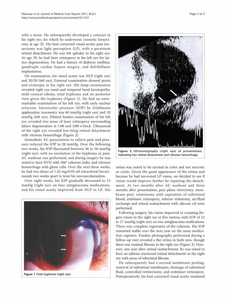
with a stone. He subsequently developed a cataract inhis right eye, for which he underwent cosmetic lensect-omy at age 25. His best corrected visual acuity post len-sectomy was light perception (LP), with a persistentretinal detachment. He was left aphakic in his right eye.At age 39, he had laser retinopexy in his left eye for lat-tice degeneration. He had a history of diabetes mellitus,quadruple cardiac bypass surgery, and defribillatorimplantation.On examination, his visual acuity was NLP (right eye)
and 20/20 (left eye). External examination showed ptosisand exotropia in his right eye. Slit lamp examinationrevealed right eye nasal and temporal band keratopathy,mild corneal edema, total hyphema and no posteriorview given the hyphema (Figure 1). He had an unre-markable examination of his left eye, with early nuclearsclerosis. Intraocular pressure (IOP) by Goldmannapplanation tonometry was 60 mmHg (right eye) and 10mmHg (left eye). Dilated fundus examination of his lefteye revealed two areas of laser retinopexy surroundinglattice degeneration at 1:00 and 3:00 o’clock. Ultrasoundof the right eye revealed low-lying retinal detachmentwith vitreous hemorrhage (Figure 2).Immediate AC paracentesis to relieve pain and pres-
sure reduced the IOP to 38 mmHg. Over the followingtwo weeks, his IOP fluctuated between 36 to 50 mmHg(right eye), with no resolution of the hyphema or pain.AC washout was performed, and during surgery he wasnoted to have NVG with 360° rubeosis iridis, and vitreoushemorrhage with ghost cells. Over the next three weeks,he had two doses of 1.25 mg/0.05 ml intravitreal bevaci-zumab two weeks apart to treat his neovascularization.Over eight weeks, his IOP gradually decreased to 15
mmHg (right eye) on four antiglaucoma medications,and his visual acuity improved from NLP to LP. His
retina was noted to be normal in color and not necroticor cystic. Given the good appearance of the retina andbecause he had recovered LP vision, we decided to see ifvision would improve further by repairing the detach-ment. At two months after AC washout and threemonths after presentation, pars plana vitrectomy, mem-brane peel, retinotomy with aspiration of subretinalblood, endolaser retinopexy, inferior iridotomy, air/fluidexchange and retinal reattachment with silicone oil wereperformed.Following surgery, his vision improved to counting fin-
gers vision in the right eye at five metres, with IOP of 12to 17 mmHg (right eye) on two antiglaucoma medications.There was complete regression of the rubeosis. His IOPremained stable over the next year on the same medica-tion regimen. Fundus photography performed during afollow-up visit revealed a flat retina in both eyes, thoughthere was residual fibrosis in the right eye (Figure 3). How-ever, one year after retinal reattachment, he was noted tohave an inferior tractional retinal detachment in the righteye with areas of subretinal fibrosis.He subsequently had a second membrane peeling,
removal of subretinal membranes, drainage of subretinalfluid, controlled retinectomy, and endolaser retinopexy.Postoperatively, his best corrected visual acuity remained
Figure 1 Total hyphema (right eye).
Figure 2 Ultrasonography (right eye) at presentation,indicating low retinal detachment and vitreous hemorrhage.
Olawoye et al. Journal of Medical Case Reports 2011, 5:221http://www.jmedicalcasereports.com/content/5/1/221
Page 2 of 4
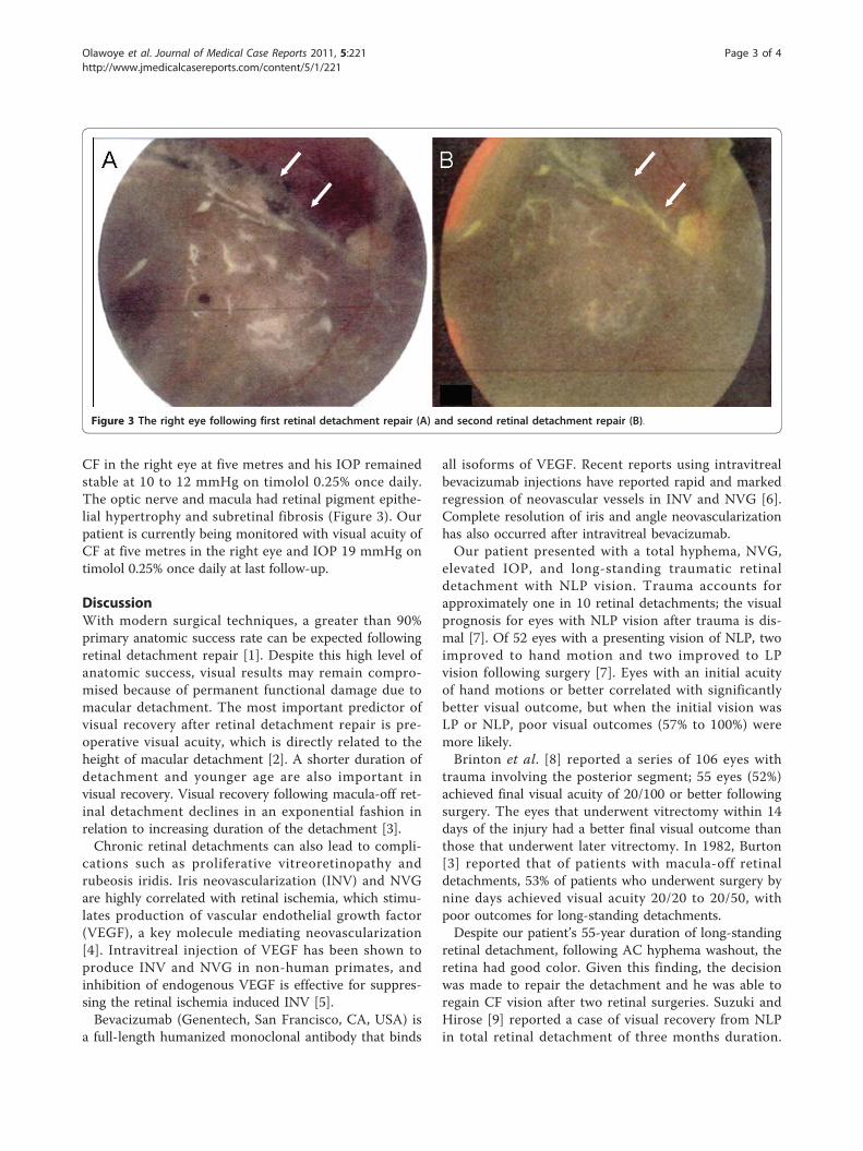
CF in the right eye at five metres and his IOP remainedstable at 10 to 12 mmHg on timolol 0.25% once daily.The optic nerve and macula had retinal pigment epithe-lial hypertrophy and subretinal fibrosis (Figure 3). Ourpatient is currently being monitored with visual acuity ofCF at five metres in the right eye and IOP 19 mmHg ontimolol 0.25% once daily at last follow-up.
DiscussionWith modern surgical techniques, a greater than 90%primary anatomic success rate can be expected followingretinal detachment repair [1]. Despite this high level ofanatomic success, visual results may remain compro-mised because of permanent functional damage due tomacular detachment. The most important predictor ofvisual recovery after retinal detachment repair is pre-operative visual acuity, which is directly related to theheight of macular detachment [2]. A shorter duration ofdetachment and younger age are also important invisual recovery. Visual recovery following macula-off ret-inal detachment declines in an exponential fashion inrelation to increasing duration of the detachment [3].Chronic retinal detachments can also lead to compli-
cations such as proliferative vitreoretinopathy andrubeosis iridis. Iris neovascularization (INV) and NVGare highly correlated with retinal ischemia, which stimu-lates production of vascular endothelial growth factor(VEGF), a key molecule mediating neovascularization[4]. Intravitreal injection of VEGF has been shown toproduce INV and NVG in non-human primates, andinhibition of endogenous VEGF is effective for suppres-sing the retinal ischemia induced INV [5].Bevacizumab (Genentech, San Francisco, CA, USA) is
a full-length humanized monoclonal antibody that binds
all isoforms of VEGF. Recent reports using intravitrealbevacizumab injections have reported rapid and markedregression of neovascular vessels in INV and NVG [6].Complete resolution of iris and angle neovascularizationhas also occurred after intravitreal bevacizumab.Our patient presented with a total hyphema, NVG,
elevated IOP, and long-standing traumatic retinaldetachment with NLP vision. Trauma accounts forapproximately one in 10 retinal detachments; the visualprognosis for eyes with NLP vision after trauma is dis-mal [7]. Of 52 eyes with a presenting vision of NLP, twoimproved to hand motion and two improved to LPvision following surgery [7]. Eyes with an initial acuityof hand motions or better correlated with significantlybetter visual outcome, but when the initial vision wasLP or NLP, poor visual outcomes (57% to 100%) weremore likely.Brinton et al. [8] reported a series of 106 eyes with
trauma involving the posterior segment; 55 eyes (52%)achieved final visual acuity of 20/100 or better followingsurgery. The eyes that underwent vitrectomy within 14days of the injury had a better final visual outcome thanthose that underwent later vitrectomy. In 1982, Burton[3] reported that of patients with macula-off retinaldetachments, 53% of patients who underwent surgery bynine days achieved visual acuity 20/20 to 20/50, withpoor outcomes for long-standing detachments.Despite our patient’s 55-year duration of long-standing
retinal detachment, following AC hyphema washout, theretina had good color. Given this finding, the decisionwas made to repair the detachment and he was able toregain CF vision after two retinal surgeries. Suzuki andHirose [9] reported a case of visual recovery from NLPin total retinal detachment of three months duration.
Figure 3 The right eye following first retinal detachment repair (A) and second retinal detachment repair (B).
Olawoye et al. Journal of Medical Case Reports 2011, 5:221http://www.jmedicalcasereports.com/content/5/1/221
Page 3 of 4
Their patient was able to regain CF vision after two sur-geries and postulated that some retinal receptors wereable to escape deterioration.We believe that our patient was able to regain vision
because of the low height of the long-standing retinaldetachment. Previous studies have shown a positive rela-tionship between the extent of the macular elevationand final visual acuity [3]. In experimental detachmentsin owl monkeys, Machemer [10] found that photorecep-tor cell degeneration increased as the distance betweenthe pigment epithelial layer and the photoreceptorsincreased. Our patient likely had areas of neurosensoryretina intact, which allowed him to have some visualrecovery after the retinal procedures.Additionally, IOP control likely contributed to the
improvement of vision. Wittstrom et al. [11] reportedthat a significant lowering of IOP seemed to improvethe function of the central retina, as demonstrated byincreased amplitudes and reduced implicit timesassessed with multi-focal electroretinography.To the best of our knowledge, there has been no pre-
vious similar report of visual recovery in a patient withlong-standing traumatic retinal detachment. We hopethat with future advances, stem cells and retinal pro-genitor cells may be transplanted into diseased retinasto integrate and develop synaptic connections with hostcells, and further improve visual function.
ConclusionFunctional visual recovery is possible despite long-stand-ing retinal detachment with NLP vision.
ConsentWritten informed consent was obtained from the patientfor publication of this case report and any accompany-ing images. A copy of the written consent is availablefor review by the Editor-in-Chief of this journal.
AcknowledgementsThis study was supported by the Arthur and Phyllis Bargonetti fund of theNew York Glaucoma Research Institute, New York, NY. OO was anInternational Council of Ophthalmology Fellow.
Author details1Einhorn Clinical Research Center, The New York Eye and Ear Infirmary, NewYork, NY, USA. 2Departments of Ophthalmology, New York Medical College,Valhalla, NY, USA. 3New York University Medical Center, New York, NY, USA.4Columbia University College of Physicians and Surgeons, New York, NY,USA.
Authors’ contributionsOO and CCT were involved in acquiring data, conception, design andwriting the manuscript; US and FAL were involved in patient care andmanuscript preparation; RR and JML were involved in patient care,conception, design, drafting and revising the manuscript. All authors haveread and approved the final manuscript.
Competing interestsThe authors declare that they have no competing interests.
Received: 29 November 2009 Accepted: 17 June 2011Published: 17 June 2011
References1. Ross WH: Visual recovery after macula-off retinal detachment. Eye 2002,
16:440-446.2. Davidorf FH, Havener WH, Lang JR: Macular vision following retinal
detachment surgery. Ophthalmic Surg 1975, 6:74-81.3. Burton TC: Recovery of visual acuity after retinal detachment involving
the macula. Trans Am Ophthalmol Soc 1982, 80:475-497.4. Tripathi RC, Li J, Tripathi BJ, Chalam KV, Adamis AP: Increased level of
vascular endothelial growth factor in aqueous humor of patients withneovascular glaucoma. Ophthalmology 1998, 105:232-237.
5. Adamis AP, Shima DT, Tolentino MJ, Gragoudas ES, Ferrara N, Folkman J,D’Amore PA, Miller JW: Inhibition of vascular endothelial growth factorprevents retinal ischemia-associated iris neovascularization in anonhuman primate. Arch Ophthalmol 1996, 114:66-71.
6. Avery RL: Regression of retinal and iris neovascularization afterintravitreal bevacizumab (Avastin) treatment. Retina 2006, 26:352-354.
7. Matthews GP, Das A, Brown S: Visual outcome and ocular survival inpatients with retinal detachments secondary to open- or closed-globeinjuries. Ophthalmic Surg Lasers 1998, 29:48-54.
8. Brinton GS, Aaberg TM, Reeser FH, Topping TM, Abrams GW: Surgicalresults in ocular trauma involving the posterior segment. Am JOphthalmol 1982, 93:271-278.
9. Suzuki R, Hirose T: Visual recovery from no light perception in totalretinal detachment with massive subretinal haemorrhage. Br JOphthalmol 1997, 81:705.
10. Machemer R: Experimental retinal detachment in the owl monkey. II.Histology of retina and pigment epithelium. Am J Ophthalmol 1968,66:396-410.
11. Wittström E, Schatz P, Lövestam-Adrian M, Ponjavic V, Bergström A,Andréasson S: Improved retinal function after trabeculectomy inglaucoma patients. Graefes Arch Clin Exp Ophthalmol 2010, 248:485-495.
doi:10.1186/1752-1947-5-221Cite this article as: Olawoye et al.: Visual recovery in a patient with totalhyphema, neovascular glaucoma, long-standing retinal detachment andno light perception vision: a case report. Journal of Medical Case Reports2011 5:221.
Submit your next manuscript to BioMed Centraland take full advantage of:
• Convenient online submission
• Thorough peer review
• No space constraints or color figure charges
• Immediate publication on acceptance
• Inclusion in PubMed, CAS, Scopus and Google Scholar
• Research which is freely available for redistribution
Submit your manuscript at www.biomedcentral.com/submit
Olawoye et al. Journal of Medical Case Reports 2011, 5:221http://www.jmedicalcasereports.com/content/5/1/221
Page 4 of 4