Embed Size (px)
Citation preview
Case ReportPulmonary Hemorrhage Secondary to DisseminatedStrongyloidiasis in a Patient with Systemic Lupus Erythematosus
Erika P. Plata-Menchaca,1 V. M. De la Puente-Diaz de Leon,1
Adriana G. Peña-Romero,2 and Eduardo Rivero-Sigarroa1
1Department of Critical Care Medicine, Instituto Nacional de Ciencias Medicas y Nutricion Salvador Zubiran (INCMNSZ),14000 Mexico City, DF, Mexico2Department of Dermatology, Instituto Nacional de Ciencias Medicas y Nutricion Salvador Zubiran (INCMNSZ),14000 Mexico City, DF, Mexico
Correspondence should be addressed to V. M. De la Puente-Diaz de Leon; vic [email protected]
Received 20 April 2015; Accepted 13 May 2015
Academic Editor: Gil Klinger
Copyright © 2015 Erika P. Plata-Menchaca et al.This is an open access article distributed under the Creative Commons AttributionLicense, which permits unrestricted use, distribution, and reproduction in anymedium, provided the originalwork is properly cited.
Introduction. Pulmonary hemorrhage secondary to disseminated strongyloidiasis is an unusual, well-recognized entity inimmunocompromised patients with autoimmune disease, which is associated with the hyperinfection syndrome, sepsis, and ahigh mortality rate. Case Presentation. We present a case of a 44-year-old Mexican woman with systemic lupus erythematosus andacute bacterial meningitis who developed pulmonary hemorrhage with acute respiratory failure requiring mechanical ventilation,treated with broad spectrum systemic antibiotics and high dose methylprednisolone, who subsequently developed a characteristicpurpuric skin eruption and septic shock and died two days later of refractory hypoxemia caused by massive pulmonary bleeding.The postmortem examination reports filariform larvae of S. stercolaris in lung, skin, and other organs. Conclusion. This casehighlights the importance of considering disseminated strongyloidiasis in the differential diagnosis of diffuse alveolar hemorrhagein systemic lupus erythematosus, and screening for S. stercolaris infection before initiation of immunosuppressive therapy shouldbe considered, especially in endemic areas. Disseminated strongyloidiasis has a high mortality rate, explained in part by absence ofclinical suspicion.
1. Introduction
Disseminated strongyloidiasis (DS) is the systemic infec-tion in immunocompromised hosts of Strongyloides ster-coralis, a parasite that commonly causes limited gastroin-testinal disease in immunocompetent hosts. S. stercoralisis a widespread, soil-transmitted intestinal nematode com-mon in tropical and subtropical areas affecting the lowersocioeconomic groups [1]. Risk factors associated with DSare those that affect cellular immunity, such as hematologicmalignances, HTLV-1 infection, bone marrow transplanta-tion, anti-TNF medication, HIV, and glucocorticoids [2].The latter associated with impairment of the intestinal wallthickness, concomitant bacterial translocation, and provok-ing superinfection syndrome, which refers to the widespreaddissemination of the parasites from the gut to diverse organs[3]. Approximately 30% of cases of DS have been reported
in immunocompromised patients with autoimmune disease.The diagnosis is usually made in concentrated repeated stoolexaminations; serial testing is recommended to increase thesensitivity of the diagnostic test [4], although the serologicaltesting by the ELISA method is the best option for thispurpose [5].
2. Case Presentation
A 44-year-old woman, fromVeracruz,Mexico, of low socioe-conomic income, was admitted to the hospital with one-weekhistory of progressive headache, phonophobia, high-gradefever, vomiting, and watery diarrhea. She had systemic lupuserythematosus (SLE) since 2003, with cutaneous, joint, andrenal involvement. In the previous month, SLE-associatedthrombotic thrombocytopenic purpura (TTP)was diagnosed
Hindawi Publishing CorporationCase Reports in Critical CareVolume 2015, Article ID 310185, 3 pageshttp://dx.doi.org/10.1155/2015/310185
2 Case Reports in Critical Care
(a) (b) (c)
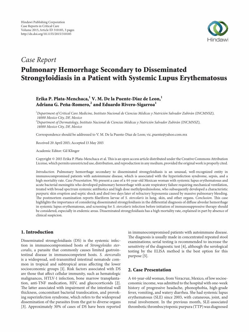
Figure 1: Purpuric dermatosis and skin biopsy. Punctiform periumbilical purpuric macules (a). 24 hours later, dermatosis worsened and wasconfluent with persistence of periumbilical involvement (“Thumb print sign”) (b). Filariform larvae were observed in the dermis (c).
(a) (b) (c)
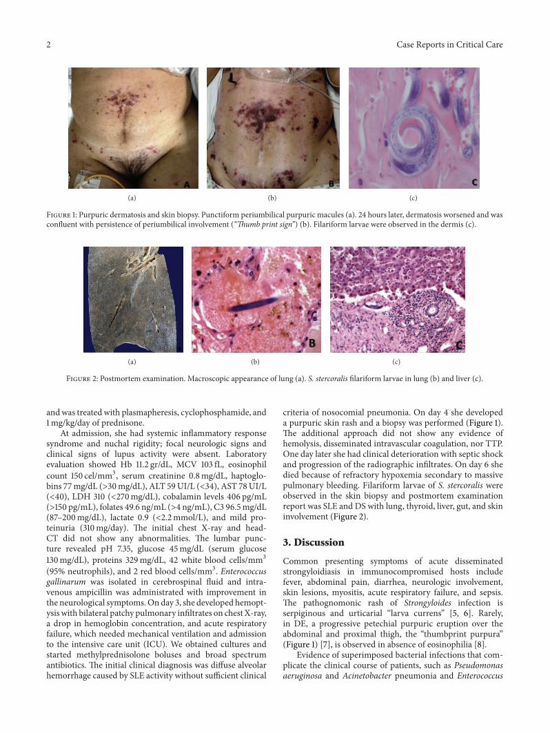
Figure 2: Postmortem examination. Macroscopic appearance of lung (a). S. stercoralis filariform larvae in lung (b) and liver (c).
andwas treatedwith plasmapheresis, cyclophosphamide, and1mg/kg/day of prednisone.
At admission, she had systemic inflammatory responsesyndrome and nuchal rigidity; focal neurologic signs andclinical signs of lupus activity were absent. Laboratoryevaluation showed Hb 11.2 gr/dL, MCV 103 fL, eosinophilcount 150 cel/mm3, serum creatinine 0.8mg/dL, haptoglo-bins 77mg/dL (>30mg/dL), ALT 59UI/L (<34), AST 78UI/L(<40), LDH 310 (<270mg/dL), cobalamin levels 406 pg/mL(>150 pg/mL), folates 49.6 ng/mL (>4 ng/mL), C3 96.5mg/dL(87–200mg/dL), lactate 0.9 (<2.2mmol/L), and mild pro-teinuria (310mg/day). The initial chest X-ray and head-CT did not show any abnormalities. The lumbar punc-ture revealed pH 7.35, glucose 45mg/dL (serum glucose130mg/dL), proteins 329mg/dL, 42 white blood cells/mm3(95% neutrophils), and 2 red blood cells/mm3. Enterococcusgallinarum was isolated in cerebrospinal fluid and intra-venous ampicillin was administrated with improvement inthe neurological symptoms.Onday 3, she developed hemopt-ysis with bilateral patchy pulmonary infiltrates on chest X-ray,a drop in hemoglobin concentration, and acute respiratoryfailure, which needed mechanical ventilation and admissionto the intensive care unit (ICU). We obtained cultures andstarted methylprednisolone boluses and broad spectrumantibiotics. The initial clinical diagnosis was diffuse alveolarhemorrhage caused by SLE activity without sufficient clinical
criteria of nosocomial pneumonia. On day 4 she developeda purpuric skin rash and a biopsy was performed (Figure 1).The additional approach did not show any evidence ofhemolysis, disseminated intravascular coagulation, nor TTP.One day later she had clinical deterioration with septic shockand progression of the radiographic infiltrates. On day 6 shedied because of refractory hypoxemia secondary to massivepulmonary bleeding. Filariform larvae of S. stercoralis wereobserved in the skin biopsy and postmortem examinationreport was SLE and DS with lung, thyroid, liver, gut, and skininvolvement (Figure 2).
3. Discussion
Common presenting symptoms of acute disseminatedstrongyloidiasis in immunocompromised hosts includefever, abdominal pain, diarrhea, neurologic involvement,skin lesions, myositis, acute respiratory failure, and sepsis.The pathognomonic rash of Strongyloides infection isserpiginous and urticarial “larva currens” [5, 6]. Rarely,in DE, a progressive petechial purpuric eruption over theabdominal and proximal thigh, the “thumbprint purpura”(Figure 1) [7], is observed in absence of eosinophilia [8].
Evidence of superimposed bacterial infections that com-plicate the clinical course of patients, such as Pseudomonasaeruginosa and Acinetobacter pneumonia and Enterococcus
Case Reports in Critical Care 3
faecalis meningitis, has been described [8, 9] which oftenprogress to septic shock and are the direct cause of death in80% of cases.
Differential diagnosis of DS includes SLE flare, vasculitis,inflammatory bowel disease [10], other systemic infections,and drug reactions. Treatment of DS has been done withivermectin and albendazole, showing cure rate for immuno-compromised patients of approximately 60%–70% [11–13].
The most common cause of diffuse alveolar hemorrhagein SLE is pulmonary capillaritis, often associated with bacte-rial pneumonia. The mortality rate has been declined sincethe rapid recognition and beginning of aggressive treatmentwith high dose methylprednisolone and cyclophosphamide.Because immunosuppressive therapy may provoke super-infection syndrome, screening for S. stercoralis infectionbefore initiation of immunosuppressive therapy should beconsidered, especially in endemic areas [5–9].
4. Conclusion
Pulmonary hemorrhage secondary to DS is an unusual, well-recognized entity in immunocompromised patients that isassociated with a high mortality rate, explained by diseaseseverity and absence of clinical suspicion. It should be consid-ered in the differential diagnosis of pulmonary hemorrhagein a patient with SLE before initiating steroids and especiallywhen the clinical status worsens rapidly with steroids.
Consent
Written informed consent was obtained from the patient forpublication of this case report and accompanying images.
Conflict of Interests
The authors declare that they have no competing interests.
Authors’ Contribution
Erika P. Plata-Menchaca andV.M.De la Puente-Diaz de Leonanalyzed and interpreted the patient data and were the majorcontributors in writing the paper. V. M. De la Puente-Diazde Leon, Adriana G. Pena-Romero, and Eduardo Rivero-Sigarroa analyzed and interpreted the patient data. Allauthors read and approved the final paper.
Acknowledgments
The authors wish to thank Martınez Braulio M.D., ConsueloRodrıguez M.D., Niembro Dolores M.D., Galindo ArturoM.D., and Ponce de Leon Alfredo M.D.
References
[1] P. B. Keiser and T. B. Nutman, “Strongyloides stercoralis inthe immunocompromised population,” Clinical MicrobiologyReviews, vol. 17, no. 1, pp. 208–217, 2004.
[2] C. G. Wambier, F. B. M. Lemos, M. A. Cappel, F. Bellissimo-Rodrigues, and N. T. Foss, “Generalized serpiginous eruptionduring immunosuppressive treatment for leprosy reactive neu-ritis,” PLoS Neglected Tropical Diseases, vol. 5, no. 12, Article IDe1357, 2011.
[3] M. N. Ly, S. L. Bethel, A. S. Usmani, and D. R. Lambert,“Cutaneous Strongyloides stercoralis infection: an unusual pre-sentation,” Journal of the American Academy of Dermatology,vol. 49, no. 2, pp. S157–S160, 2003.
[4] Y. Sato, J. Kobayashi, H. Toma, and Y. Shiroma, “Efficacy ofstool examination for detection of Strongyloides infection,”TheAmerican Journal of Tropical Medicine and Hygiene, vol. 53, no.3, pp. 248–250, 1995.
[5] T. L. Meinking, C. N. Burkhart, and C. G. Burkhart, “Changingparadigms in parasitic infections: common dermatologicalhelminthic infections and cutaneousmyiasis,”Clinics inDerma-tology, vol. 21, no. 5, pp. 407–416, 2003.
[6] S. L.Mackey andK. F.Wagner, “Dermatologicmanifestations ofparasitic diseases,” Infectious Disease Clinics of North America,vol. 8, no. 3, pp. 713–743, 1994.
[7] R. S. Purvis, E. L. Beightler, D. G. Diven, R. L. Sanchez, and S. K.Tyring, “Strongyloides stercoralis hyperinfection,” InternationalJournal of Dermatology, vol. 31, no. 3, pp. 160–164, 1992.
[8] J. I. F. Salluh, F. A. Bozza, T. S. Pinto, L. Toscano, P. F.Weller, andM. Soares, “Cutaneous periumbilical purpura in disseminatedstrongyloidiasis in cancer patients: a pathognomonic featureof potentially lethal disease?” Brazilian Journal of InfectiousDiseases, vol. 9, no. 5, pp. 419–424, 2005.
[9] C. S. Lam, M. K. H. Tong, K. M. Chan, and Y. P. Siu,“Disseminated strongyloidiasis: a retrospective study of clinicalcourse and outcome,” European Journal of Clinical Microbiologyand Infectious Diseases, vol. 25, no. 1, pp. 14–18, 2006.
[10] G. M. Hayden and S. A. Atlas, “Strongyloidiasis masqueradingas inflammatory bowel disease in a patient with lupus erythe-matosis: a case report,” Connecticut Medicine, vol. 59, no. 11, pp.649–650, 1995.
[11] K.-D. Lessnau, S. Can, andW. Talavera, “Disseminated Strongy-loides stercoralis in human immunodeficiency virus-infectedpatients: treatment failure and a review of the literature,” Chest,vol. 104, no. 1, pp. 119–122, 1993.
[12] N. C. Iriemenam, A. O. Sanyaolu, W. A. Oyibo, and A. F.Fagbenro-Beyioku, “Strongyloides stercoralis and the immuneresponse,” Parasitology International, vol. 59, no. 1, pp. 9–14,2010.
[13] R. Toledo, C.Munoz-Antoli, and J. G. Esteban, “Strongyloidiasiswith emphasis on human infections and its different clinicalforms,” Advances in Parasitology, vol. 88, pp. 165–241, 2015.

Submit your manuscripts athttp://www.hindawi.com
Stem CellsInternational
Hindawi Publishing Corporationhttp://www.hindawi.com Volume 2014
Hindawi Publishing Corporationhttp://www.hindawi.com Volume 2014
MEDIATORSINFLAMMATION
of
Hindawi Publishing Corporationhttp://www.hindawi.com Volume 2014
Behavioural Neurology
EndocrinologyInternational Journal of
Hindawi Publishing Corporationhttp://www.hindawi.com Volume 2014
Hindawi Publishing Corporationhttp://www.hindawi.com Volume 2014
Disease Markers
Hindawi Publishing Corporationhttp://www.hindawi.com Volume 2014
BioMed Research International
OncologyJournal of
Hindawi Publishing Corporationhttp://www.hindawi.com Volume 2014
Hindawi Publishing Corporationhttp://www.hindawi.com Volume 2014
Oxidative Medicine and Cellular Longevity
Hindawi Publishing Corporationhttp://www.hindawi.com Volume 2014
PPAR Research
The Scientific World JournalHindawi Publishing Corporation http://www.hindawi.com Volume 2014
Immunology ResearchHindawi Publishing Corporationhttp://www.hindawi.com Volume 2014
Journal of
ObesityJournal of
Hindawi Publishing Corporationhttp://www.hindawi.com Volume 2014
Hindawi Publishing Corporationhttp://www.hindawi.com Volume 2014
Computational and Mathematical Methods in Medicine
OphthalmologyJournal of
Hindawi Publishing Corporationhttp://www.hindawi.com Volume 2014
Diabetes ResearchJournal of
Hindawi Publishing Corporationhttp://www.hindawi.com Volume 2014
Hindawi Publishing Corporationhttp://www.hindawi.com Volume 2014
Research and TreatmentAIDS
Hindawi Publishing Corporationhttp://www.hindawi.com Volume 2014
Gastroenterology Research and Practice
Hindawi Publishing Corporationhttp://www.hindawi.com Volume 2014
Parkinson’s Disease
Evidence-Based Complementary and Alternative Medicine
Volume 2014Hindawi Publishing Corporationhttp://www.hindawi.com


![Successful anticoagulant therapy for disseminated ......including sepsis, disseminated intravascular coagulation (DIC), massive hemorrhage, and uterine infection [13, 14]. We report](https://img.pdfslide.net/doc/110x75/60f79fd72dadc86c41591dfc/successful-anticoagulant-therapy-for-disseminated-including-sepsis-disseminated.jpg)


