Embed Size (px)
Citation preview
CASE REPORT
Rare association of cutaneous vasculitis, IgA nephro-pathy and antiphospholipid antibody syndrome withtuberculous lymphadenitisRoberto Bueno Filho,I Alberto Pinto Cordeiro,I Flavia Tremeschin de Almeida,II Catarina Shaletich,III Roberto
Silva Costa,III Ana Maria F. RoselinoI
I University of Sao Paulo, School of Medicine of Ribeirao Preto, Department of Medical Clinics, Division of Dermatology, Ribeirao Preto/SP, Brazil.II University of Sao Paulo, School of Medicine of Ribeirao Preto, Department of Medical Clinics, Division of Dermatology, Biomedical, Laboratory of
Molecular Biology, Ribeirao Preto/SP, Brazil. III University of Sao Paulo, School of Medicine of Ribeirao Preto, Hospital of Clinics, Department of Pathology,
Ribeirao Preto/SP, Brazil.
Email: [email protected]
Tel.: 55 16 3602-2447
INTRODUCTION
Mycobacterium tuberculosis infections can be associated withseveral immune mechanisms. Some of these mechanisms, suchas vasculitis associated with tuberculosis, are rare and canchallenge dermatologists when making differential diagnoses.The association between pulmonary tuberculosis (Tb) andvasculitis was first described by Parish and Rhodes (1) in 1967.These associations fall into three main types: pulmonary Tb/cutaneous leukocytoclastic vasculitis (CLV), pulmonary Tb/Henoch-Schonlein purpura (HSP) and pulmonary Tb/vasculi-tis secondary to rifampicin (2-7). The existence of circulatingimmune complexes in pulmonary Tb has been demonstrated,and the levels of these complexes are related to disease activity.The mechanism of vascular damage is attributed to immunecomplexes rather than to direct damage caused by M.tuberculosis (8,9). Another differential diagnosis is CLV due torifampicin therapy (7). In cases of Tb-related vasculitis, skinlesions usually improve following the administration of aspecific Tb treatment; an anti-inflammatory therapy is notrequired (2-5,10). Immune complexes are also responsible forrenal injury, which is associated with increased levels ofimmunoglobulins (mainly IgA against the A-60 antigen of M.tuberculosis), and mesangial deposition, which leads to theactivation of the alternative complement and lecithin pathways,resulting in glomerular damage (IgA nephropathy) (11). Finally,Tb is associated with the development of anti-phospholipidantibody syndrome (APS) by inducing the production of auto-antibodies (18,19). The association of tuberculous lymphadeni-tis, CLV, IgA nephropathy and APS in a single patient has notbeen reported yet.
CASE DESCRIPTION
A 45-year-old woman from Ribeirao Preto (northeasternregion of Sao Paulo State, Brazil) presented to our clinic withpainful necrotic lesions on both feet, mainly on the toes,
which had recently increased in number and size. She had ahistory of headaches and seizures, an ischemic stroke fiveyears earlier (resulting in facial motor sequelae) and fivepregnancies, which consisted of four normal deliveries andone abortion at 22 years of age. On examination, there werecervical adenomegalies with bulky and coalescing lymphnodes (the largest measuring 3 cm in diameter) and crustedlesions on the dorsal feet and tips of the toes with purulentexudates and interdigital maceration (Figure 1). The periph-eral sensitivity test yielded normal results. Laboratory testsshowed hypochromic anemia with microcytosis (hemoglo-bin: 11.0 g/dL; NR: 12.0-15.5 g/dL), increased inflammatoryactivity (ESR: 30 mm/1st hour; NR: ,10 mm/1st hour;C-reactive protein: 3.46 mg/dL; NR: up to 0.5 mg/dL;alpha1-acid glycoprotein: 156 mg/dL; NR: 50-120 mg/dL),increased levels of immunoglobulins (IgA: 1,070 mg/dL; NR:134-297 mg/dl; IgG: 1,900 mg/dL; NR: 770-1,510 mg/dl;IgM: 222 mg/dL; NR: 67-208 mg/dl), positive autoantibo-dies (antinucleolar antibody (ANA)-positive 1:100 withnucleolar pattern; lupus anticoagulant (PIL and dRVVT)-positive; anticardiolipin IgG: 14.9 GPL/mL (weak positive);NR: up to 14.0 GPL/ml; p-ANCA-positive), urinary dis-orders (microscopic hematuria: 25-30 RBCs/field; NR: 3-5 RBCs/field; urinary erythrocytes: 90% total dysmorphiccells, 19% acanthocytes; NR: up 4% acanthocytes; proteinuria:255 mg/24 h; NR: up to 150 mg/24 h), and intradermalreaction test positive for Tb (PPD: 45 mm with necrosis).Cervical, thoracic and abdominal computed tomographiesshowed cervical adenomegaly (up to 1.8 cm in length) withcentral necrosis, a left axillary 2.3-cm lymph node, andseveral retroperitoneal lymph nodes (up to 0.9 cm in length).A cranial MRI showed a cerebral infarction on the left parietalregion and lacunar infarctions in the region of capsularnuclei. Histopathology showed the following: (1) cervicallymph node - chronic granulomatous lymphadenitis withcaseous necrosis; (2) fifth left toe - focal granulomatous inaddition to leukocytoclastic vasculitis and direct immuno-fluorescence (DIF) strongly positive for anti-fibrinogen serum(3+) on capillary walls; and (3) renal biopsy - focal andsegmental sclerosis with mild focal and chronic tubulointer-stitial damage, characterized by mesangial deposition of IgAon DIF, which may correspond to primary IgA nephropathy(Berger’s Disease) or Henoch-Schonlein purpura (Figure 2).
Copyright � 2012 CLINICS – This is an Open Access article distributed underthe terms of the Creative Commons Attribution Non-Commercial License (http://creativecommons.org/licenses/by-nc/3.0/) which permits unrestricted non-commercial use, distribution, and reproduction in any medium, provided theoriginal work is properly cited.
No potential conflict of interest was reported.
CLINICS 2012;67(12):1497-1500 DOI:10.6061/clinics/2012(12)24
1497
PCR with DNA that was extracted from paraffin blocks oflymph node and skin biopsies confirmed Mycobacterium sp.only in the lymph node (Figure 3). The final diagnosisconsisted of Tb lymphadenitis, CLV, primary IgA nephro-pathy and APS. Ciprofloxacin (500 mg) was prescribedtwice per day for ten days, and Fluconazole (150 mg) wasprescribed once per week for eight weeks for the secondaryinfection and interdigital maceration on the feet.Thereafter, aspirin (200 mg daily) and warfarin (for antic-oagulation) were prescribed. The patient was evaluatedby physicians in the Departments of Neurology,Ophthalmology, Nephrology and Infectious Diseases. Tbtreatment was initiated with RIPE (rifampicin, isoniazid,pyrazinamide and ethambutol). Because an adverse drugreaction to pyrazinamide was observed, administration ofthis medication was suspended. Because of the worseningnecrosis on her toes, the patient’s left fifth toe wasamputated with no complications. The patient finishedthe Tb treatment, which was followed by weight recovery,
normalization of lymph node size, absence of newvasculitic lesions and significant improvement of previousskin lesions. ANA and PIL were negative, but glomerularhematuria and proteinuria remained positive.
DISCUSSION
There are few reported cases of an association between Tband CLV (nine cases) and Tb and IgA nephropathy (sixcases) (2-6,13,14). Only 10% of CLV cases are attributed to
Figure 1 - Clinical presentation of a patient with cutaneousvasculitis. The upper figures show progressive necrotic lesions onthe dorsal feet, mainly on the toes. The lower figures show theamputation of the left fifth toe and the improvement ofvasculitic skin lesions following RIPE treatment.
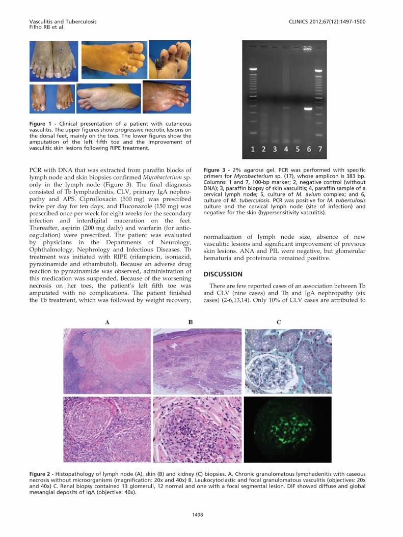
Figure 2 - Histopathology of lymph node (A), skin (B) and kidney (C) biopsies. A. Chronic granulomatous lymphadenitis with caseousnecrosis without microorganisms (magnification: 20x and 40x) B. Leukocytoclastic and focal granulomatous vasculitis (objectives: 20xand 40x) C. Renal biopsy contained 13 glomeruli, 12 normal and one with a focal segmental lesion. DIF showed diffuse and globalmesangial deposits of IgA (objective: 40x).
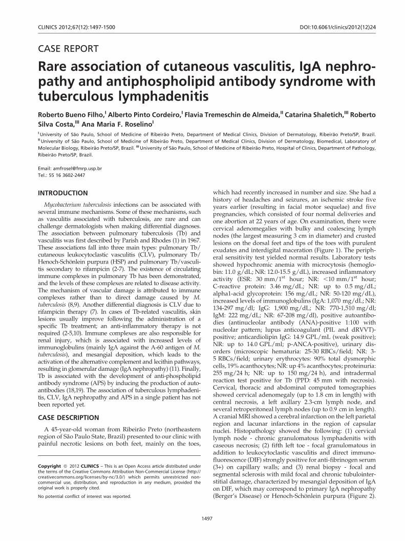
Figure 3 - 2% agarose gel. PCR was performed with specificprimers for Mycobacterium sp. (17), whose amplicon is 383 bp.Columns: 1 and 7, 100-bp marker; 2, negative control (withoutDNA); 3, paraffin biopsy of skin vasculitis; 4, paraffin sample of acervical lymph node; 5, culture of M. avium complex; and 6,culture of M. tuberculosis. PCR was positive for M. tuberculosisculture and the cervical lymph node (site of infection) andnegative for the skin (hypersensitivity vasculitis).
Vasculitis and TuberculosisFilho RB et al.
CLINICS 2012;67(12):1497-1500
1498
drugs and infections, and 61% are considered idiopathiccases. Considering the existence of circulating immunecomplexes in pulmonary Tb and the relationship betweenimmune complex levels and disease activity, the damagemechanism that has been proposed for this type ofvasculitis is the deposition of immune complexes that areformed by antibodies against antigens of the bacterium onthe vascular wall rather than direct aggression by thebacterium (8,9). Up to 56% of Tb patients have circulatingimmune complexes, and there is evidence of increasedimmunoglobulin levels in these patients, mainly IgA andIgG, as was observed in our patient. These immunoglobu-lins are produced against the A-60 antigen of themycobacterium, leading to the formation of immunecomplexes (11).
Another differential diagnosis is the onset of vasculitisfollowing rifampicin therapy, as demonstrated in some casereports, where CLV occurred due to this medication (7).However, our patient had skin lesions before using thisdrug. In cases of Tb-related vasculitis, skin lesions improvewith RIPE treatment alone; no specific anti-inflammatorytherapy is required (2-7,10,15).
In 1985, Cohen and Rosenstein (12) described a case of anassociation between Tb and IgA nephropathy in which renalinvolvement improved after Tb treatment. In this case, HSPneeded to be considered. HSP is a type of systemic vasculitisthat is more common in children, and both renal involve-ment, defined as IgA nephropathy, and palpable purpuraappear in 50% of cases (6,13,14). Some specialists defend thetheory that Berger’s disease is a restricted form of HSP, andthere have been five reported cases of HSP associated withTb. However, our patient presented with CLV with anti-fibrinogen, not IgA, deposition in the capillary walls, whichrules out a diagnosis of HSP.
In addition, PCR with primers specific to Mycobacterium(17) was performed in cervical lymph node and skinsamples, which confirmed the etiology of Mycobacteriumsp. in the lymph node sample but not in the skin sample.The DNA that was extracted from the paraffin skin samplewas amplified with keratin primers, confirming its integrity(data not shown). These results strongly suggest a hyper-sensitivity form of CLV.
None of the case reports of Tb associated with CLV or Tbassociated with IgA nephropathy in the literature includeddescriptions of positive autoantibodies. In our patient,ANA, p-ANCA and lupus anticoagulant (PIL and dRVVT)tests were positive, and there was a prior medical history ofischemic stroke (confirmed by MRI). These findingsreinforce the diagnosis of an autoimmune disease and leadus to a diagnosis of APS secondary to Tb.
The production of anti-phospholipid antibodies (aPLs)could have either an autoimmune or infectious origin. Thelatter origin does not involve anti-b2-glycoprotein I (anti-b2GPI) activity and usually does not cause thrombosis.However, there have been recently described cases oflepromatous leprosy patients with genetically determinedanti-b2GPI activity followed by thrombosis (18). By indu-cing a specific cellular immune response and secondaryantibody production (as noted by the strong positive PPDand increased immunoglobulin levels), tuberculosis stimu-lates the production of autoantibodies (including aPLs) andprocoagulant factors, such as plasma fibrinogen (factor I),factor VIII and D-dimers. Thus, M. tuberculosis can be
considered an etiologic factor in aPL production and thetriggering of APS (19).
In the literature, there is a single report of concomitantCLV, Sweet syndrome, cutaneous polyarteritis nodosa andcervical adenopathy caused by M. fortuitum (20). Theassociation of Tb, focal granulomatous, CLV and IgAnephropathy in a single patient has not been reported. Thiscase is complex, demonstrating rare manifestations of anendemic disease in Brazil, and serves as a warning todermatologists to be cautious in the differential diagnosisof patients with vasculitic presentations.
AUTHOR CONTRIBUTIONS
Bueno Filho R contributed to the writing, literature review, data
interpretation, and data and picture collection and was clinically responsible
for the patient. Cordeiro AP contributed to the collection of pictures and
data and was clinically responsible for the patient. Almeida FT was involved
in data collection and was responsible for the PCR experiment. Shaletich C
was involved in the review and data interpretation processes. Costa RS was
involved in the review, data interpretation, and writing processes. Roselino
AM was involved in the review, writing, and data interpretation processes
and was the coordinator of the Granulomatous Skin Diseases Clinic and
coordinator of the Biomolecular Laboratory of Dermatology.
REFERENCES
1. Parish WE, Rhodes EL. Bacterial antigens and aggregated gammaglobulin in the lesions of nodular vasculitis. Br J Dermatol.1967;79(3):131-47.
2. Ekenstam E, Callen JP. Cutaneous leukocytoclastic vasculitis. Clinical andlaboratory features of 82 patients seen in private practice. Arch Dermatol.1984;120(4):484-9, http://dx.doi.org/10.1001/archderm.1984.01650400066014.
3. Kim HM, Park YB, Maeng HY, Lee SK. Cutaneous leukocytoclasticvasculitis with cervical tuberculous lymphadenitis: a case report andliterature review. Rheumatol Int. 2006;26(12):1154-7, http://dx.doi.org/10.1007/s00296-006-0152-1.
4. Sais G, Vidaller A, Jucgla A, Peyrı J. Tuberculous lymphadenitis presentingwith cutaneous leucocytoclastic vasculitis. Clin Exp Dermatol.1996;21(1):65-6, http://dx.doi.org/10.1111/j.1365-2230.1996.tb00018.x.
5. Carvalho M, Dominoni RL, Senchechen D, Fernandes AF, Burigo IP,Doubrawa E. Cutaneous leukocytoclastic vasculitis accompanied bypulmonary tuberculosis. J Bras Pneumol. 2008;34(9):745-8.
6. Han BG, Choi SO, Shin SJ, Kim HY, Jung SH, Lee KH. A case of Henoch-Schonlein purpura in disseminated tuberculosis. Korean J Intern Med.1995;10(1):54-9.
7. Iredale JP, Sankaran R, Wathen CG. Cutaneous vasculitis associated withrifampicin therapy. Chest. 1989;96(1):215-6, http://dx.doi.org/10.1378/chest.96.1.215.
8. Johnson NMcI, McNicol MW, Burton Kee EJ, Mowbray JF. Circulatingimmune complexes in tuberculosis. Thorax. 1981;36(8):610-7, http://dx.doi.org/10.1136/thx.36.8.610.
9. Brostoff J. Immune complexes in the spectrum of tuberculosis. Tubercle.1981;62(3):169-73, http://dx.doi.org/10.1016/0041-3879(81)90002-7.
10. Chan CH, Chong YW, Sun AJ, Hoheisel GB. Cutaneous vasculitis associatedwith tuberculosis and its treatment. Tubercle. 1990;71(4):297-300.
11. Alifano M, Sofia M, Mormile M, Micco A, Mormile AF, Del Pezzo M,et al. IgA immune response against the mycobacterial antigen A60 inpatients with active pulmonary tuberculosis. Respiration. 1996;63(5):292-7, http://dx.doi.org/10.1159/000196563.
12. Berger J. IgA glomerular deposits in renal disease. Transplant Proc.1969;1(4):939-44.
13. De Siati L, Paroli M, Ferri C, Muda AO, Bruno G, Barnaba V. IgANephropathy and Pulmonary Tuberculosis. Ann Diagn Pathol. 1999;3(5):300-3, http://dx.doi.org/10.1016/S1092-9134(99)80026-4.
14. Cohen AJ, Rosenstein ED. IgA nephropathy associated with dissemi-nated tuberculosis. Arch Intern Med 1985;145(3):554-6, http://dx.doi.org/10.1001/archinte.1985.00360030206036.
15. Jennette JC, Falk RJ. Small vessels vasculitis. N Engl J Med.1997;337(21):1512-23.
16. Singh SP, Misra GC, Prusty PK, Das RK. Tubercular lymphadenitis withpurpura. J Indian Med Assoc 1986;84(8):247-9.
17. Brisson-Noel A, Aznar C, Chureau C, Nguyen S, Pierre C, Bartoli M, et al.Diagnosis of tuberculosis by DNA amplification in clinical practice
CLINICS 2012;67(12):1497-1500 Vasculitis and TuberculosisFilho RB et al.
1499

evaluation. Lancet 1991;338(8763):364-6, http://dx.doi.org/10.1016/0140-6736(91)90492-8.
18. Brochado MJ, Figueiredo JF, Mendes-Junior CT, Louzada-Junior P, KimOM, Roselino AM. Correlation between beta-2-glycoprotein I genepolymorphism and anti-beta-2 glycoprotein I antibodies in patients withmultibacillary leprosy. Arch Dermatol Res. 2010;302(8):583-91, http://dx.doi.org/10.1007/s00403-010-1032-9.
19. Naithani R, Agrawal N, Choudhary VP. Deep venous thrombosisassociated with tuberculosis. Blood Coagul Fibrinolysis. 2007;18(4):377-80, http://dx.doi.org/10.1097/MBC.0b013e3280d942b4.
20. Chen HH, Hsiao CH, Chiu HC. Successive development of cutaneouspolyarteritis nodosa, leucocytoclastic vasculitis and Sweet’s syndrome in apatient with cervical lymphadenitis caused by Mycobacterium fortuitum.Br J Dermatol. 2004;151(5):1096-100.
Vasculitis and TuberculosisFilho RB et al.
CLINICS 2012;67(12):1497-1500
1500