Embed Size (px)
Citation preview
Hindawi Publishing CorporationCase Reports in DentistryVolume 2013, Article ID 930345, 5 pageshttp://dx.doi.org/10.1155/2013/930345
Case ReportRehabilitation of Posterior Maxilla with Zygomatic andDental Implant after Tumor Resection: A Case Report
Faysal Ugurlu, Coskun YJldJz, B. C. Sener, and Atilla Sertgoz
Department of Oral and Maxillofacial Surgery, Dentistry Faculty, Marmara University, 34365 Istanbul, Turkey
Correspondence should be addressed to Faysal Ugurlu; [email protected]
Received 18 January 2013; Accepted 5 February 2013
Academic Editors: R. S. Brown, D. Cogulu, and R. A. de Mesquita
Copyright © 2013 Faysal Ugurlu et al. This is an open access article distributed under the Creative Commons Attribution License,which permits unrestricted use, distribution, and reproduction in any medium, provided the original work is properly cited.
Zygomatic implants have been used for dental rehabilitation in patients with insufficient bone in the posterior upper jaw, due to, forexample, tumor resection, trauma, or atrophy. Zygomatic implants are an alternative to complex free or vascularized bone graftingand distraction osteogenesis. A 42-year-old male patient with a severe defect in the right posterior maxilla, starting from the firstcanine region, which had occurred after tumor resection 3 years earlier, was referred to our department. One zygomatic implant(Brenemark System, Nobel Biocare, Goteborg, Sweden) to the zygoma and one dental implant to the canine region were placed.After a 5-month osseointegration period, a fixed denture was fabricated and adapted to the implants. Although the surgical andprosthetic procedures for zygoma implants are not easy, the final outcomes can be successful with appropriate planning.
1. Introduction
Maxillary posterior defects that occur after tumor resectionor trauma are challenging to reconstruct and rehabilitate.Theaim of rehabilitation is not only to provide a cosmeticallyacceptable appearance, but also to restore oral functions, suchas deglutition, mastication, and phonation. To provide betterfunction, an implant-supported prosthesis can be fabricated.However, implant placement in the zygoma and pterygoidbone is difficult due to the variable anatomy and varyingdegrees of atrophy possible in the maxillofacial region [1, 2].The surgery is not without risk because the drill path is closeto critical anatomical structures, such as the maxillary sinus,the nasal cavity, and the eyes [2, 3]. A small angular errormayresult in significant positional errors at the end of the tooltrajectory. Furthermore, the limited intraoperative visibility,especially given the anatomical intricacies of the curvedzygomatic bone, makes this kind of surgery a demandingprocedure.
In recent years, preoperative 3D planning of implanttreatment that takes into account both the quality and quan-tity of the jawbone and prosthodontic considerations has hada major influence on final implant treatment outcome. Inparticular, before zygoma implant placement, 3D imaging
and planning should be performed to ensure the safety ofsurgery and rehabilitation. This case study describes theclinical management of prosthetic rehabilitation of a patientwho underwent zygoma implant placement.
2. Case Report
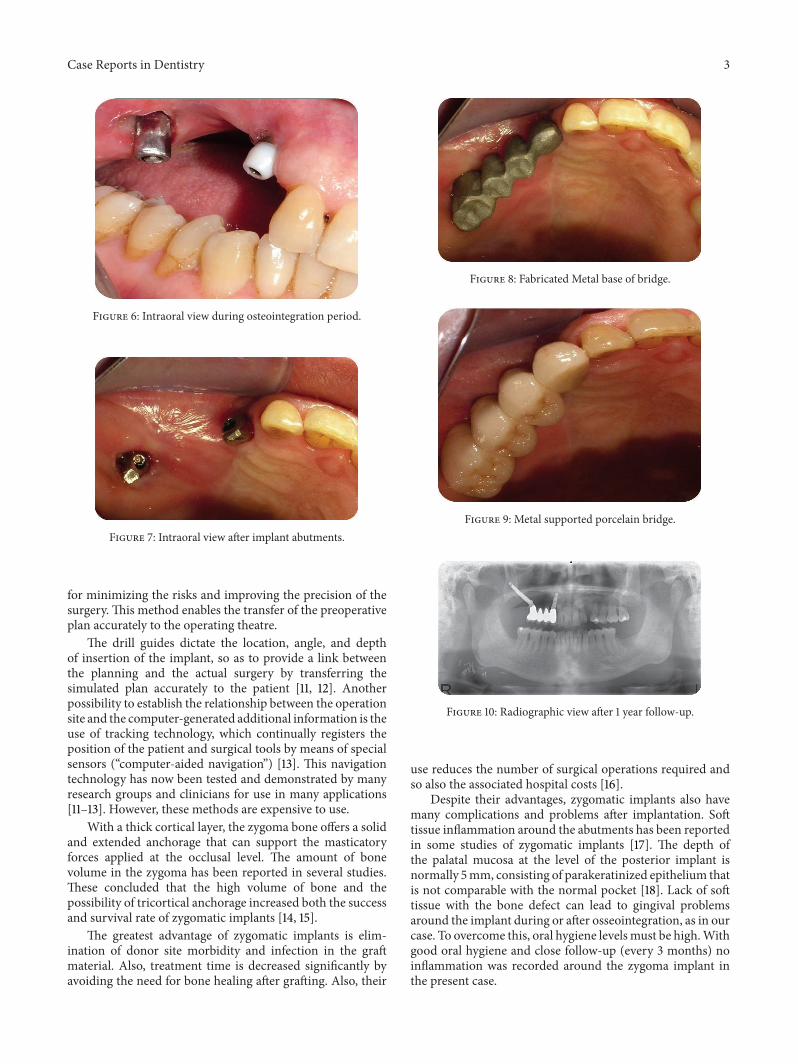
A 42-year-old male patient was referred to our departmentwith functional and esthetic issues. His anamnesis revealedthat tumor resection had been performed to his rightposterior maxilla 3 years earlier. His clinical examinationrevealed a severe defect in the right posteriormaxilla, startingfrom the canine region (Figure 1). To be able to make animplant-supported fixed prosthesis, a zygoma implant wasplanned for the posterior maxilla, due to the huge bonedefect in that area, and one dental implant was planned(Figures 2 and 3). The patient was evaluated preoperativelywith respect to jaw size, bone volume, bone density, jawrelationships, intermaxillary distance, occlusal relation, andcondition of the opposing dentition. Preoperative analysis ofthe anatomical conditions and possible maxillary pathologywas evaluated using panoramic radiographs and a cone-beamCT scan.
The operation was performed under intravenous seda-tion. A crestal incision was made and a mucoperiosteal flap
2 Case Reports in Dentistry
Figure 1: Preoperative intraoral view.
Figure 2: 3D view of defect.
was elevated up to the zygomatic buttress (Figure 4). Thezygoma implant was drilled and placed to the posterior; onedental implant was placed in the premolar region (Figure 5).The soft tissueswere readapted and sutured back into positionwith silk sutures. An antibiotic (clindamycin, 600mg/day)was prescribed for 10 days postoperatively. The remainingsutures were removed after 10 days.
Permanent prosthetic rehabilitation was initiated 5months after implant placement (Figure 6). Metal impressioncopings were screwed onto the implants after removal of thehealing screws. An impression was taken using an open-tray technique. Metal-supported porcelain restorations wereconstructed using conventional methods (Figures 7 and 8).
The patient was followed up at 5 years after the prostheticrehabilitation. The last clinical and radiographic assessmentswere uneventful, and the patient’s satisfaction with theesthetic result was excellent (Figures 9 and 10).
3. Discussion
Reconstruction of posterior maxillary defects is a challenge.Many procedures, such as onlay grafts [4], free or microvas-cular bone grafts [5, 6], transport distraction osteogenesis [7],and apposition grafts with or without a Le Fort I osteotomy[8] are well documented and have success rates of 60–90%.However, these often involve invasive and lengthy surgeries,long treatment time, and some morbidity. Microvascular
Figure 3: Preoperative simulation of implant planning.
Figure 4: Intraoperative view of zygoma after flap elevation.
Figure 5: Implant placement of zygomatic implant.
bone grafts are complex and risky operations. In addition,these conventional techniques cause donor site morbidityduring harvesting of soft tissues and bone grafts [4]. Fur-thermore, free bone grafts are commonly associated withunpredictable resorption during healing [4, 5, 9].
Zygomatic implants, an alternative to these techniques,have survival rates of 98–100% [10]. Although no randomizedclinical trial that compared sinus grafts or onlay grafts andzygomatic implants has been reported, the survival ratessuggest that zygomatic implants should be considered analternative to bone grafting and applied in routine rehabili-tation of the totally edentulous patient with high resorptionof the maxilla and severe defects in the posterior maxillaafter tumor resection. However, the placement of zygomaimplants is not without risk, due to the anatomically complexoperation site. Implant-guided surgery is a suitable method
Case Reports in Dentistry 3
Figure 6: Intraoral view during osteointegration period.
Figure 7: Intraoral view after implant abutments.
for minimizing the risks and improving the precision of thesurgery. This method enables the transfer of the preoperativeplan accurately to the operating theatre.
The drill guides dictate the location, angle, and depthof insertion of the implant, so as to provide a link betweenthe planning and the actual surgery by transferring thesimulated plan accurately to the patient [11, 12]. Anotherpossibility to establish the relationship between the operationsite and the computer-generated additional information is theuse of tracking technology, which continually registers theposition of the patient and surgical tools by means of specialsensors (“computer-aided navigation”) [13]. This navigationtechnology has now been tested and demonstrated by manyresearch groups and clinicians for use in many applications[11–13]. However, these methods are expensive to use.
With a thick cortical layer, the zygoma bone offers a solidand extended anchorage that can support the masticatoryforces applied at the occlusal level. The amount of bonevolume in the zygoma has been reported in several studies.These concluded that the high volume of bone and thepossibility of tricortical anchorage increased both the successand survival rate of zygomatic implants [14, 15].
The greatest advantage of zygomatic implants is elim-ination of donor site morbidity and infection in the graftmaterial. Also, treatment time is decreased significantly byavoiding the need for bone healing after grafting. Also, their
Figure 8: Fabricated Metal base of bridge.
Figure 9: Metal supported porcelain bridge.
Figure 10: Radiographic view after 1 year follow-up.
use reduces the number of surgical operations required andso also the associated hospital costs [16].
Despite their advantages, zygomatic implants also havemany complications and problems after implantation. Softtissue inflammation around the abutments has been reportedin some studies of zygomatic implants [17]. The depth ofthe palatal mucosa at the level of the posterior implant isnormally 5mm, consisting of parakeratinized epithelium thatis not comparable with the normal pocket [18]. Lack of softtissue with the bone defect can lead to gingival problemsaround the implant during or after osseointegration, as in ourcase. To overcome this, oral hygiene levelsmust be high.Withgood oral hygiene and close follow-up (every 3 months) noinflammation was recorded around the zygoma implant inthe present case.
4 Case Reports in Dentistry
Sinusitis has been reported by a number of authors [19,20]. The incidence was 14–30%. In some cases, patients withan oroantral fistula may develop suppuration with or withoutsinusitis. Treatment involves the administration of antibioticsand/or repositioning soft tissue, without removal of the stablezygomatic implant. Causes of sinusitis include perforation ofthe sinus membrane and leakage at the level of the maxilladue to a hole in the zygomatic implant, leading to migrationof bacteria from the mouth to the sinus [16]. In the presentcase, no sinusitis or oroantral fistula occurred.
The zygomatic implant-supported prosthesis requiresspecial care due to the biomechanical forces that affectthe long-term stability of an implant-supported restoration.Use of a stiff prosthesis is necessary because flexing of thematerials can cause deformation and deviation, resulting inloss of implant or loosening of the junction between theprosthesis and fixation. The success rates of implants in thezygomatic bone vary from 95% to 97% with 12–124 monthsof follow-up observation [21, 22], and the patient satisfactionrate is 80% 1 year after installation of the prosthesis [23].
4. Conclusions
Zygoma implants are a unique alternative for rehabilitationof the posterior maxilla after tumor resection or trauma. Dueto both the anatomical intricacies of the zygomatic bone andthe implant length, the placement of zygoma implants stillrepresents a challenge to prosthodontists. To minimize therisks of surgery, 3D reconstruction, preoperative planning,registration, surgical implant guidance, and a motion track-ing algorithm should be used.
References
[1] L. Vrielinck, C. Politis, S. Schepers, M. Pauwels, and I. Naert,“Image-based planning and clinical validation of zygoma andpterygoid implant placement in patients with severe boneatrophy using customized drill guides. Preliminary results froma prospective clinical follow-up study,” International Journal ofOral and Maxillofacial Surgery, vol. 32, no. 1, pp. 7–14, 2003.
[2] Y. Uchida, M. Goto, T. Katsuki, and T. Akiyoshi, “Measurementof the maxilla and zygoma as an aid in installing zygomaticimplants,” Journal of Oral and Maxillofacial Surgery, vol. 59, no.10, pp. 1193–1198, 2001.
[3] S. M. Parel, P. I. Branemark, L. O. Ohrnell, and B. Svensson,“Remote implant anchorage for the rehabilitation of maxillarydefects,” Journal of Prosthetic Dentistry, vol. 86, no. 4, pp. 377–381, 2001.
[4] J. Clavero and S. Lundgren, “Ramus or chin grafts for maxillarysinus inlay and local onlay augmentation: comparison of donorsite morbidity and complications,” Clinical Implant Dentistryand Related Research, vol. 5, no. 3, pp. 154–160, 2003.
[5] M. J. Yaremchuk, “Vascularized bone grafts for maxillofacialreconstruction,” Clinics in Plastic Surgery, vol. 16, pp. 29–39,1989.
[6] M. Sjostrom, L. Sennerby, H. Nilson, and S. Lundgren, “Recon-struction of the atrophic edentulous maxilla with free iliac crestgrafts and implants: a 3-year report of a prospective clinicalstudy,” Clinical Implant Dentistry and Related Research, vol. 9,no. 1, pp. 46–59, 2007.
[7] L. K. Cheung, Q. Zhang, Z. G. Zhang, and M. C. M. Wong,“Reconstruction of maxillectomy defect by transport distrac-tion osteogenesis,” International Journal of Oral and Maxillofa-cial Surgery, vol. 32, no. 5, pp. 515–522, 2003.
[8] E. Nystrom, H. Nilson, J. Gunne, and S. Lundgren, “Recon-struction of the atrophic maxilla with interpositional bonegrafting/Le Fort I osteotomy and endosteal implants: a 11–16year follow-up,” International Journal of Oral and MaxillofacialSurgery, vol. 38, no. 1, pp. 1–6, 2009.
[9] R. G. Triplett and S. R. Schow, “Autologous bone grafts andendosseous implants: complementary techniques,” Journal ofOral andMaxillofacial Surgery, vol. 54, no. 4, pp. 486–494, 1996.
[10] M. Del Fabbro, T. Testori, L. Francetti, and R. Weinstein,“Systematic review of survival rates for implants placed in thegrafted maxillary sinus,” International Journal of Periodonticsand Restorative Dentistry, vol. 24, no. 6, pp. 565–577, 2004.
[11] L. Vrielinck, C. Politis, S. Schepers, M. Pauwels, and I. Naert,“Image-based planning and clinical validation of zygoma andpterygoid implant placement in patients with severe boneatrophy using customized drill guides. Preliminary results froma prospective clinical follow-up study,” International Journal ofOral and Maxillofacial Surgery, vol. 32, no. 1, pp. 7–14, 2003.
[12] K. Lal, G. S. White, D. N. Morea, and R. F. Wright, “Useof stereolithographic templates for surgical and prosthodonticimplant planning and placement. Part I. The concept,” Journalof Prosthodontics, vol. 15, no. 1, pp. 51–58, 2006.
[13] R. Ewers, K. Schicho, G. Undt et al., “Basic research and 12 yearsof clinical experience in computer-assisted navigation technol-ogy: a review,” International Journal of Oral and MaxillofacialSurgery, vol. 34, no. 1, pp. 1–8, 2005.
[14] L. R. Duarte, H. N. Filho, C. E. Francischone, L. G. Peredo, andP. I. Branemark, “The establishment of a protocol for the totalrehabilitation of atrophic maxillae employing four zygomaticfixtures in an immediate loading system—a 30-month clinicaland radiographic follow-up,” Clinical Implant Dentistry andRelated Research, vol. 9, no. 4, pp. 186–196, 2007.
[15] P. Malo, M. de Araujo Nobre, and I. Lopes, “A new approach torehabilitate the severely atrophic maxilla using extramaxillaryanchored implants in immediate function: a pilot study,” Journalof Prosthetic Dentistry, vol. 100, no. 5, pp. 354–366, 2008.
[16] M. Stievenart and C. Malevez, “Rehabilitation of totally atro-phied maxilla by means of four zygomatic implants and fixedprosthesis: a 6-40-month follow-up,” International Journal ofOral and Maxillofacial Surgery, vol. 39, pp. 358–363, 2010.
[17] B. Al-Nawas, J. Wegener, C. Bender, and W. Wagner, “Criticalsoft tissue parameters of the zygomatic implant,” Journal ofClinical Periodontology, vol. 31, no. 7, pp. 497–500, 2004.
[18] M. Stievenart and C. Malevez, “Rehabilitation of totally atro-phied maxilla by means of four zygomatic implants and fixedprosthesis: a 6-40-month follow-up,” International Journal ofOral and Maxillofacial Surgery, vol. 39, pp. 358–363, 2010.
[19] J. P. Becktor, S. Isaksson, P. Abrahamsson, and L. Sennerby,“Evaluation of 31 zygomatic implants and 74 regular dentalimplants used in 16 patients for prosthetic reconstruction of theatrophic maxilla with cross-arch fixed bridges,” Clinical ImplantDentistry and Related Research, vol. 7, no. 3, pp. 159–165, 2005.
[20] C. Malevez, M. Abarca, F. Durdu, and P. Daelemans, “Clinicaloutcome of 103 consecutive zygomatic implants: a 6-48 monthsfollow-up study,” Clinical Oral Implants Research, vol. 15, no. 1,pp. 18–22, 2004.
[21] J. P. Urgell, V. R. Gutierrez, and C. G. Escoda, “Rehabilitation ofatrophic maxilla: a review of 101 zygomatic implants,”Medicina

Case Reports in Dentistry 5
Oral, Patologia Oral y Cirugia Bucal, vol. 13, no. 6, Article ID10489698, pp. E363–E370, 2008.
[22] C. A. Landes, “Zygoma implant-supported midfacial prostheticrehabilitation: a 4-year follow-up study including assessment ofquality of life,” Clinical Oral Implants Research, vol. 16, no. 3, pp.313–325, 2005.
[23] M. Penarrocha, C. Carrillo, A. Boronat, and E. Martı, “Level ofsatisfaction in patients withmaxillary full-arch fixed prostheses:zygomatic versus conventional implants,” International Journalof Oral and Maxillofacial Implants, vol. 22, no. 5, pp. 769–773,2007.

Submit your manuscripts athttp://www.hindawi.com
Hindawi Publishing Corporationhttp://www.hindawi.com Volume 2014
Oral OncologyJournal of
DentistryInternational Journal of
Hindawi Publishing Corporationhttp://www.hindawi.com Volume 2014
Hindawi Publishing Corporationhttp://www.hindawi.com Volume 2014
International Journal of
Biomaterials
Hindawi Publishing Corporationhttp://www.hindawi.com Volume 2014
BioMed Research International
Hindawi Publishing Corporationhttp://www.hindawi.com Volume 2014
Case Reports in Dentistry
Hindawi Publishing Corporationhttp://www.hindawi.com Volume 2014
Oral ImplantsJournal of
Hindawi Publishing Corporationhttp://www.hindawi.com Volume 2014
Anesthesiology Research and Practice
Hindawi Publishing Corporationhttp://www.hindawi.com Volume 2014
Radiology Research and Practice
Environmental and Public Health
Journal of
Hindawi Publishing Corporationhttp://www.hindawi.com Volume 2014
The Scientific World JournalHindawi Publishing Corporation http://www.hindawi.com Volume 2014
Hindawi Publishing Corporationhttp://www.hindawi.com Volume 2014
Dental SurgeryJournal of
Drug DeliveryJournal of
Hindawi Publishing Corporationhttp://www.hindawi.com Volume 2014
Hindawi Publishing Corporationhttp://www.hindawi.com Volume 2014
Oral DiseasesJournal of
Hindawi Publishing Corporationhttp://www.hindawi.com Volume 2014
Computational and Mathematical Methods in Medicine
ScientificaHindawi Publishing Corporationhttp://www.hindawi.com Volume 2014
PainResearch and TreatmentHindawi Publishing Corporationhttp://www.hindawi.com Volume 2014
Preventive MedicineAdvances in
Hindawi Publishing Corporationhttp://www.hindawi.com Volume 2014
EndocrinologyInternational Journal of
Hindawi Publishing Corporationhttp://www.hindawi.com Volume 2014
Hindawi Publishing Corporationhttp://www.hindawi.com Volume 2014
OrthopedicsAdvances in