Embed Size (px)
Citation preview
Journal of Krishna Institute of Medical Sciences University
JKIMSU, Vol. 3, No. 2, July-Dec 2014
CASE REPORT
ISSN 2231-4261
ÓÓ
Abstract:Serous Microcystic Adenoma of Pancreas (SMCA) is a rare tumour of exocrine pancreas. It has a striking predilection for elderly women and consists of small cysts surrounding central stellate scar. Histologically cysts are lined by glycogen rich cuboidal epithelium. In view of its benign nature and excellent prognosis this tumour needs to be accurately diagnosed. This report documents two cases of SMCA occurring in 45 and 36 years old females.
Keywords: Pancreatic Exocrine Tumour, Serous Microcystic Adenoma
Introduction:Serous Microcystic Adenoma of Pancreas (SMCA) is a cystic epithelial tumour composed of glycogen rich ductal type epithelial cells which produce watery fluid similar to serum. Most are benign occurring in elderly women. Synonyms include microcystic adenoma and glycogen rich cystadenoma [1]. It is uncommon pancreatic neoplasm with an incidence of 1 -2 % of all pancreatic tumours [2]. It occupies a unique place among cystic tumours of pancreas because it has little or no malignant potential [3]. With the advent of improvement in CT and USG, the number of SMCA lesions is being recognized with increas-ing frequency [2, 3].
Case Report:Herein we are presenting two cases of SMCA. First case was a 45 year old female who presented with lump in abdomen since 15 years and pain in abdomen since 1 month. On USG, a mass lesion measuring 12.5 x 11.8 x 8.8 cm was reported, suggestive of mesenchymal tumour.
Glycogen Rich Adenoma,
157
Serous Microcystic Adenoma of Pancreas1* 1 1 1Smita S. Pudale , Gopal A. Pandit , Sunita S. Dantkale , Ravish G. Fangari
1Department of Pathology, Dr. V.M. Govt. Medical College, Civil Chowk, Solapur - 413003
(Maharashtra) India.
Second case was of 36 years female who came with complaints of aching pain in abdomen since 6 months. CT scan revealed well circumscribed multilocular cystic mass measuring 5.2 x 5 cm in head of pancreas. Exploratory laparotomy with resection of the tumour was performed. In both the cases, haemogram, blood sugar levels and liver function tests were within normal limits.
Pathological Findings:Macroscopic examination from both cases consisted of large well circumscribed multicystic masses measuring 15 x 10 x 8 cm and 8 x 6 x 4 cm respectively, having bosselated external surface. Cut surfaces of both the specimens showed numerous small cysts of size 0.1 to 2 cm in diame-ter filled with serous fluid giving honeycomb or spongy appearance. In the first case along with the mass, a segment of small intestine measuring 9 cm was received. Serosal and mucosal surfaces of small intestine were unremarkable. Histological examination from the masses of both the cases revealed multicystic tumour composed of multiple cysts lined by cuboidal to flattened epithelium with uniform round nuclei and eosinophilic vacuolated cytoplasm. The cyst lumina contained pale eosinophilic secretions. The intervening stroma was fibrovascular, at places fibrocollagenous. In focal areas, entrapped islets of pancreas were seen in the stroma. Foci of calcification were also noted in the first case. Periodic Acid Schiff stain without diastase diges-tion was positive whereas Periodic Acid Schiff stain with diastase was negative, which confirmed the presence of intracytoplasmic glycogen.
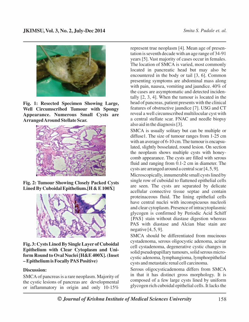
Fig. 1: Resected Specimen Showing Large, Well Circumscribed Tumour with Spongy Appearance. Numerous Small Cysts are Arranged Around Stellate Scar.
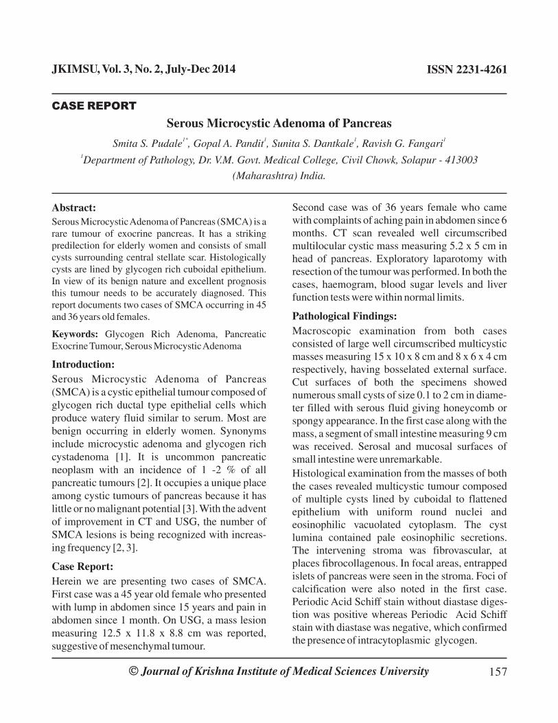
Fig. 2: Tumour Showing Closely Packed Cysts Lined By Cuboidal Epithelium.[H & E 100X]
Fig. 3: Cysts Lined By Single Layer of Cuboidal Epithelium with Clear Cytoplasm and Uni-form Round to Oval Nuclei [H&E 400X]. (Inset – Epithelium is Focally PAS Positive)
Discussion: SMCA of pancreas is a rare neoplasm. Majority of the cystic lesions of pancreas are developmental or inflammatory in origin and only 10-15%
Journal of Krishna Institute of Medical Sciences UniversityÓÓ 158
JKIMSU, Vol. 3, No. 2, July-Dec 2014 Smita S. Pudale et. al.
represent true neoplasm [4]. Mean age of presen-tation is seventh decade with an age range of 34-91 years [5]. Vast majority of cases occur in females. The location of SMCA is varied, most commonly located in pancreatic head but may also be encountered in the body or tail [3, 6]. Common presenting symptoms are abdominal mass along with pain, nausea, vomiting and jaundice. 40% of the cases are asymptomatic and detected inciden-tally [2, 3, 4]. When the tumour is located in the head of pancreas, patient presents with the clinical features of obstructive jaundice [7]. USG and CT reveal a well circumscribed multilocular cyst with a central stellate scar. FNAC and needle biopsy also aid in the diagnosis [3].SMCA is usually solitary but can be multiple or diffuse1. The size of tumour ranges from 1-25 cm with an average of 6-10 cm. The tumour is encapsu-lated, slightly bosselated, round lesion. On section the neoplasm shows multiple cysts with honey-comb appearance. The cysts are filled with serous fluid and ranging from 0.1-2 cm in diameter. The cysts are arranged around a central scar [4, 5, 9].Microscopically, innumerable small cysts lined by single row of cuboidal to flattened epithelial cells are seen. The cysts are separated by delicate acellular connective tissue septae and contain proteinaceous fluid. The lining epithelial cells have central nuclei with inconspicuous nucleoli and clear cytoplasm. Presence of intracytoplasmic glycogen is confirmed by Periodic Acid Schiff {PAS} stain without diastase digestion whereas PAS with diastase and Alcian blue stain are negative [4, 5, 9]. SMCA should be differentiated from mucinous cystadenoma, serous oligocystic adenoma, acinar cell cystadenoma, degenerative cystic changes in solid pseudopapillary tumours, solid serous micro-cystic adenoma, lymphangioma, lymphoepithelial cysts and metastatic renal cell carcinoma.Serous oligocysticadenoma differs from SMCA in that it has distinct gross morphology. It is composed of a few large cysts lined by uniform glycogen rich cuboidal epithelial cells. It lacks the
central stellate scar, has no sex predilection and also occurs in children. Mucinous pancreatic neoplasms should be differentiated from SMCA because it has malignant potential. In mucinous cystadenomas, the cysts are lined by tall, colum-nar cells with basal nuclei with abundant intracellular mucin [8].Acinar cell cystadenoma of pancreas is character-ized by multicystic spongy appearance, but it lacks central stellate scar and it has acinar cell differentiation. Degenarative cystic changes in solid pseudopapillary tumour of pancreas may cause confusion, but these cystic spaces lack epithelial lining and they occur predominantly in young women [1]. Solid serous microcystic adenoma is extremely rare having histologic and immunohistologic features similar to SMCA but lacks a secretory functionality [8]. Lymphangiomas show relatively large cystic spaces lined by flattened cells. These cells are glycogen and keratin negative. Lymphoepithelial cysts of pancreas are unilocular, contain keratinous material and are lined by squamous epithelium, supported by lymphoid stroma.
*Author for Correspondence: Dr. Smita S. Pudale, Department of Pathology, Dr. V.M. Govt. Medical College, Civil Chowk, Solapur - 413003 Maharashtra.
Cell: 9881597877, 9226494786 Email: [email protected], [email protected]
1. Jacob S, Rawat P, Mark RP. Serous microcystic adenoma (glycogen rich cystadenoma) of the pancreas. Indian J Pathol Microbiol 2010; 53(1):106-108.
2. Mandvekar AS, Amarapurkar AD, Shenoy AS, Balsarkar DJ. Serous microcystic adenoma of pan-creas. Indian J Gastroenterol 2011; 30(4):183-184.
3. Pyke CM, van Heerden JA, Colby TV, Sarr MG, Weaver AL. The spectrum of serous cystadenoma of the pancreas. Clinical, pathologic, and surgical aspects. Ann Surg 1992; 215(2):132-139.
4. Heatley MK, McCrory DC, O'Hara MD. Microcystic Adenoma of Pancreas. Ulster Med J 1991; 60(1):111-113.
5. Slukvin II, Hafez GR, Niederhuber JE, Warner TF. Combined serous microcystic adenoma and well-differentiated endocrine pancreatic neoplasm: a case report and review of the literature. Arch Pathol Lab
References:
Journal of Krishna Institute of Medical Sciences UniversityÓÓ 159
JKIMSU, Vol. 3, No. 2, July-Dec 2014 Smita S. Pudale et. al.
Metastatic renal cell carcinoma is characterized by small tubular structures composed of glycogen containing cells. The cells also contain cytoplas-mic lipid. Nuclei are irregular with distinct nucleoli [1]. Multiple sections should be taken to rule out the possibility of endocrine neoplasia associated with SMCA because SMCA admixed with pancreatic endocrine neoplasm has a higher malignant potential than pure SMCA [5].
Conclusion:SMCA of the pancreas is an uncommon tumour with rather unique gross, microscopic morphol-ogy and generally benign course. It is identified by advances in imaging techniques. The differentia-tion from other cystic tumours as well as non-neoplastic cysts is very important because of the great difference in their management.In the light of recent findings of rare instances of malignant transformation and co-existent poten-tially malignant tumours, a thorough sampling of the specimen and post-operative follow-up by regular CT surveillance is advocated.
Med 2003; 127(10):1369-1372.6. Köksal AS, Asil M, Turhan N, Yolcu OF, Küçükay F,
Akoğlu M, Sahin B. Serous microcystic adenoma of the pancreas: case report and review of the literature. Turk J Gastroenterol 2004; 15(3):183-6.
7. Genevieve LB, Chew FS. Radiologic-Pathologic Conferences of the Massachusetts General Hospital Serous Cystadenoma of the Pancreas. AJR 1993;161:786.
8. Stern JR, Frankel WL, Ellison EC, Bloomston M. Solid serous microcystic adenoma of the pancreas. World J Surg Oncol 2007; 5:26.
9. Agarwal N, Kumar S, Dass J, Arora VK, Rathi V. Diffuse pancreatic serous cystadenoma associated with neuroendocrine carcinoma: a case report and review of literature. JOP 2009; 10(1):55-58.


![3 LASOP Case 3 2006.ppt [Read-Only]lasop.com/pgs/hdouts/2006-03_Case3.pdfmeningioma (WHO grade II) • Microcystic gliomas • Hemangioblastomas • Myxoid schwannomas. Microcystic](https://img.pdfslide.net/doc/110x75/5e3bd740d005aa51c76678a8/3-lasop-case-3-2006ppt-read-onlylasopcompgshdouts2006-03case3pdf-meningioma.jpg)