Embed Size (px)
Citation preview
IP International Journal of Maxillofacial Imaging 2020;6(3):83–88
Content available at: https://www.ipinnovative.com/open-access-journals
IP International Journal of Maxillofacial Imaging
Journal homepage: www.ipinnovative.com
Case Report
Treatment of Class II div.1 malocclusion with impacted mandibular canine: A casereport
Rashi Yadav1,*, Bhavesh Kothari1, Kamlesh Garg1, Parmender Vaidik1, Bhakti Rajdev1,Tulip Chakravarty2
1Dept. of Orthodontics and Dentofacial Orthopaedics, Pacific Dental College and Hospital, Udaipur, Rajasthan, India2Dept. of Public Health Dentistry, Pacific Dental College and Hospital, Udaipur, Rajasthan, India
A R T I C L E I N F O
Article history:Received 25-06-2020Accepted 26-06-2020Available online 07-10-2020
Keywords:Class II malocclusionPremolar extractionImpacted mandibular canine
A B S T R A C T
Class II malocclusion in nongrowing patient is a great challenge in treatment. Class II div. 1 malocclusioncan be the result of a retrognathic mandible, a prognathic maxillary or both. The present article discussed acase of 17 year old female patient presented with Proclined upper anterior, crowding in lower anterior,impacted mandibular canine with increased overjet and overbite, treatment was initiated using fixedorthodontic appliances followed by extraction of upper 1st premolar and retained decidous teeth andexposure of impacted canine. Case was finished with good intercuspation of upper and lower teeth, idealoverjet and overbite were obtained.
© 2020 Published by Innovative Publication. This is an open access article under the CC BY-NC license(https://creativecommons.org/licenses/by-nc/4.0/)
1. Introduction
Patient suffering from class II malocclusion are usuallyaffected by their problem which make them the mostcommon cases that seek orthodontic treatment. Angle
′s
class II div 1 malocclusion usually characterized bymaxillary protrusion and /or mandibular retrusion.1 This isusually accompanied by protrusion of upper anterior teeth,narrow upper arch and incompetent upper lip in non growingpatient, this malocclusion can be treated by 1 of 2 methods– orthodontic camouflage which usually involves extractionof upper premolars or even upper and lower premolar ororthognathic surgery to reposition the mandible and /ormaxilla in normal position.2
2. Case Report
A 17 years old female patient with chief complaintof forwardly placed upper front teeth. Patient hadno medical history, dental history of restoration w.r.t.36 and 46. Extraoral examination showed that patient
* Corresponding author.E-mail address: [email protected] (R. Yadav).
had convex profile and incompetent lips, 100% incisorexposure on smile and patient had gummy smile of 4mm.Intraoral examination revealed class II molar and caninerelation, crowding in lower anterior with an Overjet of15mm,overbite of 7mm, missing 43, retained decidous w.r.t.83, crossbite w.r.t. 17,47 cephalometric examination showedskeletal class II jaw base relationship with retrognathicmandible (SNA- 79, SNB-76, ANB- 3), Hypodivergentgrowth pattern (Go-Sn- Gn -27, FMA-22), Proclined upperincisor. OPG revealed an impacted 43.
3. Treatment Objectives
1. Correction of Proclined upper anteriors.2. Correction of gummy smile.3. Relieving lower anterior crowding.4. Achieving class I canine relation.5. Extraction of retained decidous teeth 83.6. Surgical exposure of 43.7. Correction of posterior crossbite w.r.t 17, 47.8. Achieving normal Overjet and overbite.9. Achieving balanced soft tissue profile.
https://doi.org/10.18231/j.ijmi.2020.0212581-382X/© 2020 Innovative Publication, All rights reserved. 83

84 Yadav et al. / IP International Journal of Maxillofacial Imaging 2020;6(3):83–88
Fig. 1: Power arm based intrusion and retraction
Fig. 2: Pretreatment extraoraland intraoral photograph
Yadav et al. / IP International Journal of Maxillofacial Imaging 2020;6(3):83–88 85
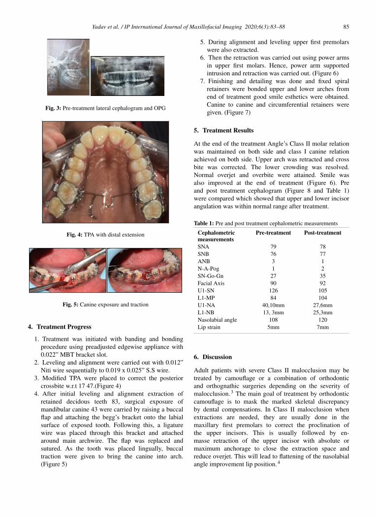
Fig. 3: Pre-treatment lateral cephalogram and OPG
Fig. 4: TPA with distal extension
Fig. 5: Canine exposure and traction
4. Treatment Progress
1. Treatment was initiated with banding and bondingprocedure using preadjusted edgewise appliance with0.022” MBT bracket slot.
2. Leveling and alignment were carried out with 0.012”Niti wire sequentially to 0.019 x 0.025” S.S wire.
3. Modified TPA were placed to correct the posteriorcrossbite w.r.t 17 47.(Figure 4)
4. After initial leveling and alignment extraction ofretained decidous teeth 83, surgical exposure ofmandibular canine 43 were carried by raising a buccalflap and attaching the begg’s bracket onto the labialsurface of exposed tooth. Following this, a ligaturewire was placed through this bracket and attachedaround main archwire. The flap was replaced andsutured. As the tooth was placed lingually, buccaltraction were given to bring the canine into arch.(Figure 5)
5. During alignment and leveling upper first premolarswere also extracted.
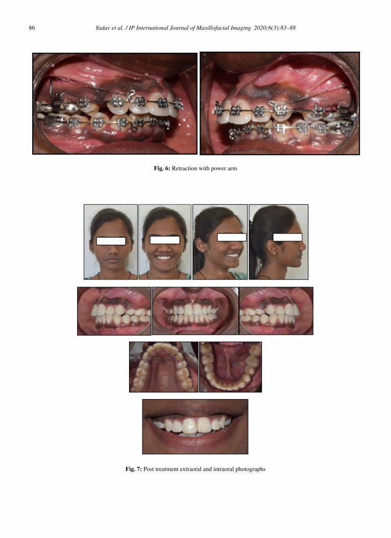
6. Then the retraction was carried out using power armsin upper first molars. Hence, power arm supportedintrusion and retraction was carried out. (Figure 6)
7. Finishing and detailing was done and fixed spiralretainers were bonded upper and lower arches fromend of treatment good smile esthetics were obtained.Canine to canine and circumferential retainers weregiven. (Figure 7)
5. Treatment Results
At the end of the treatment Angle’s Class II molar relationwas maintained on both side and class I canine relationachieved on both side. Upper arch was retracted and crossbite was corrected. The lower crowding was resolved.Normal overjet and overbite were attained. Smile wasalso improved at the end of treatment (Figure 6). Preand post treatment cephalogram (Figure 8 and Table 1)were compared which showed that upper and lower incisorangulation was within normal range after treatment.
Table 1: Pre and post treatment cephalometric measurements
Cephalometricmeasurements
Pre-treatment Post-treatment
SNA 79 78SNB 76 77ANB 3 1N-A-Pog 1 2SN-Go-Gn 27 35Facial Axis 90 92U1-SN 126 105L1-MP 84 104U1-NA 40,10mm 27,6mmL1-NB 13, 3mm 25,3mmNasolabial angle 108 120Lip strain 5mm 7mm
6. Discussion
Adult patients with severe Class II malocclusion may betreated by camouflage or a combination of orthodonticand orthognathic surgeries depending on the severity ofmalocclusion.3 The main goal of treatment by orthodonticcamouflage is to mask the marked skeletal discrepancyby dental compensations. In Class II malocclusion whenextractions are needed, they are usually done in themaxillary first premolars to correct the proclination ofthe upper incisors. This is usually followed by en-masse retraction of the upper incisor with absolute ormaximum anchorage to close the extraction space andreduce overjet. This will lead to flattening of the nasolabialangle improvement lip position.4
86 Yadav et al. / IP International Journal of Maxillofacial Imaging 2020;6(3):83–88
Fig. 6: Retraction with power arm
Fig. 7: Post treatment extraoral and intraoral photographs

Yadav et al. / IP International Journal of Maxillofacial Imaging 2020;6(3):83–88 87
Fig. 8: Post treatment lateral cephalogram and OPG
Fig. 9: Overall superimposition
88 Yadav et al. / IP International Journal of Maxillofacial Imaging 2020;6(3):83–88
In this case, it was decided to use the power arm toimprove the gummy smile. Construction of power arm doneby using 19*25 S.S. wire. As the center of resistance isconsidered the most important point for tooth movement.Power arm moves the point of force application closeto center of resistance, it provided posterior and superiorvector of force which was required for intrusion of anteriorteeth. This not only helps to avoid labial tipping of anteriorteeth but also bring about effective intrusion to minimizegummy smile. (Figure 1)
An attempt to level the buccally placed maxillary 2nd
molar by using TPA with distal extension and E-chaincrosses over the occlusion surface of maxillary 2nd molar.It helps to apply an isolated forces on buccally placedmaxillary 2nd molar without disturbing anchor unit andany undesirable movement on the dentition. This kind ofintraarch crossbite correction does not interfere with thephysiologic eruption of teeth in the opposite arch.5
Success of treatment of mandibular impacted caninedepends upon the position, impaction level and age of thepatient. In this case as the canine was in favorable positionand since canines are considered important keystones in thedental arch, we decided to orthodontically bring it into itsideal position. The needful application of force in differentdirection is required for not only successfully guiding theeruption of an impacted canine but also aligning it inits correct position. Here in this case overall Treatmenttime was 30 months with improved smile & Profile.Upper incisors were retracted to achieve normal incisorangulations, Overjet & overbite. Lips became competentand lower lip controlled upper incisors successfully, whichis very important for incisor stability in class II division 1malocclusion.
7. Conclusion
Dental camouflage orthodontic treatment could be a veryimportant alternative method of managing malocclusionrather than through conventional way of approach.Managing such way would be a much more effective clinicalway to solve complex malocclusions. Gummy smile wastreated with power arm through intrusion of the upperincisors, also patient’s profile was improved by correction ofoverjet using power arm. Treatment was successful with therecovery of impacted canine, correction of crossbite, There
was overall significant improvement in the facial appearanceand soft tissue profile of the patient. Esthetically, balancedresults were achieved.
8. Source of Funding
None.
9. Conflict of Interest
None.
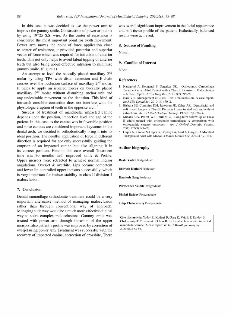
References1. Naragond A, Kenganal S, Sagarkar SR. Orthodontic Camouflage
Treatment in an Adult Patient with a Class II, Division 1 Malocclusion– A Case Report. J Clin Diag Res. 2013;7(2):395–98.
2. Naik VR. Management of Class II div I malocclusion- A case report.Int J Clin Dental Sci. 2010;1(1):70–2.
3. Bishara SE, Cummins DM, Jakobsen JR, Zaher AR. Dentofacial andsoft tissue changes in Class II, Division 1 cases treated with and withoutextractions. Am J Orthod Dentofac Orthop. 1995;107(1):28–37.
4. Mihalik CA, Proffit WR, Phillips C. Long-term follow-up of ClassII adults treated with orthodontic camouflage: A comparison withorthognathic surgery outcomes. Am J Orthod Dentofac Orthop.2003;123(3):266–78.
5. Gupta A, Kannan S, Gupta G, Goyaliya A, Kaul A, Garg N. A ModifiedTranspalatal Arch with Sleeve. J Indian Orthod Soc. 2013;47(2):112.
Author biography
Rashi Yadav Postgraduate
Bhavesh Kothari Professor
Kamlesh Garg Professor
Parmender Vaidik Postgraduate
Bhakti Rajdev Postgraduate
Tulip Chakravarty Postgraduate
Cite this article: Yadav R, Kothari B, Garg K, Vaidik P, Rajdev B,Chakravarty T. Treatment of Class II div.1 malocclusion with impactedmandibular canine: A case report. IP Int J Maxillofac Imaging2020;6(3):83-88.