Embed Size (px)
Citation preview
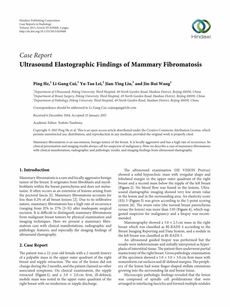
Case ReportUltrasound Elastographic Findings of Mammary Fibromatosis
Ping He,1 Li-Gang Cui,1 Yu-Tao Lei,2 Jian-Ying Liu,3 and Jin-Rui Wang1
1Department of Ultrasound, Peking University Third Hospital, 49 North Garden Road, Haidian District, Beijing 100191, China2Department of Breast Surgery, Peking University Third Hospital, 49 North Garden Road, Haidian District, Beijing 100191, China3Department of Pathology, Peking University Third Hospital, 49 North Garden Road, Haidian District, Beijing 100191, China
Correspondence should be addressed to Li-Gang Cui; [email protected]
Received 6 December 2014; Accepted 25 January 2015
Academic Editor: Yoshito Tsushima
Copyright © 2015 Ping He et al. This is an open access article distributed under the Creative Commons Attribution License, whichpermits unrestricted use, distribution, and reproduction in any medium, provided the original work is properly cited.
Mammary fibromatosis is an uncommon, benign tumor of the breast. It is locally aggressive and has a high rate of recurrence. Itsclinical presentation and imaging results always call for suspicion of malignancy. Here we describe a case of mammary fibromatosiswith clinical manifestation, radiographic and pathologic results, and imaging findings from ultrasound elastography.
1. Introduction
Mammary fibromatosis is a rare and locally aggressive benigntumor of the breast. It originates from fibroblasts and myofi-broblasts within the breast parenchyma and does not metas-tasize. It often occurs as an extension of lesions arising fromthe pectoral fascia [1]. Mammary fibromatosis accounts forless than 0.2% of all breast lesions [2]. Due to its infiltrativenature, mammary fibromatosis has a high rate of recurrence(ranging from 21% to 27% [3–5]) after inadequate surgicalexcision. It is difficult to distinguish mammary fibromatosisfrom malignant breast tumors by physical examination andimaging techniques. Here we present a mammary fibro-matosis case with clinical manifestations, radiographic andpathologic features, and especially the imaging findings ofultrasound elastography.
2. Case Report
The patient was a 22-year-old female with a 2-month historyof a palpable mass in the upper outer quadrant of the rightbreast and nipple retraction. The size of the lesion did notchange during the 2months, and the patient claimed no otherassociated symptoms. On clinical examination, the nippleretracted (Figure 1), and a 3.0 × 2.0 cm firm, ill-defined,mobile mass was noted in the upper outer quadrant of theright breast with no tenderness or nipple discharge.
The ultrasound examination (HI VISION Preirus)showed a solid hypoechoic mass with irregular shape andlobulated margin in the upper outer quadrant of the rightbreast and a second mass below the nipple of the left breast(Figure 2). No blood flow was found in the lesions. Ultra-sound elastographic imaging showed very low strain valuein the lesion and in the surrounding area. An elasticity score(ES) 5 (Figure 3) was given according to the 5-point scoringsystem [6]. The strain ratio (the normal breast parenchymaversus the lesion) was more than 3.05 (Figure 4), which sug-gested suspicion for malignancy and a biopsy was recom-mended.
Mammography showed a 3.0 × 2.5 cm mass in the rightbreast which was classified as BI-RADS 4 according to theBreast Imaging Reporting and Data System, and a nodule inthe left breast was classified as BI-RADS 3.
An ultrasound guided biopsy was performed but theresults were indeterminate and initially interpreted as hyper-plasia of interstitial tissue.The patient then underwent partialmastectomy of the right breast. Gross pathologic examinationof the specimen showed a 5.0 × 3.0 × 3.0 cm firm mass withnonuniform cut surfaces and ill-definedmargins.Theperiph-ery of the lesion had some finger-shaped stellate extensionsgrowing into the surrounding fat and breast tissue.
Microscopic pathologic findings revealed that the lesionwas composed of spindle cell proliferations that werearranged in interlacing fascicles and formedmultiple nodules
Hindawi Publishing CorporationCase Reports in RadiologyVolume 2015, Article ID 829468, 4 pageshttp://dx.doi.org/10.1155/2015/829468

2 Case Reports in Radiology
Figure 1: The nipple retraction was present, but there was no tenderness, redness, swelling, skin rupture, or nipple discharge.
(a) (b)
Figure 2: The ultrasound examination demonstrated a solid hypoechoic mass with an irregular shape and a lobulated margin both in theupper outer quadrant of the right breast (a) and below the nipple of the left breast (b).
Figure 3: Ultrasound elastographic imaging showed very low strainvalue in the tumor and the surrounding area, scored as ES 5 basedon the 5-point scoring system.
with amoderate amount of collagen (Figure 5). Some residualducts and lobules were observed within and between thenodules, but they did not form foliation. Cellular atypia wasnot obvious and mitotic figures were uncommon.
Immunohistochemical staining results showed thatthe spindle cells were positive for 𝛽-catenin, SMA, andVIMENTIN but were negative for DESMIN, S-100, and CK.
Figure 4:The strain ratio (the normal breast parenchyma versus thetumor) was more than 3.05.
The histology and immunohistochemical staining resultssupported the diagnosis of mammary fibromatosis.
3. Discussion
Mammary fibromatosis primarily affects females with an agerange between 13 and 80 years (average age 46, median age40), but it ismore common in the childbearing age group than

Case Reports in Radiology 3
Figure 5:Microscopic pathological findings revealed that the lesionwas composed of a spindle cell proliferation that was arranged ininterlacing fascicles and formed multiple nodules with a moderateamount of collagen.
the perimenopausal and postmenopausal groups [1]. A fewcases have also been reported in males [7]. Bilateral mam-mary fibromatosis has rarely been reported, with most casesoccurring synchronously except for one case in which thelesions appeared asynchronously with a 2-year interval inbetween [8].
Mammary fibromatosis is usually painless and the pre-senting symptom is always a palpable, firm breast mass. Skindimpling and nipple retraction may be present. Nipple dis-charge is uncommon.The etiology of mammary fibromatosisis unknown. Some cases occur after trauma or surgical pro-cedures such as breast reduction or breast augmentation withsaline or silicone implants [9]. However, it can also happenin patients with familial adenomatous polyposis (FAP) syn-drome, Gardner syndrome, or hereditary desmoid diseasessuch as familial multicentric fibromatosis [8]. Mutations inthe adenomatous polyposis coli (APC) and 𝛽-catenin path-way play an important role in the pathogenesis of mammaryfibromatosis [8].
The reported lesion size mammary fibromatosis rangesfrom 0.5 to 10 cm (average 2.5–3.0 cm), and the tumor isalways ill-defined with a firm, white-grey, or tan cut surface.Well-circumscribed cases are occasionally seen. On ultra-sound images, it typically appears as an ill-circumscribed,lobulated, irregular, and solid hypoechoic mass with straight-ening and tethering of Cooper ligaments, which imitatesmalignant tumors. Because of its infiltrative growth pattern,the pectoralis major or intercostal muscles may be involved.However, unlike breast cancer, mammary fibromatosis doesnot have acoustic shadowing, echogenic halo, or microcalci-fication, and its orientation is usually parallel.
Ultrasound elastography is an imaging technique thatcan measure the stiffness of the soft tissue. It can be usedto differentiate between benign and malignant breast lesionsbased on the principle that the stiffness of different tissues atdifferent pathological states follows a general rule: normal fat< normal glandular < fibrous tissue < breast carcinoma. Theelastographic images of breast lesions are usually scored bya 5-point scoring system described by Zhu et al. [6]. Benignlesions tend to have an ES of 1 or 2, whereas most malignantlesions have an ES of 4 or 5. A lesion with an ES of 3 couldbe either benign or malignant. Zhi et al. [10] suggested thatthe strain ratio measurement could be used for evaluatingthe hardness or stiffness of breast lesions semiquantitatively.
When the cutoff value of the strain ratio was set at 3.05, ultra-sound elastography showed a sensitivity of 92.4%, a specificityof 91.1%, and an accuracy of 91.4%.However, in themammaryfibromatosis case we reported here that both ES value (5)and strain ratio (>3.05) indicated malignant tumor, whichwas overruled by the final pathological results. This suggeststhat the ultrasound elastography may not be an ideal methodto discriminate between mammary fibromatosis and malig-nant tumors in the breast, because the composition of mam-mary fibromatosis lesion makes it stiffer than normal breasttissues and may lead to a false diagnosis of malignant tumorbased on the elastographic results.
Mammographically, mammary fibromatosis appears as aspiculated mass without microcalcifications which may beassessed as BI-RADS 3, 4, or 5.MRI is the best way to evaluatetumor extent and the involvement of the chest wall.
The diagnosis of mammary fibromatosis can be madefrom the microscopic findings on routine hematoxylin andeosin stained sections. In general, the lesion does not havemalignancy features such as high mitotic rate, cellular atypia,necrosis, or vascular invasion. Lymphocytic infiltrates wereoften noted at the periphery of the lesion.
Since there are no specific immunomarkers for the mam-mary fibromatosis, immunohistochemical staining is notrequired for making the final diagnosis.
To prevent or reduce the recurrence, the recommendedtreatment for mammary fibromatosis is wide local resection.
Conflict of Interests
The authors declare that there is no conflict of interestsregarding the publication of this paper.
References
[1] M. Drijkoningen, F. A. Tavassoli, and G. Magro, Pathology andGenetics of Tumours of the Breast and Female Genital Organs,World Health Organization Classification of Tumours, WHO,Geneva, Switzerland, 2003.
[2] G. S. Schwarz, M. Drotman, R. Rosenblatt, L. Milner, J. Sha-monki, and M. P. Osborne, “Fibromatosis of the breast: casereport and current concepts in the management of an uncom-mon lesion,” Breast Journal, vol. 12, no. 1, pp. 66–71, 2006.
[3] E. S. Wargotz, H. J. Norris, R. M. Austin, and F. M. Enzinger,“Fibromatosis of the breast. A clinical and pathological study of28 cases,”TheAmerican Journal of Surgical Pathology, vol. 11, no.1, pp. 38–45, 1987.
[4] F. E. Gump, M. J. Sternschein, and M. Wolff, “Fibromatosis ofthe breast,” Surgery Gynecology and Obstetrics, vol. 153, no. 1, pp.57–60, 1981.
[5] P. P. Rosen and D. Ernsberger, “Mammary fibromatosis. Abenign spindle-cell tumor with significant risk for local recur-rence,” Cancer, vol. 63, no. 7, pp. 1363–1369, 1989.
[6] Q.-L. Zhu, Y.-X. Jiang, J.-B. Liu et al., “Real-time ultrasoundelastography: its potential role in assessment of breast lesions,”Ultrasound inMedicine andBiology, vol. 34, no. 8, pp. 1232–1238,2008.
[7] I. Rudan, N. Rudan, T. Skoric, and B. Sarcevic, “Fibromatosis ofmale breast,” Acta Medica Croatica, vol. 50, no. 3, pp. 157–159,1996.

4 Case Reports in Radiology
[8] M. E. McMenamin, K. DeSchryver, and C. D. M. Fletcher,“Fibrous lesions of the breast: a review,” International Journalof Surgical Pathology, vol. 8, no. 2, pp. 99–108, 2000.
[9] K.N.Glazebrook andC.A. Reynolds, “Mammary fibromatosis,”The American Journal of Roentgenology, vol. 193, no. 3, pp. 856–860, 2009.
[10] H. Zhi, X.-Y. Xiao, H.-Y. Yang, B. Ou, Y.-L.Wen, and B.-M. Luo,“Ultrasonic elastography in breast cancer diagnosis. Strain ratiovs 5-point scale,” Academic Radiology, vol. 17, no. 10, pp. 1227–1233, 2010.

Submit your manuscripts athttp://www.hindawi.com
Stem CellsInternational
Hindawi Publishing Corporationhttp://www.hindawi.com Volume 2014
Hindawi Publishing Corporationhttp://www.hindawi.com Volume 2014
MEDIATORSINFLAMMATION
of
Hindawi Publishing Corporationhttp://www.hindawi.com Volume 2014
Behavioural Neurology
EndocrinologyInternational Journal of
Hindawi Publishing Corporationhttp://www.hindawi.com Volume 2014
Hindawi Publishing Corporationhttp://www.hindawi.com Volume 2014
Disease Markers
Hindawi Publishing Corporationhttp://www.hindawi.com Volume 2014
BioMed Research International
OncologyJournal of
Hindawi Publishing Corporationhttp://www.hindawi.com Volume 2014
Hindawi Publishing Corporationhttp://www.hindawi.com Volume 2014
Oxidative Medicine and Cellular Longevity
Hindawi Publishing Corporationhttp://www.hindawi.com Volume 2014
PPAR Research
The Scientific World JournalHindawi Publishing Corporation http://www.hindawi.com Volume 2014
Immunology ResearchHindawi Publishing Corporationhttp://www.hindawi.com Volume 2014
Journal of
ObesityJournal of
Hindawi Publishing Corporationhttp://www.hindawi.com Volume 2014
Hindawi Publishing Corporationhttp://www.hindawi.com Volume 2014
Computational and Mathematical Methods in Medicine
OphthalmologyJournal of
Hindawi Publishing Corporationhttp://www.hindawi.com Volume 2014
Diabetes ResearchJournal of
Hindawi Publishing Corporationhttp://www.hindawi.com Volume 2014
Hindawi Publishing Corporationhttp://www.hindawi.com Volume 2014
Research and TreatmentAIDS
Hindawi Publishing Corporationhttp://www.hindawi.com Volume 2014
Gastroenterology Research and Practice
Hindawi Publishing Corporationhttp://www.hindawi.com Volume 2014
Parkinson’s Disease
Evidence-Based Complementary and Alternative Medicine
Volume 2014Hindawi Publishing Corporationhttp://www.hindawi.com