Embed Size (px)
Citation preview
Hindawi Publishing CorporationCase Reports in Obstetrics and GynecologyVolume 2013, Article ID 401962, 4 pageshttp://dx.doi.org/10.1155/2013/401962
Case ReportUterine Carcinosarcoma in a Patient with Didelphys Uterus
C. Iavazzo, F. Kokka, A. Sahdev, N. Singh, and K. Reynolds
Gynaecological Department, St Bartholomews Hospital, London EC1A 7BE, UK
Correspondence should be addressed to C. Iavazzo; [email protected]
Received 27 January 2013; Accepted 18 February 2013
Academic Editors: D. Hochner-Celnikier, E. F. C. Murta, B. Piura, and A. Semczuk
Copyright © 2013 C. Iavazzo et al. This is an open access article distributed under the Creative Commons Attribution License,which permits unrestricted use, distribution, and reproduction in any medium, provided the original work is properly cited.
Background. Didelphys uterus is a noncommon finding in women. Till now, few cases with benignmesenchymal tumors in patientswith didelphys uterus are described. We present a case of a patient with carcinosarcoma arising in a didelphys uterus. Case. A 73-year-old patient presented with profuse watery postmenopausal bleeding. On examination under anesthesia, left and right cervixeswere identified. Tumor extended from the left cervix into the lower third of the vagina andwas adherent to the right vaginal sidewall.There was no evidence of parametrial extension. Tissue was sent for biopsy which revealed high-grade uterine carcinosarcoma.Two uterine fundi and two vaginas in keeping with uterine didelphys were identified on imaging. The patient underwent vaginalexcision of the protruding tumormeasuring 8×6 cmwith harmonic scalpel followed by total abdominal hysterectomy and bilateralsalpingooophorectomy. Although a number of pelvic and paraaortic lymph nodes were also identified on imaging, she was notplanned for lymphadenectomy after MDT (multidisciplinary team) discussion because of her comorbidities. The final histologyconfirmed the diagnosis. Conclusion. According to our knowledge, this is the second case of carcinosarcoma arising in didelphysuterus in the world literature.
1. Introduction
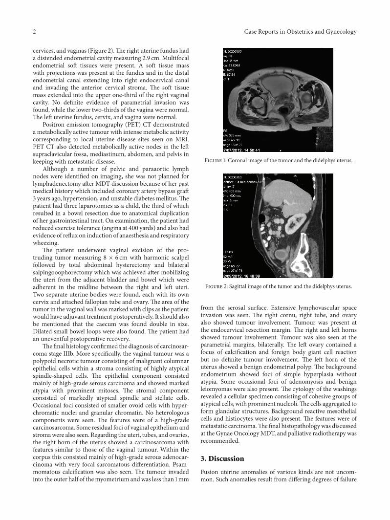
Persistent Mullerian duct syndrome presenting as didelphysuterus is a very rare anomaly which is estimated to occur in1/3,000 women [1]. However, it might go undetected in theabsence of medical and reproductive complications and sothe incidence may be underestimated. Fibroids are benignmesenchymal tumors which are commonly associated withcases of didelphys uterus and are present in premenopausalwomen [2]. However, sarcoma—themalignantmesenchymaltumor—is an extremely rare finding in a woman with adidephys uterus. We are only aware of one case previouslydescribed in the literature [3]. Moreover, very few reportsof endometrial cancer arising in patients with uterine mal-formations could be also found in the literature [4–7]. Wepresent a case of carcinosarcoma found in a woman witha didelphys uterus (Figure 3). A review of the literaturewas performed to clarify the diagnostic pathway and themanagement of this rare entity.
2. Case
A 73-year-fold patient, para 5, presented with profuse waterypostmenopausal discharge and frank red bleeding per vaginafor 2 weeks. On examination under anesthesia, left andright cervixes were identified. Tumor extended from the leftcervix into the lower third of the vagina and was adherentto the right vaginal sidewall. The uterus was otherwisemobile, and there was no evidence of parametrial extension.Tissue was sent for biopsy which revealed high-grade uterinecarcinosarcoma.
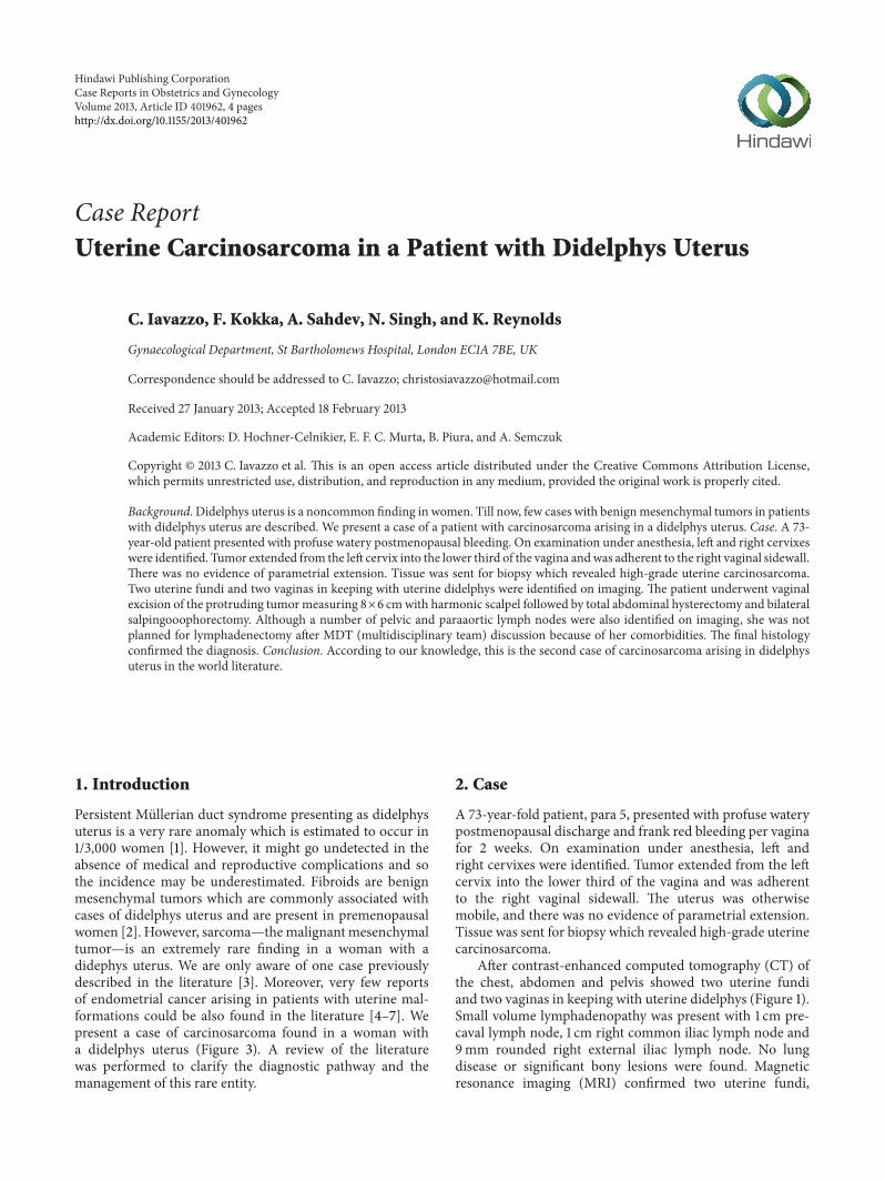
After contrast-enhanced computed tomography (CT) ofthe chest, abdomen and pelvis showed two uterine fundiand two vaginas in keeping with uterine didelphys (Figure 1).Small volume lymphadenopathy was present with 1 cm pre-caval lymph node, 1 cm right common iliac lymph node and9mm rounded right external iliac lymph node. No lungdisease or significant bony lesions were found. Magneticresonance imaging (MRI) confirmed two uterine fundi,
2 Case Reports in Obstetrics and Gynecology
cervices, and vaginas (Figure 2).The right uterine fundus hada distended endometrial cavity measuring 2.9 cm. Multifocalendometrial soft tissues were present. A soft tissue masswith projections was present at the fundus and in the distalendometrial canal extending into right endocervical canaland invading the anterior cervical stroma. The soft tissuemass extended into the upper one-third of the right vaginalcavity. No definite evidence of parametrial invasion wasfound, while the lower two-thirds of the vagina were normal.The left uterine fundus, cervix, and vagina were normal.
Positron emission tomography (PET) CT demonstrateda metabolically active tumour with intense metabolic activitycorresponding to local uterine disease sites seen on MRI.PET CT also detected metabolically active nodes in the leftsupraclavicular fossa, mediastinum, abdomen, and pelvis inkeeping with metastatic disease.
Although a number of pelvic and paraaortic lymphnodes were identified on imaging, she was not planned forlymphadenectomy after MDT discussion because of her pastmedical history which included coronary artery bypass graft3 years ago, hypertension, and unstable diabetes mellitus.Thepatient had three laparotomies as a child, the third of whichresulted in a bowel resection due to anatomical duplicationof her gastrointestinal tract. On examination, the patient hadreduced exercise tolerance (angina at 400 yards) and also hadevidence of reflux on induction of anaesthesia and respiratorywheezing.
The patient underwent vaginal excision of the pro-truding tumor measuring 8 × 6 cm with harmonic scalpelfollowed by total abdominal hysterectomy and bilateralsalpingooophorectomy which was achieved after mobilizingthe uteri from the adjacent bladder and bowel which wereadherent in the midline between the right and left uteri.Two separate uterine bodies were found, each with its owncervix and attached fallopian tube and ovary. The area of thetumor in the vaginal wall wasmarked with clips as the patientwould have adjuvant treatment postoperatively. It should alsobe mentioned that the caecum was found double in size.Dilated small bowel loops were also found. The patient hadan uneventful postoperative recovery.
The final histology confirmed the diagnosis of carcinosar-coma stage IIIb. More specifically, the vaginal tumour was apolypoid necrotic tumour consisting of malignant columnarepithelial cells within a stroma consisting of highly atypicalspindle-shaped cells. The epithelial component consistedmainly of high-grade serous carcinoma and showed markedatypia with prominent mitoses. The stromal componentconsisted of markedly atypical spindle and stellate cells.Occasional foci consisted of smaller ovoid cells with hyper-chromatic nuclei and granular chromatin. No heterologouscomponents were seen. The features were of a high-gradecarcinosarcoma. Some residual foci of vaginal epithelium andstromawere also seen. Regarding the uteri, tubes, and ovaries,the right horn of the uterus showed a carcinosarcoma withfeatures similar to those of the vaginal tumour. Within thecorpus this consisted mainly of high-grade serous adenocar-cinoma with very focal sarcomatous differentiation. Psam-momatous calcification was also seen. The tumour invadedinto the outer half of themyometrium andwas less than 1mm
Figure 1: Coronal image of the tumor and the didelphys uterus.
Figure 2: Sagittal image of the tumor and the didelphys uterus.
from the serosal surface. Extensive lymphovascular spaceinvasion was seen. The right cornu, right tube, and ovaryalso showed tumour involvement. Tumour was present atthe endocervical resection margin. The right and left hornsshowed tumour involvement. Tumour was also seen at theparametrial margins, bilaterally. The left ovary contained afocus of calcification and foreign body giant cell reactionbut no definite tumour involvement. The left horn of theuterus showed a benign endometrial polyp. The backgroundendometrium showed foci of simple hyperplasia withoutatypia. Some occasional foci of adenomyosis and benignleiomyomas were also present. The cytology of the washingsrevealed a cellular specimen consisting of cohesive groups ofatypical cells, with prominent nucleoli.The cells aggregated toform glandular structures. Background reactive mesothelialcells and histiocytes were also present. The features were ofmetastatic carcinoma.The final histopathology was discussedat the Gynae OncologyMDT, and palliative radiotherapy wasrecommended.
3. Discussion
Fusion uterine anomalies of various kinds are not uncom-mon. Such anomalies result from differing degrees of failure
Case Reports in Obstetrics and Gynecology 3
Figure 3: A posterior view of a didelphys uterus from a 73-year-oldwoman with uterine carcinosarcoma is shown.
of fusion of the two Mullerian ducts at about 9 weeks ofgestation. A congenital anomaly syndrome with didelphysuterus and ipsilateral renal agenesis was first reported in 1922[8]. Since then, over 180 cases were reported in the literature[5].
Most of these anomalies do not reduce the female fertility,and this was demonstrated in our patient as she had fivenormal deliveries. However, complete duplication of theuterus and cervix may prevent descent of the fetal headin late pregnancy or obstruct labor by the nonpregnanthorn, something that was mentioned in our patient’s medicalhistory.
Usually, women with a congenital uterine anomaly havean increased risk of renal anomalies and should undergo renaltract imaging. Our case was not correlated with significantrenal anomalies, but she had a history of gastrointestinaltract anomalies (duplicate colon).The other finding from thegenitourinary tract was that our patient had normal urethra,as well as a second “blind” urethra.
Uterine malformations may cause a delayed diagnosisof gynecological malignancies. Imaging studies includingultrasound, 3D ultrasound, CT, and MRI could help inthe diagnosis of uterine malignancies in cases of didelphysuterus [5]. We used ultrasound and MRI for the diagnosis ofdidelphys uterus, aswell as PET scan for clarification of lymphnode metastases. However, it should be mentioned that thediagnosis is based on the histological findings.
APubmed andGoogle searchwas conducted for internet-based resources and open access publications with the termsdidelphys uterus or double uterus and cancer or sarcomaor malignancy and we identified 17 cases with endometrialcancer and another case with carcinosarcoma between theyears 1952 and 2012 [3–21]. So according to, our knowledgeour case is the second described in the literature withcarcinosarcoma in a patient with didelphys uterus. The firstcase of carcinosarcoma of the uterus is related with tamoxifenuse in a 72-year-old patient with a history of breast cancer [3].The patient similarly to our case presented with a large pelvicmass, but the tumor was not recognized preoperatively. Thispatient died of the disease 5 months after diagnosis. So bothcases presented in postmenopausal women over 70 years withrather large tumors.
The question of why the patient underwent MRI couldbe raised as the role of MRI is to identify patients likely tobe at risk of nodal disease and thereby select patients for
lymphadenectomy. CT or PETCT alone would have sufficedfor the detection of distant disease, whilst local staging wassurgical. However, we performed first the imaging and afterthat the MDM decided to avoid lymphadenectomy.
Carcinosarcomas are also called mixedMullerian tumorswhich are characterized by a typical biphasic patternwith car-cinomatous and sarcomatous elements (either homologousor heterologous). In our case, no heterologous componentswere found.The behavior of carcinosarcomas is characterizedmainly by the carcinomatous component, and so mainlythey give lymph node metastases. In our case, pelvic andparaaortic lymph nodes were identified, but because ofthe comorbidity, a lymphadenectomy was not performed.Radiotherapy is suggested in the literature as another optionfor treatment of uterine didelphys malignancy [3]. In general,it is known that the overall 5-year survivals of patientswith uterine carcinosarcoma receiving radiotherapy versusno irradiation were 41.5% and 33.2%, respectively. Combinedadjuvant radiotherapy and chemotherapy may prolong evenmore the 5-year survival.
4. Conclusion
Didelphys uterus is a rare finding in women. It is even morerare to be complicatedwith carcinosarcoma.According to ourknowledge this is the second case of carcinosarcoma arisingin didelphys uterus in the world literature.
References
[1] G. F. Grimbizis, M. Camus, B. C. Tarlatzis, J. N. Bontis, and P.Devroey, “Clinical implications of uterine malformations andhysteroscopic treatment results,” Human Reproduction Update,vol. 7, no. 2, pp. 161–174, 2001.
[2] M. L. Johnsrud, “Successful pregnancy outcome in uterus didel-phys with leiomyoma uteri,” Acta Obstetricia et GynecologicaScandinavica, vol. 73, no. 2, pp. 158–160, 1994.
[3] C. Kunos, C. Woods, V. C. Colussi, F. W. Abdul-Karim, andS. Waggoner, “Low-dose-rate brachytherapy for treatment ofuterine didelphysmalignancy,” Journal of Clinical Oncology, vol.29, no. 5, pp. e104–e106, 2011.
[4] C. Y. Chen, M. S. Yen, M. J. Yang, and Y. C. Wu, “Uterusdidelphyswith adenocarcinoma in the right cavity diagnosed by2-dimensional sonography and magnetic resonance imaging,”Journal of Ultrasound in Medicine, vol. 27, no. 12, pp. 1802–1803,2008.
[5] F. Fanfani, A. Fagotti, G. Restaino, M. Guerriero, and G. Scam-bia, “Endometrial cancer arising in both horns of didelphysuterus in a Down’s syndrome woman,” Gynecologic Oncology,vol. 101, no. 3, pp. 537–539, 2006.
[6] R. Bhalla, H. Evans, L. Berger, J. Crow, M. Deheragoda, and Y.Taper, “A uterus didelphys bicollis, with endometrial cancer inboth uteruses,” Journal of Obstetrics and Gynaecology, vol. 25,no. 8, pp. 823–825, 2005.
[7] C. E. Purslow, “A case of unilateral haematocopos, haematome-tra and haematosalpinx,” BJOG: An International Journal ofObstetrics & Gynaecology, vol. 29, no. 4, p. 643, 1922.
[8] J. Fealy and J. H. Nelson, “Adenocarcinoma in one-half ofa uterus didelphys,” The Medical Annals of the District ofColumbia, vol. 26, no. 2, pp. 76–77, 1957.
4 Case Reports in Obstetrics and Gynecology
[9] R. D. Braun, “Uterus didelphys and endometrial carcinoma. Acase report,”Obstetrics and Gynecology, vol. 35, no. 1, pp. 93–95,1970.
[10] K. B. Grant and R. L. Sedlacek, “Uterus didelphys with adeno-carcinoma in one fundus—a case report,” Journal of the IowaMedical Society, vol. 60, no. 5, pp. 324–325, 1970.
[11] A. D. Anneberg, “Double vagina with double uterus (didelphys)containing endometrial adenocarcinoma. Report of a case,”Journal of the Iowa Medical Society, vol. 61, no. 11, pp. 674–675,1971.
[12] E. Eichner and K. A. Simak, “Uterus didelphys unicollis withadenocarcinoma in one horn and atypical endometrial hyper-plasia in the other: case report,” American Journal of Obstetricsand Gynecology, vol. 139, no. 2, pp. 222–225, 1981.
[13] P. Suprasert and S. Khunamornpong, “Carcinosarcoma arisingin uterine didelphys after tamoxifen therapy for breast cancer: acase report,” Journal of the Medical Association of Thailand, vol.93, no. 5, pp. 608–612, 2010.
[14] A. G. Thomas, L. Deligdisch, and M. Goldstein, “Endometrialcarcinoma with uterus didelphys,” Mount Sinai Journal ofMedicine, vol. 55, no. 5, pp. 411–413, 1988.
[15] M. S. Woods, R. G. Sheppard, D. A. Hardman, and H. J. Woods,“Congenital genitourinary anomalies: is there a predilection formultiple primary malignant neoplasms?” Cancer, vol. 69, no. 2,pp. 546–549, 1992.
[16] F. P. Chen and K. K. Ng, “Term pregnancy at the site of atresiafollowing vaginal canalization in a case of uterus didelphys withhemivaginal atresia and ipsilateral renal agenesis,” TaiwaneseJournal of Obstetrics and Gynecology, vol. 45, no. 4, pp. 366–368,2006.
[17] D. V. Pojman and J. B. Taxy, “Images in clinical medicine.Double uterus with adenocarcinoma,”TheNew England Journalof Medicine, vol. 333, no. 11, p. 709, 1995.
[18] A. Kosinski and M. Dini, “Endometrial cancer in a doubleuterus: a report of two cases,” The Journal of ReproductiveMedicine, vol. 39, no. 11, pp. 926–927, 1994.
[19] N. V. Vial’tsev, N. V. Sokolova, and A. G. Pronin, “Developmentof endometrial cancer in one half of a double uterus,” ArkhivPatologii, vol. 53, no. 3, pp. 62–64, 1991.
[20] Y. Tsukahara, Y. Fukamatsu, K. Tomita, T. Shiozawa, H. Iinuma,and T. Fukuta, “Endometrial carcinoma arising from a doubleuterus,” Gynecologic and Obstetric Investigation, vol. 29, no. 4,pp. 311–312, 1990.
[21] F. J. van Assen, “An exceptional case of endometrial carcinomaor a double tumour of the uterus?”Nederlandsch Tijdschrift voorVerloskunde en Gynaecologie, vol. 70, no. 2, pp. 155–164, 1970.

Submit your manuscripts athttp://www.hindawi.com
Stem CellsInternational
Hindawi Publishing Corporationhttp://www.hindawi.com Volume 2014
Hindawi Publishing Corporationhttp://www.hindawi.com Volume 2014
MEDIATORSINFLAMMATION
of
Hindawi Publishing Corporationhttp://www.hindawi.com Volume 2014
Behavioural Neurology
EndocrinologyInternational Journal of
Hindawi Publishing Corporationhttp://www.hindawi.com Volume 2014
Hindawi Publishing Corporationhttp://www.hindawi.com Volume 2014
Disease Markers
Hindawi Publishing Corporationhttp://www.hindawi.com Volume 2014
BioMed Research International
OncologyJournal of
Hindawi Publishing Corporationhttp://www.hindawi.com Volume 2014
Hindawi Publishing Corporationhttp://www.hindawi.com Volume 2014
Oxidative Medicine and Cellular Longevity
Hindawi Publishing Corporationhttp://www.hindawi.com Volume 2014
PPAR Research
The Scientific World JournalHindawi Publishing Corporation http://www.hindawi.com Volume 2014
Immunology ResearchHindawi Publishing Corporationhttp://www.hindawi.com Volume 2014
Journal of
ObesityJournal of
Hindawi Publishing Corporationhttp://www.hindawi.com Volume 2014
Hindawi Publishing Corporationhttp://www.hindawi.com Volume 2014
Computational and Mathematical Methods in Medicine
OphthalmologyJournal of
Hindawi Publishing Corporationhttp://www.hindawi.com Volume 2014
Diabetes ResearchJournal of
Hindawi Publishing Corporationhttp://www.hindawi.com Volume 2014
Hindawi Publishing Corporationhttp://www.hindawi.com Volume 2014
Research and TreatmentAIDS
Hindawi Publishing Corporationhttp://www.hindawi.com Volume 2014
Gastroenterology Research and Practice
Hindawi Publishing Corporationhttp://www.hindawi.com Volume 2014
Parkinson’s Disease
Evidence-Based Complementary and Alternative Medicine
Volume 2014Hindawi Publishing Corporationhttp://www.hindawi.com