Embed Size (px)
Citation preview
� CASE REPORTS
Anesthesiology 2001; 95:1023–4 © 2001 American Society of Anesthesiologists, Inc. Lippincott Williams & Wilkins, Inc.
Extremely Prolonged Vecuronium Clearance in a Brain Death CaseMotoshi Kainuma, M.D., Ph.D.,* Toshiyuki Miyake, M.D., Ph.D.,† Tetsuo Kanno M.D., Ph.D.‡
VECURONIUM is commonly used in the critical caresetting. One reason is its relatively short duration ofaction, which allows clinical assessment of patientswithin a few hours after discontinuing administration ofthe drug. We recently encountered a case in whichextremely prolonged vecuronium action was observed.
Case Report
An otherwise healthy 61-yr-old woman weighing 42 kg lost con-sciousness in her bathroom in June 2000. At the time of arrival in theemergency room, she had a Glasgow Coma Scale score of 4, a bloodpressure of 178/72 mmHg, a heart rate of 78 beats/min, and a respi-ratory rate of 22 breaths/min. She underwent tracheal intubation, andcomputerized tomography of the head was performed. The scanshowed subarachnoid and intraventricular hemorrhage. Because of herpoor neurologic condition, conservative therapy with controlled ven-tilation was chosen. Propofol and vecuronium were intravenouslyadministered at 70 and 4 mg/h, respectively, for 15 h 40 min to ensureimmobility and to control blood pressure. Neuromuscular functionwas not monitored. Rectal temperature was maintained between 34and 35°C. Phenytoin, 250 mg/day, was administered, and in addition,she received hydrocortisone, cefotiam, aztreonam, and mannitol. Thefamily reported no history of renal disturbance. Blood urea nitrogenand serum creatinine concentrations were 15 and 0.7 mg/dl, respec-tively, at the time of admission.
On the second hospital day, her pupils were fully dilated, and herelectroencephalogram was isoelectric. Her family voluntarily pre-sented an organ donor card in the patient’s own writing. To allow adiagnosis of brain death, the propofol and vecuronium infusions werestopped. At the time of their discontinuation, blood urea nitrogen andserum creatinine concentrations were 27 and 1.6 mg/dl, respectively.Serum concentrations of alanine aminotransferase, aspartate amino-transferase, and bilirubin were within normal limits.
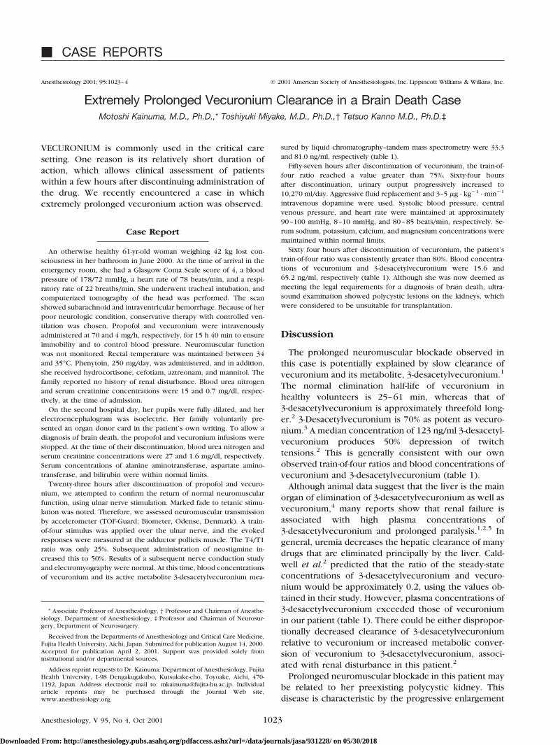
Twenty-three hours after discontinuation of propofol and vecuro-nium, we attempted to confirm the return of normal neuromuscularfunction, using ulnar nerve stimulation. Marked fade to tetanic stimu-lation was noted. Therefore, we assessed neuromuscular transmissionby accelerometer (TOF-Guard; Biometer, Odense, Denmark). A train-of-four stimulus was applied over the ulnar nerve, and the evokedresponses were measured at the adductor pollicis muscle. The T4/T1ratio was only 25%. Subsequent administration of neostigmine in-creased this to 50%. Results of a subsequent nerve conduction studyand electromyography were normal. At this time, blood concentrationsof vecuronium and its active metabolite 3-desacetylvecuronium mea-
sured by liquid chromatography–tandem mass spectrometry were 33.3and 81.0 ng/ml, respectively (table 1).
Fifty-seven hours after discontinuation of vecuronium, the train-of-four ratio reached a value greater than 75%. Sixty-four hoursafter discontinuation, urinary output progressively increased to10,270 ml/day. Aggressive fluid replacement and 3–5 �g · kg�1 · min�1
intravenous dopamine were used. Systolic blood pressure, centralvenous pressure, and heart rate were maintained at approximately90–100 mmHg, 8–10 mmHg, and 80–85 beats/min, respectively. Se-rum sodium, potassium, calcium, and magnesium concentrations weremaintained within normal limits.
Sixty four hours after discontinuation of vecuronium, the patient’strain-of-four ratio was consistently greater than 80%. Blood concentra-tions of vecuronium and 3-desacetylvecuronium were 15.6 and65.2 ng/ml, respectively (table 1). Although she was now deemed asmeeting the legal requirements for a diagnosis of brain death, ultra-sound examination showed polycystic lesions on the kidneys, whichwere considered to be unsuitable for transplantation.
Discussion
The prolonged neuromuscular blockade observed inthis case is potentially explained by slow clearance ofvecuronium and its metabolite, 3-desacetylvecuronium.1
The normal elimination half-life of vecuronium inhealthy volunteers is 25–61 min, whereas that of3-desacetylvecuronium is approximately threefold long-er.2 3-Desacetylvecuronium is 70% as potent as vecuro-nium.3 A median concentration of 123 ng/ml 3-desacetyl-vecuronium produces 50% depression of twitchtensions.2 This is generally consistent with our ownobserved train-of-four ratios and blood concentrations ofvecuronium and 3-desacetylvecuronium (table 1).
Although animal data suggest that the liver is the mainorgan of elimination of 3-desacetylvecuronium as well asvecuronium,4 many reports show that renal failure isassociated with high plasma concentrations of3-desacetylvecuronium and prolonged paralysis.1,2,5 Ingeneral, uremia decreases the hepatic clearance of manydrugs that are eliminated principally by the liver. Cald-well et al.2 predicted that the ratio of the steady-stateconcentrations of 3-desacetylvecuronium and vecuro-nium would be approximately 0.2, using the values ob-tained in their study. However, plasma concentrations of3-desacetylvecuronium exceeded those of vecuroniumin our patient (table 1). There could be either dispropor-tionally decreased clearance of 3-desacetylvecuroniumrelative to vecuronium or increased metabolic conver-sion of vecuronium to 3-desacetylvecuronium, associ-ated with renal disturbance in this patient.2
Prolonged neuromuscular blockade in this patient maybe related to her preexisting polycystic kidney. Thisdisease is characteristic by the progressive enlargement
* Associate Professor of Anesthesiology, † Professor and Chairman of Anesthe-siology, Department of Anesthesiology, ‡ Professor and Chairman of Neurosur-gery, Department of Neurosurgery.
Received from the Departments of Anesthesiology and Critical Care Medicine,Fujita Health University, Aichi, Japan. Submitted for publication August 14, 2000.Accepted for publication April 2, 2001. Support was provided solely frominstitutional and/or departmental sources.
Address reprint requests to Dr. Kainuma: Department of Anesthesiology, FujitaHealth University, 1-98 Dengakugakubo, Kutsukake-cho, Toyoake, Aichi, 470-1192, Japan. Address electronic mail to: [email protected]. Individualarticle reprints may be purchased through the Journal Web site,www.anesthesiology.org.
Anesthesiology, V 95, No 4, Oct 2001 1023
Downloaded From: http://anesthesiology.pubs.asahq.org/pdfaccess.ashx?url=/data/journals/jasa/931228/ on 05/30/2018
of a portion of renal tubule segments, typically causingrenal insufficiency by the fifth or sixth decade of life.6 Itis often accompanied by cerebral aneurysms.6 Potentialrenal parenchymal ischemia may be caused by cyst ex-pansion, which might worsen along with progression ofdiabetes insipidus in this case.
Other possible coexistent factors might influenceclearance of vecuronium and 3-desacetylvecuronium inthis patient. Metabolic acidosis and electrolyte abnormal-ities, including hypermagnesemia, are reportedly vari-ables associated with prolonged neuromuscular block-ade.1 However, these values were meticulouslycorrected. Phenytoin might alter the neuromuscular re-sponse to vecuronium.7 Administration of corticoste-roids to patients with long-term use of nondepolarizingneuromuscular agents has been implicated as a cause ofprolonged muscle weakness.8,9 Mannitol might also in-fluence the clearance of vecuronium.10 However, insuf-ficient data are available regarding these issues to permitfurther speculation.
In Japan, the diagnosis of brain death requires confir-mation of no residual effect of muscle relaxants becausethese are believed to potentially influence the decision-
making process, e.g., interfering with examination of theswallowing reflex.11 The measurement of blood concen-trations of vecuronium and 3-desacetylvecuronium werenot mandated as part of legal decision making in thiscase, but their measurement fortuitously revealed theextremely prolonged clearance of vecuronium and itsprimary metabolite.
The authors thank Naosuke Sugai, M.D., Ph.D. (Department of Anesthesiology,Chigasaki Tokushukai Medical Center, Kanagawken, Japan), for valuable com-ments in preparing the manuscript.
References
1. Segredo V, Caldwell JE, Matthay MA, Sharma ML, Gruenke LD, Miller RD:Persistent paralysis in critically ill patients after long-term administration ofvecuronium. N Engl J Med 1992; 327:524–8
2. Caldwell JE, Szenohradszky J, Segredo V, Wright PMC, McLoughlin C,Sharma ML, Gruenke LD, Fisher DM, Miller RD: The pharmacodynamics andpharmacokinetics of the metabolite 3-desacetylvecuronium (ORG 7268) and itsparent compound, vecuronium, in human volunteers. J Pharmacol Exp Therap1994; 270:1216–22
3. Marshall IG, Gibb AJ, Durant NN: Neuromuscular and vagal blocking actionsof pancuronium bromide, its metabolites, and vecuronium bromide (Org NC45)and its potential metabolites in the anesthetized cat. Br J Anesth 1983; 55:703–14
4. Segredo V, Shin YS, Sharma ML, Gruenke LD, Caldwell JE, Khuenl-Brady KS,Agoston S, Miller RD: Pharmacokinetics, neuromuscular effects, and biodisposi-tion of 3-desacetylvecuronium (Org 7268) in cats. ANESTHESIOLOGY 1991; 74:1052–9
5. Lynam DP, Cronnelly R, Castagnoli KP, Canfell PC, Caldwell J, Arden J,Miller RD: The pharmacodynamics and pharmacokinetics of vecuronium inpatients anesthetized with isoflurane with normal renal function or with renalfailure. ANESTHESIOLOGY 1988; 69:227–31
6. Grantham JJ: Polycystic kidney disease: A predominance of giant nephrons.Am J Physiol 1983; 244:F3–10
7. Platt Pr, Thackray NM: Phenytoin-induced resistance to vecuronium. An-aesth Intensive Care 1993; 21:185–91
8. Barohn RJ, Jackson CE, Rogers SJ, Ridings LW, McVey AL: Prolongedparalysis due to nondepolarizing neuromuscular blocking agents and corticoste-roids. Muscle Nerve 1994; 17:647–54
9. Kindler CH, Verotta D, Grey AT, Gropper MA, Yost CS: Additive inhibitionof nicotinic acetylcholine receptors by corticosteroids and the neuromuscularblocking drug vecuronium. ANESTHESIOLOGY 2000; 92:821–32
10. Matteo RS, Nishitateno K, Pua EK, Spector S: Pharmacokinetics of d-tubocurarine in man: effect of an osmotic diuretic on urinary excretion. ANESTHE-SIOLOGY 1980; 52:335–8
11. Isono S, Ide K, Kochi T, Mizuguchi T, Nishino T: Effects of partial paralysison the swallowing reflex in conscious humans. ANESTHESIOLOGY 1991; 75:980–4
Table 1. Blood Concentrations of Vecuronium and 3-Desacetylvecuronium after Cessation of IntravenousVecuronium Administration
Time afterDiscontinuation
(h)TOF Ratio
(%)Vecuronium
(ng/ml)3-Desacetylvecuronium
(ng/ml)
23 (serum) 25 33.3 81.043 (serum) 28 31.1 91.145 (serum) 45 27.2 84.245 (urine) 45 47.1 127.364 (serum) 82 15.6 65.2
TOF � train-of-four.
1024 CASE REPORTS
Anesthesiology, V 95, No 4, Oct 2001
Downloaded From: http://anesthesiology.pubs.asahq.org/pdfaccess.ashx?url=/data/journals/jasa/931228/ on 05/30/2018
Anesthesiology 2001; 95:1025–6 © 2001 American Society of Anesthesiologists, Inc. Lippincott Williams & Wilkins, Inc.
Successful Resuscitation of a Child after Exsanguination Due toAortoesophageal Fistula from Undiagnosed Foreign Body
Eckehard A. E. Stuth, M.D.,* Astrid G. Stucke, M.D.,† Roger D. Cohen, M.D.,‡ Robert D. B. Jaquiss, M.D.,§Subra Kugathasan, M.D.,� S. Bert Litwin, M.D.#
FOREIGN bodies in the esophagus can cause pressurenecrosis of the esophageal wall that may lead to perfo-ration, mediastinitis, and formation of an aortoesopha-geal fistula (AEF).1 Patients may present with the typical“Chiari triad” of midthoracic pain, a sentinel hemorrhageof bright red blood, and exsanguination hours to dayslater.2
We report the successful resuscitation of a child whohad exsanguination from an undiagnosed AEF caused byunsuspected foreign body ingestion. We believe that ourpatient’s survival was due to immediate, aggressive vol-ume resuscitation and temporary control of the fistula byinflation of an intraoperatively placed esophageal bal-loon catheter.
Case Report
A 7-yr-old, 35-kg girl with a history of epilepsy treated with valproicacid presented to an outlying hospital after she had vomited smallamounts of dark brown blood. Her only complaints were moderatemidepigastric pain on deep palpation. The patient’s hematocrit was 31.After a second emesis of moderate amounts of dark brown blood, shewas transferred to Children’s Hospital of Wisconsin (Milwaukee, WI).With vigorous hydration of 45 ml/kg isotonic fluid in the emergencyroom, her hematocrit dropped to 18. Blood was ordered, a nasogastrictube was placed while the patient was in the emergency room, andsaline lavage yielded only scantly blood-stained fluid. A tentative diag-nosis of gastric ulcer was made, and she was transported to theoperating room for urgent endoscopy without any further routineworkup or radiography. Transfusion of 250 ml cross-matched packederythrocytes was begun in the preoperative area. Suddenly, the childsat up and began to vomit bright red blood profusely. She was rushedto the operating room, and resuscitation with blood and fluids wasintensified. Throughout, the child was conscious and followedcommands.
A rapid-sequence induction with intravenous ketamine (60 mg) andsuccinylcholine (40 mg) was performed. The trachea was intubatedwith a cuffed 6.0-mm-ID endotracheal tube. Midazolam (2 mg) androcuronium (50 mg) were administered, and the patient underwent
ventilation with 100% oxygen. The patient continued to lose massiveamounts of blood from the mouth, and despite ongoing fluid resusci-tation through two peripheral intravenous cannulae (22 and 20 gauge)in the left and right arms, she sustained a cardiac arrest within minutes.Central pulses were absent, the pulse oximeter failed, and end-tidalcarbon dioxide values decreased to single digits, while the electrocar-diogram showed a sinus tachycardia of 180 beats/min. Chest compres-sions were begun and were continued for about 5 min. During 17 minof resuscitation, 900 mg CaCl2, 1 mg epinephrine, 750 ml packederythrocytes, and 500 ml albumin, 5%, were administered, and anepinephrine infusion (0.1–1.0 �g · kg�1 · min�1) was started. Thesemeasures resulted in a return of faint femoral pulses and end-tidalcarbon dioxide but also in an increase in oral blood loss, which madeendoscopy impossible. An emergency laparotomy was performed withthe presumption that the bleeding source was gastroduodenal.
The stomach was massively distended with clotted and fresh blood.No source of gastric bleeding was found, but there was a steady flowof bright red blood from the esophagus. After attempted esophagos-copy, a dark, stained 25-cent coin was found in the opened stomach.The esophageal bleeding remained brisk, and a pumping bleedingsource was noticed by the endoscopist in the mid esophagus about18 cm from the incisors. A 24-French Foley balloon catheter wasintroduced retrograde by the surgeon, and after 15-ml balloon inflation,the bleeding markedly decreased. This measure allowed the anesthesiateam to catch up with the blood loss and allowed the surgical team toestablish invasive monitoring. The epinephrine infusion was stopped.Another surgical attempt to locate the source of bleeding by deflationof the Foley catheter resulted in very brisk rebleeding and wasabandoned.
The cardiothoracic surgeons were consulted. They made a presump-tive diagnosis of an AEF and approached the aorta via left thoracot-omy. Extensive friable granulation tissue was encountered betweenthe esophagus and the distal aortic arch. To better visualize the oper-ating field without compromising distal perfusion during aortic cross-clamping, the patient was placed on partial cardiopulmonary bypasswith cannulae in the left atrium and the mid descending thoracic aorta.Throughout the whole bypass procedure, the heart continued to beat,providing perfusion to the cerebral and coronary circulations. Thepatient was fully heparinized (14,000 IU) and was kept mildly hypo-thermic (34–35°C). Distal perfusion pressure was monitored by asurgically placed femoral arterial line, and proximal pressures wereobtained noninvasively by oscillotonometry. A right radial arterial linewould have been ideal during partial bypass, but could not be placed.A left radial arterial line was easily placed but was significantly bluntedduring aortic cross-clamping. A 4-mm AEF was found 0.5 cm distal tothe left subclavian artery. The fistula was isolated between clampsplaced on the mid transverse arch, i.e., between the left carotid andsubclavian arteries, and the descending thoracic aorta, respectively.The esophagus was closed primarily using a small segment of aorticwall, which was left attached to the esophagus. The diseased segmentof aorta was replaced with a 14-mm Dacron graft, presoaked in anti-biotic solution. Aortic clamp time was 35 min, and partial bypass timewas 40 min. The patient was weaned from partial bypass withoutdifficulty. After closure of the thorax, a feeding gastrostomy tube wasplaced, and the abdomen was closed. The total volume of fluids andblood products infused was 3,000 ml packed erythrocytes, 500 mlfresh frozen plasma, 3 four-donor units (� 150 ml) of platelets,
* Associate Professor, † Research Fellow, Department of Anesthesiology, ‡ As-sociate Professor, Department of Anesthesiology, Division of Pediatric Surgery,§ Assistant Professor, Division of Cardiothoracic Surgery, � Assistant Professor, De-partment of Pediatrics, # Clinical Professor, Division of Cardiothoracic Surgery,Medical College of Wisconsin.
Received from the Sections of Pediatric Anesthesia, Pediatric Surgery, andPediatrics, Children’s Hospital of Wisconsin, Milwaukee, Wisconsin, and theDepartment of Anesthesiology and Anesthesia Research Service, Medical Collegeof Wisconsin, Milwaukee, Wisconsin. Submitted for publication December 28,2000. Accepted for publication April 2, 2001. Support was provided solely frominstitutional and/or departmental sources.
Address reprint requests to Dr. Stuth: Children’s Hospital of Wisconsin, Sec-tion of Pediatric Anesthesia, Milwaukee, Wisconsin 53226. Address electronicmail to: [email protected]. Individual article reprints may be purchased throughthe Journal Web site, www.anesthesiology.org.
1025CASE REPORTS
Anesthesiology, V 95, No 4, Oct 2001
Downloaded From: http://anesthesiology.pubs.asahq.org/pdfaccess.ashx?url=/data/journals/jasa/931228/ on 05/30/2018
1,500 ml albumin (5%), and 4,000 ml lactated Ringer’s solution, total-ing an estimated blood loss replacement of 2.5 blood volumes. Thepatient underwent extubation on postoperative day 2 and was dis-charged home after 2 weeks. Follow-up clinical examination andesophagography 3 months later were unremarkable.
Discussion
An AEF is a rare cause of bleeding in the upper gastro-intestinal tract. The few cases reported in children wereassociated with congenital vascular rings or foreign bodyingestion.3–5 All reported children with AEF after foreignbody ingestion died except for an 8-month-old child inwhich an extended laparotomy and esophagotomy wereperformed until the bleeding site was found and manu-ally compressed.5 In our case, the AEF was diagnosedafter gastrotomy, and the bleeding was successfully tam-ponaded with a Foley catheter. The fistula was thenrepaired through a left thoracotomy using partial cardio-pulmonary bypass.
Foreign body ingestion is common in children and maypresent with atypical symptoms.3 In many cases, parentsreport suspicion of foreign body ingestion. However, inthis child, the bleeding was attributed to a gastric ulcer,and no specific questions were asked. In addition, chestradiography was omitted in both emergency depart-ments. This case suggests that in the absence of a defi-nite cause, a portable chest radiograph should be ob-tained routinely and early in a child with uppergastrointestinal bleeding. It does not delay further ther-apy and might yield unexpected results. Placement of anasogastric tube with the significant risk of dislodgingthe foreign body and causing additional bleeding clearlywould have been avoided.
In the current case, the sequence of events was deter-mined by the profuse bleeding that started in the preop-erative area and demanded vigorous fluid resuscitation.We assume that the bleeding slowed down during thearrest period because of tamponade or closure of theAEF with clot or surrounding tissue. After the source ofthe bleeding was identified, balloon tamponade of the
esophagus with a Foley catheter proved to be an effec-tive method to tamponade the bleeding site and allowedus to reestablish an adequate circulating blood volume.Esophageal compression, usually with a Sengstaken-Blakemore tube, has been described in adults for thesame purpose and allowed for surgery to close thefistula.1,6
Had radiography been performed in this child, theknowledge of a foreign body in the esophagus, togetherwith hematemesis, could have led to the tentative diag-nosis of vascular erosion. However, given the rare oc-currence of AEF, the most likely next step would havebeen a rigid esophagoscopy and an attempt to removethe coin. This probably would have led to rebleedingwith possible exsanguination as has been described byGilchrist et al.3 Placement of a balloon catheter fromabove may have also tamponaded such bleeding. Esoph-ageal balloon tamponade seems to offer the best chanceto stop massive arterial bleeding from an AEF so thatsurgical repair can be performed.
In summary, we report the successful resuscitation andrecovery of a child with a previously undiagnosed aor-toesophageal fistula. Despite initial exsanguination,massive volume resuscitation, cardiac massage, andesophageal balloon tamponade allowed temporary car-diovascular stabilization of the child and the perfor-mance of a definitive surgical repair.
References
1. Amin S, Luketich J, Wald A: Aortoesophageal fistula: Case report and reviewof the literature. Dig Dis Sci 1998; 43:1665–71
2. Hollander JE, Quick G: Aortoesophageal fistula: A comprehensive review ofthe literature. Am J Med 1991; 91:279–86
3. Gilchrist BF, Valerie EP, Nguyen M, Coren C, Klotz D, Ramenofsky ML:Pearls and perils in the management of prolonged, peculiar, penetrating esoph-ageal foreign bodies in children. J Pediatr Surg 1997; 32:1429–31
4. Sigalet DL, Laberge JM, DiLorenzo M, Adolph V, Nguyen LT, Youssef S,Guttman FM: Aortoesophageal fistula: Congenital and acquired causes. J PediatrSurg 1994; 29:1212–4
5. McComas BC, van Miles P, Katz BE: Successful salvage of an 8-month-oldchild with an aortoesophageal fistula. J Pediatr Surg 1991; 26:1394–5
6. Heckstall RL, Hollander JE: Aortoesophageal fistula: Recognition and diag-nosis in the emergency department. Ann Emerg Med 1998; 32:502–5
1026 CASE REPORTS
Anesthesiology, V 95, No 4, Oct 2001
Downloaded From: http://anesthesiology.pubs.asahq.org/pdfaccess.ashx?url=/data/journals/jasa/931228/ on 05/30/2018
Anesthesiology 2001; 95:1027–8 © 2001 American Society of Anesthesiologists, Inc. Lippincott Williams & Wilkins, Inc.
Sequential Compression Devices Can Cause Erroneous CardiacOutput Measurements
Seda B. Akinci, M.D.,* Peter Pronovost, M.D., Ph.D.,† Todd Dorman, M.D., F.C.C.M.‡
LOWER limb pneumatic compression devices are recom-mended to prevent venous thromboembolism in manyintensive care unit patients.1 These pneumatic stockingsincrease lower limb venous return, causing acute buttransient decrease in pulmonary artery blood tempera-ture. Although they increased the variability betweenindividual measurements, the use of these stockings wasreported not to affect the accuracy of thermodilutioncardiac output (CO) measurements made using 10 mlroom temperature injectate.2,3
We present a case in which the use of a pneumaticsequential compression device affected CO measure-ments, thereby delaying decision making, misguidingtherapy, and increasing resource use.
Case Report
A 70-yr-old man was transferred emergently to the surgical intensivecare unit from an outside hospital for management of a perforatedesophagus secondary to dilatation for a peptic stricture. He had asignificant medical history for hypertension and for coronary arterydisease with coronary artery bypass grafting 21 and 11 yr previously.All saphenous vein grafts and native vessels were occluded. His inter-nal mammary graft was his only patent cardiac vessel. His left ventric-ular function was estimated to be normal (57%). He had poor exercisetolerance and frequent angina; his medications included atenolol, hy-drochlorothiazide, nitroglycerine, and furosemide.
A sequential compression device (SCD) with long sleeves with45 mmHg (Sequel model 7325; Kendall Co., Mansfield, MA) was ap-plied to each leg as part of routine practice for admission to oursurgical intensive care unit.
He was taken to the operating room for a diverting cervical esopha-gostomy, exploratory laparotomy, and reduction of diaphragmatic her-nia as well as placement of a gastrostomy tube. His intraoperativecourse was uneventful. He arrived back in the intensive care unitintubated, and mechanical ventilation was started.
During the next 6 h, a significant capillary leak developed in thepatient, requiring large-volume crystalloid resuscitation (total intake �total output � �6 l), after which we decided to place a pulmonaryartery catheter to assist with fluid management. A pulmonary arterythermodilution catheter (PAC; Baxter Swan Ganz ref. 131HF7, ther-
modilution catheter with antimicrobial coating (AMC) thromboshield-antimicrobial heparin coating 7F; Baxter Healthcare Corp., EdwardsCritical Care Division, Irvine, CA) was placed uneventfully by rewiringof the previously placed right internal jugular central venous catheter.The PAC was connected to the CO module (7200 TRAM AR model No.S7200 Tram Module) of the monitor (series 7010 monitors; MarquetteElectronics Inc., Milwaukee, WI).
After placement of the PAC, the patient had an arterial blood pres-sure of 91/46 mmHg, a heart rate of 100 beats/min, a pulmonary arterypressure of 39/20 mmHg, a central venous pressure of 6 mmHg, atemperature of 38.8°C, a CO of 3.7 l/min with an index of 1.8, apulmonary artery wedge pressure of 6 mmHg, a stroke volume of37 ml, and a systemic vascular resistance of 1,189 dyn · s�1 · cm5.
The effect of further fluid resuscitation and vasoactive support onCO could not be assessed because the nursing staff reported variabilityof the CO measurements. The individual CO measurements variedbetween 1.8 and 10.4 l/min. Different physicians and nurses sawerratic waveforms with multiple attempts to measure CO. Withoutinfusing injectate, the bedside cardiac monitor showed cycling outputwaveforms that varied between 3.5 and 5.0 l/min. Despite changingthe monitor, changing the PAC, and having a biomedical engineerinvestigate the monitor, the problem persisted.
During this struggle with the CO measurements, care for the patientwas guided by clinical assessment. After 4 h, the cause of the erraticwaveforms was found to be from the cycling of the SCDs. We turnedthe SCDs off, and CO was measured to be 5.7 l/min with an indexof 2.7, stroke volume was 63 ml, systemic vascular resistance was814 dyn · s�1 · cm5 when blood pressure was 100/51 mmHg, heart ratewas 90 beats/min, pulmonary artery pressure was 39/22 mmHg, andcentral venous pressure was 11 mmHg. Core temperature measured byPAC was 38.3°C, and injectate temperature was 24.7°C. The skin of thelower extremities was cool, with a toe temperature of 33.1°C, and thepatient had 2� pitting edema of the lower extremities.
We took several steps to establish a causal relation between SCDcompression and the autonomous CO. First, we applied the SCDs toone leg and obtained an output from the CO computer of 5.8 l/min,with a stable baseline and good waveforms. Second, we reapplied theSCDs to two legs again; erratic waveforms and cycling autonomousoutput from CO computer reoccurred again. Third, when the full-legSCDs were replaced with calf SCDs, erratic waveforms disappeared.Finally, we reapplied the full-leg SCDs, and the autonomous output COcomputer reappeared and varied between 3.5 and 5.3 l/min. The restof the CO measurements were obtained while the SCDs were tempo-rarily turned off. The patient resolved the inflammatory response andwas discharged from the intensive care unit 5 days later.
Discussion
Lower limb pneumatic compression devices are com-monly used in intensive care units to prevent venousthromboembolism. We present a patient in whom webelieve sequential compression devices caused errone-ous CO measurements.
When using a PAC, CO is inversely proportional to thearea under the thermodilution curve and is determinedby injectate volume, blood temperature, density factor,
* Critical Care Fellow, Department of Anesthesiology and Critical Care Medi-cine, † Assistant Professor, Departments of Anesthesiology and Critical CareMedicine, Surgery, and Health Policy and Management, ‡ Associate Professor,Departments of Anesthesiology and Critical Care Medicine, Surgery and Medi-cine, The Johns Hopkins University School of Medicine; joint appointment: JohnsHopkins University School of Nursing.
Received from The Johns Hopkins Medical Institution, Baltimore, Maryland.Submitted for publication December 12, 2000. Accepted for publication April 12,2001. Supported by the Department of Anesthesiology and Critical Care Medi-cine, Johns Hopkins School of Medicine, Baltimore, Maryland.
Address reprint requests to Dr. Akinci: Hacettepe University, Department ofAnesthesiology, Sihhiye Ankara, Turkey 06100. Address electronic mail to:[email protected]. Individual article reprints may be purchased through theJournal Web site, www.anesthesiology.org.
1027CASE REPORTS
Anesthesiology, V 95, No 4, Oct 2001
Downloaded From: http://anesthesiology.pubs.asahq.org/pdfaccess.ashx?url=/data/journals/jasa/931228/ on 05/30/2018
and computation constant. In the current patient, themeasure of CO was significantly impacted by the use ofSCDs. Given that the thermistor on the PAC measurestemperature changes, we hypothesize that the inflationcycle of the compression stockings brought cool bloodfrom the legs into the central circulation, resulting in adecrease in pulmonary artery blood temperature. In thisextreme example, because the patient was febrile andedematous with a low stroke volume, the volume of coolblood from the two legs squeezed by the long-sleeveSCD was enough to create a CO waveform and COcalculation by the computer. Neither one-leg SCD norcalf SCD compression produced enough volume to cre-ate a CO waveform by itself. We postulate that sepsis andthe ensuing volume resuscitation may have resulted incool extremities and that the patient’s low flow stateamplified the impact of cool fluid being delivered to thecentral circulation.
Horiuchi et al.2 found a larger coefficient of variationon the pulmonary artery pressures when the calf pneu-matic compression devices were used. In one patient inwhom temperature changes in pulmonary artery bloodwere compared between the compression device onone or both legs, less temperature change was notedwhen the device was applied to one leg. They postulatedthat the larger the amount of cool blood that returns, thegreater the variability in temperature. Despite this vari-ation, CO was unaffected by the use of an intermittentcompression device. Nonetheless, the variability was
greater when they used room temperature injectate in-stead of iced injectate. With the higher temperature ofinjectate, the decrease in pulmonary artery temperaturewould be less than that observed with ice-coldinjectate.2
In the current patient, the impact of the transientdecrease in pulmonary artery temperature from the pe-ripheral pooled venous blood was perceived as a newCO curve by the computer. One would also expect thatlower stroke volumes would result in less warm bloodbeing ejected from the heart, thus increasing the effectof cool blood being returned from the limbs.3
The use of SCDs is important for preventing deepvenous thromboses in critically ill patients and will likelyremain a mainstay of patient care in intensive care units.Physicians need to be aware of the potential for full-legSCDs to impact thermodilution CO measurements andmay need to consider temporarily turning SCDs off dur-ing CO measurements when these measurements arehighly variable.
References
1. Goldhaber SZ: Medical progress: Pulmonary embolism. N Engl J Med 1998;339:93–104
2. Horiuchi K, Johnson R, Weissman C: Influence of lower limb pneumaticcompression on pulmonary artery temperature: Effect on cardiac output mea-surements. Crit Care Med 1999; 27:1096–9
3. Hickey R: Pneumatic compression stockings increase the variability ofthermodilution cardiac output measurements: Do they truly affect cardiac out-put? Crit Care Med 1999; 27:1039–41
Anesthesiology 2001; 95:1028–31 © 2001 American Society of Anesthesiologists, Inc. Lippincott Williams & Wilkins, Inc.
Anesthesia for Cesarean Delivery in a Patient with anUndiagnosed Traumatic Diaphragmatic Hernia
Cynthia L. Dietrich, D.O.,* Charles E. Smith, M.D., F.R.C.P.C.†
DELAYED diagnosis of traumatic diaphragmatic injuriesmay result in gastric or intestinal herniation and intesti-nal obstruction.1,2 If the hernia becomes strangulated,ischemia, necrosis, and sepsis may develop. Pulmonaryand cardiac complications occur because of compres-sion of the lung and heart by the herniated organs orherniation of organs into the pericardial sac.3–5 Rarely,perforation of herniated organs may lead to pneumotho-rax and pneumomediastinum.6
Delayed presentation of traumatic diaphragmatic her-nia occurs because there are no distinctive symptoms orsigns associated with diaphragmatic injuries and becausethese injuries can be difficult to diagnose without lapa-rotomy, which is usually performed for other acute in-juries.7 Occasionally, herniation may only begin longafter the initial injury, and the hernia may then rapidlyenlarge within days or weeks.2,8 Increased intraabdomi-nal pressure during the second and third trimesters ofpregnancy and during labor may also predispose to her-niation and strangulation.8 We report a patient withundiagnosed diaphragmatic hernia who required urgentcesarean delivery for nonreassuring fetal status. The di-agnosis was first suspected during preanesthetic evalua-tion of the patient. The postoperative course was com-plicated by respiratory failure, pericardial tamponade,intestinal ischemia, and sepsis.
* Assistant Professor, † Associate Professor.
Received from the Faculty of Medicine, Case Western Reserve University,Department of Anesthesia, MetroHealth Medical Center, Cleveland, Ohio.Submitted for publication January 31, 2001. Accepted for publication April 16,2001. Funding for library, photography, and computer services was provided bythe Department of Anesthesia, MetroHealth Medical Center.
Address reprint requests to Dr. Smith: Department of Anesthesia, MetroHealthMedical Center, 2500 MetroHealth Drive, Cleveland, Ohio 44124. Address elec-tronic mail to: [email protected]. Individual article reprints may be pur-chased through the Journal Web site, www.anesthesiology.org.
1028 CASE REPORTS
Anesthesiology, V 95, No 4, Oct 2001
Downloaded From: http://anesthesiology.pubs.asahq.org/pdfaccess.ashx?url=/data/journals/jasa/931228/ on 05/30/2018
Case Report
A 19-yr-old gravida 3 para 0 woman, 361⁄7 weeks’ gestational age inactive labor, was transferred to our institution with a 2-day history ofsevere left upper quadrant pain and dyspnea. She had sustained a stabwound to the left side of the chest 3 yr ago and did not require surgicalintervention. Past history included appendectomy and asthma. Therehad been no asthma exacerbations for the past 2 yr, and she was nottaking daily asthma medications. Her previous two pregnancies had endedin miscarriages at 5 months (3 yr previously) and at 4 months (1 yrpreviously), respectively. Medications were prenatal vitamins and iron.
Blood pressure was 121/83 mmHg, pulse was 131 beats/min, respi-ration was 28 breaths/min, temperature was 36.7°C, oxygen saturationwas 96% on 4 l O2 via nasal cannula. Breath sounds were absent on theleft side, and the trachea was deviated to the right. There was nowheezing. Cardiovascular examination showed tachycardia, and thepoint of maximal impulse was displaced to the right. Fetal heart ratewas decreased with late decelerations. The patient was transferred tothe operating room for urgent cesarean delivery. Left-sided uterinedisplacement was performed. However, because of concerns for ma-ternal safety, a portable chest radiograph was obtained, which showedtotal opacification of the left hemithorax with rightward displacementof mediastinal structures.
The membranes were artificially ruptured, and internal fetal heartrate monitoring was instituted. Thoracic and trauma surgery staff wereconsulted. Meperidine, 25 mg, was administered intravenously, and aleft-sided chest tube was inserted using local anesthesia. Approxi-mately 1,200 ml blood-tinged fluid was drained, and clinically, thepatient was less dyspneic. Nonreassuring fetal status persisted, and thescalp pH was 7.20. Thirty milliliters sodium citrate was administeredorally. Spinal anesthesia was performed with the patient in the sittingposition with 12 mg hyperbaric bupivacaine, and a T6 level wasobtained. Forty milligrams intravenous ephedrine, in incrementaldoses, was required to maintain blood pressure between 105/40and 120/50 mmHg. Heart rate was 130–140 beats/min, and oxygensaturation measured by pulse oximetry (SpO2) was 97–100% with10 l O2/min via a nonrebreathing face mask. A healthy female infantwas delivered 4 min after incision. Twenty milligrams intravenousketamine was required for analgesia during delivery.
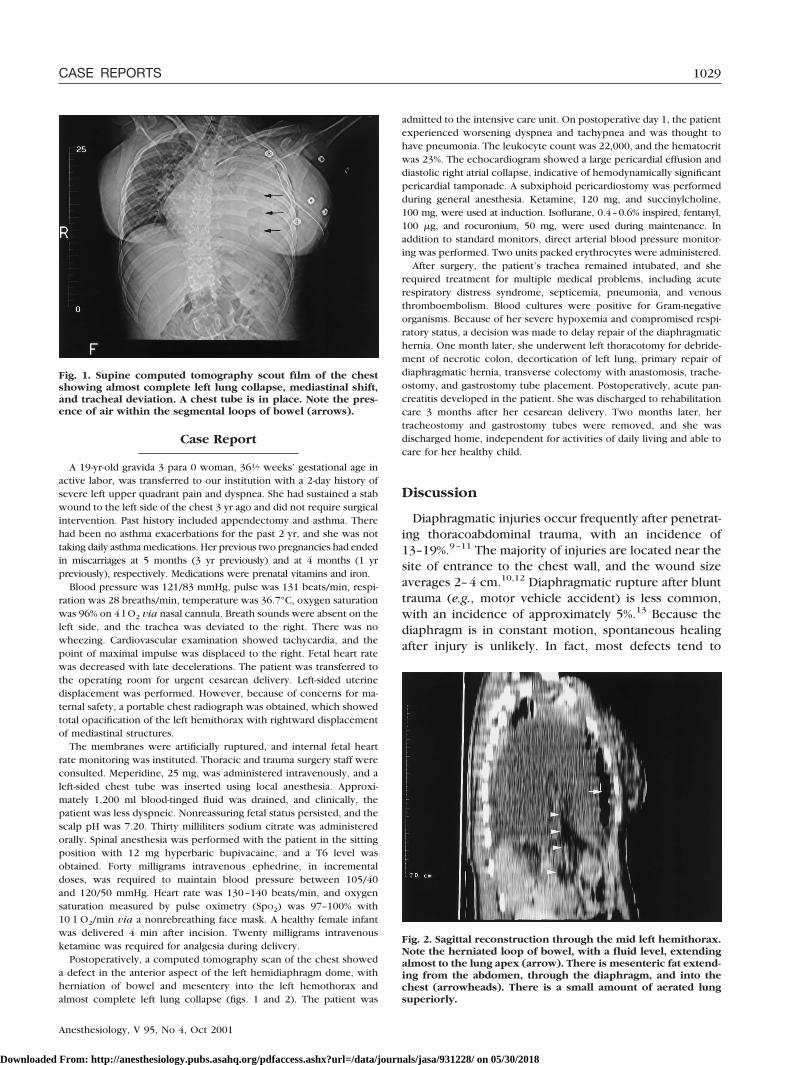
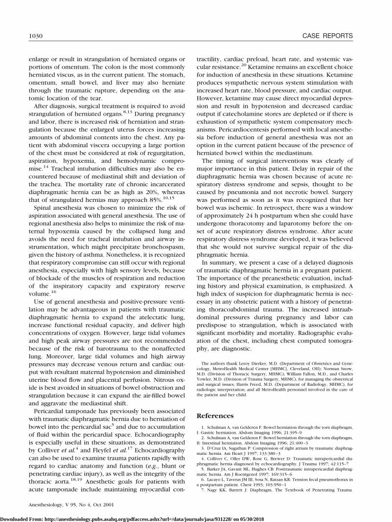
Postoperatively, a computed tomography scan of the chest showeda defect in the anterior aspect of the left hemidiaphragm dome, withherniation of bowel and mesentery into the left hemothorax andalmost complete left lung collapse (figs. 1 and 2). The patient was
admitted to the intensive care unit. On postoperative day 1, the patientexperienced worsening dyspnea and tachypnea and was thought tohave pneumonia. The leukocyte count was 22,000, and the hematocritwas 23%. The echocardiogram showed a large pericardial effusion anddiastolic right atrial collapse, indicative of hemodynamically significantpericardial tamponade. A subxiphoid pericardiostomy was performedduring general anesthesia. Ketamine, 120 mg, and succinylcholine,100 mg, were used at induction. Isoflurane, 0.4–0.6% inspired, fentanyl,100 �g, and rocuronium, 50 mg, were used during maintenance. Inaddition to standard monitors, direct arterial blood pressure monitor-ing was performed. Two units packed erythrocytes were administered.
After surgery, the patient’s trachea remained intubated, and sherequired treatment for multiple medical problems, including acuterespiratory distress syndrome, septicemia, pneumonia, and venousthromboembolism. Blood cultures were positive for Gram-negativeorganisms. Because of her severe hypoxemia and compromised respi-ratory status, a decision was made to delay repair of the diaphragmatichernia. One month later, she underwent left thoracotomy for debride-ment of necrotic colon, decortication of left lung, primary repair ofdiaphragmatic hernia, transverse colectomy with anastomosis, trache-ostomy, and gastrostomy tube placement. Postoperatively, acute pan-creatitis developed in the patient. She was discharged to rehabilitationcare 3 months after her cesarean delivery. Two months later, hertracheostomy and gastrostomy tubes were removed, and she wasdischarged home, independent for activities of daily living and able tocare for her healthy child.
Discussion
Diaphragmatic injuries occur frequently after penetrat-ing thoracoabdominal trauma, with an incidence of13–19%.9–11 The majority of injuries are located near thesite of entrance to the chest wall, and the wound sizeaverages 2–4 cm.10,12 Diaphragmatic rupture after blunttrauma (e.g., motor vehicle accident) is less common,with an incidence of approximately 5%.13 Because thediaphragm is in constant motion, spontaneous healingafter injury is unlikely. In fact, most defects tend to
Fig. 1. Supine computed tomography scout film of the chestshowing almost complete left lung collapse, mediastinal shift,and tracheal deviation. A chest tube is in place. Note the pres-ence of air within the segmental loops of bowel (arrows).
Fig. 2. Sagittal reconstruction through the mid left hemithorax.Note the herniated loop of bowel, with a fluid level, extendingalmost to the lung apex (arrow). There is mesenteric fat extend-ing from the abdomen, through the diaphragm, and into thechest (arrowheads). There is a small amount of aerated lungsuperiorly.
1029CASE REPORTS
Anesthesiology, V 95, No 4, Oct 2001
Downloaded From: http://anesthesiology.pubs.asahq.org/pdfaccess.ashx?url=/data/journals/jasa/931228/ on 05/30/2018
enlarge or result in strangulation of herniated organs orportions of omentum. The colon is the most commonlyherniated viscus, as in the current patient. The stomach,omentum, small bowel, and liver may also herniatethrough the traumatic rupture, depending on the ana-tomic location of the tear.
After diagnosis, surgical treatment is required to avoidstrangulation of herniated organs.8,13 During pregnancyand labor, there is increased risk of herniation and stran-gulation because the enlarged uterus forces increasingamounts of abdominal contents into the chest. Any pa-tient with abdominal viscera occupying a large portionof the chest must be considered at risk of regurgitation,aspiration, hypoxemia, and hemodynamic compro-mise.14 Tracheal intubation difficulties may also be en-countered because of mediastinal shift and deviation ofthe trachea. The mortality rate of chronic incarcerateddiaphragmatic hernia can be as high as 20%, whereasthat of strangulated hernias may approach 85%.10,15
Spinal anesthesia was chosen to minimize the risk ofaspiration associated with general anesthesia. The use ofregional anesthesia also helps to minimize the risk of ma-ternal hypoxemia caused by the collapsed lung andavoids the need for tracheal intubation and airway in-strumentation, which might precipitate bronchospasm,given the history of asthma. Nonetheless, it is recognizedthat respiratory compromise can still occur with regionalanesthesia, especially with high sensory levels, becauseof blockade of the muscles of respiration and reductionof the inspiratory capacity and expiratory reservevolume.16
Use of general anesthesia and positive-pressure venti-lation may be advantageous in patients with traumaticdiaphragmatic hernia to expand the atelectatic lung,increase functional residual capacity, and deliver highconcentrations of oxygen. However, large tidal volumesand high peak airway pressures are not recommendedbecause of the risk of barotrauma to the nonaffectedlung. Moreover, large tidal volumes and high airwaypressures may decrease venous return and cardiac out-put with resultant maternal hypotension and diminisheduterine blood flow and placental perfusion. Nitrous ox-ide is best avoided in situations of bowel obstruction andstrangulation because it can expand the air-filled boweland aggravate the mediastinal shift.
Pericardial tamponade has previously been associatedwith traumatic diaphragmatic hernia due to herniation ofbowel into the pericardial sac5 and due to accumulationof fluid within the pericardial space. Echocardiographyis especially useful in these situations, as demonstratedby Colliver et al.4 and Fleyfel et al.17 Echocardiographycan also be used to examine trauma patients rapidly withregard to cardiac anatomy and function (e.g., blunt orpenetrating cardiac injury), as well as the integrity of thethoracic aorta.18,19 Anesthetic goals for patients withacute tamponade include maintaining myocardial con-
tractility, cardiac preload, heart rate, and systemic vas-cular resistance.20 Ketamine remains an excellent choicefor induction of anesthesia in these situations. Ketamineproduces sympathetic nervous system stimulation withincreased heart rate, blood pressure, and cardiac output.However, ketamine may cause direct myocardial depres-sion and result in hypotension and decreased cardiacoutput if catecholamine stores are depleted or if there isexhaustion of sympathetic system compensatory mech-anisms. Pericardiocentesis performed with local anesthe-sia before induction of general anesthesia was not anoption in the current patient because of the presence ofherniated bowel within the mediastinum.
The timing of surgical interventions was clearly ofmajor importance in this patient. Delay in repair of thediaphragmatic hernia was chosen because of acute re-spiratory distress syndrome and sepsis, thought to becaused by pneumonia and not necrotic bowel. Surgerywas performed as soon as it was recognized that herbowel was ischemic. In retrospect, there was a windowof approximately 24 h postpartum when she could haveundergone thoracotomy and laparotomy before the on-set of acute respiratory distress syndrome. After acuterespiratory distress syndrome developed, it was believedthat she would not survive surgical repair of the dia-phragmatic hernia.
In summary, we present a case of a delayed diagnosisof traumatic diaphragmatic hernia in a pregnant patient.The importance of the preanesthetic evaluation, includ-ing history and physical examination, is emphasized. Ahigh index of suspicion for diaphragmatic hernia is nec-essary in any obstetric patient with a history of penetrat-ing thoracoabdominal trauma. The increased intraab-dominal pressures during pregnancy and labor canpredispose to strangulation, which is associated withsignificant morbidity and mortality. Radiographic evalu-ation of the chest, including chest computed tomogra-phy, are diagnostic.
The authors thank Leroy Dierker, M.D. (Department of Obstetrics and Gyne-cology, MetroHealth Medical Center [MHMC], Cleveland, OH); Norman Snow,M.D. (Division of Thoracic Surgery, MHMC); William Fallon, M.D., and CharlesYowler, M.D. (Division of Trauma Surgery, MHMC), for managing the obstetricaland surgical issues; Harris Freed, M.D. (Department of Radiology, MHMC), forradiologic interpretation; and all MetroHealth personnel involved in the care ofthe patient and her child.
References
1. Schulman A, van Gelderen F: Bowel herniation through the torn diaphragm,I: Gastric herniation. Abdom Imaging 1996; 21:395–9
2. Schulman A, van Gelderen F: Bowel herniation through the torn diaphragm,II: Intestinal herniation. Abdom Imaging 1996; 21:400–3
3. D’Cruz IA, Sugathan P: Compression of right atrium by traumatic diaphrag-matic hernia. Am Heart J 1997; 133:380–3
4. Colliver C, Oller DW, Rose G, Brewer D: Traumatic intrapericardial dia-phragmatic hernia diagnosed by echocardiography. J Trauma 1997; 42:115–7
5. Barker JA, Gavant ML, Hughes CB: Posttraumatic intrapericardial diaphrag-matic hernia. Am J Roentgenol 1997; 169:315–6
6. Lacayo L, Taveras JM III, Sosa N, Ratzan KR: Tension fecal pneumothorax ina postpartum patient. Chest 1993; 103:950–1
7. Nagy KK, Barrett J: Diaphragm, The Textbook of Penetrating Trauma.
1030 CASE REPORTS
Anesthesiology, V 95, No 4, Oct 2001
Downloaded From: http://anesthesiology.pubs.asahq.org/pdfaccess.ashx?url=/data/journals/jasa/931228/ on 05/30/2018
Edited by Ivatury RR, Cayten CG. Baltimore, Williams & Wilkins, 1996, pp564–70
8. Dudley AG, Teaford H, Gatewood TS Jr: Delayed traumatic rupture of thediaphragm in pregnancy. Obstet Gynecol 1979; 53(suppl 3):25S–27S
9. Mariadason JG, Parsa MH, Ayuyao A, Freeman HP: Management of stabwounds to the thoracoabdominal region: A clinical approach. Ann Surg 1988;207:335–40
10. Madden MR, Paull DE, Finkelstein JL, Goodwin CW, Marzulli V, Yurt RW,Shires GT: Occult diaphragmatic injury from stab wounds to the lower chest andabdomen. J Trauma 1989; 29:292–8
11. Moore JB, Moore EE, Thompson JS: Abdominal injuries associated withpenetrating trauma in the lower chest. Am J Surg 1980; 140:724–30
12. Adamthwaite DN: Penetrating injuries of the diaphragm. Injury 1982;14:151–8
13. Barnett PS, Van Dongen LGR, Bremner CG: Traumatic diaphragmatichernia presenting in pregnancy. S Afr Med J 1979; 55:94–5
14. Katz RI, Belenker SL, Poppers PJ: Intraoperative management of a patient
with a chronic, previously undiagnosed traumatic diaphragmatic hernia: J ClinAnesth 1998; 10:506–9
15. Saber WL, Moore EE, Hopeman AR, Aragon WE: Delayed presentation oftraumatic diaphragmatic hernia. J Emerg Med 1986; 4:1–7
16. Freund FG, Bonica JJ, Ward RJ, Akamatsu TJ, Kennedy WF Jr: Ventilatoryreserve and level of motor block during high spinal and epidural anesthesia:ANESTHESIOLOGY 1967; 28:834–7
17. Fleyfel M, Ferreira JF, Gonzalez de Linares H, Merlier O, Harchaoui A:Cardiac tamponade after intrapericardial diaphragmatic hernia. Br J Anaesth1994; 73:249-51
18. Frazee RC, Mucha P, Farnell MB, Miller FA: Objective evaluation of bluntcardiac trauma. J Trauma 1986; 26:510–19
19. Goarin JP, Cluzel P, Gosgnach M, Lamine K, Coriat P, Riou B: Evaluation oftransesophageal echocardiography for diagnosis of traumatic aortic injury. ANES-THESIOLOGY 2000; 93:1373–7
20. Stoelting RK, Dierdorf SF: Anesthesia and Co-Existing Disease, 3rd edition.New York, Churchill Livingstone, 1993, pp 107–12
Anesthesiology 2001; 95:1031–3 © 2001 American Society of Anesthesiologists, Inc. Lippincott Williams & Wilkins, Inc.
Spinal–Epidural Hematoma following Epidural Anesthesia in thePresence of Antiplatelet and Heparin Therapy
Rainer J. Litz, M.D.,* Matthias Hübler, M.D.,† Thea Koch, M.D.,‡ D. Michael Albrecht, M.D.§
ANTIPLATELET drugs are widely used in various catego-ries of patients1; therefore, knowledge of their impact oncoagulation is important for the patients’ perioperativecare. The American Society of Regional Anesthesia andPain Medicine does not consider antiplatelet drugs, bythemselves, as risk factors for the development of spinalhematoma in patients having neuraxial blocks, but con-current use of other medications that affect clottingmechanisms may increase the risk of bleeding complica-tions.2,3 We report a case of an epidural hematoma in apatient to whom low-molecular-weight heparin (LMWH)was administered perioperatively as prophylaxis againstdevelopment of venous thromboembolism and who addi-tionally had taken oral ibuprofen for pain relief on her own.
Case Report
A 63-yr-old woman (weight, 75 kg; height, 165 cm) with AmericanSociety of Anesthesiologists physical status III presented for reimplan-tation of a prosthesis of the right knee. One year previously, theprosthetic joint had been removed because of an infection. At thistime, the patient also had spondylodiscitis of the lumbar vertebrae 4and 5 and of the thoracic vertebra 10 and a paravertebral abscess.Clinically and radiologically, the patient had recovered from this com-plication. The patient’s other significant medical history included anischemic heart disease of grade II (classification of angina of the
Canadian Cardiovascular Society), allergies to several antibiotics, andarthritic pain of both shoulders. Her preoperative oral medicationsincluded 5 mg propanolol twice daily and 150 mg ranitidine oncedaily. Additionally, one subcutaneous injection of LMWH (nadroparin,3,800 IU/day) was administered to the patient as a prophylactic anti-thrombotic treatment. Preoperatively, the activated partial thrombo-plastin time (PTT) was 33 s (normal range, 30–40 s), the prothrombintime (PT) was 84% (normal range, 70–120%), and the InternationalNormalized Ratio (INR) was 1.01 (therapeutic level, 2–4.5). The plate-let count was 151 � 109/l, at the lower limit of the normal range(150 � 109/l–400 � 109/l).
On the day of surgery, the patient did not receive the subcutaneousinjection of LMWH, the last dose of which had been administered 12 hpreviously. She was taken to the operating room, and an indwellingepidural catheter via an 18-gauge Tuohy-type needle (MedimexHolfeld, Hamburg, Germany) was placed at the L3–L4 interspace usingthe “loss-of-resistance” technique. Epidural puncture and insertion ofthe catheter were uneventful, and no blood or spinal fluid wereobserved. A test dose of 4 ml bupivacaine, 0.5%, was injected. Theblock was then extended up to the T6 dermatome using 0.5% bupiv-acaine. General anesthesia was induced, the trachea was intubated,and surgery was performed with a combined regional–general anes-thesia. At the end of surgery, the tracheal tube was removed. Thepatient was awake and free of pain. The sensory level of anesthesiareached T9, and motor block was Bromage scale 2. After reversal of themotor block, a continuous epidural infusion of 0.25% bupivacaine(8 ml/h) was initiated. Thromboprophylactic treatment with LMWHwas reestablished 6 h after the end of surgery. Eighteen hours later, thepatient reported pain of increasing intensity at the site of the surgicalincisions and back pain. The epidural infusion rate was increased to12 ml/h, and two boluses of 12 ml bupivacaine, 0.25%, were adminis-tered, without yielding a sufficient level of analgesia. Additionally, shereceived 400 mg ibuprofen rectally for the back pain, which decreasedin intensity afterward. The epidural bolus administered after this treat-ment was painful during injection. Therefore, the epidural infusionwas stopped and the catheter was removed approximately 7 h after thesecond postoperative LMWH administration. At this time, the coagula-tion parameters of activated partial thromboplastin time, InternationalNormalized Ratio, and prothrombin time were within normal ranges.The platelet count was 118 � 109/l. After catheter removal, the patientreceived patient-controlled intravenous analgesia using piritramid.
Ten hours after removal of the epidural catheter, the intensity of theback pain increased again, and voiding difficulties developed. Neuro-
* Staff Anesthesiologist, † Resident, ‡ Professor of Anesthesiology, § Professorand Chairman.
Received from the Department of Anesthesiology and Intensive Care Medi-cine, Carl-Gustav-Carus University Hospital, Dresden, Germany. Submitted forpublication February 5, 2001. Accepted for publication April 16, 2001. Supportwas provided solely from institutional and/or departmental sources. Presented atthe German Annual Meeting of Anaesthesiologists (DAK), Wiesbaden, Germany,May 5–8, 1999.
Address reprint requests to: Dr. Hübler: Department of Anesthesiologyand Intensive Care Medicine, Carl Gustav Carus University Hospital, Fetscherstr.74, 01307 Dresden, Germany. Address electronic mail to: [email protected]. Individual article reprints may be purchased through theJournal Web site, www.anesthesiology.org.
1031CASE REPORTS
Anesthesiology, V 95, No 4, Oct 2001
Downloaded From: http://anesthesiology.pubs.asahq.org/pdfaccess.ashx?url=/data/journals/jasa/931228/ on 05/30/2018
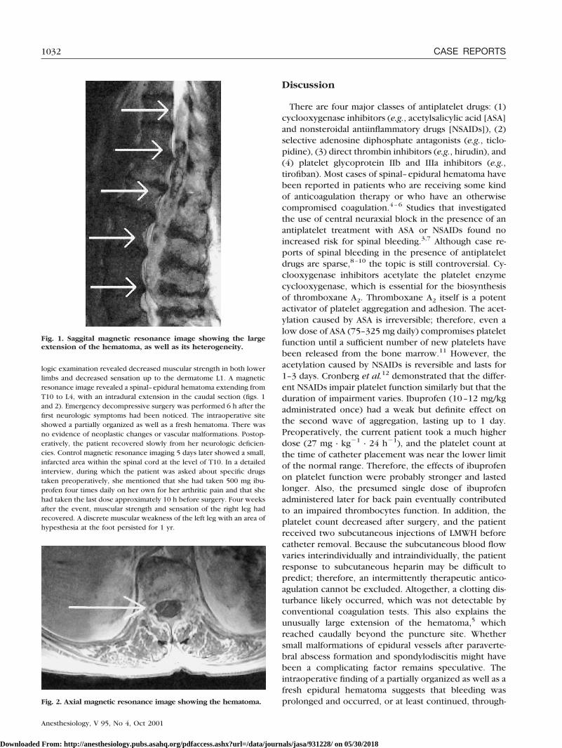
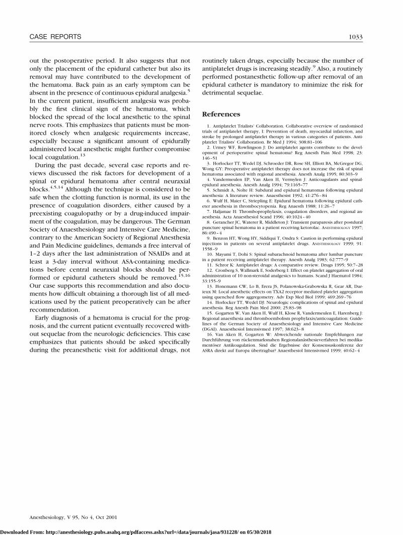
logic examination revealed decreased muscular strength in both lowerlimbs and decreased sensation up to the dermatome L1. A magneticresonance image revealed a spinal–epidural hematoma extending fromT10 to L4, with an intradural extension in the caudal section (figs. 1and 2). Emergency decompressive surgery was performed 6 h after thefirst neurologic symptoms had been noticed. The intraoperative siteshowed a partially organized as well as a fresh hematoma. There wasno evidence of neoplastic changes or vascular malformations. Postop-eratively, the patient recovered slowly from her neurologic deficien-cies. Control magnetic resonance imaging 5 days later showed a small,infarcted area within the spinal cord at the level of T10. In a detailedinterview, during which the patient was asked about specific drugstaken preoperatively, she mentioned that she had taken 500 mg ibu-profen four times daily on her own for her arthritic pain and that shehad taken the last dose approximately 10 h before surgery. Four weeksafter the event, muscular strength and sensation of the right leg hadrecovered. A discrete muscular weakness of the left leg with an area ofhypesthesia at the foot persisted for 1 yr.
Discussion
There are four major classes of antiplatelet drugs: (1)cyclooxygenase inhibitors (e.g., acetylsalicylic acid [ASA]and nonsteroidal antiinflammatory drugs [NSAIDs]), (2)selective adenosine diphosphate antagonists (e.g., ticlo-pidine), (3) direct thrombin inhibitors (e.g., hirudin), and(4) platelet glycoprotein IIb and IIIa inhibitors (e.g.,tirofiban). Most cases of spinal–epidural hematoma havebeen reported in patients who are receiving some kindof anticoagulation therapy or who have an otherwisecompromised coagulation.4–6 Studies that investigatedthe use of central neuraxial block in the presence of anantiplatelet treatment with ASA or NSAIDs found noincreased risk for spinal bleeding.3,7 Although case re-ports of spinal bleeding in the presence of antiplateletdrugs are sparse,8–10 the topic is still controversial. Cy-clooxygenase inhibitors acetylate the platelet enzymecyclooxygenase, which is essential for the biosynthesisof thromboxane A2. Thromboxane A2 itself is a potentactivator of platelet aggregation and adhesion. The acet-ylation caused by ASA is irreversible; therefore, even alow dose of ASA (75–325 mg daily) compromises plateletfunction until a sufficient number of new platelets havebeen released from the bone marrow.11 However, theacetylation caused by NSAIDs is reversible and lasts for1–3 days. Cronberg et al.12 demonstrated that the differ-ent NSAIDs impair platelet function similarly but that theduration of impairment varies. Ibuprofen (10–12 mg/kgadministrated once) had a weak but definite effect onthe second wave of aggregation, lasting up to 1 day.Preoperatively, the current patient took a much higherdose (27 mg · kg�1 · 24 h�1), and the platelet count atthe time of catheter placement was near the lower limitof the normal range. Therefore, the effects of ibuprofenon platelet function were probably stronger and lastedlonger. Also, the presumed single dose of ibuprofenadministered later for back pain eventually contributedto an impaired thrombocytes function. In addition, theplatelet count decreased after surgery, and the patientreceived two subcutaneous injections of LMWH beforecatheter removal. Because the subcutaneous blood flowvaries interindividually and intraindividually, the patientresponse to subcutaneous heparin may be difficult topredict; therefore, an intermittently therapeutic antico-agulation cannot be excluded. Altogether, a clotting dis-turbance likely occurred, which was not detectable byconventional coagulation tests. This also explains theunusually large extension of the hematoma,5 whichreached caudally beyond the puncture site. Whethersmall malformations of epidural vessels after paraverte-bral abscess formation and spondylodiscitis might havebeen a complicating factor remains speculative. Theintraoperative finding of a partially organized as well as afresh epidural hematoma suggests that bleeding wasprolonged and occurred, or at least continued, through-Fig. 2. Axial magnetic resonance image showing the hematoma.
Fig. 1. Saggital magnetic resonance image showing the largeextension of the hematoma, as well as its heterogeneity.
1032 CASE REPORTS
Anesthesiology, V 95, No 4, Oct 2001
Downloaded From: http://anesthesiology.pubs.asahq.org/pdfaccess.ashx?url=/data/journals/jasa/931228/ on 05/30/2018
out the postoperative period. It also suggests that notonly the placement of the epidural catheter but also itsremoval may have contributed to the development ofthe hematoma. Back pain as an early symptom can beabsent in the presence of continuous epidural analgesia.5
In the current patient, insufficient analgesia was proba-bly the first clinical sign of the hematoma, whichblocked the spread of the local anesthetic to the spinalnerve roots. This emphasizes that patients must be mon-itored closely when analgesic requirements increase,especially because a significant amount of epidurallyadministered local anesthetic might further compromiselocal coagulation.13
During the past decade, several case reports and re-views discussed the risk factors for development of aspinal or epidural hematoma after central neuraxialblocks.4,5,14 Although the technique is considered to besafe when the clotting function is normal, its use in thepresence of coagulation disorders, either caused by apreexisting coagulopathy or by a drug-induced impair-ment of the coagulation, may be dangerous. The GermanSociety of Anaesthesiology and Intensive Care Medicine,contrary to the American Society of Regional Anesthesiaand Pain Medicine guidelines, demands a free interval of1–2 days after the last administration of NSAIDs and atleast a 3-day interval without ASA-containing medica-tions before central neuraxial blocks should be per-formed or epidural catheters should be removed.15,16
Our case supports this recommendation and also docu-ments how difficult obtaining a thorough list of all med-ications used by the patient preoperatively can be afterrecommendation.
Early diagnosis of a hematoma is crucial for the prog-nosis, and the current patient eventually recovered with-out sequelae from the neurologic deficiencies. This caseemphasizes that patients should be asked specificallyduring the preanesthetic visit for additional drugs, not
routinely taken drugs, especially because the number ofantiplatelet drugs is increasing steadily.9 Also, a routinelyperformed postanesthetic follow-up after removal of anepidural catheter is mandatory to minimize the risk fordetrimental sequelae.
References
1. Antiplatelet Trialists’ Collaboration. Collaborative overview of randomisedtrials of antiplatelet therapy, I: Prevention of death, myocardial infarction, andstroke by prolonged antiplatelet therapy in various categories of patients. Anti-platelet Trialists’ Collaboration. Br Med J 1994; 308:81–106
2. Urmey WF, Rowlingson J: Do antiplatelet agents contribute to the devel-opment of perioperative spinal hematoma? Reg Anesth Pain Med 1998; 23:146–51
3. Horlocker TT, Wedel DJ, Schroeder DR, Rose SH, Elliott BA, McGregor DG,Wong GY: Preoperative antiplatelet therapy does not increase the risk of spinalhematoma associated with regional anesthesia. Anesth Analg 1995; 80:303–9
4. Vandermeulen EP, Van Aken H, Vermylen J: Anticoagulants and spinal-epidural anesthesia. Anesth Analg 1994; 79:1165–77
5. Schmidt A, Nolte H: Subdural and epidural hematomas following epiduralanesthesia: A literature review. Anaesthesist 1992; 41:276–84
6. Wulf H, Maier C, Striepling E: Epidural hematoma following epidural cath-eter anesthesia in thrombocytopenia. Reg Anaesth 1988; 11:26–7
7. Haljamae H: Thromboprophylaxis, coagulation disorders, and regional an-aesthesia. Acta Anaesthesiol Scand 1996; 40:1024–40
8. Gerancher JC, Waterer R, Middleton J: Transient paraparesis after postduralpuncture spinal hematoma in a patient receiving ketorolac. ANESTHESIOLOGY 1997;86:490–4
9. Benzon HT, Wong HY, Siddiqui T, Ondra S: Caution in performing epiduralinjections in patients on several antiplatelet drugs. ANESTHESIOLOGY 1999; 91:1558–9
10. Mayumi T, Dohi S: Spinal subarachnoid hematoma after lumbar puncturein a patient receiving antiplatelet therapy. Anesth Analg 1983; 62:777–9
11. Schror K: Antiplatelet drugs: A comparative review. Drugs 1995; 50:7–2812. Cronberg S, Wallmark E, Soderberg I: Effect on platelet aggregation of oral
administration of 10 non-steroidal analgesics to humans. Scand J Haematol 1984;33:155–9
13. Honemann CW, Lo B, Erera JS, Polanowska-Grabowska R, Gear AR, Dur-ieux M: Local anesthetic effects on TXA2 receptor mediated platelet aggregationusing quenched flow aggregometry. Adv Exp Med Biol 1999; 469:269–76
14. Horlocker TT, Wedel DJ: Neurologic complications of spinal and epiduralanesthesia. Reg Anesth Pain Med 2000; 25:83–98
15. Gogarten W, Van Aken H, Wulf H, Klose R, Vandermeulen E, Harenberg J:Regional anaesthesia and thromboembolism prophylaxis/anticoagulation: Guide-lines of the German Society of Anaesthesiology and Intensive Care Medicine(DGAI). Anaesthesiol Intensivmed 1997; 38:623–8
16. Van Aken H, Gogarten W: Abweichende nationale Empfehlungen zurDurchführung von rückenmarksnahen Regionalanästhesieverfahren bei medika-mentöser Antikoagulation. Sind die Ergebnisse der Konsensuskonferenz derASRA direkt auf Europa übertragbar? Anaesthesiol Intensivmed 1999; 40:62–4
1033CASE REPORTS
Anesthesiology, V 95, No 4, Oct 2001
Downloaded From: http://anesthesiology.pubs.asahq.org/pdfaccess.ashx?url=/data/journals/jasa/931228/ on 05/30/2018
Anesthesiology 2001; 95:1034–5 © 2001 American Society of Anesthesiologists, Inc. Lippincott Williams & Wilkins, Inc.
Anaphylaxis Due to Airborne Exposure to Latex in a PrimigravidaGifford V. Eckhout, Jr., M.D.,* Sabry Ayad, M.D.†
LATEX sensitivity in the surgical population has beenwell-described, including anaphylactic reactions fromcontact with latex products in the operating suite.1–4
The use of powdered latex examination gloves is asso-ciated with increased levels of latex particles in theair.5,6 Exposure to airborne latex antigen can triggerallergic reactions in sensitized individuals.5,7,8 We de-scribe a case of a parturient with known latex sensitivitywho had an anaphylactic reaction in the obstetric triageroom after unintentional exposure to airborne latex par-ticles. This reaction precipitated fetal distress and thepotential need for emergency cesarean delivery.
Case Report
A 32-yr-old gravida 1 para 0 patient at 32 weeks’ gestational age pre-sented to the triage room of the obstetric unit for evaluation of possiblepreterm labor. At the time of her arrival, the obstetric nurse approachedthe patient while wearing a pair of powdered latex examination gloves(Perry X-AM®; Ansell Healthcare Products, Inc., Massillon, OH). Question-ing of the patient before physical contact revealed a history of latexallergy. She reported contact dermatitis to latex in the past and hadundergone skin-prick testing to latex antigen, the results of which shereported as positive. She had not had a previous life-threatening reactionto latex. In her prenatal visits, she had been treated with latex-safeprecautions. The obstetric staff member removed the gloves in the imme-diate vicinity of the patient at that time. No contact with the patient wasmade, nor were medications administered. Within several minutes, thepatient began to complain of tightness in her chest and dyspnea. Asadditional staff were summoned, the dyspnea progressed, and audiblewheezing was appreciated. A generalized urticarial rash was noted overher face and trunk. Vital signs were obtained, and oxygen delivery via facemask, left uterine displacement, and insertion of an intravenous cannulawere accomplished. Initial blood pressure was 80/40 mmHg, pulse was98 beats/min, and respiratory rate was 24 breaths/min. Fetal heart ratewas externally assessed by Doppler ultrasonography and showed fetalbradycardia in the range of 80–90 beats/min.
The obstetrician in attendance instituted initial treatment, whichconsisted of a rapid 500-ml bolus of lactated Ringer’s solution, 50 mgdiphenhydramine, and 100 mg hydrocortisone acetate intravenously asthe obstetric anesthesia team was summoned to the triage room. At thetime of our arrival, the patient was hypotensive, with cuff blood pressure
of 78/40 mmHg and persistent fetal bradycardia. Given the patient’shistory of previous latex allergy, the presumptive diagnosis of anaphylac-tic reaction caused by accidental exposure to latex antigen was made.Ephedrine, 10 mg intravenously, was administered for treatment of ma-ternal hypotension because of its immediate availability. Because of pro-longed fetal bradycardia (approximately 80 beats/min), the patient wasimmediately transferred to the operating suite for emergent operativedelivery of the fetus. Because the operative suite had not beenprepared as a latex-safe environment, the patient underwent pre-oxygenation in the operating room via the latex-free anesthesiaventilator bellows. Reexamination of the patient at this time (approx-imately 10 min after initial symptoms) revealed a blood pressure of110/55 mmHg, a heart rate of 100 beats/min, and a respiratory rate of20 breaths/min. Symptomatically, the dyspnea was less pronounced,and reexamination showed the fetal heart rate to be 100 beats/min andincreasing. The patient was observed in the operating room for anadditional 15–20 min, during which time her vital signs and symptomsof dyspnea and urticaria resolved. She was transferred to a patientroom that had been ventilated and prepared as latex-safe, and wassubsequently discharged from the hospital later that day. The patientreturned several times in the ensuing weeks for obstetric examinationwithout further difficulties. Strict latex-safe precautions were followedfor her subsequent care. She subsequently had a spontaneous vaginaldelivery, with delivery of a normal term fetus. The patient declinedfurther laboratory investigation of her latex sensitivity because of herprevious positive allergy testing.
Discussion
Latex use is ubiquitous in the medical field. The inci-dence of potentially life-threatening allergic reaction tolatex has been increasing since the late 1980s.9 Health-care providers at all levels are aware of the need forexposure avoidance in the care of the latex-allergicpatient. Nevertheless, with mandatory implementationof universal precautions for bodily fluid avoidance, latexis still commonly found throughout the hospitalenvironment.
Airborne exposure to latex can lead to systemic reactionsin sensitized individuals, including bronchospasm, angio-edema, and hypotension. The use of latex examinationgloves, in particular powdered gloves, is associated with ameasurable increase in airborne latex particles.10 Anaphy-laxis after airborne exposure has been previously reportedduring inhalational latex challenge testing.11
Systemic anaphylaxis is a potentially life-threateningcondition. Symptoms may include flushing, urticaria,bronchospasm, hypotension, and seizures.12 In the ob-stetric population, hypotension with anaphylaxis hasbeen associated with poor fetal outcomes, despite ma-ternal recovery.12,13
The management of anaphylaxis in pregnancy consistsof maternal resuscitation and close monitoring of thefetal status, with preparation for immediate delivery of
* Staff, Department of General Anesthesiology, The Cleveland Clinic Founda-tion. † Director of Obstetric Anesthesia and Staff Anesthesiologist, WestgateMedical Anesthesia Group, Incorporated, Fairview Hospital, Cleveland ClinicHealth System.
Received from the Department of General Anesthesiology, The ClevelandClinic Foundation, Cleveland, Ohio, and Westgate Medical Anesthesia Group,Incorporated, Fairview Hospital, Cleveland Clinic Health System, Cleveland,Ohio. Submitted for publication February 16, 2001. Accepted for publicationApril 16, 2001. Support was provided solely from institutional and/or departmen-tal sources.
Address reprint requests to Dr. Eckhout: Department of General Anesthesiol-ogy and Critical Care Medicine/E31, The Cleveland Clinic Foundation, 9500Euclid Avenue, Cleveland, Ohio 44195. Address electronic mail to:[email protected]. Individual article reprints may be purchased through theJournal Web site, www.anesthesiology.org.
1034 CASE REPORTS
Anesthesiology, V 95, No 4, Oct 2001
Downloaded From: http://anesthesiology.pubs.asahq.org/pdfaccess.ashx?url=/data/journals/jasa/931228/ on 05/30/2018
the fetus if compromised. Treatment of the parturientdepends on the severity of the reaction and consists offluid resuscitation and administration of supplementaloxygen, epinephrine, H1 and H2 blockers, and cortico-steroids.12 Support of maternal circulation is mandatoryto prevent uteroplacental insufficiency. Epinephrine isthe first-line drug in the treatment of anaphylaxis, and itsuse in this patient was contemplated. In this case, ephed-rine was readily available and was used primarily forimmediate treatment of maternal hypotension. The useof epinephrine in the obstetric population has raisedconcerns of decreasing uteroplacental perfusion andworsening of fetal distress14; however, maternal hypo-tension in anaphylaxis must be aggressively treated withfluid resuscitation and pressors, including epinephrinefor severe reactions.15
We present a case in which exposure to airborne latexparticles triggered an anaphylactic reaction in a primi-gravida, with resulting fetal distress. The severity of thereaction nearly led to an emergency cesarean delivery ina preterm pregnancy. Our treatment of this patient con-sisted of support of maternal circulation with intrave-nous fluids, vasopressors, and oxygen. Preparations foremergent delivery of the fetus with latex-safe precau-tions were made as ongoing treatment of anaphylaxiswas undertaken. The quick resolution of her symptomsafter removal of the latex source obviated the need formore aggressive pharmacologic intervention and cesar-ean delivery. External fetal heart monitoring was insti-tuted to guide the obstetric management of thisemergency.
The high risk of anaphylactic reaction to mother andfetus leads us to suggest that a latex-safe environment,particularly in areas where an obstetric patient is initiallyexamined, be provided to avoid unanticipated exposureto latex allergens in the latex-sensitive patient. In addi-tion, medications and equipment to quickly treat allergicreactions in these patients should be readily available.
References
1. Tan BB, Lear JT, Watts J, Jones P, English JSC: Perioperative collapse:prevalence of latex allergy in patients sensitive to anaesthetic agents. ContactDermatitis 1997; 36:47–50
2. Pollard RJ, Layon AJ, Setzer N: Latex allergy in the operating room: Casereport and a brief review of the literature. J Clin Anesth 1996; 8:161–7
3. Ebo DG, Stevens WJ, DeClerck, LS: Latex anaphylaxis. Acta Clin Belg 1995;50:87–93
4. Kam PCA, Lee MSM, Thompson JF: Latex allergy: An emerging clinical andoccupational health problem. Anaesthesia 1997; 52:570–5
5. Tomazic VJ, Shampaine EL, Lamanna MS, Withrow TJ, Adkinson NF Jr,Hamilton RG: Cornstarch powder on latex products is an allergen carrier. JAllergy Clin Immunol 1994; 93:751–8
6. Page EH, Esswein EJ, Petersen MR, Lewis DM, Bledsoe TA: Natural rubberlatex: Glove use, sensitization and airborne and latent dust concentrations at aDenver hospital. J Occup Environ Med 2000; 42:613–620
7. Tarlo SM, Sussman G, Contala A, Swanson MC: Control of airborne latex byuse of powder-free latex gloves. J Allergy Clin Immunol 1994; 93:985–9
8. Masood D, Brown JE, Patterson R, Greenberger PA: Recurrent anaphylaxisdue to unrecognized latex hypersensitivity in two healthcare professionals. AnnAllergy Asthma Immunol 1995; 74:311–13
9. Floyd PT: Latex allergy update. J Perianesthesia Nurs 2000; 15:26–3010. Baur X, Chen Z, Allmers H: Can a threshold limit value for natural rubber
latex airborne allergens be defined? J Allergy Clin Immunol 1998; 101(pt 1):24–711. Palczynski C, Walusiak J, Ruta U, Gorski P: Occupational allergy to latex:
Life threatening reactions in health care workers: Report of three cases. Int JOccup Med Environ Health 1997; 10:297–301
12. Gallagher JS: Anaphylaxis in pregnancy. Obstet Gynecol 1988; 71:491–313. Konno R, Nagase S: Anaphylactic reaction to cefazolin in pregnancy. J
Obstet Gynaecol 1995; 21:577–914. Entman SS, Moise KJ: Anaphylaxis in pregnancy. South Med J 1984; 77:40215. Holzman RS, Latex allergy, an emerging operating room problem. Anesth
Analg 1993; 76:635–41
Anesthesiology 2001; 95:1035–7 © 2001 American Society of Anesthesiologists, Inc. Lippincott Williams & Wilkins, Inc.
Complete Heart Block during Anesthetic Management in aPatient with Mucopolysaccharidosis Type VII
Yuichiro Toda, M.D.,* Mamoru Takeuchi, M.D.,* Kiyoshi Morita, M.D.,† Tatsuo Iwasaki, M.D.,* Katsunori Oe, M.D.,*Masataka Yokoyama, M.D.,‡ Masahisa Hirakawa, M.D.§
IT is believed that complete heart block is unlikely tooccur in patients without preexisting left bundle branchblock. We describe the occurrence of complete heartblock during attempted placement of a central venouscatheter in a child with mucopolysaccharidosis (MPS)
type VII (Sly syndrome) without preexisting left bundlebranch block. Numerous reports of anesthesia in pa-tients with MPS have described airway management andrespiratory complications. However, cardiac problemsduring anesthesia in patients with MPS should be con-sidered important because of the possibility of preexist-ing cardiomyopathy or coronary stenosis.
Case Report
The patient was a 4-yr-old girl with MPS type VII. Patient height andweight were 92 cm and 13 kg, respectively. She was born in the 36thweek of pregnancy, and her weight at delivery was 2,552 g. Sheunderwent phototherapy and an exchange blood transfusion for ic-terus neonatorum and polycythemia. The patient had retarded speechand motor development, gargoyle facies, and hepatosplenomegaly.
* Staff Anesthesiologist, † Associate Professor, ‡ Assistant Professor, § Profes-sor and Chairman.
Received from the Department of Anesthesiology and Resuscitology, OkayamaUniversity Medical School, Okayama City, Japan. Submitted for publication De-cember 27, 2000. Accepted for publication April 18, 2001. Support was providedsolely from institutional and/or departmental sources.
Address reprint requests to Dr. Toda: Department of Anesthesiology andResuscitology, Okayama University Medical School, 2-5-1, Shikata-Cho, OkayamaCity, Japan, 700-8558. Address electronic mail to: [email protected]. Indi-vidual article reprints may be purchased through the Journal Web site,www.anesthesiology.org.
1035CASE REPORTS
Anesthesiology, V 95, No 4, Oct 2001
Downloaded From: http://anesthesiology.pubs.asahq.org/pdfaccess.ashx?url=/data/journals/jasa/931228/ on 05/30/2018
Consequently, she was diagnosed with MPS type VII. At the age of 3 yr,echocardiography was performed because of hypertension (bloodpressure, 160/90 mmHg), and angiography revealed severe stenosis ofthe aorta from the descending to the abdominal part, which is calledatypical coarctation of the aorta. After a preoperative examination, thechild underwent uneventful aortic bypass grafting from the descendingto the abdominal part without an intracardiac procedure on January14, 2000. No difficulty in intubation was encountered. Postoperativelychest radiography revealed pulmonary atelectasis, and the bronchiwere often occluded by viscous sputum. Therefore, mechanical venti-lation was maintained, and physiatric intervention was performed for5 days. The patient was discharged from the intensive care unit 7 dayslater.
On March 14, 2000, when she was 4 yr old, she was readmitted forbone marrow transplantation because of MPS and was scheduled toundergo insertion of a subcutaneously tunneled central venous cathe-ter during general anesthesia. Her blood pressure was 138/72 mmHg,and no abnormal values were observed in routine preoperative bloodtests. No abnormalities, including atelectasis, were revealed by preop-erative radiography. The electrocardiogram showed left ventricularhypertrophy but did not reveal any conduction defects.
The patient was premedicated with 3 mg oral midazolam 30 minbefore anesthesia. Her blood pressure and heart rate were160/76 mmHg and 140 beats/min, measured by cuff and electrocar-diograph, respectively, before anesthesia induction. Anesthesia wascautiously induced with 5% sevoflurane and nitrous oxide in 50%oxygen. A peripheral intravenous catheter was placed on the dorsumof the left hand. Systolic blood pressure of approximately 160 mmHgwas observed during induction of anesthesia. Anesthesia was main-tained with 1% inhaled sevoflurane via face mask, with spontaneousassisted ventilation. Oxygen saturation (SpO2) and expired carbondioxide (PETCO2) were continuously measured, and there was noevidence of hypoxia or hypercapnia during the procedure. A Hick-man® 7-French dual lumen central venous catheter (C.R. Bard, SaltLake City, UT) was selected because long-term catheter placement wasexpected in this patient. The subclavian approach was used afterinjection of topical lidocaine. The subclavian vein was readily accessi-ble. Thereafter, the guide wire was smoothly inserted and advanced tothe superior vena cava during fluoroscopic control. We tried but failedto insert the introducer with vessel dilator because of difficulty nego-tiating the infraclavicular space. After a while, blood pressure suddenlydecreased to 50 mmHg, and heart rate decreased to 65 beats/min.Nitrous oxide and sevoflurane were discontinued, the fraction of in-spired oxygen (FIO2) was increased to 1.0, and 0.2 mg atropine wasadministered intravenously. Fluoroscopy revealed that the guide wirehad entered the right ventricle; thus, it was withdrawn into the supe-rior vena cava. Despite subsequent intravenous atropine administration(total dose, 3 mg) and 4 mg ephedrine administration, the patient’sheart rate decreased to 40 beats/min, and blood pressure was notmeasurable. The electrocardiogram displayed complete heart block.Cardiac compression was initiated, and the trachea was intubatedwithout difficulty. Fifty micrograms epinephrine was administeredintravenously, but circulatory collapse continued. Because hypoten-sion and bradycardia had continued for approximately 10 min, resus-citation had been continued all the while. After administration of anadditional 50 �g epinephrine, the patient was successfully resusci-tated. Blood pressure increased to 170/94 mmHg, and heart rateincreased to 108 beats/min. Immediately after resuscitation, arterialblood gas analysis showed a pH of 7.226, an arterial carbon dioxidetension (PaCO2) of 29.1 mmHg, an arterial oxygen tension (PaO2) of463.3 mmHg, and a bicarbonate concentration of 11 mM. Sodiumbicarbonate was administered for treatment of metabolic acidosis.
Attempts to insert the Hickman® catheter were aborted. A conven-tional central venous catheter was introduced into the right internaljugular vein without difficulty. No abnormalities, including pneumo-thorax and hemothorax, were evident from postoperative chest radi-ography. After the procedure, the patient’s trachea remained intu-
bated, and she was subsequently transferred to the intensive care unit.She regained consciousness at the time of her arrival in the intensivecare unit, and her trachea was then successfully extubated. The post-operative course was uneventful, and her consciousness level wassimilar to that observed preoperatively. Postoperative electrocardiog-raphy did not show any changes compared with the preoperativetracing.
Discussion
Previous reports1–3 have shown that airway manage-ment during anesthesia is important in patients withMPS. However, cardiovascular complications during an-esthesia in patients with MPS have been described inonly a few reports. Cardiovascular complications shouldbe considered as a potential consequence of valvularinsufficiency, cardiomyopathy, and coronary stenosis.Belani et al.4 described the occurrence of cardiac arrest intwo patients with Hurler syndrome. In both cases, autopsyrevealed almost complete occlusion of the coronary arter-ies. Furthermore, Okada et al.5 reported that infiltration ofthe cardiac conduction system by gargoyle cells waspresent at the time of autopsy in patients with MPS.
Sinus bradycardia followed by complete heart blockwas most likely a consequence of guide wire–inducedtrauma of the conduction system, including the atrioven-tricular node, the His bundle, and the right bundlebranch. Complete heart block was difficult to reversebecause of possibly associated disease in the coronaryarteries and the conduction system in the current pa-tient. Eissa and Kvetan6 reported a case of completeheart block caused by direct trauma to the His bundle.Sprung et al.7 described a case of complete heart blockduring pulmonary artery catheterization. They suggestedthat the conduction system may have been compro-mised by ischemia caused by coronary disease. Althoughit is believed that the risk of complete heart block ishigher in patients with left bundle branch block than inpatients without this conduction disturbance, Castella-nos et al.8 reported a case of left bundle branch blockduring right-sided heart catheterization. Therefore, com-plete heart block may occur in patients without preex-isting left bundle branch block. In the current patient,coronary blood flow most likely decreased as a result ofhypotension secondary to sinus bradycardia and thencomplete heart block; this may have been aggravated bycoronary narrowing. Additionally, despite descendingaortic bypass grafting, hypertension was still present inthe patient. This may have led to altered autoregulationsuch that the decrease in blood pressure noted may havecompounded the decrease in coronary blood flow, caus-ing severe ischemia of the gargoyle-infiltrated conduc-tion system. Cautious advancement of the guide wire bymore vigilant fluoroscope use would have prevented thismorbidity if the guide wire had been prevented fromentering the right side of the heart.
1036 CASE REPORTS
Anesthesiology, V 95, No 4, Oct 2001
Downloaded From: http://anesthesiology.pubs.asahq.org/pdfaccess.ashx?url=/data/journals/jasa/931228/ on 05/30/2018
In conclusion, we describe the anesthetic manage-ment in a child with MPS type VII. Previous reports haveemphasized airway precautions. In this report, we alertclinicians to the likelihood of cardiac complications.
References
1. Diaz JH, Belani KG: Perioperative management of children with mucopo-lysaccharidoses. Anesth Analg 1993; 77:1261–70
2. Baines D, Keneally J: Anaesthetic implications of the mucopolysaccharido-ses: A fifteen-year experience in a children’s hospital. Anaesth Intens Care 1983;11:198–202
3. Herrick IA, Rhine EJ: The mucopolysaccharidoses and anaesthesia: A reportof clinical experience. Can J Anaesth 1988; 35:67–73
4. Belani KG, Krivit W, Carpenter BLM, Braunlin E, Buckley JJ, Liao JC, FloydT, Leonard AS, Summers CG, Levin S, Whitley CB: Children with mucopolysac-charidosis: Perioperative care, morbidity, mortality, and new findings. J PediatrSurg 1993; 28:403–10
5. Okada R, Rosenthal IM, Scaravelli G, Lev M: A histopathologic study of theheart in gargoylism. Arch Path 1967; 84:20–30
6. Eissa NT, Kvetan V: Guide wire as a cause of complete heart block in patientwith preexisting left bundle branch block. ANESTHESIOLOGY 1990; 73:772–4
7. Sprung CL, Elser B, Schein RMH, Marcial EH, Schrager BR: Risk of rightbundle-branch block and complete heart block during pulmonary artery cathe-terization. Crit Care Med 1989; 17:1–3
8. Castellanos A, Ramirez AV, Mayorga-Cortes A, Pefkaros K, Rozanski JJ,Sprung C, Myerburg RJ: Left fascicular blocks during right-heart catheterizationusing the Swan-Ganz catheter. Circulation 1981; 64:1271–6
1037CASE REPORTS
Anesthesiology, V 95, No 4, Oct 2001
Downloaded From: http://anesthesiology.pubs.asahq.org/pdfaccess.ashx?url=/data/journals/jasa/931228/ on 05/30/2018