Embed Size (px)
Citation preview
1
Case Studies in Electrolyte Management
Julie Miller, RN, BSN, CCRN
Objectives
• Link altered electrolytes to potential life threatening complications
• Evaluate emergent treatment options for altered electrolyte disturbances
Disclosures
• NONE! The Lytes & ECG’s
Danger Signs!!
Potassium
• Normal:
• 3.5 - 5.3 mEq/L
K+ K+
K+ K+
K+ K+
Hyperkalemia
• Serum level > 5.3
• Causes:
–Crush injury
–Acidosis
–Renal failure
–Rhabdomyolysis
K+ K+
K+ K+ K+ K+
K+ K+
K+ K+
K+ K+
H+ H+ H+ H+
2
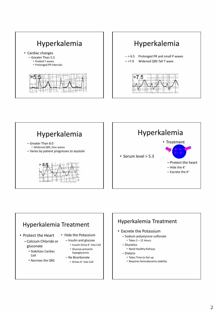
Hyperkalemia • Cardiac changes
– Greater Than 5.5 • Peaked T waves • Prolonged PR Intervals
Hyperkalemia
– > 6.5 Prolonged PR and small P waves
– >7.0 Widened QRS Tall T wave
Hyperkalemia – Greater Than 8.0
• Widened QRS, Sine waves
– Varies by patient progresses to asystole
Hyperkalemia
• Serum level > 5.3
• Treatment
–Protect the heart – Hide the K+
– Excrete the K+
K+ K+
K+ K+
Hyperkalemia Treatment
• Protect the Heart
–Calcium Chloride or gluconate
• Stabilizes Cardiac Cell
• Narrows the QRS
• Hide the Potassium
– Insulin and glucose
• Insulin Drive K+ Into Cell
• Glucose prevents hypoglycemia
– Na Bicarbonate
• Drives K+ Into Cell
Hyperkalemia Treatment
• Excrete the Potassium – Sodium polystyrene sulfonate
• Takes 2 – 12 Hours
– Diuretics • Need Healthy Kidneys
– Dialysis • Takes Time to Set-up
• Requires hemodynamic stability
3
Protect the
Heart
Hide the
potassium
Excrete the
potassium
Emergency
Hyperkalemia
Treatment
Calcium
Chloride or
gluconate
intravenous
(CaCl has
three times
more available
Ca than
gluconate)
Insulin and
dextrose and
sodium
bicarbonate.
Insulin and
sodium bicarb
drive potassium
into the cell
temporarily
Sodium polystyrene
sulfonate, dialysis,
diuretics
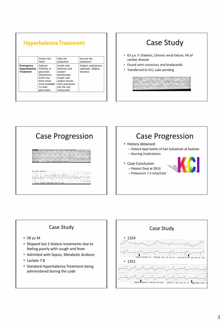
Hyperkalemia Treatment Case Study
• 63 y.o. F. Diabetic, Chronic renal failure, HX of cardiac disease
• Found semi-conscious and bradycardic
• Transferred to ICU, Labs pending
Case Progression Case Progression • History obtained:
– Patient kept bottle of Salt Substitute at bedside
– Nursing Implications
• Case Conclusion – Patient Died at 0910
– Potassium 7.3 mEq/Liter
Case Study
• 58 yo M
• Skipped last 2 dialysis treatments due to feeling poorly with cough and fever
• Admitted with Sepsis, Metabolic Acidosis
• Lactate 7.8
• Standard Hyperkalemia Treatment being administered during the code
Case Study
• 1324
• 1351
4
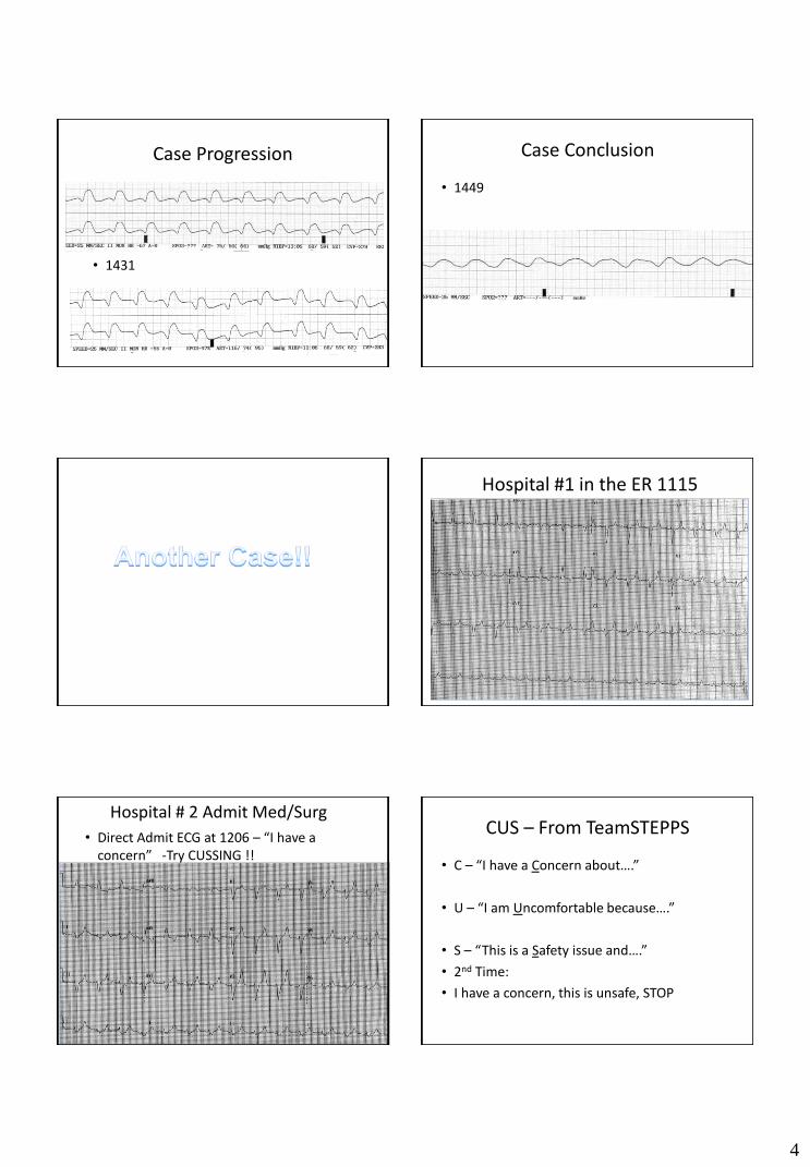
Case Progression
• 1412
• 1431
Case Conclusion
• 1449
Hospital #1 in the ER 1115
Hospital # 2 Admit Med/Surg
• Direct Admit ECG at 1206 – “I have a concern” -Try CUSSING !!
CUS – From TeamSTEPPS
• C – “I have a Concern about….”
• U – “I am Uncomfortable because….”
• S – “This is a Safety issue and….”
• 2nd Time:
• I have a concern, this is unsafe, STOP
5
Hospital # 2 Medical Surgical • ICU at 1821 after dialysis
Hypokalemia
• Serum levels < 3.5
• Causes:
–Diuresis
–Gastric loss
– Insulin
–NaHCO3¯
• Treatment • Protect the Heart!
–Replace losses
Hypokalemia Case Study
• At risk for PVC’s, V Tach, V Fib
Case Study
• 54 y.o. M c/o 3 day history of diarrhea and vomiting.
• Has continued Lasix tablets but stopped KCL because it upset his stomach
Case Study
Guess the potassium?
6
Case Study
• Potassium: 1.8 mEq/Liter
• Treatment?
–Replace the losses
–IV: Not faster than 10 mEq/Hour
Hypokalemia
• Patient complains of feeling dizzy and palpitations…. – See the “R on T” ?
Case Study
• Treatment
– Continue to replace potassium
– Antidysrhythmics
• Amiodarone – Dosing ?
• Lidocaine
• Procainamide
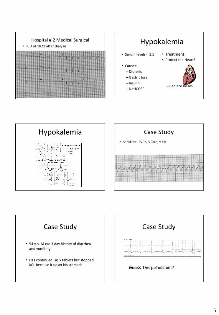
Magnesium
• Normal:
–Text book 1.2 - 2.9 mEq/L
–1.8 – 2.3 mEq/L
K+ K+
Mg++ K+
K+ Mg++
Hypomagnesemia • Serum level < 1.2 mEq/L (< 1.8)
• Symptoms:
– Muscle tremors
– Nausea
– Cardiac dysrhythmias?
• Prolonged QT interval
7
Hypomagnesemia VT & Torsades – Prolonged QT Causes
• Conditions & Medications that Prolong QT – Antidysrhythmics:
amiodarone, procainamide, sotalol, ibutilide
– Tricyclic Anti-Depressants
– Haldol, Geodon
• Phenothiazines (Compazine, Thorazine)
• Hypomagnesemia, Hypocalcemia, Hypokalemia
• Hypothyroidism
• Liquid Protein Diets
• Antibiotics - Levaquin
Measuring the QT Interval
• QTc = QT measured / Square Root of the R-R interval
– Should be Less 0.45 seconds
• Simplified Formula
– QTc = Less than ½ the preceding R-R interval
– Works with Regular Rhythms
Prolonged QT & Torsades
• History: – 40 yo admitted with Hx. of Methamphetamine
Addiction • Placed on Telemetry
– Haldol 10 mg IV prn for agitation • Doses given at 0330, 0430, 0610
– AM Labs – Potassium and Magnesium • Potassium 2.7 mEq/Liter • Magnesium 1.7 mEq/Liter • Low levels of both of these put patient’s at risk for prolonged
QT interval
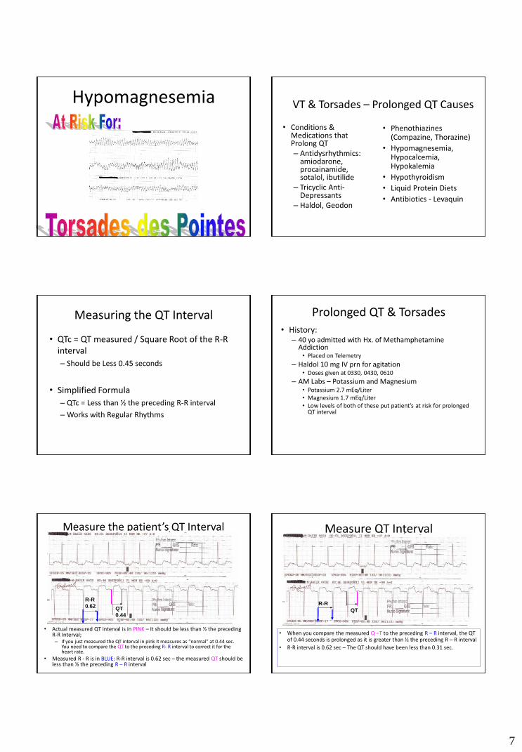
Measure the patient’s QT Interval
• Actual measured QT interval is in PINK – It should be less than ½ the preceding R-R Interval; – if you just measured the QT interval in pink it measures as “normal” at 0.44 sec.
You need to compare the QT to the preceding R- R interval to correct it for the heart rate.
• Measured R - R is in BLUE: R-R interval is 0.62 sec – the measured QT should be less than ½ the preceding R – R interval
QT
0.44
R-R
0.62
Measure QT Interval
• When you compare the measured Q –T to the preceding R – R interval, the QT of 0.44 seconds is prolonged as it is greater than ½ the preceding R – R interval
• R-R interval is 0.62 sec – The QT should have been less than 0.31 sec.
QT
R-R
8
10 minutes after 4th Dose of prn Haldol Nursing Implications
• Be AWARE of the numerous medications which prolong the QT
• Amiodarone, Levaquin, Haldol, Geodon, anti-depressants, etc. see table below
• When giving a medication that may prolong the QT interval
– Measure the QT and correct it for the HR = QTc
– QT calculated or QTc =
QT (measured) ÷ √R-R interval (seconds)
– Evaluate your electrolytes and correct
Table Medications that Prolong the QT interval
Medications implicated in torsades de pointes Proocainamide
Chlorpromazine
Disopyramide
Quinidine
Class III antiarrhythmics
Sotalol
Dofetilide
Amiodarone
Ibutilide
Antimicrobials
Antiprotozoals
Pentamidine
Macrolides
Clarithromycin
Erythromycin
Antimalarials
Chloroquine
Halofantrine
Adapted from AJCC AMERICAN JOURNAL OF CRITICAL CARE, January
2008, Volume 17, No. 1 Antipsychotics
Phenothiazine neuroleptics Mesoridazine
Thioridazine
Droperidol
Pimozide
Haloperidol
Diphenylpiperidine neuroleptics
Butyrophenone neuroleptics
Others
Methadone
Arsenic trioxide
Amiodarone & Geodon should
not be used together
Update your knowledge with the latest AHA
Scientific statement found at:
http://circ.ahajournals.org/content/121/8/1047.full
.pdf+html
AACN Article:
http://ajcc.aacnjournals.org/content/17/1/77.full.p
df+html?sid=5f1913f6-9e05-4271-9f65-
c283c99bd8a3
Hypermagnesemia • Serum level
– > 2.9 mEq/L
• Symptoms:
– Respiratory depression
– ECG is similar to hyperkalemia
Potassium and Magnesium
• Electrolytes – Rule of Thumb
– Potassium > 4.0
– Magnesium > 2.0
• Normal:
–Total: 8.6 – 10 mEq/Liter
–Ionized: 1.16 – 1.32 mEq/Liter
9
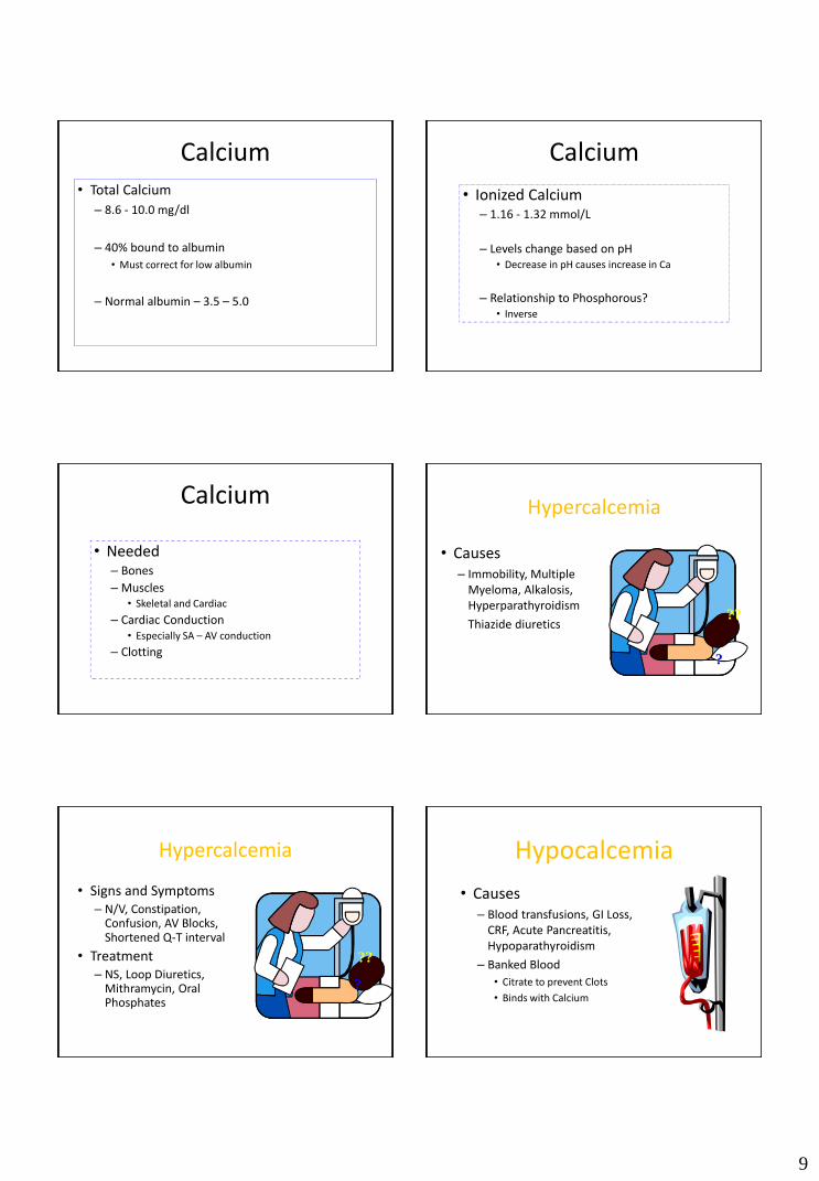
Calcium • Total Calcium
– 8.6 - 10.0 mg/dl
– 40% bound to albumin
• Must correct for low albumin
– Normal albumin – 3.5 – 5.0
Calcium
• Ionized Calcium – 1.16 - 1.32 mmol/L
– Levels change based on pH • Decrease in pH causes increase in Ca
– Relationship to Phosphorous? • Inverse
Calcium
• Needed – Bones
– Muscles • Skeletal and Cardiac
– Cardiac Conduction • Especially SA – AV conduction
– Clotting
Hypercalcemia
• Causes – Immobility, Multiple
Myeloma, Alkalosis, Hyperparathyroidism
Thiazide diuretics
??
?
Hypercalcemia
• Signs and Symptoms – N/V, Constipation,
Confusion, AV Blocks, Shortened Q-T interval
• Treatment – NS, Loop Diuretics,
Mithramycin, Oral Phosphates
??
?
Hypocalcemia
• Causes – Blood transfusions, GI Loss,
CRF, Acute Pancreatitis, Hypoparathyroidism
– Banked Blood
• Citrate to prevent Clots
• Binds with Calcium
10
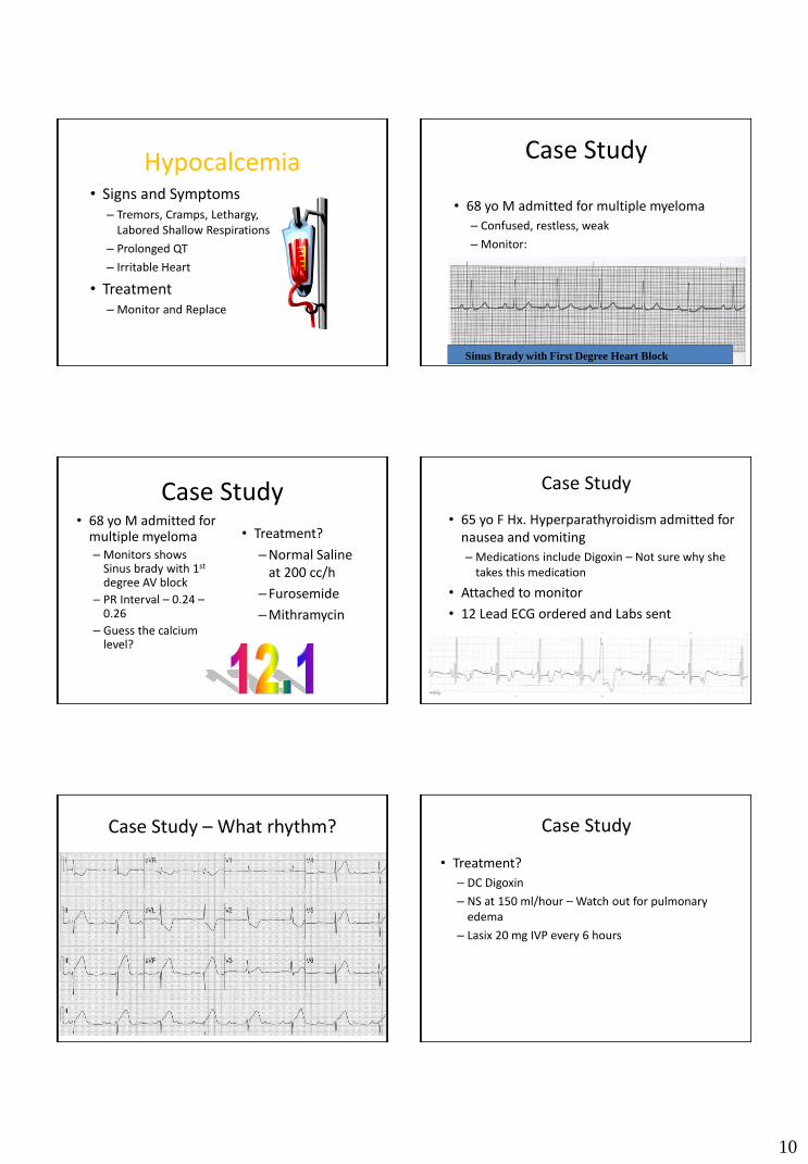
Hypocalcemia • Signs and Symptoms
– Tremors, Cramps, Lethargy, Labored Shallow Respirations
– Prolonged QT
– Irritable Heart
• Treatment – Monitor and Replace
Case Study
• 68 yo M admitted for multiple myeloma
– Confused, restless, weak
– Monitor:
Sinus Brady with First Degree Heart Block
Case Study • 68 yo M admitted for
multiple myeloma – Monitors shows
Sinus brady with 1st degree AV block
– PR Interval – 0.24 – 0.26
– Guess the calcium level?
• Treatment?
–Normal Saline at 200 cc/h
– Furosemide
–Mithramycin
Case Study
• 65 yo F Hx. Hyperparathyroidism admitted for nausea and vomiting
– Medications include Digoxin – Not sure why she takes this medication
• Attached to monitor
• 12 Lead ECG ordered and Labs sent
Case Study – What rhythm?
Case Study
• Treatment?
– DC Digoxin
– NS at 150 ml/hour – Watch out for pulmonary edema
– Lasix 20 mg IVP every 6 hours
11
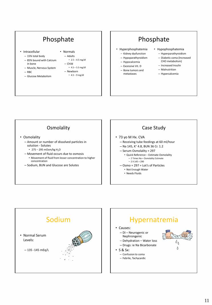
Phosphate
• Intracellular
– 15% total body
– 85% bound with Calcium in bone
– Muscle, Nervous System
– RBC
– Glucose Metabolism
• Normals
– Adults • 2.5 – 4.5 mg/dl
– Child • 4.5 – 5.5 mg/dl
– Newborn • 4.5 – 9 mg/dl
Phosphate • Hyperphosphatemia
– Kidney dysfunction
– Hypoparathyroidism
– Hypocalcemia
– Excessive Vit. D
– Bone tumors and metastases
• Hypophosphatemia
– Hyperparathyroidism
– Diabetic coma (Increased CHO metabolism)
– Increased Insulin
– Malnutrition
– Hypercalcemia
Osmolality
• Osmolality – Amount or number of dissolved particles in
solution - Solutes • 275 – 295 mOsm/kg H2O
– Movement of fluid occurs due to osmosis • Movement of fluid from lesser concentration to higher
concentration
– Sodium, BUN and Glucose are Solutes
Case Study
• 73 yo M Hx. CVA – Receiving tube feedings at 60 ml/hour
– Na 145, K+ 4.8, BUN 36 Cr. 1.2
– Serum Osmolality = 297 • Quick Reference – Estimate Osmolality
– 2 Times Na = Osmolality Estimate
– 2 X 145 = 290
– Osmo = 297 = Lot’s of Particles • Not Enough Water
• Needs Fluids
Sodium
• Normal Serum Levels:
– 135 -145 mEq/L
Hypernatremia • Causes:
– DI – Neurogenic or Nephrongenic
– Dehydration – Water loss
– Drugs: ie Na Bicarbonate
• S & Sx: – Confusion to coma
– Febrile, Tachycardic
12
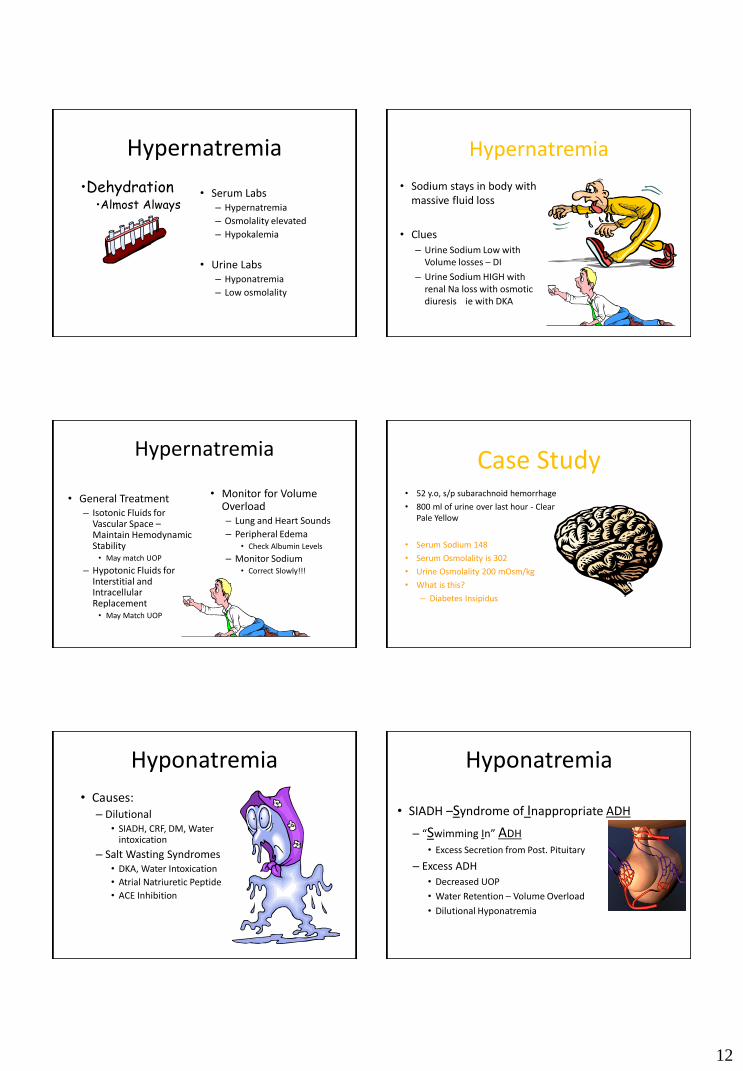
Hypernatremia
• Serum Labs – Hypernatremia
– Osmolality elevated
– Hypokalemia
• Urine Labs – Hyponatremia
– Low osmolality
•Dehydration •Almost Always
Hypernatremia
• Sodium stays in body with massive fluid loss
• Clues
– Urine Sodium Low with Volume losses – DI
– Urine Sodium HIGH with renal Na loss with osmotic diuresis ie with DKA
Hypernatremia
• General Treatment – Isotonic Fluids for
Vascular Space – Maintain Hemodynamic Stability • May match UOP
– Hypotonic Fluids for Interstitial and Intracellular Replacement • May Match UOP
• Monitor for Volume Overload – Lung and Heart Sounds
– Peripheral Edema • Check Albumin Levels
– Monitor Sodium • Correct Slowly!!!
Case Study • 52 y.o, s/p subarachnoid hemorrhage
• 800 ml of urine over last hour - Clear Pale Yellow
• Serum Sodium 148
• Serum Osmolality is 302
• Urine Osmolality 200 mOsm/kg
• What is this?
– Diabetes Insipidus
Hyponatremia
• Causes: – Dilutional
• SIADH, CRF, DM, Water intoxication
– Salt Wasting Syndromes • DKA, Water Intoxication
• Atrial Natriuretic Peptide
• ACE Inhibition
Hyponatremia
• SIADH –Syndrome of Inappropriate ADH
– “Swimming In” ADH
• Excess Secretion from Post. Pituitary
– Excess ADH
• Decreased UOP
• Water Retention – Volume Overload
• Dilutional Hyponatremia
13
Hyponatremia
• Salt wasting – Atrial Natriuretic Factor
• Hormone produces sodium excretion
• Associated with Neurologic damage
– Results in: • High urine output
• Low serum sodium
• Water loss = Dehydration – Decreased CVP & PAOP
Hyponatremia
• Treat Underlying Cause
– SIADH
• Fluid Restrict, Diuretics, Hypertonic Saline
– Salt Wasting • Isotonic Fluid Replacement
• Hypertonic Saline
• S & Sx
– Headache, muscle cramps, confusion, Tachycardia, Seizures
– Dilutional
• Increased CVP & PAOP
– Saline Loss
• Volume Loss
• Decreased CVP & PAOP
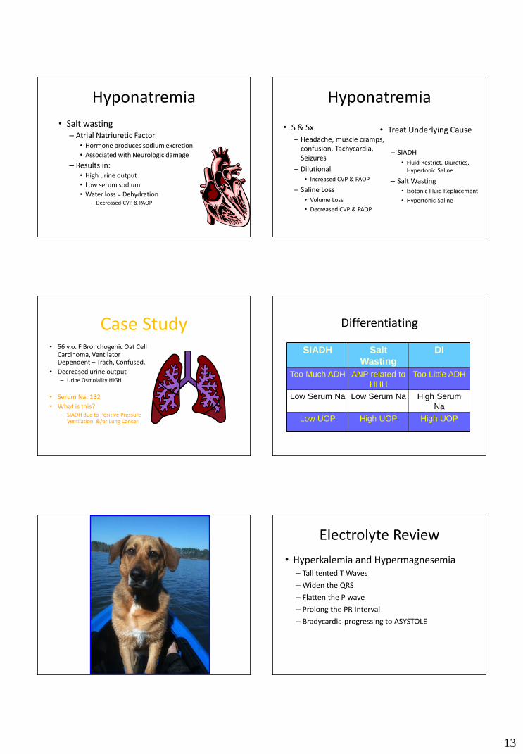
Case Study • 56 y.o. F Bronchogenic Oat Cell
Carcinoma, Ventilator Dependent – Trach, Confused.
• Decreased urine output – Urine Osmolality HIGH
• Serum Na: 132
• What is this? – SIADH due to Positive Pressure
Ventilation &/or Lung Cancer
Differentiating
SIADH Salt
Wasting
DI
Too Much ADH ANP related to
HHH
Too Little ADH
Low Serum Na Low Serum Na High Serum
Na
Low UOP High UOP High UOP
Electrolyte Review
• Hyperkalemia and Hypermagnesemia – Tall tented T Waves
– Widen the QRS
– Flatten the P wave
– Prolong the PR Interval
– Bradycardia progressing to ASYSTOLE
14
Electrolyte Review
• Hypokalemia and Hypomagnesemia – Irritate the Heart
– PVC’s
– V Tach
– V Fib
– Torsades des Pointes due to prolonged QT interval
Electrolyte Review
• Hypercalcemia – Prolong the PR Interval
– Brady and Blocks
– Shortens the Q-T
– Slows muscle contraction
– Slows electrical conduction
Electrolyte Review
• Hypocalcemia – Irritable Ventricle
– Decreased Contractility
– Prolonged QT – May lead to Torsades!
– Alters the clotting cascade – at risk for bleeding
– Irritable muscles – Tremors
• Positive Chvostek’s and Trosseau’s
Electrolyte Review
• Hypernatremia
– Almost always associated with dehydration
• Hyponatremia
– Correct Sodium levels slowly
Questions
• 28 yo pt. Admitted for 4 days N/V and diarrhea, unable to keep any food or liquid down. The electrolyte disturbance you might expect is:
a. Hyperkalemia
b. Hypercalcemia
c. Hypokalemia
d. Hyponatremia
Questions
• 48 yo pt admitted with renal insufficiency. Has been recently diagnosed with hyperparathyroidism. The electrolyte disturbance you might expect is:
a. Hypercalcemia
b. Hypokalemia
c. Hypocalcemia
d. Hypernatremia

15
Questions
• You are caring for a patient with renal failure who is receiving antacids. Telemetry tech calls to report your patient is displaying a prolonged PR interval, widened QRS and tall peaked T-Wave. Labs are drawn and you expect to find:
a. Hypernatremia & Hyperkalemia
b. Hypokalemia & Hypomagnesemia
c. Hyperkalemia & Hypermagnesemia
d. Hyponatremia & Hypokalemia
Questions
• You are caring for a patient with acute pancreatitis. Neurologic assessment shows confusion and short term memory loss. Patient c/o of “Jittery” muscles. You expect the labs to show:
a. Hypocalcemia
b. Hypercalcemia
c. Hypermagnesemia
d. Hypophosphatemia
Speaker Contact
• Julie Miller, RN, BSN, CCRN
• President and Founder – Paws To Learn
– Empowering Nurses with Education!
Phone: 903-245-1223
Email: [email protected]
Bibliography / Webliography • http://circ.ahajournals.org/content/121/8/1047.full.pdf+html
• AACN Article: http://ajcc.aacnjournals.org/content/17/1/77.full.pdf+html?sid=5f1913f6-9e05-4271-9f65-c283c99bd8a3
• Alspach J, ed. Core Curriculum for Critical Care Nursing. 6th ed. Philadelphia PA:Saunders Publications; 2006.
• Criddle LM. Rhabdomyolysis: Pathophysiology, Recognition and Management. Crit Car Nurs. 2003;(23)6:14-32
• Lynn-Mchale DJ, Carlson KK, eds. AACN Procedure Manual for Critical Care. Philadelpha: W. B. Saunders, 6th ed. 2011.
• Maffei FA, Connolly H. Toxicity, Tricyclic Antidepressant. Last Updated: August 2, 2004
• http://www.emedicine.com/ped/topic2714.htm Last accessed August 13, 2005
• Urden LD, Stacy KM, Lough ME. (eds.) Critical Care Nursing: Diagnosis and Management. 7th edition. Mosby: St. Louis. 2014