Embed Size (px)
Citation preview
Case Study 9: Lost polychromy and gilding of Neo-Assyrian ivories
Summary
Antique objects are known to have been brightly coloured. However, the appearance of these
objects has changed over time and paint traces are rarely preserved. But today, only specific
alteration phenomena like cracks and discolorations can be observed on the ivory surfaces. Where
has the colour gone? Can it be reconstructed from the remaining traces on the objects, and what
might we learn about them?
Elephant-ivory carvings by highly skilled Phoenician craftsmen, from the Neo-Assyrian archaeological
site of Arslan Tash in Syria and dated to the 8th century BC, were excavated in 1928 and are now in
the collections of the Louvre Museum and the Badisches Landesmuseum.
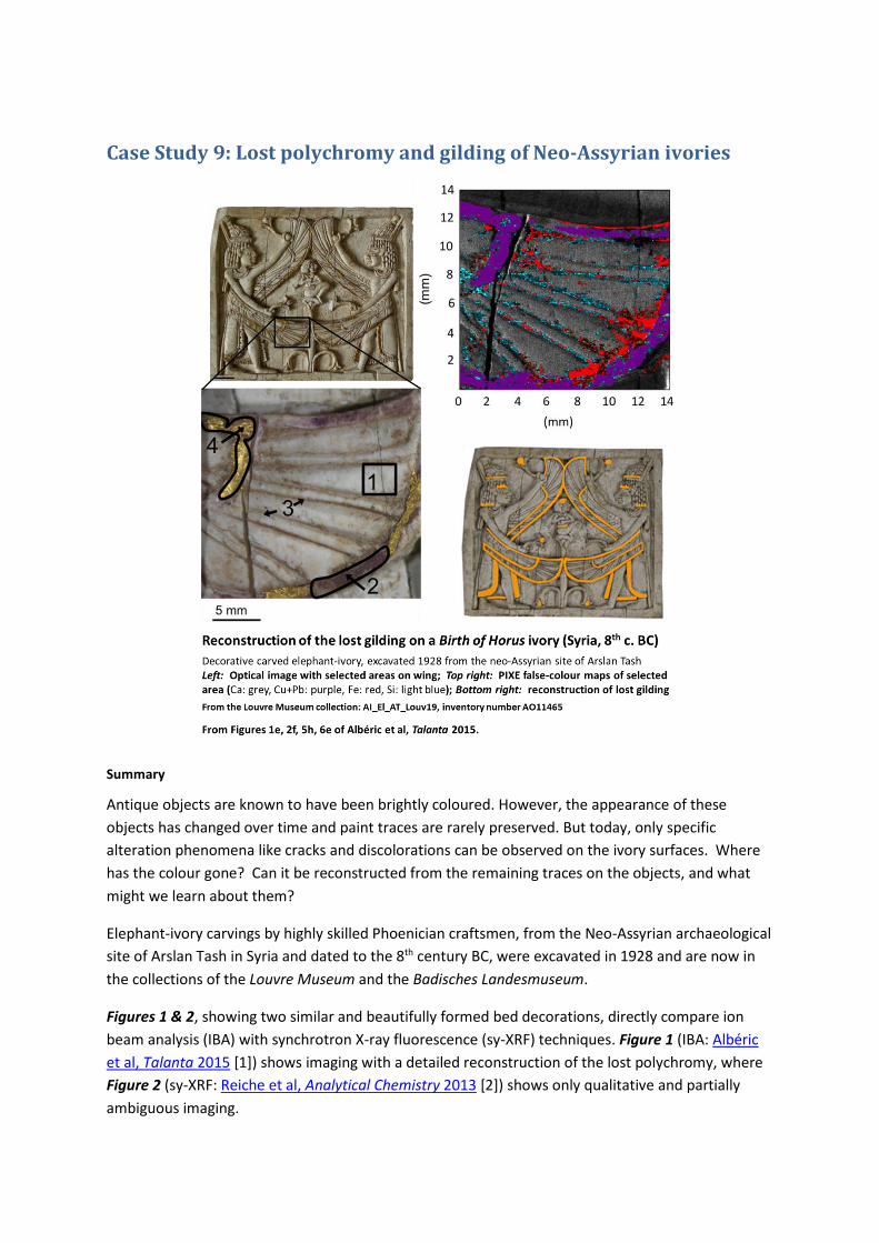
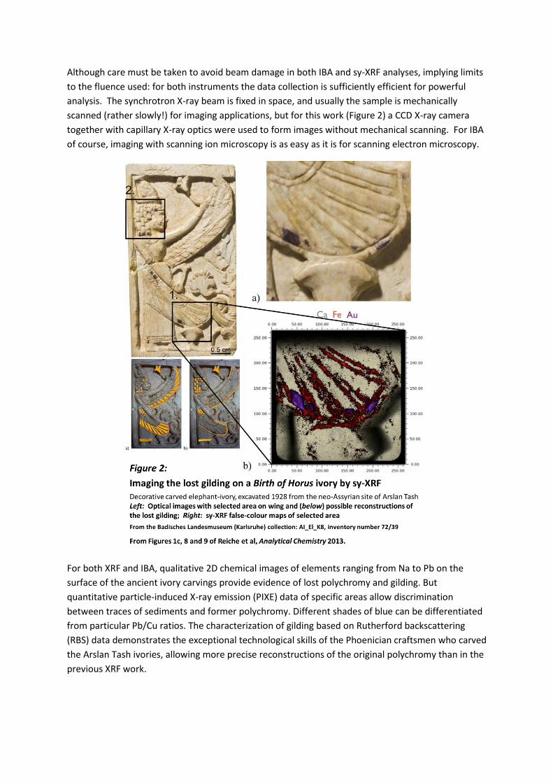
Figures 1 & 2, showing two similar and beautifully formed bed decorations, directly compare ion
beam analysis (IBA) with synchrotron X-ray fluorescence (sy-XRF) techniques. Figure 1 (IBA: Albéric
et al, Talanta 2015 [1]) shows imaging with a detailed reconstruction of the lost polychromy, where
Figure 2 (sy-XRF: Reiche et al, Analytical Chemistry 2013 [2]) shows only qualitative and partially
ambiguous imaging.
Although care must be taken to avoid beam damage in both IBA and sy-XRF analyses, implying limits
to the fluence used: for both instruments the data collection is sufficiently efficient for powerful
analysis. The synchrotron X-ray beam is fixed in space, and usually the sample is mechanically
scanned (rather slowly!) for imaging applications, but for this work (Figure 2) a CCD X-ray camera
together with capillary X-ray optics were used to form images without mechanical scanning. For IBA
of course, imaging with scanning ion microscopy is as easy as it is for scanning electron microscopy.
For both XRF and IBA, qualitative 2D chemical images of elements ranging from Na to Pb on the
surface of the ancient ivory carvings provide evidence of lost polychromy and gilding. But
quantitative particle-induced X-ray emission (PIXE) data of specific areas allow discrimination
between traces of sediments and former polychromy. Different shades of blue can be differentiated
from particular Pb/Cu ratios. The characterization of gilding based on Rutherford backscattering
(RBS) data demonstrates the exceptional technological skills of the Phoenician craftsmen who carved
the Arslan Tash ivories, allowing more precise reconstructions of the original polychromy than in the
previous XRF work.
Body
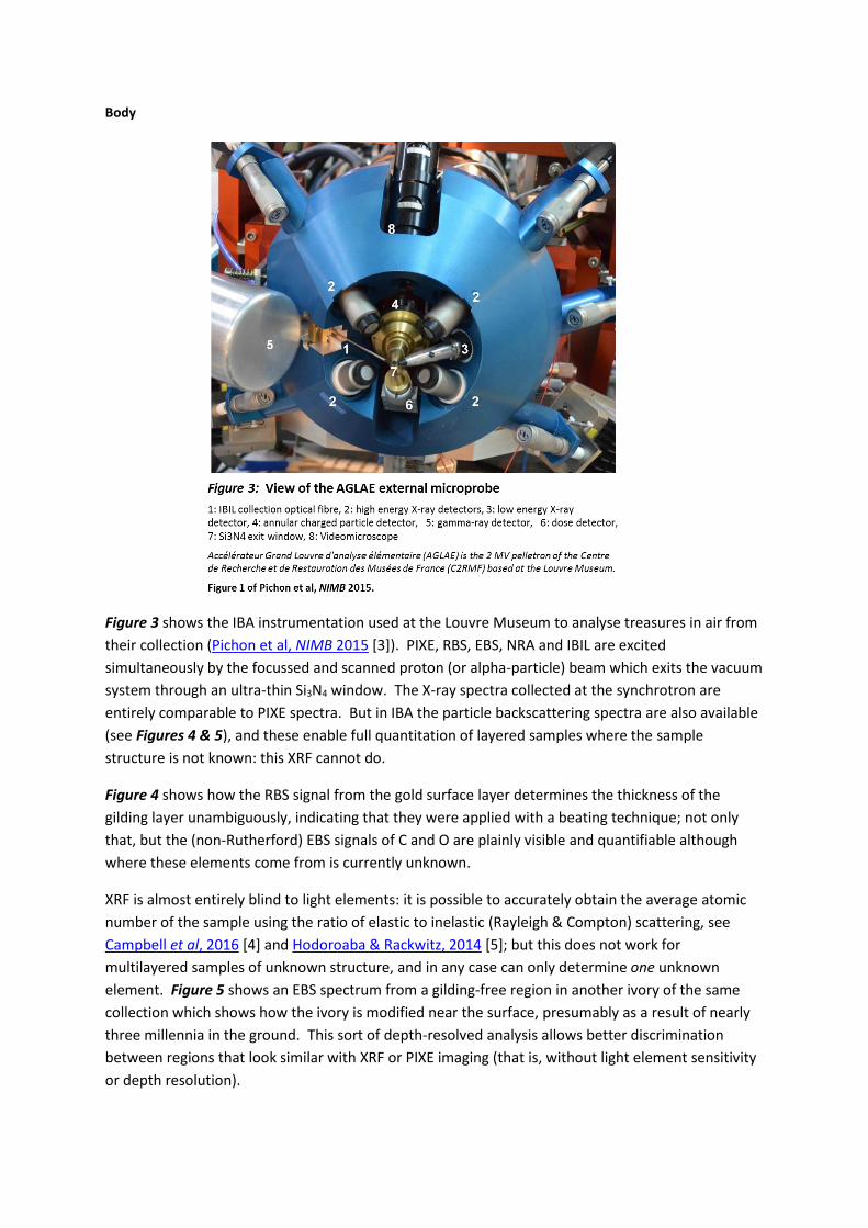
Figure 3 shows the IBA instrumentation used at the Louvre Museum to analyse treasures in air from
their collection (Pichon et al, NIMB 2015 [3]). PIXE, RBS, EBS, NRA and IBIL are excited
simultaneously by the focussed and scanned proton (or alpha-particle) beam which exits the vacuum
system through an ultra-thin Si3N4 window. The X-ray spectra collected at the synchrotron are
entirely comparable to PIXE spectra. But in IBA the particle backscattering spectra are also available
(see Figures 4 & 5), and these enable full quantitation of layered samples where the sample
structure is not known: this XRF cannot do.
Figure 4 shows how the RBS signal from the gold surface layer determines the thickness of the
gilding layer unambiguously, indicating that they were applied with a beating technique; not only
that, but the (non-Rutherford) EBS signals of C and O are plainly visible and quantifiable although
where these elements come from is currently unknown.
XRF is almost entirely blind to light elements: it is possible to accurately obtain the average atomic
number of the sample using the ratio of elastic to inelastic (Rayleigh & Compton) scattering, see
Campbell et al, 2016 [4] and Hodoroaba & Rackwitz, 2014 [5]; but this does not work for
multilayered samples of unknown structure, and in any case can only determine one unknown
element. Figure 5 shows an EBS spectrum from a gilding-free region in another ivory of the same
collection which shows how the ivory is modified near the surface, presumably as a result of nearly
three millennia in the ground. This sort of depth-resolved analysis allows better discrimination
between regions that look similar with XRF or PIXE imaging (that is, without light element sensitivity
or depth resolution).
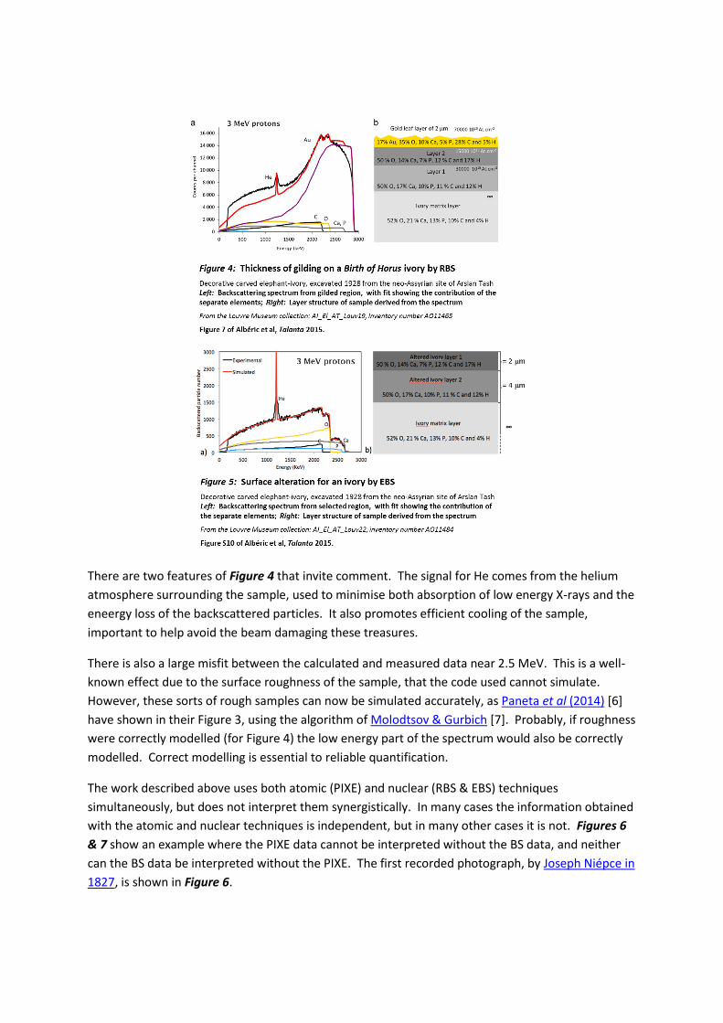
There are two features of Figure 4 that invite comment. The signal for He comes from the helium
atmosphere surrounding the sample, used to minimise both absorption of low energy X-rays and the
eneergy loss of the backscattered particles. It also promotes efficient cooling of the sample,
important to help avoid the beam damaging these treasures.
There is also a large misfit between the calculated and measured data near 2.5 MeV. This is a well-
known effect due to the surface roughness of the sample, that the code used cannot simulate.
However, these sorts of rough samples can now be simulated accurately, as Paneta et al (2014) [6]
have shown in their Figure 3, using the algorithm of Molodtsov & Gurbich [7]. Probably, if roughness
were correctly modelled (for Figure 4) the low energy part of the spectrum would also be correctly
modelled. Correct modelling is essential to reliable quantification.
The work described above uses both atomic (PIXE) and nuclear (RBS & EBS) techniques
simultaneously, but does not interpret them synergistically. In many cases the information obtained
with the atomic and nuclear techniques is independent, but in many other cases it is not. Figures 6
& 7 show an example where the PIXE data cannot be interpreted without the BS data, and neither
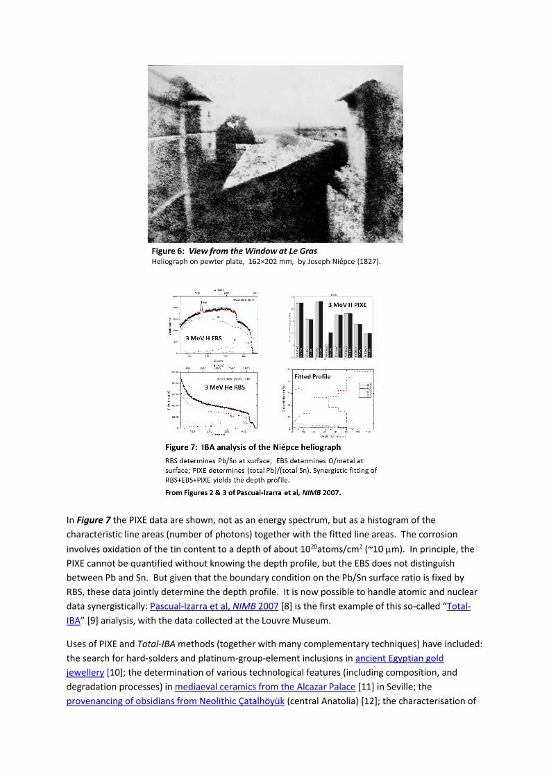
can the BS data be interpreted without the PIXE. The first recorded photograph, by Joseph Niépce in
1827, is shown in Figure 6.
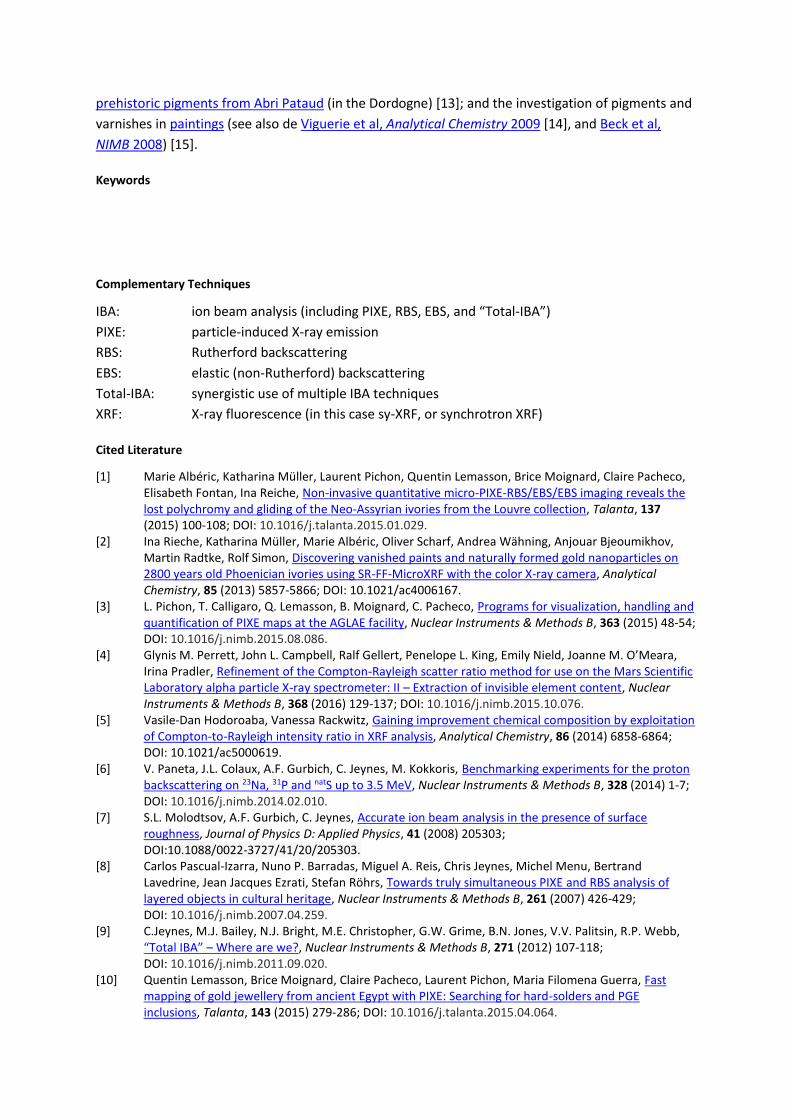
In Figure 7 the PIXE data are shown, not as an energy spectrum, but as a histogram of the
characteristic line areas (number of photons) together with the fitted line areas. The corrosion
involves oxidation of the tin content to a depth of about 1020atoms/cm2 (~10 m). In principle, the
PIXE cannot be quantified without knowing the depth profile, but the EBS does not distinguish
between Pb and Sn. But given that the boundary condition on the Pb/Sn surface ratio is fixed by
RBS, these data jointly determine the depth profile. It is now possible to handle atomic and nuclear
data synergistically: Pascual-Izarra et al, NIMB 2007 [8] is the first example of this so-called “Total-
IBA” [9] analysis, with the data collected at the Louvre Museum.
Uses of PIXE and Total-IBA methods (together with many complementary techniques) have included:
the search for hard-solders and platinum-group-element inclusions in ancient Egyptian gold
jewellery [10]; the determination of various technological features (including composition, and
degradation processes) in mediaeval ceramics from the Alcazar Palace [11] in Seville; the
provenancing of obsidians from Neolithic Çatalhöyük (central Anatolia) [12]; the characterisation of
prehistoric pigments from Abri Pataud (in the Dordogne) [13]; and the investigation of pigments and
varnishes in paintings (see also de Viguerie et al, Analytical Chemistry 2009 [14], and Beck et al,
NIMB 2008) [15].
Keywords
Complementary Techniques
IBA: ion beam analysis (including PIXE, RBS, EBS, and “Total-IBA”)
PIXE: particle-induced X-ray emission
RBS: Rutherford backscattering
EBS: elastic (non-Rutherford) backscattering
Total-IBA: synergistic use of multiple IBA techniques
XRF: X-ray fluorescence (in this case sy-XRF, or synchrotron XRF)
Cited Literature
[1] Marie Albéric, Katharina Müller, Laurent Pichon, Quentin Lemasson, Brice Moignard, Claire Pacheco, Elisabeth Fontan, Ina Reiche, Non-invasive quantitative micro-PIXE-RBS/EBS/EBS imaging reveals the lost polychromy and gliding of the Neo-Assyrian ivories from the Louvre collection, Talanta, 137 (2015) 100-108; DOI: 10.1016/j.talanta.2015.01.029.
[2] Ina Rieche, Katharina Müller, Marie Albéric, Oliver Scharf, Andrea Wähning, Anjouar Bjeoumikhov, Martin Radtke, Rolf Simon, Discovering vanished paints and naturally formed gold nanoparticles on 2800 years old Phoenician ivories using SR-FF-MicroXRF with the color X-ray camera, Analytical Chemistry, 85 (2013) 5857-5866; DOI: 10.1021/ac4006167.
[3] L. Pichon, T. Calligaro, Q. Lemasson, B. Moignard, C. Pacheco, Programs for visualization, handling and quantification of PIXE maps at the AGLAE facility, Nuclear Instruments & Methods B, 363 (2015) 48-54; DOI: 10.1016/j.nimb.2015.08.086.
[4] Glynis M. Perrett, John L. Campbell, Ralf Gellert, Penelope L. King, Emily Nield, Joanne M. O’Meara, Irina Pradler, Refinement of the Compton-Rayleigh scatter ratio method for use on the Mars Scientific Laboratory alpha particle X-ray spectrometer: II – Extraction of invisible element content, Nuclear Instruments & Methods B, 368 (2016) 129-137; DOI: 10.1016/j.nimb.2015.10.076.
[5] Vasile-Dan Hodoroaba, Vanessa Rackwitz, Gaining improvement chemical composition by exploitation of Compton-to-Rayleigh intensity ratio in XRF analysis, Analytical Chemistry, 86 (2014) 6858-6864; DOI: 10.1021/ac5000619.
[6] V. Paneta, J.L. Colaux, A.F. Gurbich, C. Jeynes, M. Kokkoris, Benchmarking experiments for the proton backscattering on 23Na, 31P and natS up to 3.5 MeV, Nuclear Instruments & Methods B, 328 (2014) 1-7; DOI: 10.1016/j.nimb.2014.02.010.
[7] S.L. Molodtsov, A.F. Gurbich, C. Jeynes, Accurate ion beam analysis in the presence of surface roughness, Journal of Physics D: Applied Physics, 41 (2008) 205303; DOI:10.1088/0022-3727/41/20/205303.
[8] Carlos Pascual-Izarra, Nuno P. Barradas, Miguel A. Reis, Chris Jeynes, Michel Menu, Bertrand Lavedrine, Jean Jacques Ezrati, Stefan Röhrs, Towards truly simultaneous PIXE and RBS analysis of layered objects in cultural heritage, Nuclear Instruments & Methods B, 261 (2007) 426-429; DOI: 10.1016/j.nimb.2007.04.259.
[9] C.Jeynes, M.J. Bailey, N.J. Bright, M.E. Christopher, G.W. Grime, B.N. Jones, V.V. Palitsin, R.P. Webb, “Total IBA” – Where are we?, Nuclear Instruments & Methods B, 271 (2012) 107-118; DOI: 10.1016/j.nimb.2011.09.020.
[10] Quentin Lemasson, Brice Moignard, Claire Pacheco, Laurent Pichon, Maria Filomena Guerra, Fast mapping of gold jewellery from ancient Egypt with PIXE: Searching for hard-solders and PGE inclusions, Talanta, 143 (2015) 279-286; DOI: 10.1016/j.talanta.2015.04.064.

[11] I. Garofano, M.D. Robador, J.L. Perez-Rodriguez, J. Castaing, C. Pacheco, A. Duran, Ceramics from the Alcazar Palace in Seville (Spain) dated between the 11th and 15th centuries: Compositions, technological features and degradation processes, Journal of the European Ceramic Society, 35 (2015) 4307-4319; DOI: 10.1016/j.jeurceramsoc.2015.07.033.
[12] Gérard Poupeau, François-Xavier Le Bourdonnec, Tristan Carter, Sarah Delerue, M. Steven Shackley, Jean-Alix Barrat, Stéphan Dubernet, Philippe Moretto, Thomas Calligaro, Marina Milić, Katsuji Kobayashi, The use of SEM-EDS, PIXE and ERXRF for obsidian provenance studies in the Near East: A case study from Neolithic Çatalhöyük (central Anatolia), Journal of Archaeological Science, 37 (2010) 2705-2720; DOI: 10.1016/j.jas.2010.06.007.
[13] L. Beck, M. Lebon, L. Pichon, M. Menu, L. Chiotti, R. Nespoulet, P. Paillet, PIXE characterisation of prehistoric pigments from Abri Pataud (Dordogne, France), X-Ray Spectrometry, 40 (2011) 219-223; DOI: 10.1002/xrs.1321.
[14] L. de Viguerie, L. Beck, J. Salomon, L. Pichon, Ph. Walter, Composition of renaissance paint layers: Simultaneous particle induced X-ray emission and backscattering spectrometry, Analytical Chemistry, 81 (2009) 7960-7966; DOI: 10.1021/ac901141v.
[15] L. Beck. C. Jeynes, N.P. Barradas, Characterisation of paint layers by simultaneous self-consistent fitting of RBS/PIXE spectra using simulated annealing, Nuclear Instruments & Methods B, 266 (2008) 1871-1874; DOI: 10.1016/j.nimb.2007.12.091.