Embed Size (px)
Citation preview
Case Study: DNALeonardo Trabuco and Elizabeth Villa
1
1 The Molecule of Life
With the 50th aniversary of the discovery of the DNA structure by JamesWatson and Francis Crick and the race for the human genome, with all itscontroversies and awe-inspiring medical implications, we have all heard a lotabout DNA, the molecule of life.
What makes DNA “the molecule of life”? DNA, or deoxyribonucleicacid, is the molecule that carries all the genetic information of an organism(except for some RNA viruses). It can be thought of as a blueprint containingthe instructions that govern the production of proteins and other moleculesessential to cell function. The collections of these instructions is called agenome. The informational units of the genome are called genes. Genesare translated into protein via the genetic code, which defines the proteinsequence. This translation process is central to life.
Despite being so important, DNA in reality is a passive molecule. As aninstruction book, all DNA does is to store information. DNA is thereforelike any other data-storing device: it needs to be read, stored, copied andotherwise manipulated by other biomolecules. These other macromoleculesexist in different forms and types, and interact with DNA in different ways,e.g., by bending it, copying it, denaturing it, packing it, walking on it, nickingit, repairing it, etc. All this is done in order to preserve and obey the setof instructions that give life to the organism. In this sense, there is no lifewithout genetic information.
As passive as it might be, the DNA molecule bears incredible physicalproperties that allow for long genomes to be stored in compact nucleii andfor extreme manipulation resulting in replication and gene regulation. In thiscase study, we will introduce you to this wonderful molecule of life. In section2, you will learn about the chemical components that form DNA. Section 3will introduce you to the different helical shapes that DNA adopts. In section4, you will learn of the tight packing that DNA needs to undergo in orderto be stored in the nucleus; the flexibility that DNA must have to make thispossible will be explored in section 5. Finally, section 6 will introduce youto some of the proteins that manipulate DNA and illustrate the mechanismsbehind protein-DNA interaction.
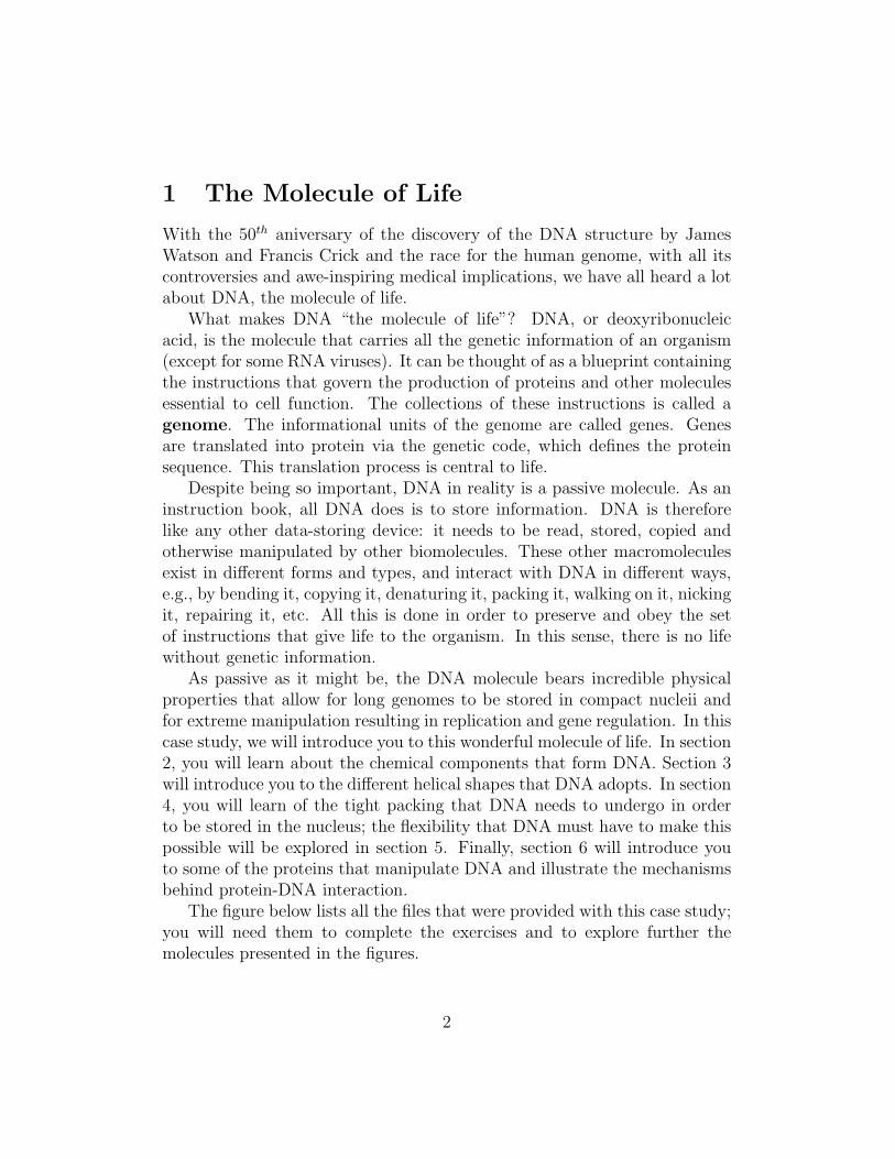
The figure below lists all the files that were provided with this case study;you will need them to complete the exercises and to explore further themolecules presented in the figures.
2
DNA case study
building-blocks
2endo.pdb3endo.pdbadenine.pdbanti-dAMP.pdbanti-deoxyadenosine.pdbcytosine.pdbdAMP.pdbdCMP.pdbdGMP.pdbdTMP.pdbguanine.pdbsyn-dAMP.pdbsyn-deoxyadenosine.pdbthymine.pdb
bases.vmdnucleotides.vmdsugar-pucker.vmdsyn-anti.vmd
dna-structure
adna.pdbbdna.pdbzdna.pdb
dna.vmd
dna-packing
bdna.pdbnucleosome.pdbnucleosome.psf
chromatin.tcldna-packing.vmd
dna-flexibility
coil.pdbcoil.psfdna-ws.psfdna-ws-eq.dcd
coil.vmddna-bend.vmdrgyr.tcl
protein-dna
1AN2.pdb1CMA.pdb1LMB.pdb1YSA.pdbat-bdna.pdber-ere-ws-ion.psber-ere-ws-ion.psfgc-bdna.pdb
dna-binding-motifs.vmddna-recognition.vmdestrogen-receptor-dna.vmd
3
2 The Building Blocks
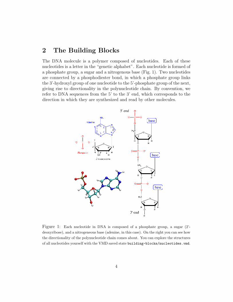
The DNA molecule is a polymer composed of nucleotides. Each of thesenucleotides is a letter in the “genetic alphabet”. Each nucleotide is formed ofa phosphate group, a sugar and a nitrogenous base (Fig. 1). Two nucleotidesare connected by a phosphodiester bond, in which a phosphate group linksthe 3’-hydroxyl group of one nucleotide to the 5’-phosphate group of the next,giving rise to directionality in the polynucleotide chain. By convention, werefer to DNA sequences from the 5’ to the 3’ end, which corresponds to thedirection in which they are synthesized and read by other molecules.
Figure 1: Each nucleotide in DNA is composed of a phosphate group, a sugar (2’-deoxyribose), and a nitrogeneous base (adenine, in this case). On the right you can see howthe directionality of the polynucleotide chain comes about. You can explore the structuresof all nucleotides yourself with the VMD saved state building-blocks/nucleotides.vmd.
4
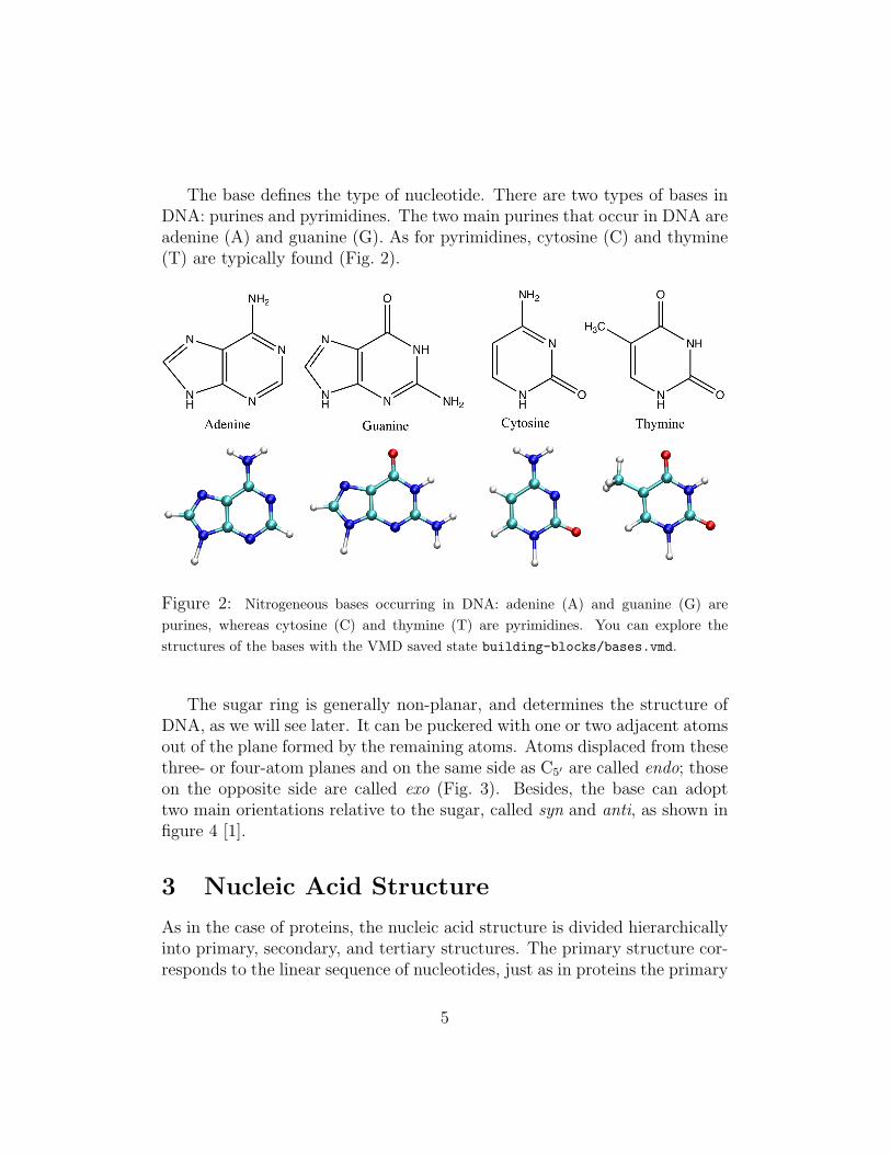
The base defines the type of nucleotide. There are two types of bases inDNA: purines and pyrimidines. The two main purines that occur in DNA areadenine (A) and guanine (G). As for pyrimidines, cytosine (C) and thymine(T) are typically found (Fig. 2).
Figure 2: Nitrogeneous bases occurring in DNA: adenine (A) and guanine (G) arepurines, whereas cytosine (C) and thymine (T) are pyrimidines. You can explore thestructures of the bases with the VMD saved state building-blocks/bases.vmd.
The sugar ring is generally non-planar, and determines the structure ofDNA, as we will see later. It can be puckered with one or two adjacent atomsout of the plane formed by the remaining atoms. Atoms displaced from thesethree- or four-atom planes and on the same side as C5′ are called endo; thoseon the opposite side are called exo (Fig. 3). Besides, the base can adopttwo main orientations relative to the sugar, called syn and anti, as shown infigure 4 [1].
3 Nucleic Acid Structure
As in the case of proteins, the nucleic acid structure is divided hierarchicallyinto primary, secondary, and tertiary structures. The primary structure cor-responds to the linear sequence of nucleotides, just as in proteins the primary
5
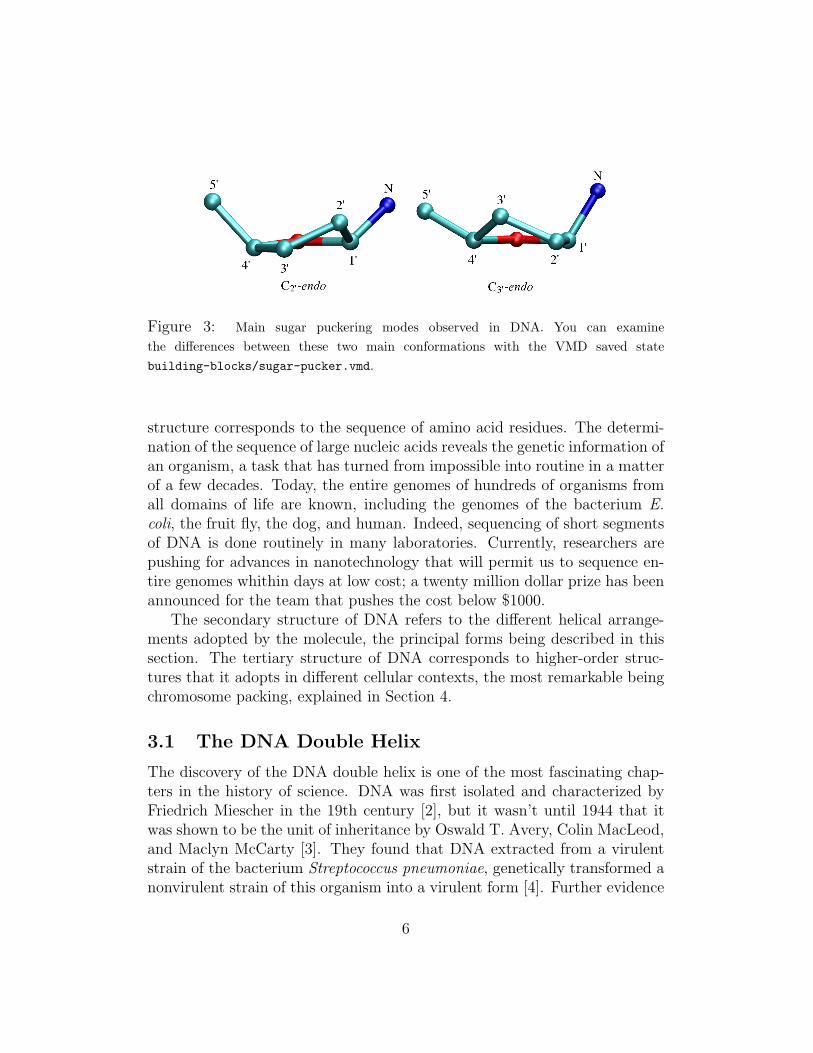
Figure 3: Main sugar puckering modes observed in DNA. You can examinethe differences between these two main conformations with the VMD saved statebuilding-blocks/sugar-pucker.vmd.
structure corresponds to the sequence of amino acid residues. The determi-nation of the sequence of large nucleic acids reveals the genetic information ofan organism, a task that has turned from impossible into routine in a matterof a few decades. Today, the entire genomes of hundreds of organisms fromall domains of life are known, including the genomes of the bacterium E.coli, the fruit fly, the dog, and human. Indeed, sequencing of short segmentsof DNA is done routinely in many laboratories. Currently, researchers arepushing for advances in nanotechnology that will permit us to sequence en-tire genomes whithin days at low cost; a twenty million dollar prize has beenannounced for the team that pushes the cost below $1000.
The secondary structure of DNA refers to the different helical arrange-ments adopted by the molecule, the principal forms being described in thissection. The tertiary structure of DNA corresponds to higher-order struc-tures that it adopts in different cellular contexts, the most remarkable beingchromosome packing, explained in Section 4.
3.1 The DNA Double Helix
The discovery of the DNA double helix is one of the most fascinating chap-ters in the history of science. DNA was first isolated and characterized byFriedrich Miescher in the 19th century [2], but it wasn’t until 1944 that itwas shown to be the unit of inheritance by Oswald T. Avery, Colin MacLeod,and Maclyn McCarty [3]. They found that DNA extracted from a virulentstrain of the bacterium Streptococcus pneumoniae, genetically transformed anonvirulent strain of this organism into a virulent form [4]. Further evidence
6
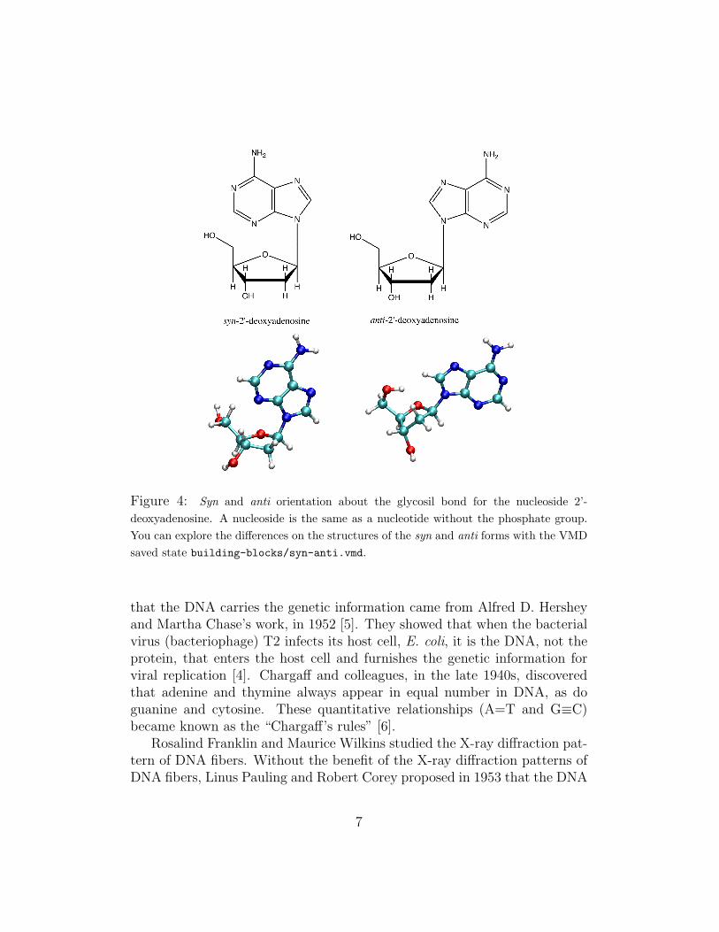
Figure 4: Syn and anti orientation about the glycosil bond for the nucleoside 2’-deoxyadenosine. A nucleoside is the same as a nucleotide without the phosphate group.You can explore the differences on the structures of the syn and anti forms with the VMDsaved state building-blocks/syn-anti.vmd.
that the DNA carries the genetic information came from Alfred D. Hersheyand Martha Chase’s work, in 1952 [5]. They showed that when the bacterialvirus (bacteriophage) T2 infects its host cell, E. coli, it is the DNA, not theprotein, that enters the host cell and furnishes the genetic information forviral replication [4]. Chargaff and colleagues, in the late 1940s, discoveredthat adenine and thymine always appear in equal number in DNA, as doguanine and cytosine. These quantitative relationships (A=T and G≡C)became known as the “Chargaff’s rules” [6].
Rosalind Franklin and Maurice Wilkins studied the X-ray diffraction pat-tern of DNA fibers. Without the benefit of the X-ray diffraction patterns ofDNA fibers, Linus Pauling and Robert Corey proposed in 1953 that the DNA
7
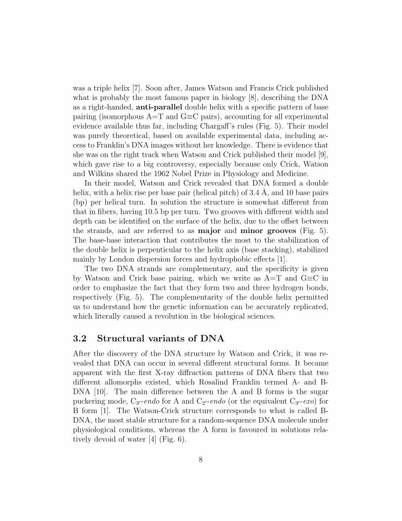
was a triple helix [7]. Soon after, James Watson and Francis Crick publishedwhat is probably the most famous paper in biology [8], describing the DNAas a right-handed, anti-parallel double helix with a specific pattern of basepairing (isomorphous A=T and G≡C pairs), accounting for all experimentalevidence available thus far, including Chargaff’s rules (Fig. 5). Their modelwas purely theoretical, based on available experimental data, including ac-cess to Franklin’s DNA images without her knowledge. There is evidence thatshe was on the right track when Watson and Crick published their model [9],which gave rise to a big controversy, especially because only Crick, Watsonand Wilkins shared the 1962 Nobel Prize in Physiology and Medicine.
In their model, Watson and Crick revealed that DNA formed a doublehelix, with a helix rise per base pair (helical pitch) of 3.4 A, and 10 base pairs(bp) per helical turn. In solution the structure is somewhat different fromthat in fibers, having 10.5 bp per turn. Two grooves with different width anddepth can be identified on the surface of the helix, due to the offset betweenthe strands, and are referred to as major and minor grooves (Fig. 5).The base-base interaction that contributes the most to the stabilization ofthe double helix is perpenticular to the helix axis (base stacking), stabilizedmainly by London dispersion forces and hydrophobic effects [1].
The two DNA strands are complementary, and the specificity is givenby Watson and Crick base pairing, which we write as A=T and G≡C inorder to emphasize the fact that they form two and three hydrogen bonds,respectively (Fig. 5). The complementarity of the double helix permittedus to understand how the genetic information can be accurately replicated,which literally caused a revolution in the biological sciences.
3.2 Structural variants of DNA
After the discovery of the DNA structure by Watson and Crick, it was re-vealed that DNA can occur in several different structural forms. It becameapparent with the first X-ray diffraction patterns of DNA fibers that twodifferent allomorphs existed, which Rosalind Franklin termed A- and B-DNA [10]. The main difference between the A and B forms is the sugarpuckering mode, C3′-endo for A and C2′-endo (or the equivalent C3′-exo) forB form [1]. The Watson-Crick structure corresponds to what is called B-DNA, the most stable structure for a random-sequence DNA molecule underphysiological conditions, whereas the A form is favoured in solutions rela-tively devoid of water [4] (Fig. 6).
8
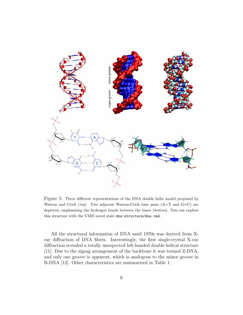
Figure 5: Three different representations of the DNA double helix model proposed byWatson and Crick (top). Two adjacent Watson-Crick base pairs (A=T and G≡C) aredepicted, emphasizing the hydrogen bonds between the bases (bottom). You can explorethis structure with the VMD saved state dna-structure/dna.vmd.
All the structural information of DNA until 1970s was derived from X-ray diffraction of DNA fibers. Interestingly, the first single-crystal X-raydiffraction revealed a totally unexpected left-handed double helical structure[11]. Due to the zigzag arrangement of the backbone it was termed Z-DNA,and only one groove is apparent, which is analogous to the minor groove inB-DNA [12]. Other characteristics are summarized in Table 1.
9
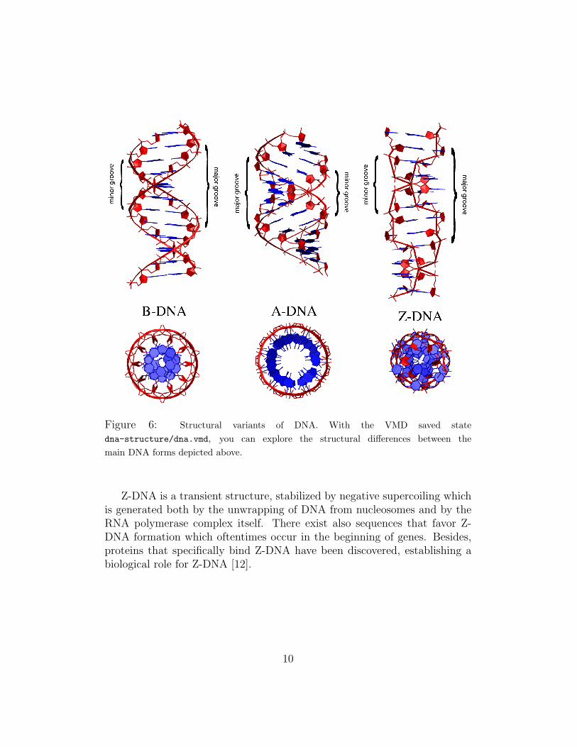
Figure 6: Structural variants of DNA. With the VMD saved statedna-structure/dna.vmd, you can explore the structural differences between themain DNA forms depicted above.
Z-DNA is a transient structure, stabilized by negative supercoiling whichis generated both by the unwrapping of DNA from nucleosomes and by theRNA polymerase complex itself. There exist also sequences that favor Z-DNA formation which oftentimes occur in the beginning of genes. Besides,proteins that specifically bind Z-DNA have been discovered, establishing abiological role for Z-DNA [12].
10
A-DNA B-DNA Z-DNAHelical sense Right handed Right handed Left handedDiameter ∼ 26A ∼ 20A ∼ 18ABase pairs per helical turn 11 10.5 12Helical rise per base pair 2.6 A 3.4 A 3.7 ABase tilt normal to the helix axis 20 ◦ 6 ◦ 7 ◦
Sugar pucker conformation C3′ -endo C2′ -endo C2′ -endo for pyrimidines;C3′ -endo for purines
Glycosyl bond conformation Anti Anti Anti for pyrimidines;syn for purines
Table 1: Summary of the differences between the three main DNA double-helical con-formations (from [4]).
Exercise 1: Double helix of DNA. In this exercise, you are going to ex-plore the various structures of double helical DNA. Load the VMD saved statedna-structure/dna.vmd, which contains the structures of A, B, and Z-DNAa.
1. Measure the distance between two contiguous base pairs (helical rise) in eachof the structures.
2. Make a H-bonds representation (use a distance cutoff of 3.2 A) for a G≡C andan A=T pair and create a snapshot. How many hydrogen bonds are there ineach case?
3. When segments of DNA are subject to high temperature, the double helix melts.There is a difference in melting temperature between segments with high A=Tand G≡C content. Which case would correspond to the higher melting temper-ature? Explain this difference based on Watson-Crick base pairing.
4. Create a van der Waals representation for each structure with one color for thebackbone (you can use the keyword “backbone” in the selection) and anotherfor the nucleoside (“not backbone”). Create a snapshot, identifying the majorand minor grooves (see text), as well as the helical sense (right or left handedhelix) for each DNA form. Measure the size of the major and minor grooves foreach DNA form.
aThe PDB files were obtained fromhttp://chemistry.gsu.edu/glactone/PDB/pdb.html
11
4 DNA packing inside the cell
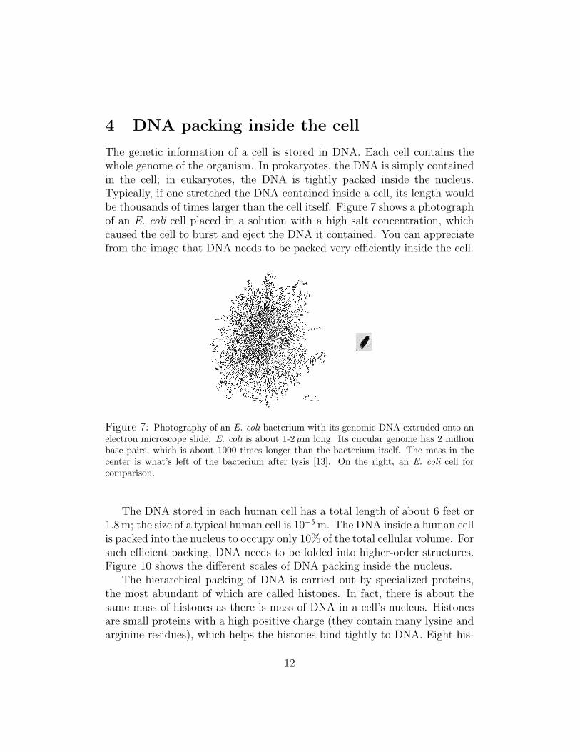
The genetic information of a cell is stored in DNA. Each cell contains thewhole genome of the organism. In prokaryotes, the DNA is simply containedin the cell; in eukaryotes, the DNA is tightly packed inside the nucleus.Typically, if one stretched the DNA contained inside a cell, its length wouldbe thousands of times larger than the cell itself. Figure 7 shows a photographof an E. coli cell placed in a solution with a high salt concentration, whichcaused the cell to burst and eject the DNA it contained. You can appreciatefrom the image that DNA needs to be packed very efficiently inside the cell.
Figure 7: Photography of an E. coli bacterium with its genomic DNA extruded onto anelectron microscope slide. E. coli is about 1-2 µm long. Its circular genome has 2 millionbase pairs, which is about 1000 times longer than the bacterium itself. The mass in thecenter is what’s left of the bacterium after lysis [13]. On the right, an E. coli cell forcomparison.
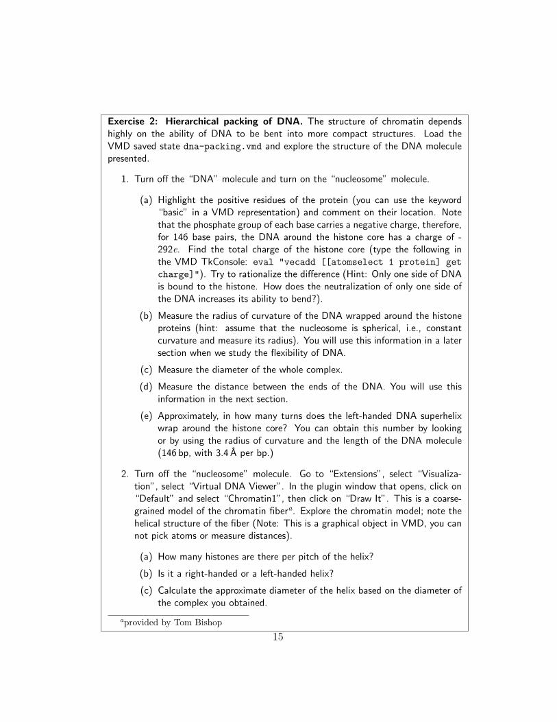
The DNA stored in each human cell has a total length of about 6 feet or1.8m; the size of a typical human cell is 10−5 m. The DNA inside a human cellis packed into the nucleus to occupy only 10% of the total cellular volume. Forsuch efficient packing, DNA needs to be folded into higher-order structures.Figure 10 shows the different scales of DNA packing inside the nucleus.
The hierarchical packing of DNA is carried out by specialized proteins,the most abundant of which are called histones. In fact, there is about thesame mass of histones as there is mass of DNA in a cell’s nucleus. Histonesare small proteins with a high positive charge (they contain many lysine andarginine residues), which helps the histones bind tightly to DNA. Eight his-
12
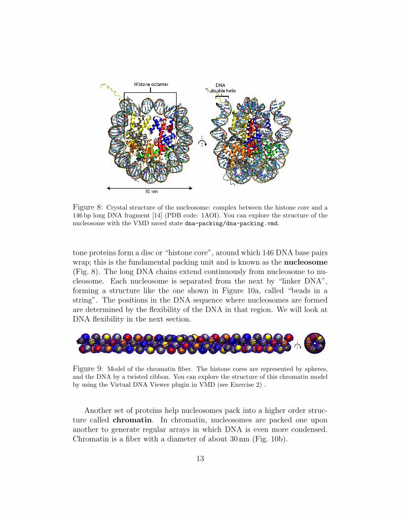
Figure 8: Crystal structure of the nucleosome: complex between the histone core and a146 bp long DNA fragment [14] (PDB code: 1AOI). You can explore the structure of thenucleosome with the VMD saved state dna-packing/dna-packing.vmd.
tone proteins form a disc or “histone core”, around which 146 DNA base pairswrap; this is the fundamental packing unit and is known as the nucleosome(Fig. 8). The long DNA chains extend continuously from nucleosome to nu-cleosome. Each nucleosome is separated from the next by “linker DNA”,forming a structure like the one shown in Figure 10a, called “beads in astring”. The positions in the DNA sequence where nucleosomes are formedare determined by the flexibility of the DNA in that region. We will look atDNA flexibility in the next section.
Figure 9: Model of the chromatin fiber. The histone cores are represented by spheres,and the DNA by a twisted ribbon. You can explore the structure of this chromatin modelby using the Virtual DNA Viewer plugin in VMD (see Exercise 2) .
Another set of proteins help nucleosomes pack into a higher order struc-ture called chromatin. In chromatin, nucleosomes are packed one uponanother to generate regular arrays in which DNA is even more condensed.Chromatin is a fiber with a diameter of about 30 nm (Fig. 10b).
13
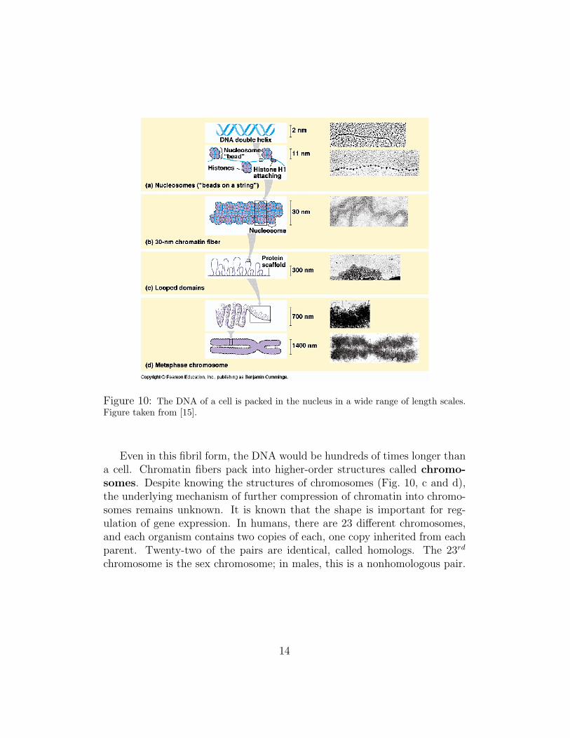
Figure 10: The DNA of a cell is packed in the nucleus in a wide range of length scales.Figure taken from [15].
Even in this fibril form, the DNA would be hundreds of times longer thana cell. Chromatin fibers pack into higher-order structures called chromo-somes. Despite knowing the structures of chromosomes (Fig. 10, c and d),the underlying mechanism of further compression of chromatin into chromo-somes remains unknown. It is known that the shape is important for reg-ulation of gene expression. In humans, there are 23 different chromosomes,and each organism contains two copies of each, one copy inherited from eachparent. Twenty-two of the pairs are identical, called homologs. The 23rd
chromosome is the sex chromosome; in males, this is a nonhomologous pair.
14
Exercise 2: Hierarchical packing of DNA. The structure of chromatin dependshighly on the ability of DNA to be bent into more compact structures. Load theVMD saved state dna-packing.vmd and explore the structure of the DNA moleculepresented.
1. Turn off the “DNA” molecule and turn on the “nucleosome” molecule.
(a) Highlight the positive residues of the protein (you can use the keyword“basic” in a VMD representation) and comment on their location. Notethat the phosphate group of each base carries a negative charge, therefore,for 146 base pairs, the DNA around the histone core has a charge of -292e. Find the total charge of the histone core (type the following inthe VMD TkConsole: eval "vecadd [[atomselect 1 protein] getcharge]"). Try to rationalize the difference (Hint: Only one side of DNAis bound to the histone. How does the neutralization of only one side ofthe DNA increases its ability to bend?).
(b) Measure the radius of curvature of the DNA wrapped around the histoneproteins (hint: assume that the nucleosome is spherical, i.e., constantcurvature and measure its radius). You will use this information in a latersection when we study the flexibility of DNA.
(c) Measure the diameter of the whole complex.
(d) Measure the distance between the ends of the DNA. You will use thisinformation in the next section.
(e) Approximately, in how many turns does the left-handed DNA superhelixwrap around the histone core? You can obtain this number by lookingor by using the radius of curvature and the length of the DNA molecule(146 bp, with 3.4 A per bp.)
2. Turn off the “nucleosome” molecule. Go to “Extensions”, select “Visualiza-tion”, select “Virtual DNA Viewer”. In the plugin window that opens, click on“Default” and select “Chromatin1”, then click on “Draw It”. This is a coarse-grained model of the chromatin fibera. Explore the chromatin model; note thehelical structure of the fiber (Note: This is a graphical object in VMD, you cannot pick atoms or measure distances).
(a) How many histones are there per pitch of the helix?
(b) Is it a right-handed or a left-handed helix?
(c) Calculate the approximate diameter of the helix based on the diameter ofthe complex you obtained.
aprovided by Tom Bishop
15
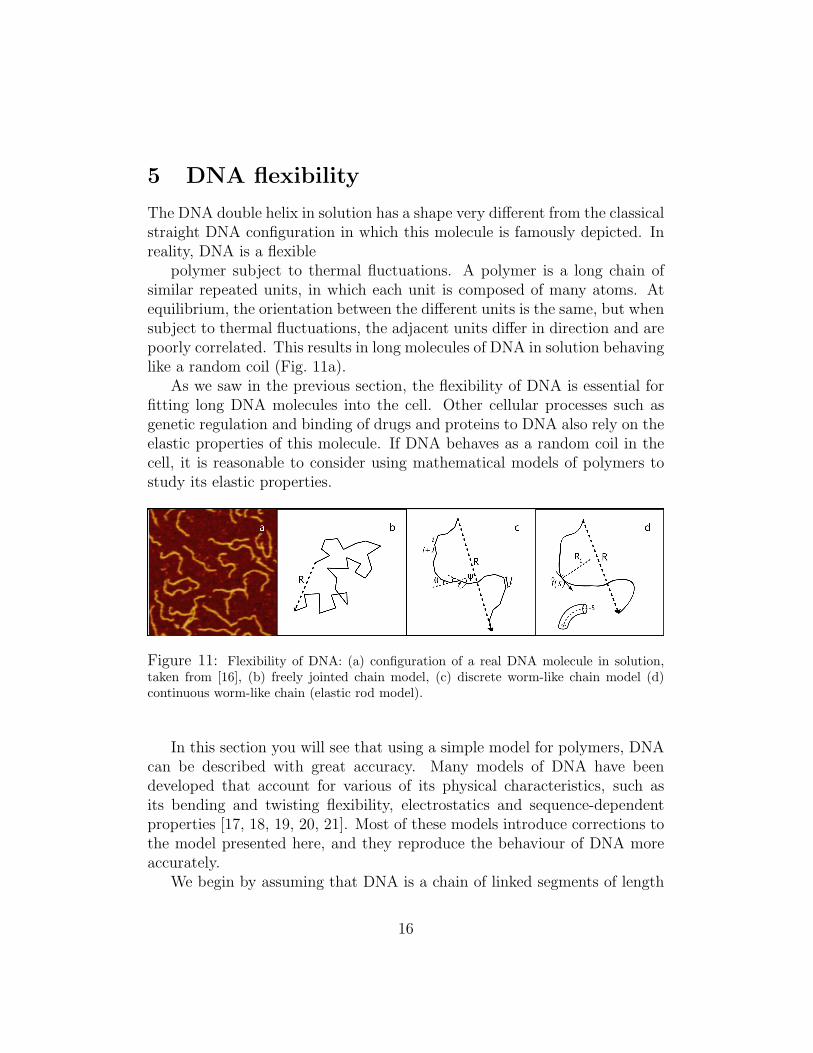
5 DNA flexibility
The DNA double helix in solution has a shape very different from the classicalstraight DNA configuration in which this molecule is famously depicted. Inreality, DNA is a flexible
polymer subject to thermal fluctuations. A polymer is a long chain ofsimilar repeated units, in which each unit is composed of many atoms. Atequilibrium, the orientation between the different units is the same, but whensubject to thermal fluctuations, the adjacent units differ in direction and arepoorly correlated. This results in long molecules of DNA in solution behavinglike a random coil (Fig. 11a).
As we saw in the previous section, the flexibility of DNA is essential forfitting long DNA molecules into the cell. Other cellular processes such asgenetic regulation and binding of drugs and proteins to DNA also rely on theelastic properties of this molecule. If DNA behaves as a random coil in thecell, it is reasonable to consider using mathematical models of polymers tostudy its elastic properties.
Figure 11: Flexibility of DNA: (a) configuration of a real DNA molecule in solution,taken from [16], (b) freely jointed chain model, (c) discrete worm-like chain model (d)continuous worm-like chain (elastic rod model).
In this section you will see that using a simple model for polymers, DNAcan be described with great accuracy. Many models of DNA have beendeveloped that account for various of its physical characteristics, such asits bending and twisting flexibility, electrostatics and sequence-dependentproperties [17, 18, 19, 20, 21]. Most of these models introduce corrections tothe model presented here, and they reproduce the behaviour of DNA moreaccurately.
We begin by assuming that DNA is a chain of linked segments of length
16
l, that are connected by flexible linkers that can rotate freely (Fig. 11b). Thecontour length of the molecule is Nl, where N is the number of segments.The end-to-end distance vector is defined as:
R =∑
i
li, (1)
where li is the vector representation of the segment i. The square of themagnitude of r can be used as a characteristic length of the chain, and isgiven by
R2 =
(n∑i
li
)(n∑j
lj
). (2)
Factoring the diagonal terms i = j and taking the average over all possibleconfigurations1 we get the mean square end-to-end distance⟨
R2⟩
= Nl2 + 2∑j>i
〈li · lj〉 . (3)
For random orientation of the segments (Fig. 11c), the second term on theright hand side is zero, thus ⟨
R2⟩
= Nl2, (4)
which is the mean square end-to-end distance for the so-called freely jointedchain (FJC) model, the simplest model for describing polymers. The FJCmodel works only for large values of N , and generally describes the confor-mations of polymers poorly, mainly because it neglects interactions betweenthe polymer with itself (the polymer self-crosses often). As an example, thismodel gives an average end-to-end displacement 〈R〉 = 0, which is not whatyou would expect to observe of a polymer in solution.
A widely used measure for the characterization of the configuration of apolymer is the radius of gyration (RG), which measures the root-mean-square distance of the collection of segments from their common center ofmass, i.e.
R2G =
1
N + 1
N∑i=0
(ri − rcm)2 (5)
1All averages hereupon are taken in the same way.
17
where rcm is the vector defining the coordinates of the center of mass (rcm =∑j mjrj, mj and rj being the mass and the position of segment j, respec-
tively). The radius of gyration can be measured from particle scatteringexperiments [22]. The radius of gyration can also be expressed in terms ofthe average mean square end-to-end distance 〈R2〉. To find this relation, weuse the theorem of Lagrange [23]:
R2G =
1
(N + 1)2
∑0≤i<j≤N
r2ij (6)
where rij is the length of the vector connecting segments i and j. Averagingover the statistical ensemble gives⟨
R2G
⟩=
1
(N + 1)2
∑0≤i<j≤N
⟨r2ij
⟩. (7)
Using (4), for N = j − i ⟨r2ij
⟩= (j − i)l2. (8)
Thus,
⟨R2
G
⟩=
l2
(N + 1)2
∑0≤i<j≤N
(j − i)
=l2
(N + 1)2
N∑j=1
j∑k=1
k
=l2
2(N + 1)2
N∑j=1
(j2 + j) (9)
where we have introduced the change of variable k = j − i. We note that
N∑j=1
(j2 + j) =N(N + 1)(2N + 1)
6+N(N + 1)
2. (10)
For large N , this is approximately N3/3, which gives, using (9) and (4),
⟨R2
G
⟩≈ Nl2
6=〈R2〉
6. (11)
18
Another useful quantity is the persistence length (lp), defined as theaverage projection of the end-to-end vector onto an arbitrary vector li, in thelimit of an infinite chain, i.e.
lp = limN→∞
⟨N∑
j=i
li ·ljl
⟩. (12)
The persistence length is a measure of the rigidity of the molecule, or ameasure of the length over which there is significant correlation between thedirections of two segments. In order to relate the persistence length to themean square end-to-end distance, we rewrite (3) in the following way:
⟨R2⟩
= Nl2 + 2∑j>i
〈li · lj〉 (13)
= Nl2 + 2N∑
j=2
〈l1 · lj〉+ 2N∑
j=i+1
N∑i=2
〈li · lj〉 (14)
For large N , the first summation above corresponds to (12), except that itstarts from j = 2. Therefore,
N∑j=2
〈l1 · lj〉 = llp − l2 (15)
It is clear that every term in the second summation of (14) also contributeswith llp − l2. Thus,⟨
R2⟩
= Nl2 + 2N(llp − l2) = 2Nllp −Nl2 (16)
or
lp =〈R2〉+Nl2
2Nl(17)
for large N . As we saw in (4), the mean square end-to-end distance for theFJC model is give by 〈R2〉 = Nl2. Thus, for this model the persistence lengthis simply given by lp = l.
Now we consider the statistical distribution of the end-to-end vector. LetΦ(R, N) be the probability distribution function of the end-to-end vector of
19
a chain of N segments being R. The conformational distribution of the chaincan be considered as a product of independent conformations of the segments
Ψ ({rn}) =N∏
n=1
ψ(rn) (18)
where ψ(rn) is the random distribution of a segment with length l
ψ(rn) =1
4πl2δ (|rn| − l) (19)
Φ (R, N) is calculated by
Φ (R, N) =
∫dr1
∫dr2 . . .
∫drNδ(R−
N∑i=1
rn)Ψ ({rn}) . (20)
Using
δ(r) =1
(2π)3
∫dke(ik·r), (21)
one can rewrite (20) as
Φ (R, N) =1
(2π)3
∫dr1
∫dr2 . . .
∫drN exp (ik · (R−
N∑i=1
rn)Ψ ({rn})
=1
(2π)3
∫dke(ik·R)
∫dr1 . . .
∫drN
N∏n=1
e(−ik·rn)ψ(rn)
=1
(2π)3
∫dke(ik·R)
[∫drne
(−ik·rn)ψ(rn)
]N
(22)
The integral over r is evaluated by introducing polar coordinates (r, θ, φ); thereference axis of θ being taken along the vector k, giving
∫dre(−ik·r)ψ(r)
=1
4πl2
∫ ∞
0
drr2
∫ 2π
0
dφ
∫ π
0
dθ sin θ exp(−ikr cos θ) δ(r − l)
=sin kl
kl(23)
20
where k = |k|. From equations (22) and (23)
Φ(R, N) =1
(2π)3
∫dk eik·R
(sin kl
kl
)N
. (24)
If N is large, (sin kl/kl)N becomes very small unless kl is small. For kl� 1,(sin kl/kl)N can be approximated as(
sin kl
kl
)N
'(
1− k2l2
6
)N
' exp
(−Nk
2l2
6
). (25)
This approximation holds also for kl ≥ 1 since both sides of equation (25)are nearly zero in such a case. Thus Φ(R, N) is calculated as
Φ(R, N) =1
(2π)3
∫dkeik·R exp
(−Nk
2l2
6
). (26)
The integral over k is a standard Gaussian integral∫ ∞
−∞dx exp(−ax2 + bx) = (π/a)1/2 exp(
b2
4a) (27)
If kα and Rα (α = x, y, z) denote the components of the vectors k and R,then,
Φ(R, N) = (2π)−3∏
α=x,y,z
[∫ ∞
−∞dkα exp(ikαRα −Nk2
αl2/6)
]
= (2π)−3∏
α=x,y,z
(6π
Nl2
)1/2
exp
(− 3
2Nl2R2
α
)= (3/2πNl2)3/2 exp
(− 3R2
2Nl2
). (28)
Thus the distribution function of the end-to-end vector is Gaussian.A correction to the FJC model is made by the freely rotating chain or
worm-like chain (WLC) model. In this model, the nth bond is connectedto the (n− 1)th bond with a fixed angle θ, but can rotate freely around the(n − 1)th bond (Fig. 11c). For this model, the second term in (3) does notaverage zero. In order to find 〈li · lj〉, we first look at adjacent links
21
〈li · li+1〉 = l2 cos θ. (29)
We can denote α = cos θ. In order to calculate the average of the projectionof bond i + k on i, we note that the inner product of li and projection ofli+k in the transverse direction of li+k−1 average zero. The inner product of liand the projection of what remains in the transverse direction of li+k−2 alsoaverage zero, and so on. Thus,
〈li · li+k〉 = l2αk, (30)
Introducing this into (3), we have
⟨R2⟩
= Nl2 + 2∑j>i
l2αj−i (31)
= Nl2 + 2l2N∑
j=1
αj
j−1∑i=1
1
αi. (32)
The sum of the first n+ 1 terms of a geometric series is given by
n∑k=0
yk =1− yn+1
1− y, (33)
so that
j−1∑i=1
(1
α
)i
=1− α−j
1− α−1− 1 =
1− α1−j
α− 1, (34)
which yields
⟨R2⟩
= Nl2 + 2l2N∑
j=1
αj
(1− α1−j
α− 1
)
= Nl2 +2l2
α− 1
[(N∑
j=1
αj
)−Nα
]
Using (33) again
22
N∑j=1
αj =1− αN+1
1− α=α− αN+1
1− α(35)
we obtain
⟨R2⟩
= Nl2 +2l2
α− 1
[α− αN+1
1− α−Nα
](36)
= Nl2 +2l2
(α− 1)2
(Nα−Nα2 + αN+1 − α
)(37)
Rearranging terms leads to
⟨R2⟩
= Nl2[1 + α
1− α+
2α(1− αN)
N(α− 1)2
](38)
For large N , we can neglect the last term, obtaining⟨R2⟩≈ Nl2
1 + α
1− α. (39)
The mean square radius of gyration for the WLC model can be obtainedby combining (38) and (7). As before, we introduce the change of variablek = j − i,
⟨R2
G
⟩=
l2
(N + 1)2
N∑j=1
j∑k=1
k
[1 + α
1− α+
2α
(α− 1)2
(1
k− αk
k
)](40)
Using (33),
⟨R2
G
⟩=
l2
(N + 1)2
N∑j=1
{(1 + α
1− α
)j(j + 1)
2+
2α
(α− 1)2
[j − 1− αj+1
1− α+ 1
]}(41)
Introducing (10),
⟨R2
G
⟩=
l2
(N + 1)2
{(1 + α
1− α
)1
2
[N(N + 1)(2N + 1)
6+N(N + 1)
2
]+
+2α
(α− 1)2
[N(N + 1)
2− N
1− α+N +
N∑j=1
αj+1
1− α
]}
23
we can evaluate the sum above using (33)
N∑j=1
αj+1 =N∑
k=2
αk =1− αN+1
1− α− 1− α =
α2 − αN+1
1− α. (42)
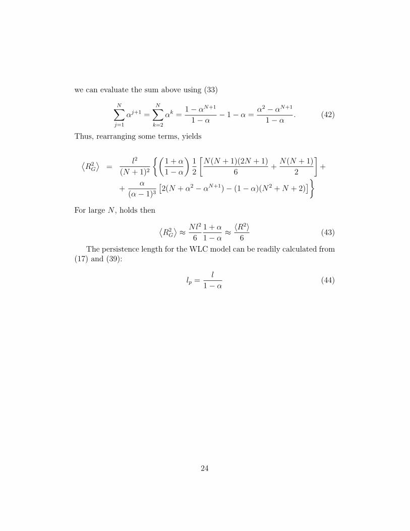
Thus, rearranging some terms, yields
⟨R2
G
⟩=
l2
(N + 1)2
{(1 + α
1− α
)1
2
[N(N + 1)(2N + 1)
6+N(N + 1)
2
]+
+α
(α− 1)3
[2(N + α2 − αN+1)− (1− α)(N2 +N + 2)
]}For large N , holds then
⟨R2
G
⟩≈ Nl2
6
1 + α
1− α≈ 〈R2〉
6(43)
The persistence length for the WLC model can be readily calculated from(17) and (39):
lp =l
1− α(44)
24
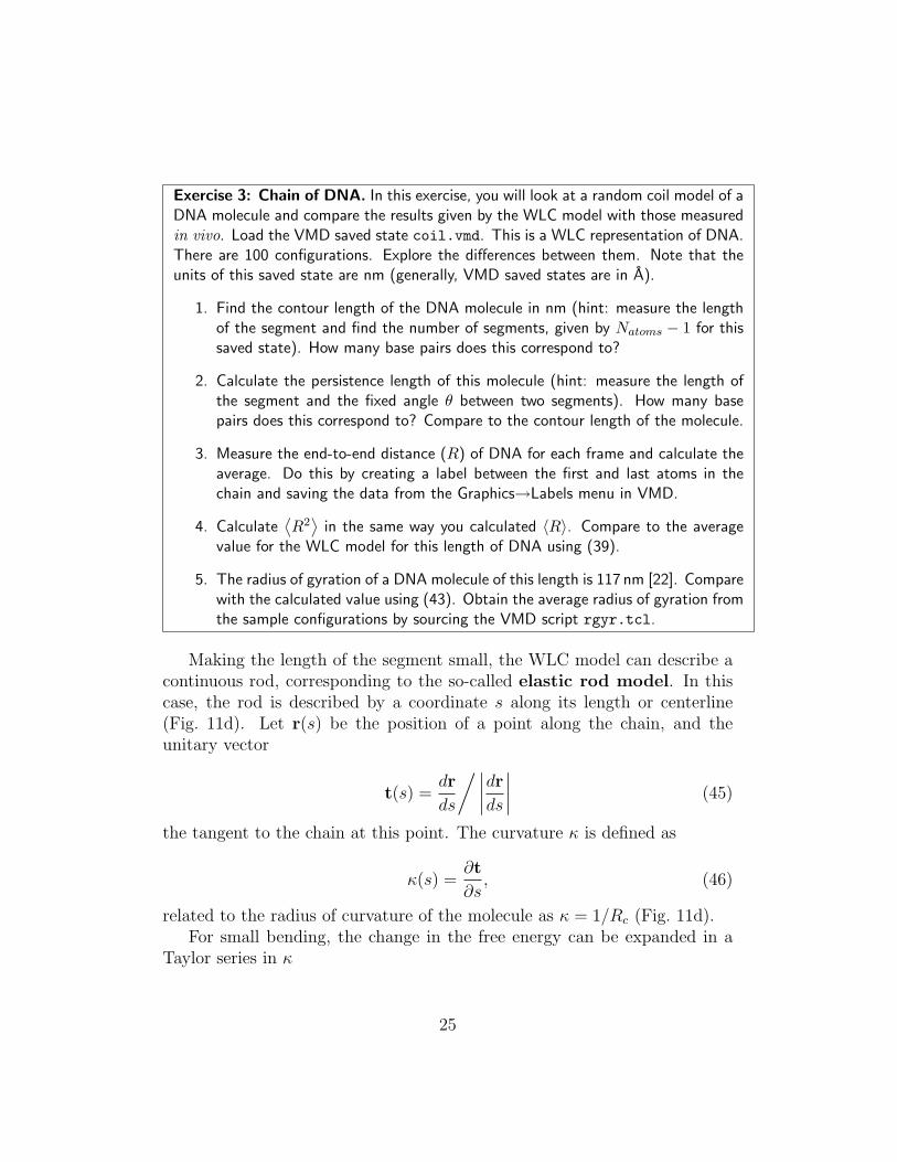
Exercise 3: Chain of DNA. In this exercise, you will look at a random coil model of aDNA molecule and compare the results given by the WLC model with those measuredin vivo. Load the VMD saved state coil.vmd. This is a WLC representation of DNA.There are 100 configurations. Explore the differences between them. Note that theunits of this saved state are nm (generally, VMD saved states are in A).
1. Find the contour length of the DNA molecule in nm (hint: measure the lengthof the segment and find the number of segments, given by Natoms − 1 for thissaved state). How many base pairs does this correspond to?
2. Calculate the persistence length of this molecule (hint: measure the length ofthe segment and the fixed angle θ between two segments). How many basepairs does this correspond to? Compare to the contour length of the molecule.
3. Measure the end-to-end distance (R) of DNA for each frame and calculate theaverage. Do this by creating a label between the first and last atoms in thechain and saving the data from the Graphics→Labels menu in VMD.
4. Calculate⟨R2⟩
in the same way you calculated 〈R〉. Compare to the averagevalue for the WLC model for this length of DNA using (39).
5. The radius of gyration of a DNA molecule of this length is 117 nm [22]. Comparewith the calculated value using (43). Obtain the average radius of gyration fromthe sample configurations by sourcing the VMD script rgyr.tcl.
Making the length of the segment small, the WLC model can describe acontinuous rod, corresponding to the so-called elastic rod model. In thiscase, the rod is described by a coordinate s along its length or centerline(Fig. 11d). Let r(s) be the position of a point along the chain, and theunitary vector
t(s) =dr
ds
/∣∣∣∣drds∣∣∣∣ (45)
the tangent to the chain at this point. The curvature κ is defined as
κ(s) =∂t
∂s, (46)
related to the radius of curvature of the molecule as κ = 1/Rc (Fig. 11d).For small bending, the change in the free energy can be expanded in a
Taylor series in κ
25
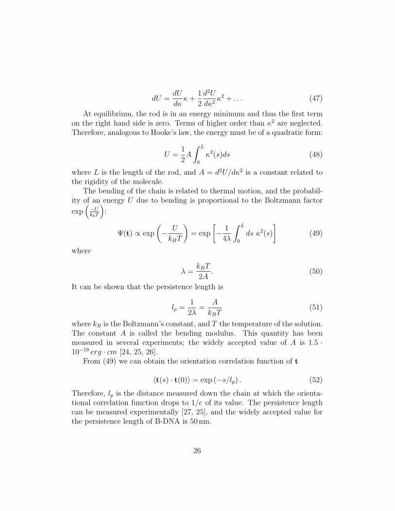
dU =dU
dκκ+
1
2
d2U
dκ2κ2 + . . . (47)
At equilibrium, the rod is in an energy minimum and thus the first termon the right hand side is zero. Terms of higher order than κ2 are neglected.Therefore, analogous to Hooke’s law, the energy must be of a quadratic form:
U =1
2A
∫ L
0
κ2(s)ds (48)
where L is the length of the rod, and A = d2U/dκ2 is a constant related tothe rigidity of the molecule.
The bending of the chain is related to thermal motion, and the probabil-ity of an energy U due to bending is proportional to the Boltzmann factor
exp(−UkbT
):
Ψ(t) ∝ exp
(− U
kBT
)= exp
[− 1
4λ
∫ L
0
ds κ2(s)
](49)
where
λ =kBT
2A. (50)
It can be shown that the persistence length is
lp =1
2λ=
A
kBT(51)
where kB is the Boltzmann’s constant, and T the temperature of the solution.The constant A is called the bending modulus. This quantity has beenmeasured in several experiments; the widely accepted value of A is 1.5 ·10−19 erg · cm [24, 25, 26].
From (49) we can obtain the orientation correlation function of t
〈t(s) · t(0)〉 = exp (−s/lp) . (52)
Therefore, lp is the distance measured down the chain at which the orienta-tional correlation function drops to 1/e of its value. The persistence lengthcan be measured experimentally [27, 25], and the widely accepted value forthe persistence length of B-DNA is 50 nm.
26
Although the WLC model presented above does not implicitly account forvolume exclusion, the introduction of an angular constraint between segmentsavoids much of the self-crossing, which only occurs between distant segments.A correction can be introduced in case this needs to be accounted for [? 23].The WLC has been a very successful model for describing DNA stretchingexperiments, and is used for data interpretation very successfully [28].
Exercise 4: Nucleosome revisited. In the previous section, you learned that DNAis wrapped around the histone core in a very compact configuration.
1. How does the persistence length of DNA given in the text compare to the lengthof the DNA in the nucleosome?
2. How does it compare to the curvature you found in the nucleosome?
3. In exercise 2, you obtained the end-to-end distance of the DNA in the nucleo-some. What is the probability that this configuration of DNA occurs in solution(no protein constraints)?
4. According to the WLC model, estimate the energy required to bend DNA intosuch compact configuration.
5. For this configuration to occur, the interaction energy between the protein andthe DNA must be of a similar magnitude. From the structure of the nucleosomeyou explored before, give examples of interactions between protein and DNA thatwould account for this.
6 Protein-DNA Interaction
So far, we have considered that a DNA segment shorter than the persistencelength behaves as a straight stiff rod. In reality, the sequence of DNA affectsits bending and twisting properties. We discussed earlier that nucleosomesform in regions where the DNA is more prone to bend. Furthermore, otherenvironmental factors such as salt concentration, temperature and pressureaffect the structure of DNA.
The deviation from the straight double helix at equilibrium can be soprominent that one can observe DNA sequences of the order of the persistencelength of DNA that form a circle without being covalently bonded at theends! (Fig. 12). The study of the sequence-dependent properties of DNA isan active field of research [29, 30, 31], in view of the fact that the sequence
27
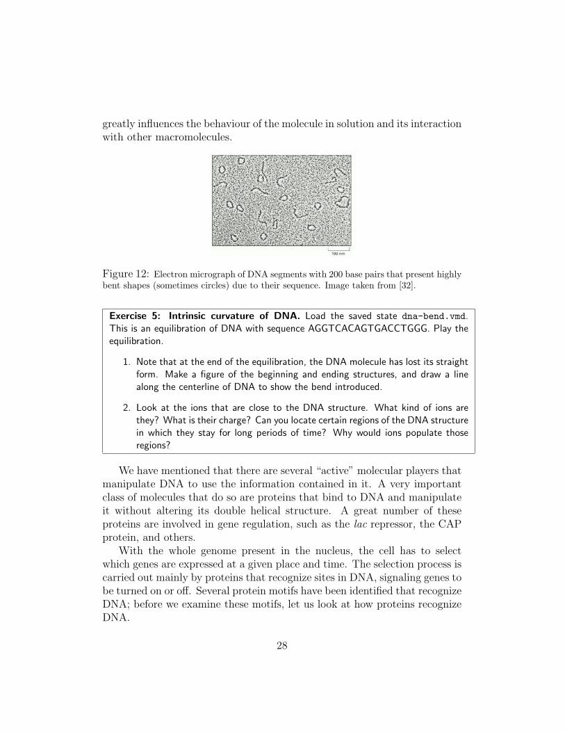
greatly influences the behaviour of the molecule in solution and its interactionwith other macromolecules.
Figure 12: Electron micrograph of DNA segments with 200 base pairs that present highlybent shapes (sometimes circles) due to their sequence. Image taken from [32].
Exercise 5: Intrinsic curvature of DNA. Load the saved state dna-bend.vmd.This is an equilibration of DNA with sequence AGGTCACAGTGACCTGGG. Play theequilibration.
1. Note that at the end of the equilibration, the DNA molecule has lost its straightform. Make a figure of the beginning and ending structures, and draw a linealong the centerline of DNA to show the bend introduced.
2. Look at the ions that are close to the DNA structure. What kind of ions arethey? What is their charge? Can you locate certain regions of the DNA structurein which they stay for long periods of time? Why would ions populate thoseregions?
We have mentioned that there are several “active” molecular players thatmanipulate DNA to use the information contained in it. A very importantclass of molecules that do so are proteins that bind to DNA and manipulateit without altering its double helical structure. A great number of theseproteins are involved in gene regulation, such as the lac repressor, the CAPprotein, and others.
With the whole genome present in the nucleus, the cell has to selectwhich genes are expressed at a given place and time. The selection process iscarried out mainly by proteins that recognize sites in DNA, signaling genes tobe turned on or off. Several protein motifs have been identified that recognizeDNA; before we examine these motifs, let us look at how proteins recognizeDNA.
28
6.1 Proteins have motifs to bind and recognize DNA
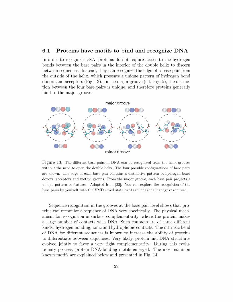
In order to recognize DNA, proteins do not require access to the hydrogenbonds between the base pairs in the interior of the double helix to discernbetween sequences. Instead, they can recognize the edge of a base pair fromthe outside of the helix, which presents a unique pattern of hydrogen bonddonors and acceptors (Fig. 13). In the major groove (c.f. Fig. 5), the distinc-tion between the four base pairs is unique, and therefore proteins generallybind to the major groove.
Figure 13: The different base pairs in DNA can be recognized from the helix grooveswithout the need to open the double helix. The four possible configurations of base pairsare shown. The edge of each base pair contains a distinctive pattern of hydrogen bonddonors, acceptors and methyl groups. From the major groove, each base pair projects aunique pattern of features. Adapted from [32]. You can explore the recognition of thebase pairs by yourself with the VMD saved state protein-dna/dna-recognition.vmd.
Sequence recognition in the grooves at the base pair level shows that pro-teins can recognize a sequence of DNA very specifically. The physical mech-anism for recognition is surface complementarity, where the protein makesa large number of contacts with DNA. Such contacts are of three differentkinds: hydrogen bonding, ionic and hydrophobic contacts. The intrinsic bendof DNA for different sequences is known to increase the ability of proteinsto differentiate between sequences. Very likely, protein and DNA structuresevolved jointly to favor a very tight complementarity. During this evolu-tionary process, protein DNA-binding motifs emerged. The most commonknown motifs are explained below and presented in Fig. 14.
29
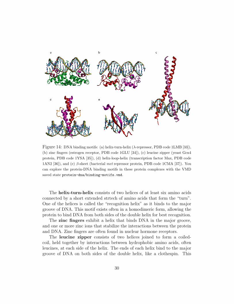
Figure 14: DNA binding motifs: (a) helix-turn-helix (λ-repressor, PDB code 1LMB [33]),(b) zinc fingers (estrogen receptor, PDB code 1GLU [34]), (c) leucine zipper (yeast Gcn4protein, PDB code 1YSA [35]), (d) helix-loop-helix (transcription factor Max, PDB code1AN2 [36]), and (e) β-sheet (bacterial met repressor protein, PDB code 1CMA [37]). Youcan explore the protein-DNA binding motifs in these protein complexes with the VMDsaved state protein-dna/binding-motifs.vmd.
The helix-turn-helix consists of two helices of at least six amino acidsconnected by a short extended strtech of amino acids that form the “turn”.One of the helices is called the “recognition helix” as it binds to the majorgroove of DNA. This motif exists often in a homodimeric form, allowing theprotein to bind DNA from both sides of the double helix for best recognition.
The zinc fingers exhibit a helix that binds DNA in the major groove,and one or more zinc ions that stabilize the interactions between the proteinand DNA. Zinc fingers are often found in nuclear hormone receptors.
The leucine zipper consists of two helices joined to form a coiled-coil, held together by interactions between hydrophobic amino acids, oftenleucines, at each side of the helix. The ends of each helix bind to the majorgroove of DNA on both sides of the double helix, like a clothespin. This
30
motif can be a heterodimer, which results in greater specificity.The helix-loop-helix, which is different from the helix-turn-helix motif,
consists of a long helix, and a loop that permits a shorter helix to fold backand pack against the first helix, allowing both helices to make contacts withDNA. This motif also exists often in a dimeric form.
Besides helices, proteins can also use β-sheets to recognize DNA; theydo so by inserting two-stranded β-sheets into the major groove of DNA.
6.2 Nuclear hormone receptors
Nuclear hormone receptors form a class of proteins that get activated by smallmolecules called hormones, that signal them to bind to specific sequences ofDNA and serve as on-off switches for expression of genes in the cell nucleus.These switches control the development and differentiation of skin, bone andbehavioral centers in the brain, as well as the continual regulation of repro-ductive tissues. Hormones are signaling molecules that exert their functionby traveling through the bloodstream and interacting with cells in a varietyof target tissues.
An example of a nuclear hormone is estrogen. The breast and the uterus,which play central roles in sexual reproduction, are two of the main targetsof estrogen. In addition, estrogen molecules act on the brain, bone, liver, andheart. Estrogen acts on these cells by binding estrogen receptors, which arefound only in cells that are targets for estrogen action. In this way, estrogencirculates in the bloodstream, but only exerts effects on the cell that containreceptors.
When an estrogen molecule enters a cell that contains receptors, it bindsto its receptor, causing it to change its shape. In this new shape, the receptorrecognizes a specific sequence of DNA, located near genes that are controlledby estrogen. The binding of the estrogen receptor to DNA induces geneexpression, influencing the cell behavior in different ways, depending on thecell type involved. In liver cells, for example, estrogen alters the productionof proteins that influence cholesterol levels in the blood.
31
Exercise 6: Changes in DNA structure induced by a protein. Load the savedstate estrogen-receptor-dna.vmd. The molecule you loaded is part of the humannuclear estrogen receptor protein [34]. This is an equilibrated structure of the estrogenreceptor-DNA complex. The DNA sequence it recognizes is the same as the one youlooked at in exercise 5. Look at the different representations of the molecule (turn onthe ones that are not shown).
1. Make a figure of the DNA similar to the one you made for exercise 5, anddraw a line through the centerline of DNA. Comment on the change of shapeintroduced into the DNA by the protein.
2. Examine the protein-DNA interface. Can you recognize any of the protein-DNAinteraction motifs mentioned above? Make a figure showing the main featuresof the motif.
3. Modify the figure you just created to show some of the amino acids that makecontact with DNA. What are the main interactions (hydrophobic, ionic, hydro-gen bonds) that govern the protein-DNA recognition in this case?
4. The zinc ions serve to stabilize the structure of the protein, and sometimes makecontacts with the DNA phosphate. Make a figure showing what kind of residuessurround the zinc ions.
5. Look for other ions close to the surface of DNA. Note that the ions you saw inexercise 5 have been replaced by protein contacts. Are there any charged aminoacids compensating for this replacement? Explain why.
References
[1] W. Saenger, editor. Principles of Nucleic Acid Structure. Springer–Verlag, New York, NY, 1984.
[2] F. Miescher. Ueber die chemische Zusammensetzung der Eiterzellen.Med.-Chem. Unters., 4:441–460, 1871.
[3] O. T. Avery, C. M. MacLeod, and M. McCarty. Studies of the chemicalnature of the substance inducing transformation of pneumococcal types.Induction of transformation by a deoxyribonucleic acid fraction isolatedfrom pneumococcus type III. J. Exp. Med., 79:137–158, 1944.
[4] D. L. Nelson and M. M. Cox. Lehninger Principles of Biochemistry.Worth Publishers, 3rd edition, 2000.
32
[5] A. D. Hershey and M. Chase. Independent functions of viral proteinsand nucleic acid in growth of bacteriophage. J. Gen. Physiol., 36:39–56,1952.
[6] E. Chargaff. Structure and function of nucleic acids as cell constituents.Fed. Proc., 10:654–659, 1951.
[7] L. Pauling and R. B. Corey. A proposed structure for the nucleic acids.Proc. Natl. Acad. Sci. USA, 39:84–97, 1953.
[8] J. D. Watson and F. H. C. Crick. A structure for deoxyribose nucleicacids. Nature, 171:737–738, 1953.
[9] B Maddox. The double helix and the ‘wronged heroine’. Nature,421:407–408, 2003.
[10] R. E. Franklin and R.G. Gosling. The structure of sodium thymonucleatefibers. I. the influence of water content. Acta Cryst., 6:673–677, 1953.
[11] A. H. Wang, G. J. Quigley, F. J. Kolpak, J. L. Crawford, J. H. van Boom,G. van der Marel, and A. Rich. Molecular structure of a left-handeddouble helical DNA fragment at atomic resolution. Nature, 282:680–686, 1979.
[12] A. Rich and S. Zhang. Timeline: Z-DNA: the long road to biologicalfunction. Nat. Rev. Genet., 4:566–572, 2003.
[13] J. Wayne, T. Siler, A. Suzanne, K. Clarke, A. Denes, M. J. Grey,R. Kavenoff, T. Kovachevich, D. Kremers, J. Newman, and M. R Rich.Models, metaphors and matter: Artists and scientists visualize scientificconcepts. ”Art Journal”, 55(1):33–43, 1996.
[14] K. Luger, A. W. Mader, R. K. Richmond, D. F. Sargent, and T. J.Richmond. Crystal structure of the nucleosome core particle at 2.8 Aresolution. Nature, 389:251–260, 1997.
[15] N. Campbell. Biology. Benjamin Cummings, 6th edition, 2001.
[16] M. Lysetska, A. Knoll, D. Boehringer, T. Hey, G. Krauss, andG. Krausch. UV light-damaged DNA and its interaction with humanreplication protein A: an atomic force microscopy study. NAR, 30:2686–2691, 2002.
33
[17] Alexander Balaeff, L. Mahadevan, and Klaus Schulten. Modeling DNAloops using the theory of elasticity. Phys. Rev. E, 73:031919, 2006. (23pages).
[18] W. K. Olson and V. B. Zhurkin. Modeling DNA deformations. Curr.Opin. Struct. Biol., 10:286–297, 2000.
[19] J. F. Marko. DNA under high tension: Overstretching, undertwisting,and relaxation dynamics. Phys. Rev. E, 57(2):2134–2149, 1998.
[20] A. V. Vologodskii and N. R. Cozzarelli. Conformational and thermo-dynamic properties of supercoiled DNA. Annu. Rev. Biophys. Biomol.Struct., 23:609–643, 1994.
[21] T. Schlick. Modeling superhelical DNA: recent analytical and dynamicapproaches. Curr. Opin. Struct. Biol., 5:245–262, 1995.
[22] P. Nelson. Biological Physics. W.H. Freeman and Company, New York,2004.
[23] P. J. Flory. Statistical Mechanics of Polymer Chains. Hanser Publishers,Cincinnati, 2003.
[24] T. R. Strick, J.-F. Allemand, D. Bensimon, and V. Croquette. Stress-induced structural transitions in DNA and proteins. Annu. Rev. Bio-phys. Biomol. Struct., 29:523–543, 2000.
[25] P. J. Hagerman. Flexibility of DNA. Annu. Rev. Biophys. Biophys.Chem., 17:265–286, 1988.
[26] P. J. Heath, J. B. Clendenning, B. S. Fujimoto, and J. M. Schurr. Effectof bending strain on the torsion elastic constant of DNA. J. Mol. Biol.,260:718–730, 1996.
[27] S. G. Baumann, S. B. Smith, V. A. Bloomfield, and Carlos Bustamante.Ionic effects on the elasticity of single DNA molecules. Proc. Natl. Acad.Sci. USA, 94(3):6185–6190, 1997.
[28] C. Bustamante, Z. Bryant, and S. B. Smith. Ten year of tension: single-molecule DNA mechanics. Nature, 421:423–427, 2003.
34
[29] Y. Yang, T. P. Westcott, S. C. Pedersen, I. Tobias, and W. K. Olson.Effects of localized bending on DNA supercoiling. Trends Biochem. Sci.,20(8):313–319, 1995.
[30] B. D. Coleman, W. W. Olson, and D. Swigon. Theory of sequence-dependent dna elasticity. J. Chem. Phys., 118(15):7127–7140, 2003.
[31] Wilma K. Olson, Andrey A. Gorin, Xiang-Jun Lu, Lynette M. Hock,and Victor B. Zhurkin. DNA sequence-dependent deformability de-duced from protein-DNA crystal complexes. Proc. Natl. Acad. Sci. USA,95:11163–11168, September 1998.
[32] B. Alberts, A. Johnson, J. Lewis, M. Raff, K. Roberts, and P. Walter.Molecular Biology of The Cell. Garland Science, New York & London,4th edition, 2002.
[33] L. J. Beamer and C. O. Pabo. Refined 1.8 A crystal structure of the arepressor-operator complex. J. Mol. Biol., 227:177–196, 1992.
[34] B.F. Luisi et al. Crystallographic analysis of the interaction of the glu-cocorticoid receptor with DNA. Nature, 352:497–505, 1991.
[35] T. E. Ellenberger and C. J. Brandl and K. Struhl and S. C. Harrison.The GCN4 basic region leucine zipper binds DNA as a dimer of unin-terrupted alpha helices: crystal structure of the protein-DNA complex.Cell, 71:1223–1237, 1992.
[36] A. R. Ferre-D’Amare and G. C. Prendergast and E. B. Ziff and S. K.Burley. Recognition by Max of its cognate DNA through a dimericb/HLH/Z domain. Nature, 363:38–45, 1993.
[37] W. S. Somers and S. E. V. Phillips. Crystal structure of the metrepressor-operator complex at 2.8 Aresolution reveals DNA recognitionby beta-strands. Nature, 359:387–393, 1992.
35


![Welcome! [] · 12.20 – 12.50: Case study I: Gene expression in rat brain 12.50 – 13.10: Case study II: DNA copy number changes (Array-CGH and SNP-analysis) 13.20 – 13.35: Break](https://img.pdfslide.net/doc/110x75/5fa383357bc1cc1bc4366d84/welcome-1220-a-1250-case-study-i-gene-expression-in-rat-brain-1250-a.jpg)