Embed Size (px)
Citation preview
Case ReportApixaban as a Rare Cause of Leukocytoclastic Vasculitis
Jenna Spears , David Alexandre Chetrit , Sina Manthey, Christopher Lee ,and Yousif Al-Saiegh
Department of Medicine, Pennsylvania Hospital, University of Pennsylvania Health System (UPHS), Philadelphia, PA, USA
Correspondence should be addressed to Jenna Spears; [email protected]
Received 14 November 2019; Accepted 27 January 2020; Published 27 February 2020
Academic Editor: Jamal Mikdashi
Copyright © 2020 Jenna Spears et al. )is is an open access article distributed under the Creative Commons Attribution License,which permits unrestricted use, distribution, and reproduction in any medium, provided the original work is properly cited.
Apixaban is a rare cause of leukocytoclastic vasculitis (LCV). To our knowledge, there is only one other reported case due toapixaban in the literature. We present a case of apixaban-induced leukocytoclastic vasculitis in a 95-year-old male. He had beenstarted on apixaban 12 days prior to presentation and developed worsening palpable purpura of his lower extremities. Possibleetiologies of this new rash were excluded, with biopsy showing extensive purpura with superficial perivascular neutrophilicinfiltrate and leukocytoclasis. Apixaban was discontinued, and the patient was started on a slow prednisone taper with subsequentresolution of his rash.
1. Introduction
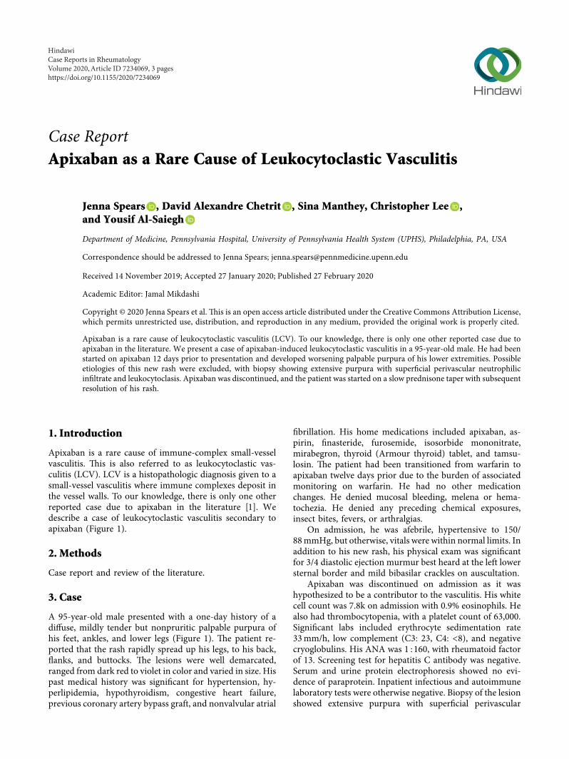
Apixaban is a rare cause of immune-complex small-vesselvasculitis. )is is also referred to as leukocytoclastic vas-culitis (LCV). LCV is a histopathologic diagnosis given to asmall-vessel vasculitis where immune complexes deposit inthe vessel walls. To our knowledge, there is only one otherreported case due to apixaban in the literature [1]. Wedescribe a case of leukocytoclastic vasculitis secondary toapixaban (Figure 1).
2. Methods
Case report and review of the literature.
3. Case
A 95-year-old male presented with a one-day history of adiffuse, mildly tender but nonpruritic palpable purpura ofhis feet, ankles, and lower legs (Figure 1). )e patient re-ported that the rash rapidly spread up his legs, to his back,flanks, and buttocks. )e lesions were well demarcated,ranged from dark red to violet in color and varied in size. Hispast medical history was significant for hypertension, hy-perlipidemia, hypothyroidism, congestive heart failure,previous coronary artery bypass graft, and nonvalvular atrial
fibrillation. His home medications included apixaban, as-pirin, finasteride, furosemide, isosorbide mononitrate,mirabegron, thyroid (Armour thyroid) tablet, and tamsu-losin. )e patient had been transitioned from warfarin toapixaban twelve days prior due to the burden of associatedmonitoring on warfarin. He had no other medicationchanges. He denied mucosal bleeding, melena or hema-tochezia. He denied any preceding chemical exposures,insect bites, fevers, or arthralgias.
On admission, he was afebrile, hypertensive to 150/88mmHg, but otherwise, vitals were within normal limits. Inaddition to his new rash, his physical exam was significantfor 3/4 diastolic ejection murmur best heard at the left lowersternal border and mild bibasilar crackles on auscultation.
Apixaban was discontinued on admission as it washypothesized to be a contributor to the vasculitis. His whitecell count was 7.8k on admission with 0.9% eosinophils. Healso had thrombocytopenia, with a platelet count of 63,000.Significant labs included erythrocyte sedimentation rate33mm/h, low complement (C3: 23, C4: <8), and negativecryoglobulins. His ANA was 1 :160, with rheumatoid factorof 13. Screening test for hepatitis C antibody was negative.Serum and urine protein electrophoresis showed no evi-dence of paraprotein. Inpatient infectious and autoimmunelaboratory tests were otherwise negative. Biopsy of the lesionshowed extensive purpura with superficial perivascular
HindawiCase Reports in RheumatologyVolume 2020, Article ID 7234069, 3 pageshttps://doi.org/10.1155/2020/7234069
neutrophilic infiltrate and leukocytoclasis. Gram stain andGrocott stains were negative for bacterial or fungal in-volvement. Immunofluorescence was not completed on thisspecimen.
Given the concern for an inflammatory process, oralprednisone 40mg daily was initiated, with a slow taper overfive weeks. )e purpuric rash and platelet count rapidlyimproved. Inflammatory markers and complement were notrechecked.
)e patient was not formally tested with an allergy testduring admission, but LCV secondary to apixaban was felt tobe the most likely diagnosis. At the time of discharge, he wasplaced back on warfarin. At his follow-up visit, he had nofurther evidence of vasculitis and was tolerating warfarin.
4. Discussion
To our knowledge, this is the second reported case ofapixaban-related leukocytoclastic vasculitis (LCV) in theliterature [1]. LCV is a small-vessel vasculitis, where immunecomplexes are deposited in small-vessel walls, particularlyinvolving the dermal postcapillary venules. )e associatedimmune response results in loss of vessel wall integrity andextravasation of erythrocytes, resulting in the characteristicpurpura [2]. )e cutaneous manifestations often present inthe lower extremities and buttocks as was the case in thispatient [3]. When small-vessel vasculitis is suspected, punchbiopsy should be completed as early as possible (within24–48 hours of symptom onset) to maximize the diagnosticyield. Direct immunofluorescence can evaluate for thepresence of immunoglobulins (such as IgA) which can bettercharacterize the vasculitis and should be done within 8–24hours of the rash [4]. Direct immunofluorescence was notcompleted on this patient’s biopsy specimen, despite thebiopsy being completed within approximately 16 hours afterthe rash developed. )e reason for this was not specified bythe pathologist. Drug-induced LCV is a diagnosis of ex-clusion after infectious, autoimmune, and inflammatoryconditions have been excluded [3]. Almost 30% of all cases ofLCV are drug-induced [5]; however, anticoagulants are arare cause of LCV [3]. Complement levels should be checked
after resolution of the rash to determine if the initial rash wasan early manifestation of a systemic syndrome such assystemic lupus erythematosus (SLE) [6].
)e interval between exposure to the offending agentand onset of the purpuric rash is variable. On review of theliterature, the majority occurred between 7 and 14 days afterinitiation of the causative medication; however, some re-actions appeared after years of use [1–3, 7–9]. )e man-agement of other cases of anticoagulant-induced LCVinvolved discontinuing the causative drug, with or withoutthe initiation of immunosuppressive therapies [1–3, 7–9].)ere is a clear role for immunosuppressive therapies incases involving skin necrosis, severe systemic vasculitis, orrefractory cases [2]. Various case reports of anticoagulant-induced LCV suggest that patients can be safely initiated onan alternative anticoagulant due to the difference in mo-lecular structures between these medications [1]. However,high-level evidence in this area is lacking [1].
Our case of apixaban-induced LCV highlights a rarecomplication of a common medication, which is importantfor physicians to be aware of.
5. Conclusion
Apixaban is a rare but important cause of leukocytoclasticvasculitis. In cases isolated to the skin, treatment is sup-portive and consists of withdrawal of the offending medi-cation. Patients should be transitioned to a differentanticoagulant. In more complicated cases involving skinnecrosis or severe systemic vasculitis, there is a role forimmunosuppressive therapy.
Conflicts of Interest
)e authors declare that they have no conflicts of interest.
References
[1] U. B. Nasir, A. Kumar, and A. Easwar, “Apixaban causingleukocytoclastic vasculitis,” �e Journal of Allergy and ClinicalImmunology: In Practice, vol. 6, no. 5, pp. 1744-1745, 2018.
(a) (b)
Figure 1: Purpuric rash of the left lower extremity (a) and the right popliteal region (b).
2 Case Reports in Rheumatology
[2] N. B. Hasbal, T. Basturk, Y. Koç, T. Sahutoglu, F. BayrakdarÇaglayan, and A. Unsal, “Leukocytoclastic vasculitis associatedwith a new anticoagulant: Rivaroxaban,” Turkish Journal ofHematology, vol. 34, no. 1, pp. 116-117, 2017.
[3] G. Chaaya, J. Jaller-Char, E. Ghaffar, and A. Castiglioni,“Rivaroxaban-induced leukocytoclastic vasculitis,” Annals ofAllergy, Asthma & Immunology, vol. 116, no. 6, pp. 577-578,2016.
[4] E. Shavit, A. Alavi, and R. G. Sibbald, “Vasculitis-what do wehave to know? A review of literature,”�e International Journalof Lower Extremity Wounds, vol. 17, no. 4, pp. 218–226, 2018.
[5] C. Sunderkotter, G. Bonsmann, A. Sindrilaru, and T. Luger,“Management of leukocytoclastic vasculitis,” Journal of Der-matological Treatment, vol. 16, no. 4, pp. 193–206, 2005.
[6] R.M. Kısla Ekinci, S. Balcı, A. Bisgin, B. Atmıs, D. Dogruel, andM. Yılmaz, “Autoimmune manifestations in heterozygote typeI complement 2 deficiency: a child eventually diagnosed withsystemic lupus erythematosus,” Archives of Rheumatology,vol. 34, pp. 96–99, 2019.
[7] E. Potolidis, C. Mandros, K. Kotsa, E. Mitsiou, D. Potolidis, andP. Fanourgiakis, “Dabigatran associated leukocytoclastic vas-culitis,” Case Reports in Medicine, vol. 2015, Article ID 616109,2 pages, 2015.
[8] D. Elantably, A. Mourad, A. Elantably, and M. Effat, “Warfarininduced leukocytoclastic vasculitis: an extraordinary side ef-fect,” Journal of �rombosis and �rombolysis, vol. 49, no. 1,pp. 149–152, 2019.
[9] C.-Y. Hsu, W.-S. Chen, and S.-H. Sung, “Warfarin-inducedleukocytoclastic vasculitis: a case report and review of litera-ture,” Internal Medicine, vol. 51, no. 6, pp. 601–606, 2012.
Case Reports in Rheumatology 3