Embed Size (px)
Citation preview

International Scholarly Research NetworkISRN OncologyVolume 2011, Article ID 531803, 3 pagesdoi:10.5402/2011/531803
Case Report
A Very Unusual Presentation of Metastatic Colon Cancer
Farzan Fahrtash,1 David Chan,2 Andrew Colebatch,3 and Joseph Rutovitz2
1 Department of Medical Oncology, Westmead Hospital, Sydney, NSW 2145, Australia2 Department of General Medicine, Hornsby Hospital, Palmerston Road, Hornsby, Sydney, NSW 2077, Australia3 Department of Anatomical Pathology, Royal North Shore Hospital, Sydney, NSW 2065, Australia
Correspondence should be addressed to Joseph Rutovitz, [email protected]
Received 20 March 2011; Accepted 26 April 2011
Academic Editors: C. Perez, E. A. Rakha, M. Talieri, and Y. Yu
Copyright © 2011 Farzan Fahrtash et al. This is an open access article distributed under the Creative Commons AttributionLicense, which permits unrestricted use, distribution, and reproduction in any medium, provided the original work is properlycited.
This case highlights two very rare complications of metastatic colorectal carcinoma. It describes a 59 year old female with bothcutaneous and endometrial metastases from colorectal carcinoma. While both of these presentations are very unusual, theyhighlight the need to be vigilant about the detection of metastatic complications during follow up.
1. Introduction
A previously well 59-year-old lady presented to her gastroen-terologist with per rectal bleeding. Apart from intermittentinhaled salbutamol for asthma, she takes no medications andhas no allergies. She is a lifelong nonsmoker and consumes30 g of alcohol per week. She has no family history of note.Colonoscopy demonstrated a tumour at the rectosigmoidjunction (see Figure 1).
A staging CT of Chest/Abdomen/Pelvis revealed noobvious distal metastatic disease. Anterior resection andcholecystectomy was performed in June 2006 withoutcomplication. Histopathology demonstrated moderately welldifferentiated rectosigmoid adenocarcinoma. Resection mar-gins were free of tumour. Both lymphatic and venousinvasion were noted; 17 of 18 regional lymph nodes includingthe apical gland contained metastatic tumour. She was stagedas T3N2M0 (IIIb). She received adjuvant FOLFOX-6 (Flu-orouracil, Oxaliplatin, bolus Fluorouracil, and Leucovorin)for six months. Full course and doses were achieved with onlymild thrombocytopaenia.
Repeat CT scan at end of chemotherapy (January 2007)demonstrated two 5 mm lesions in the left lower lobe andright middle lobe of the lung. While metastatic diseasecould not be ruled out, the lesions were thought to begranulomatous in nature. Two low attenuation areas wereidentified in the liver posteromedially, measuring 8 mm and
15 mm in diameter. A para-aortic lymph node measured6 mm.
Progress CT in July 2007 demonstrated increase in thepara-aortic lymphadenopathy. Repeated 4 months later, anew lobulated 5 mm nodule was noted in periphery ofright upper lobe along with more extensive para-aorticand left common iliac lymphadenopathy, suspicious ofrecurrent disease. PET scanning demonstrated FDG aviddisease involving lymph node groups both above and belowthe diaphragm, and in particular the left para-aortic chain.CT confirmed extensive left para-aortic lymphadenopathywith more focal nodal masses posterior to left renal vein andat common iliac bifurcation.
A needle biopsy of the para-aortic lymph node confirmedadenocarcinoma consistent with prior colonic primary.
She received a further 6-month course of FOLFOX-6from 3/3/2008 to 6/8/2008; again achieving a good partialresponse with resolution of the adenopathy and stable diseasein the other measurable lesions.
In November 2008 the patient reported left leg lym-phoedema. Progress imaging and Doppler Ultrasound oflegs revealed no new pathology. The oedema progressed andspread to the thigh, with left iliac fossa tenderness felt at thesame time. CEA had increased to 8.8.
The patient developed per vaginal bleeding in January2010. Pelvic ultrasound demonstrated a thin and regularendometrium. CT showed no evidence of recurrence at
2 ISRN Oncology
Figure 1: Original rectosigmoid adenocarcinoma.
surgical site, as well as decreased size of both right middlelobe lung lesion and left external iliac lymph nodes. She wasreferred for uterine curettage, performed in February 2010.This demonstrated scanty disintegrating adenocarcinomacells infiltrating the lower uterine segment. The malignantcells were positive for CK20 and negative for CK7, sup-porting diagnosis of metastatic colonic carcinoma. P16 wasnegative and CEA/mucin stains were difficult to interpret dueto degenerative changes.
She was referred for gynaecological assessment in Febru-ary 2010. Per vaginal examination showed the uterus to havea firm and nodular consistency.
Along with the PV bleeding, the patient reported rightupper back pain in a radicular pattern from the lower cervicalspine to the right proximal humerus. There was no neuro-logical deficit. CT on same day showed a destructive lesion inC7 vertebral body on right side with pathological fracturing.Whole body bone scan with SPECT 19/2/10 showed intenseabnormality in body C7 consistent with a metastatic lesion.MRI of the spine 19/2/10 showed pathological superiorendplate fracture associated with metastatic deposit in theC7 vertebral body. Some bone retropulsion into canal waspresent without significant canal stenosis/cord impingement.Marked right-sided C8/T1 foraminal stenosis was noted withpossible right C7 radicular impingement.
The patient commenced palliative radiotherapy to thecervical spine in 10 fractions of 30 Gy. She tolerated this fairlywell with minor dermatological reactions.
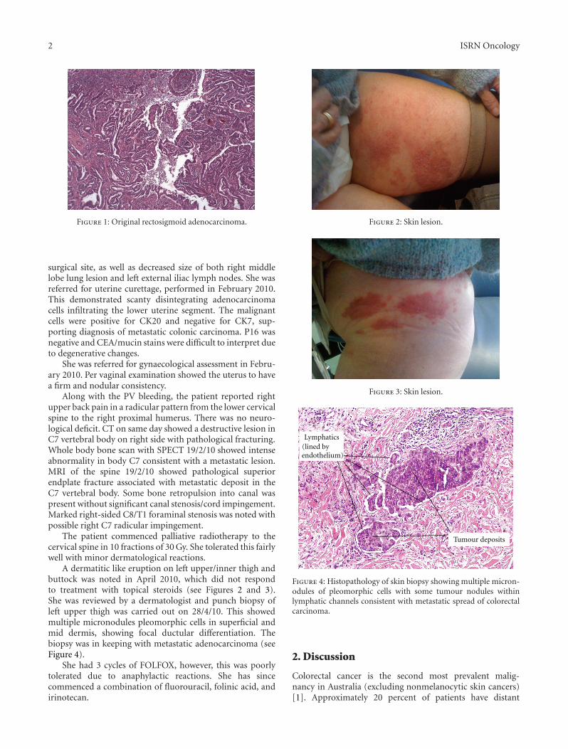
A dermatitic like eruption on left upper/inner thigh andbuttock was noted in April 2010, which did not respondto treatment with topical steroids (see Figures 2 and 3).She was reviewed by a dermatologist and punch biopsy ofleft upper thigh was carried out on 28/4/10. This showedmultiple micronodules pleomorphic cells in superficial andmid dermis, showing focal ductular differentiation. Thebiopsy was in keeping with metastatic adenocarcinoma (seeFigure 4).
She had 3 cycles of FOLFOX, however, this was poorlytolerated due to anaphylactic reactions. She has sincecommenced a combination of fluorouracil, folinic acid, andirinotecan.
Figure 2: Skin lesion.
Figure 3: Skin lesion.
Tumour deposits
Lymphatics(lined byendothelium)
Figure 4: Histopathology of skin biopsy showing multiple micron-odules of pleomorphic cells with some tumour nodules withinlymphatic channels consistent with metastatic spread of colorectalcarcinoma.
2. Discussion
Colorectal cancer is the second most prevalent malig-nancy in Australia (excluding nonmelanocytic skin cancers)[1]. Approximately 20 percent of patients have distant
ISRN Oncology 3
metastatic disease at the time of presentation [2]. Carcinomacan spread by lymphatic, haematogenous, contiguous andtransperitoneal routes. The most common metastatic sitesare the regional lymph nodes, liver, lungs, and peritoneum.Consequently, patients may present with signs or symptomsreferable to these areas. There are recognized unusualpresentations of CRC. These include local invasion causingmalignant fistula formation into adjacent organs, such asbladder or small bowel. Fever of unknown origin, intra-abdominal, retroperitoneal, or abdominal wall abscesses canoften be the first manifestation.
CRC proves to be the aetiology in 6% of adenocarcino-mas of unknown primary sites [3].
Our patient presented with spinal, endometrial, andcutaneous involvement. These presentations are very rareindividually, however, there has been no previous case reportof a patient with both endometrial and cutaneous features.
Brand et al. reported a series of 6 patients presenting withper vaginal bleeding in the setting of recurrent colon cancer[4]. Only one of the patients in that series received adjuvantchemotherapy prior to per vaginal bleeding. Despite aggres-sive chemo-radiation or surgical management, all patientshad recurrence of colonic carcinoma.
Hu et al. found 124 cutaneous metastases from a seriesof 12.146 patients with internal malignancies (1%) [5]. 16of these cases originated from colorectal cancer, equatingto a rate of cutaneous metastasis of 0.81%. Other authorshave found rates of cutaneous metastasis between 2.3 and6% [6–8]. Cutaneous metastases most often occur on a siterelatively close to the internal primary. Skin metastases fromthe breast usually occur on the chest, the lung to the chestand upper extremities, and the gastrointestinal tract to theabdomen [9]. These metastases usually manifest as a rapidlygrowing, mobile nodule [10]. On immunohistochemicalstaining, CK20 positivity often correlates with colorectaltumours [11].
This case highlights the importance of paying closeattention to symptoms and signs in patients with a pasthistory of internal malignancy. It is important to maintaina high index of suspicion to clinical features that mayrepresent a metastatic process. Furthermore, tissue diagnosisto confirm the aetiology of a symptom can prove very useful,as shown in this case.
References
[1] Australian Institute of Health and Welfare, “Cancer in Aus-tralia: an overview,” in Australasian Association of CancerRegistries, AAIoHaWAAAoC, Ed., Canberra, Australia, 2007.
[2] A. Jemal, R. Siegel, E. Ward, Y. Hao, J. Xu, and M. J. Thun,“Cancer statistics, 2009,” CA Cancer Journal for Clinicians, vol.59, no. 4, pp. 225–249, 2009.
[3] J. L. Abbruzzese, M. C. Abbruzzese, R. Lenzi, K. R. Hess, andM. N. Raber, “Analysis of a diagnostic strategy for patientswith suspected tumors of unknown origin,” Journal of ClinicalOncology, vol. 13, no. 8, pp. 2094–2103, 1995.
[4] A. Brand, J. Scurry, R. Planner, and S. Leung, “Primary andrecurrent colorectal cancer masquerading as gynaecologicalmalignancy,” Australian and New Zealand Journal of Obstetricsand Gynaecology, vol. 36, no. 2, pp. 165–167, 1996.
[5] S. C. S. Hu, G. S. Chen, C. S. Wu, C. Y. Chai, W. T. Chen, andC. C. E. Lan, “Rates of cutaneous metastases from differentinternal malignancies: experience from a Taiwanese medicalcenter,” Journal of the American Academy of Dermatology, vol.60, no. 3, pp. 379–387, 2009.
[6] H. L. Abrams, R. Spiro, and N. Goldstein, “Metastases incarcinoma; analysis of 1000 autopsied cases,” Cancer, vol. 3,no. 1, pp. 74–85, 1950.
[7] D. P. Lookingbill, N. Spangler, and F. M. Sexton, “Skininvolvement as the presenting sign of internal carcinoma. Aretrospective study of 7316 cancer patients,” Journal of theAmerican Academy of Dermatology, vol. 22, no. 1, pp. 19–26,1990.
[8] P. S. Spencer and T. N. Helm, “Skin metastases in cancerpatients,” Cutis, vol. 39, no. 2, pp. 119–121, 1987.
[9] D. G. Brodland and J. A. Zitelli, “Mechanisms of metastasis,”Journal of the American Academy of Dermatology, vol. 27, no.1, pp. 1–10, 1992.
[10] M. H. Brownstein and E. B. Helwig, “Patterns of cutaneousmetastasis,” Archives of Dermatology, vol. 105, no. 6, pp. 862–868, 1972.
[11] S. Azoulay, C. Adem, F. L. Pelletier, S. Barete, C. Frances,and F. Capron, “Skin metastases from unknown origin: roleof immunohistochemistry in the evaluation of cutaneousmetastases of carcinoma of unknown origin,” Journal ofCutaneous Pathology, vol. 32, no. 8, pp. 561–566, 2005.

Submit your manuscripts athttp://www.hindawi.com
Stem CellsInternational
Hindawi Publishing Corporationhttp://www.hindawi.com Volume 2014
Hindawi Publishing Corporationhttp://www.hindawi.com Volume 2014
MEDIATORSINFLAMMATION
of
Hindawi Publishing Corporationhttp://www.hindawi.com Volume 2014
Behavioural Neurology
EndocrinologyInternational Journal of
Hindawi Publishing Corporationhttp://www.hindawi.com Volume 2014
Hindawi Publishing Corporationhttp://www.hindawi.com Volume 2014
Disease Markers
Hindawi Publishing Corporationhttp://www.hindawi.com Volume 2014
BioMed Research International
OncologyJournal of
Hindawi Publishing Corporationhttp://www.hindawi.com Volume 2014
Hindawi Publishing Corporationhttp://www.hindawi.com Volume 2014
Oxidative Medicine and Cellular Longevity
Hindawi Publishing Corporationhttp://www.hindawi.com Volume 2014
PPAR Research
The Scientific World JournalHindawi Publishing Corporation http://www.hindawi.com Volume 2014
Immunology ResearchHindawi Publishing Corporationhttp://www.hindawi.com Volume 2014
Journal of
ObesityJournal of
Hindawi Publishing Corporationhttp://www.hindawi.com Volume 2014
Hindawi Publishing Corporationhttp://www.hindawi.com Volume 2014
Computational and Mathematical Methods in Medicine
OphthalmologyJournal of
Hindawi Publishing Corporationhttp://www.hindawi.com Volume 2014
Diabetes ResearchJournal of
Hindawi Publishing Corporationhttp://www.hindawi.com Volume 2014
Hindawi Publishing Corporationhttp://www.hindawi.com Volume 2014
Research and TreatmentAIDS
Hindawi Publishing Corporationhttp://www.hindawi.com Volume 2014
Gastroenterology Research and Practice
Hindawi Publishing Corporationhttp://www.hindawi.com Volume 2014
Parkinson’s Disease
Evidence-Based Complementary and Alternative Medicine
Volume 2014Hindawi Publishing Corporationhttp://www.hindawi.com