Embed Size (px)
Citation preview

1
Cases presented with teleradiology and telepathology by the local team
W. Demey (oncology), P. Van Dam (surgery), I. Biltjes (radiology), P. Dirix (radiation oncology), C. Colpaert (pathology)

Case 1: JA °12/3/1968 (46 years)
• Medical History
– Mother (32y) and grandmother (50y) breast cancer
– Hemangioma liver
• Oncologic history
– Spring 2009: investigation “sensation of swelling” of the right breast
– Clinical examination: no mass
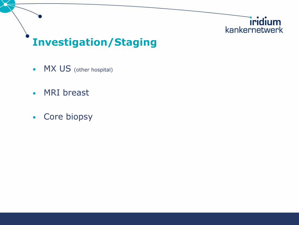
Investigation/Staging
• MX US (other hospital)
• MRI breast
• Core biopsy
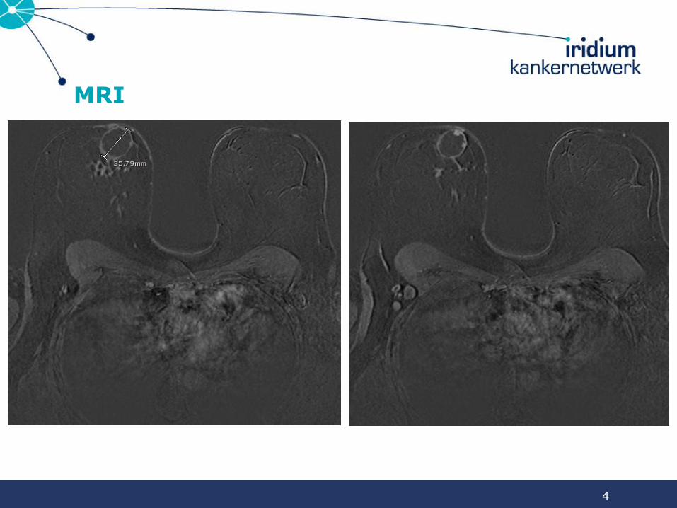
MRI
4

MRI

TRU CUT mass
• April 2009 : tumorectomy right breast+Sn procedure: pT1apN0M0, extensive DCIS with 3 invasive foci IDA G2 (2, 3 and 5 mm), margins involved, ER 2/8 PR 2/8 HER2 0
• Sn: 0/6
• BRCA : -
What would you do?
• Mastectomy
• Radiotherapy
• Chemotherapy
• Hormonal treatment
• Other

Pathology
• Tumorectomy: extensive DCIS with 3 invasive foci IDA G2 ( 2, 3 and 5 mm)
• Mastectomy 19/5/2009
• Mastectomy (no residual tu) –ALND (0/12)
• 6 cycles CEF
• Tamoxifen

• 2/2014 : rising CA 15.3 : 77, nl liver tests
• Investigation
− CT scan chest – liver
− PET scan
− MRI : liver lesion : right lobe
Imaging : MRI liver
• 24/3/2014 (CT scan)
• 29/10/2014
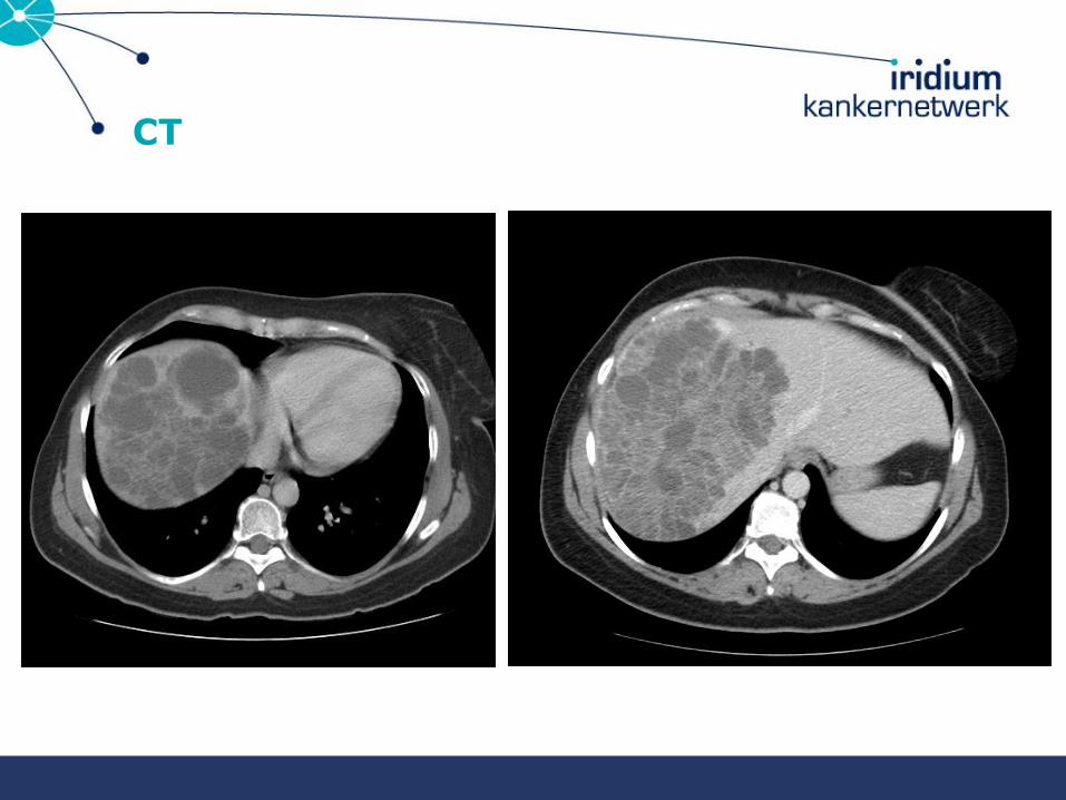
CT

PET-CT
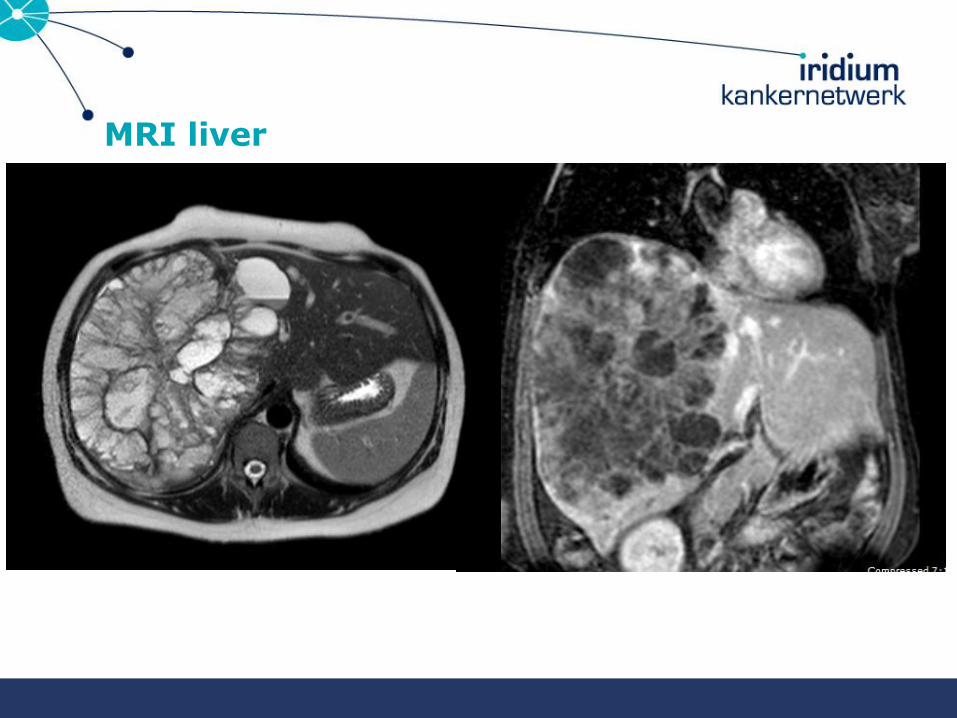
MRI liver

Pathology liver
• Second opinion (UZ Gent):
– Liver metastasis from breast cancer: GATA3+, ER+,
– 3 liver specific antigens -

What would you do
• Chemotherapy / systemic treatment
• Resection
• Chemo and resection
• Other

• Evaluation after 12 weeks Paclitaxel
– Stable disease
• Evaluation after 3 cycles Carboplatinum- Vinorelbine
– Stable disease

• Resection was performed:
– Right hemi hepatectomy: metastatic breast cancer, ER+; PR-, Her2neu negative, margins are free
• What to do next week?

Case 2: SVH (°02/06/1961).
• Relevant patient history:
– Family: paternal aunt with breast cancer at 60 yrs.
– Medical: hypertension.
– Surgical: -
– A0P0G0.
– Medication: Nobiten.
– Allergy: -
– Smoking: -

Case 2: SVH (°02/06/1961).
• Investigations:
– Clinical examination: palpable, mobile nodule in left breast at 2h (2.0 x 2.0 cm), no skin retraction, cN0.
– MX US (23/10/2014): tumor (20 mm) peripherally at 2h in the left breast with a little satellite nodule of 7 mm. cN0.
– MRI (24/10/2014): solitary tumor in the upper, outer quadrant of the left breast with maximal diameter of 1.9 cm. No other lesions. No adenopathy.
– US abdomen (30/10/2014): normal.
– Chest X-ray (30/10/2014): normal.
– Bone scan (29/10/2014): normal.

Screening MX 2 years before
RCC LCC RMLO LMLO

MX
23/10/2014
RCC LCC RMLO LMLO
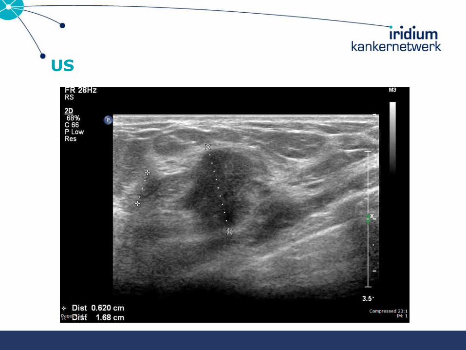
US
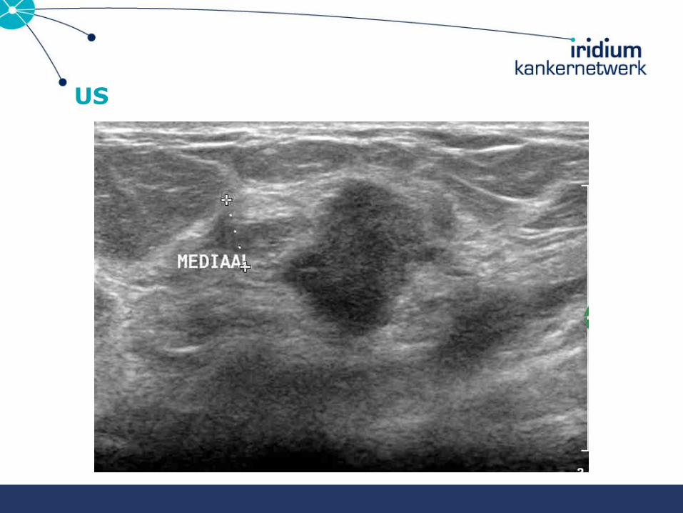
US

MRI

CORE BIOPSY
Pre-operative staging: conclusion.
• IDA grade 2, ER 8/8, PR 8/8, HER-2 2+ but SISH negative.
• cT1c cN0 cM0.

BCS + SLND on 04/11/2014.
• IDA grade 2, maximal diameter 1.8 cm.
• LVI+.
• ER 8/8, PR 8/8, HER-2 2+ but SISH negative.
• One of two SLNs positive with macroM+ of 3.5 mm.
• pT1c pN1a(sn).
• Further ALND or not?
• Adjuvant chemotherapy or not?
• Radiotherapy: • Whole-breast radiotherapy alone.
• Whole-breast + IM-MS radiotherapy.
• Whole-breast + axillary radiotherapy.
• Whole-breast + IM-MS + axillary radiotherapy.
• Hormonal therapy.
What now?

Multidisciplinary discussion.
• No further ALND.
• No adjuvant chemotherapy.
• Whole-breast radiotherapy alone, without explicit nodal irradiation. Not even what is called “high tangents”.
• Hormonal treatment: 5 yrs AI + 5 yrs Tam.
RT fields (1).
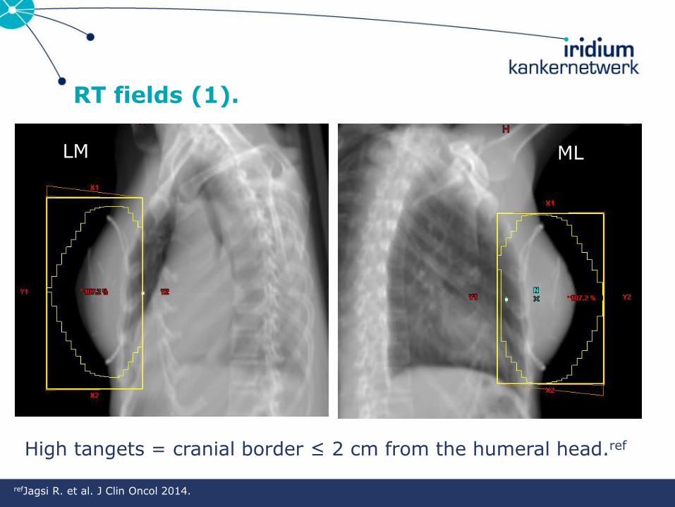
High tangets = cranial border ≤ 2 cm from the humeral head.ref
ML LM
refJagsi R. et al. J Clin Oncol 2014.
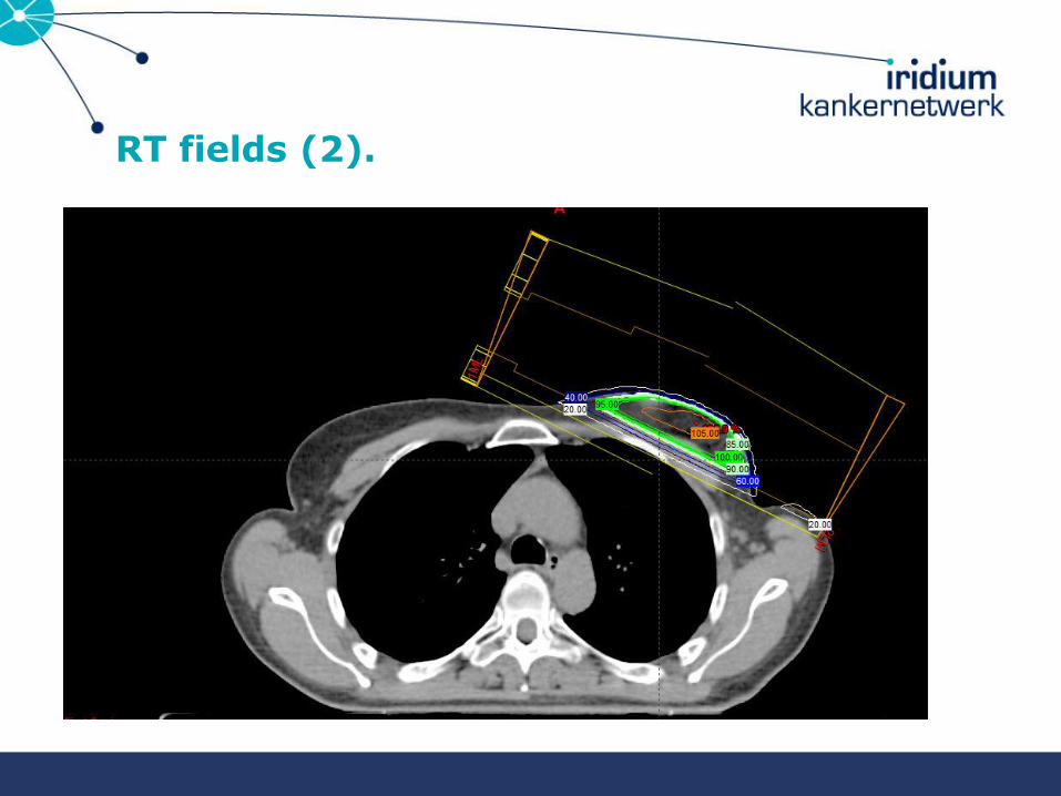
RT fields (2).
ML LM

Case 3: HW °28/04/1980
• Previous oncological history: nil
• Medical history: G0P0A0
• Familial history: nil
• Current problem: palpates mass in left breast, refferred because diagnosis IDC
• Physical examination 2/8/2013:
– Normal palpation of the right breast and no lymphadeneopathy
– Mass 3 cm superolateral in the left breast, suspicous left axillary nodes
– T2N1Mx
• Imaging:
– MX US (other hospital): lesion SL left breast and suspicious lymph node 9 mm left axilla
– MRI 12/8/2013: lesion 50 mm lateral part left breast
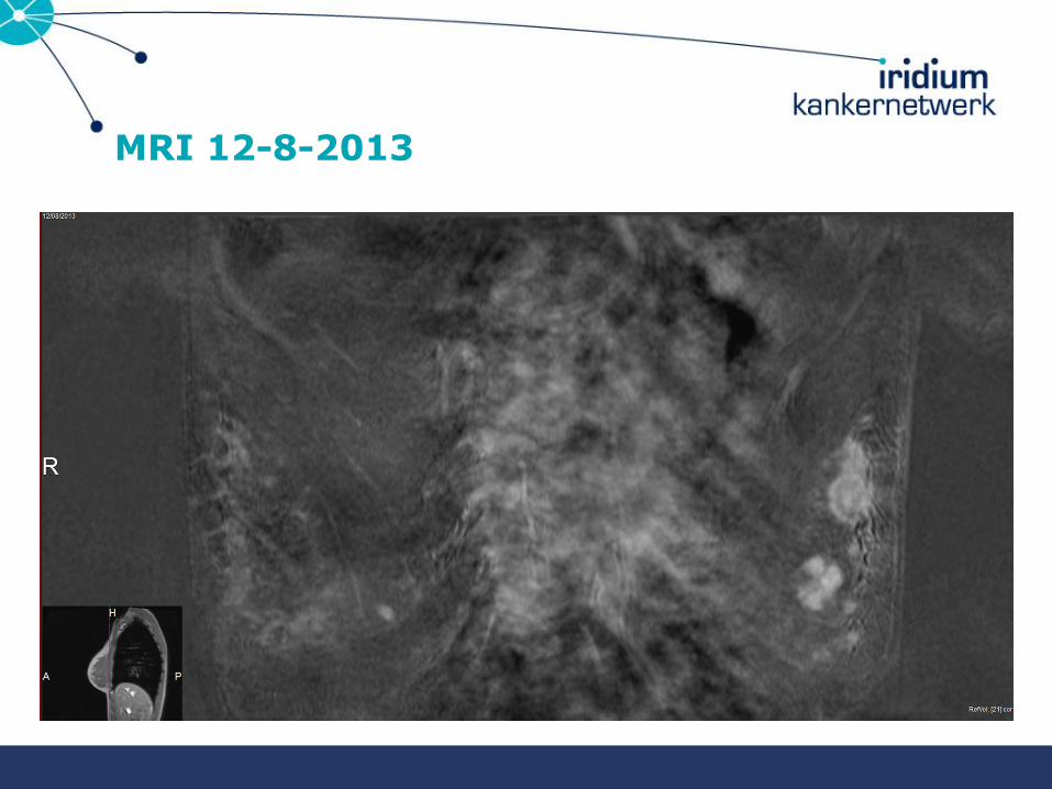
MRI 12-8-2013
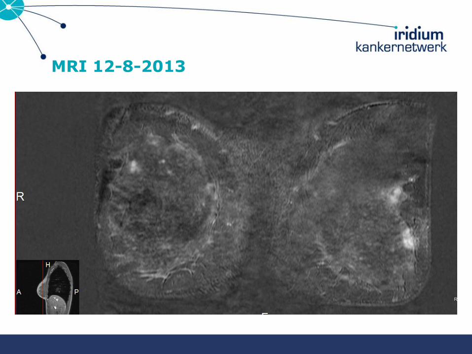
MRI 12-8-2013

CORE BIOPSY: mass left +, LN +, right breast: FA

Case 3: HW °28/04/1980
• Ultrasound guided core biopsy:
– Grade III IDC, ER 50%, and PR 10%, Her-2 1+, Ki67 > 80%
– FNAC axillary lymph node: metastatic cells
• Staging:
– PET-CT 12/8/2013: suspicious mass in the left breast, dsuspicious FDG captation of 3 left axillary lymph nodes, no distant metastasis
– Bonscintigraphy 13/8/2013, chest Xray 7/8/2013 and ultrasound liver normal
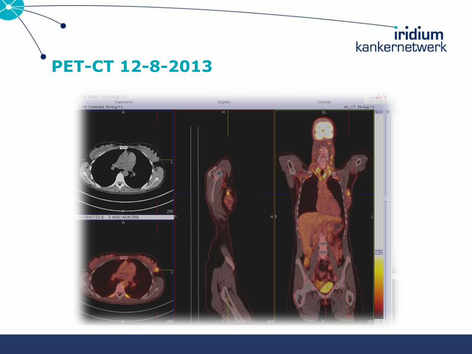
PET-CT 12-8-2013

Case 3: HW °28/04/1980
– What would you do ?
• Primary BCS with sentinel node biopsy procedure
• Primary BCS with sentinel node biopsy and complete axillary clearence
• Primary mastectomy with sentinel node biopsy procedure
• Primary mastectomy and complete axillary clearence
• Neoadjuvant chemotherapy
Is there a need for genetic testing ?
Case 3: HW °28/04/1980
27/8/2013: Start dose dense AC 4x followed by docetaxel with Neulasta support
1/10/2013 PETCT: partial respons
20/10/13 MRI breasts: partial respons
5/11/2013: switch to taxol weekly (9 cycles) because of grade III hand-foot syndrome
7/1/2014 MRI breasts: further respons
23/1/2014: wide local excision after guide wire localisation, sentinel node biopsy and axillary clearence level I-II
Pathology result:
Focus 2 mm DCIS completely excised, fibrous tissue tumor bed. Sentinel node and 7 further nodes negative : yPTisPN0M0
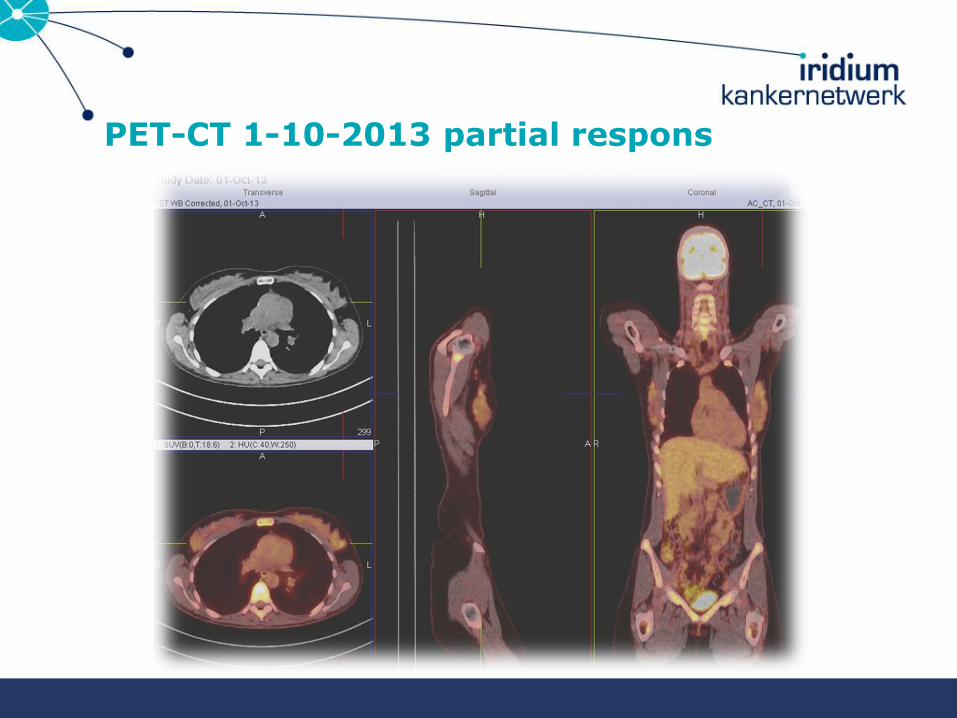
PET-CT 1-10-2013 partial respons
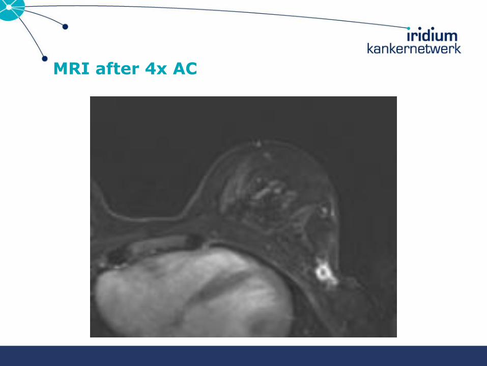
MRI after 4x AC

MRI after TAXOL

Case 3: HW °28/04/1980
Pathology result:
Focus 2 mm DCIS completely excised, fibrous tissue tumor bed. Sentinel node and 7 further nodes negative : ypTisPN0M0
2/2013 and 3/2014: external beam radiotherapy on breast and tributary nodal areas (50 Gy in 25 sessions, boost tumor bed 10 Gy in 10 sessions)
BRCA and check-2 analysis negative
MOC advice: Zoladex and tamoxifen
Case 3: HW °28/04/1980
Patient refuses hormonal treatment because she wants to get pregnant !!!
What would you advice ?
* do not become pregnant during the first 5 y, then it can be considered if no metastasis
* agree with pregnancy and no hormonal therapy
* never become pregnant later in life (pregnancy life-long contraindicated)
Case 3: HW °28/04/1980
23/11/2014: at follow up visit increasing back- and hip pain.
23/11/14: Physical examination tender lumbar vertebrae. Palpation breasts and axillae normal
21/11/14 CA15.3: 6.7 (has always been normal)
5/12/14 PET CT: diffuse bone metastases
17/12/14 bonescintigraphy: bone metastases
MOC advice: bone biopsy, X-geva, castration, JPBL study (Faslodex with or without CDK4/6 inhibitor)

PET-CT 5-12-2014 (almost year after surgery)
Case 4: MvL °8/8/1967 (47 years)
• Medical History: Insulin dependent DM – premenopausal patient
• Familial history: 0
• Nov 2013 : presented at the breast clinic: tumor in the left breast +/- 4 cm
• Clinical examination: cT2N0 tumor of the left breast

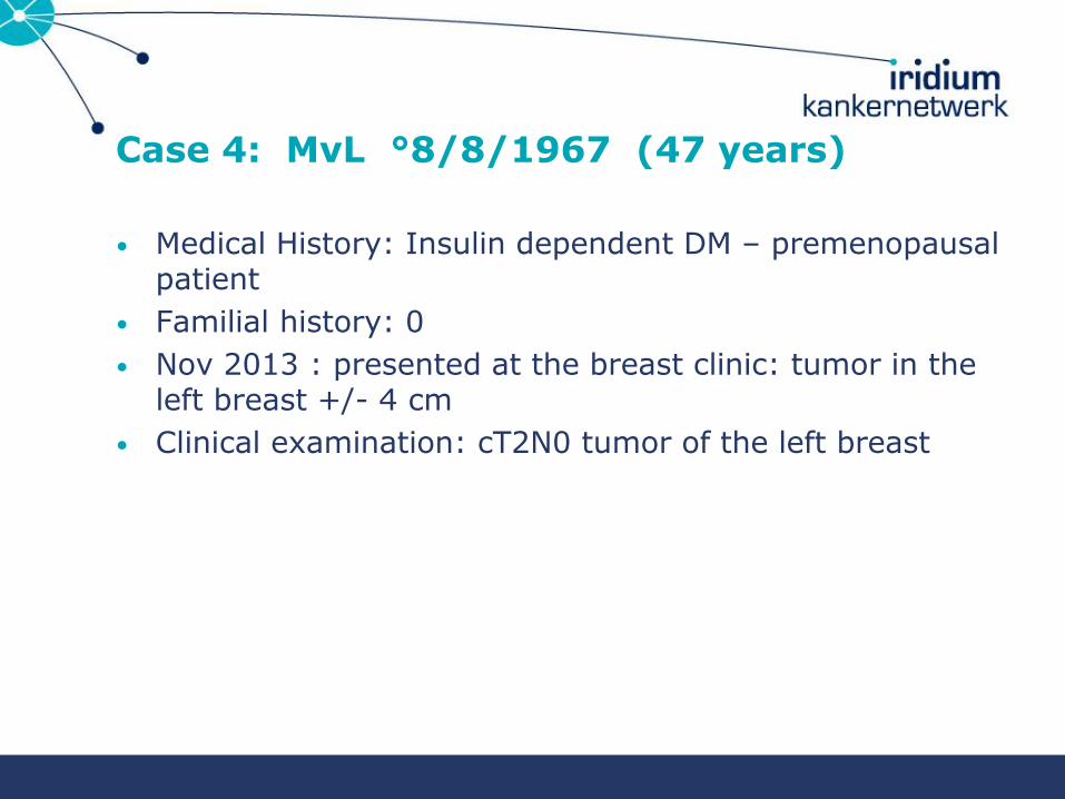
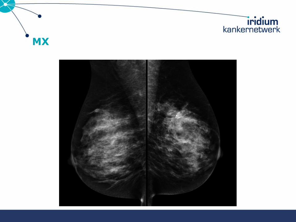
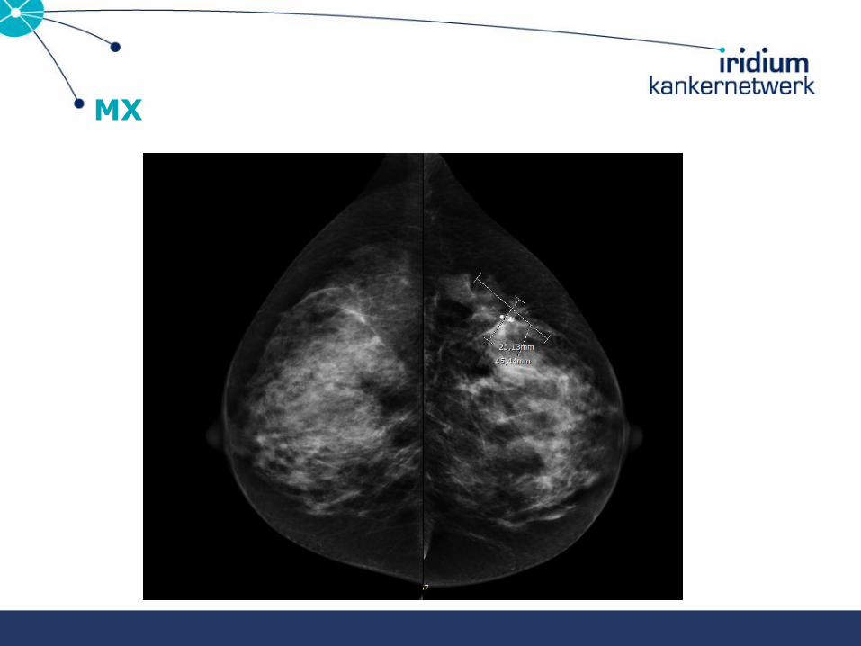
Staging
• MX US
• MRI
• CT chest – liver
• Core biopsy
MX
MX

US
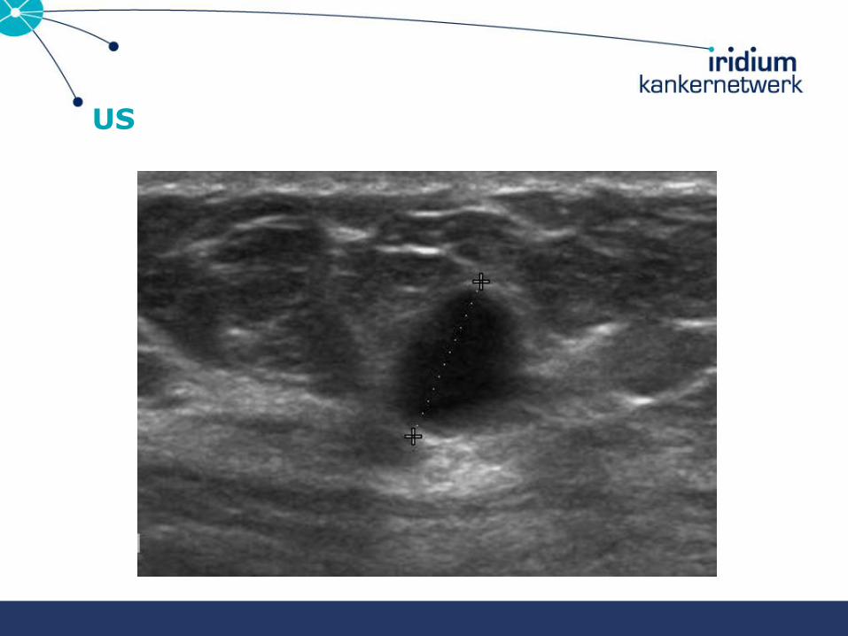
US

MRI

MRI

CT CHEST – LIVER
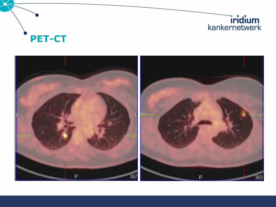
PET-CT

CORE BIOPSY

After staging…
• cT2N1M1 (2 lung lesions)
• IDA G3, ER 0 PR 0 HER2 3+,SISH HER2 + , Ki 67: 75%
• PET/CT-scan: Breast tu left, axillary lymph nodes, 2 lung lesions
• What would you do?
- Start palliative systemic therapy?
- Surgery: ME-ALND?
- Neoadjuvant systemic therapy?
• R/ neoadjuvant chemo: 4 x CEF, 4 x Taxotere-Herceptin
• Evaluation after chemo NMR/PET scan: PR
• PET scan: no residual activity in the lung lesions
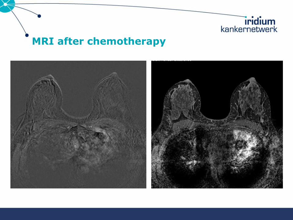
MRI after chemotherapy

MRI after chemotherapy

CT THORAX after chemotherapy

PET-CT after chemotherapy

Multidisciplinary discussion
• Mastectomy and LN dissection ?
• Radiotherapy ?
• Herceptin 18 x
• Herceptin until progression
• Other

Evolution
April 2014:
• Mastectomy and axillary clearance ypT0N0
August 2014:
• Headache
• CT and NMR
• Bone scan and CT chest – liver: OK
68

Brain CT aug 2014

Brain MRI aug 2014

Brain MRI after resection aug 2014 and nov 2014

Pathologic evaluation after resection
• M+ Breast cancer; Her2neu+++
Multidisciplinary discussion
• Radiotherapy?
• Systemic treatment
– Herceptin mono
– Combination with Lapatinib
– Chemotherapy ?
– ……………?
Radiotherapy
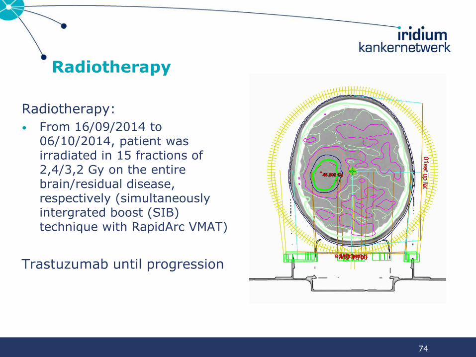
Radiotherapy:
• From 16/09/2014 to 06/10/2014, patient was irradiated in 15 fractions of 2,4/3,2 Gy on the entire brain/residual disease, respectively (simultaneously intergrated boost (SIB) technique with RapidArc VMAT)
Trastuzumab until progression
74
Case 5: CM °14/11/1964
• Previous oncological history: (elswhere)
• 2/2008: mass right breast: IDC, ER and PR negative, Her-2 negative, MAI > 50
• Neoadjuvant chemotherapy: 4xCEF, 4x taxotere
• 4/2009: wide local excision and axillary lymphadenectomy
• 7/5/2009 till 19/6/2009: external beam radiotherapy 60 Gy in 30 sessions on right breast and 45 Gy in 25 sesions on axilla, supracalvicular and mediastinal lymph nodes
• Medical history: G0P0A0
• Familial history: father lungcancer 55y, grandmother (M) oesophagal cancer

Case 5: CM °14/11/1964
• Current problem: 25/4/2014: routine follow-up: feels excellent
• Physical examination:
– Normal palpation of the right breast and no lymphadeneopathy
– Vague dense area (2cm diameter) superolateral in the left breast
• Imaging:
– MX US: lesion 16x13 mm SL and a small lesion 5 mm left breast
– MRI

MX
77

US
78
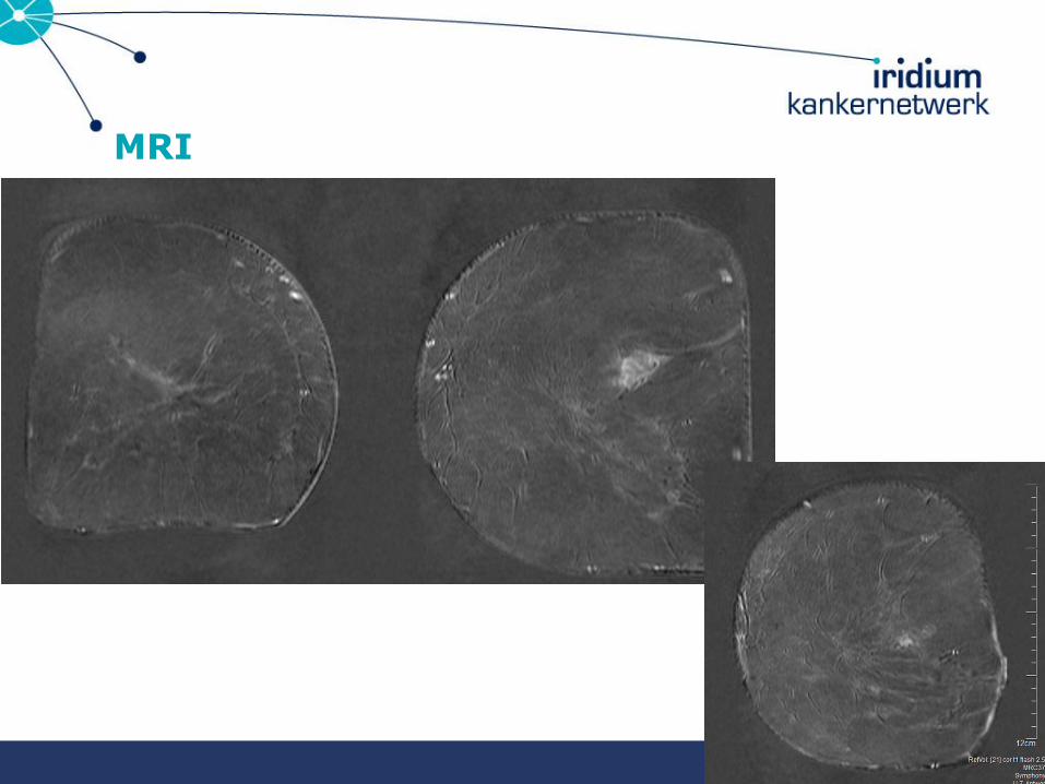
MRI

CORE BIOPSY
Case 5: CM °14/11/1964
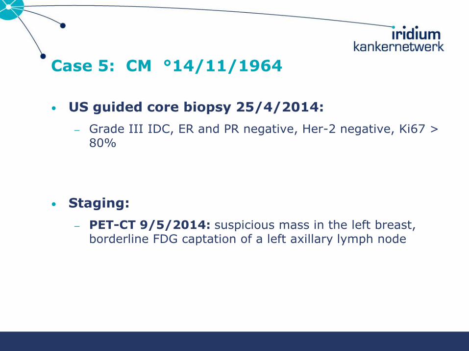
• US guided core biopsy 25/4/2014:
– Grade III IDC, ER and PR negative, Her-2 negative, Ki67 > 80%
• Staging:
– PET-CT 9/5/2014: suspicious mass in the left breast, borderline FDG captation of a left axillary lymph node

PET-CT
Case 5: CM °14/11/1964
• What would you do ?
– Primary BCS with sentinel node biopsy procedure
– Primary BCS with sentinel node biopsy and complete axillary clearence
– Primary mastectomy with sentinel node biopsy procedure
– Primary mastectomy and complete axillary clearence
– Neoadjuvant chemotherapy
• Is there a need for genetic testing ?

Case 5: CM °14/11/1964
• Is there a role for genetic testing ?
– Will the result influence your surgical approach ?
– Will the result influence your systemic treatment ?
84

Case 5: CM °14/11/1964
26/05/2014: start weekly neoadjuvant carbo AUC 2/taxol 80 mg/m
9/7/2014 PETCT: excellent respons with normalisation of the FDG captation in the left breast and axilla
25/8/2014 MRI scan breast: execellent respons, remaining suspicious area 6 mm
19/9/2014 mastectomy with sentinel node procedure (DEP Prep negative )and level 1 axillary lymphadenectomy
29/9/2014 final pathology: tumor bed without malignant cells, 2 sentinel nodes and 5 additional nodes normal ypT0N0M0
13/11/2014 till 5/12/20114 chest wall radiotherapy (40 Gy in 15 sessions)
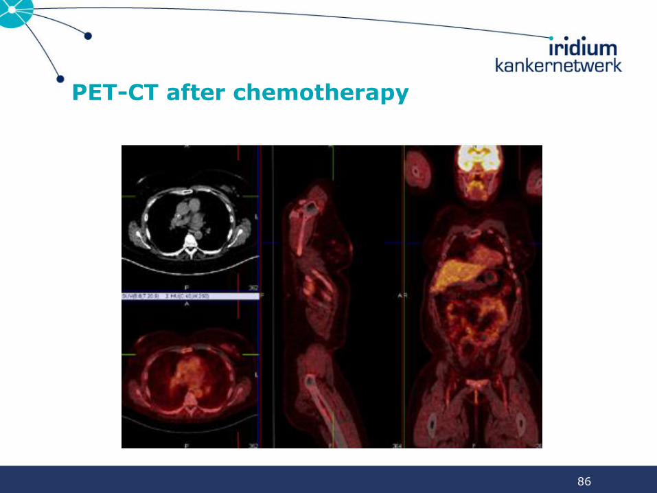
PET-CT after chemotherapy
86

MRI after chemotherapy
87
Case 5: CM °14/11/1964
10/2014 BRCA mutation analysis: BRCA1 positive
15/1/2014: laparoscopic salpingooophorectomy
A contralateral prophylactic mastectomy with bilateral DIEP flap reconstruction is planned in the winter 2014-2015
Case 3: SVH (°02/06/1961).
Piet Dirix MD, PhD
Radiation Oncology
Iridium Kankernetwerk

Case 3: SVH (°02/06/1961).
• Relevant patient history:
– Family: paternal aunt with breast cancer at 60 yrs.
– Medical: hypertension.
– Surgical: -
– A0P0G0.
– Medication: Nobiten.
– Allergy: -
– Smoking: -

Case 3: SVH (°02/06/1961).
• Technical investigations:
– Clinical examination: palpable, mobile nodule in left breast at 2h (2.0 x 2.0 cm), no skin retraction, cN0.
– Mammo-US (23/10/2014): tumour (20 mm) peripherally at 2h in the left breast with a little satellite nodule of 7 mm. cN0.
– MRI (24/10/2014): solitary tumour in the upper, outer quadrant of the left breast with maximal diameter of 1.9 cm. No other lesions. No adenopathy.
– US abdomen (30/10/2014): normal.
– Chest X-ray (30/10/2014): normal.
– Bone scan (29/10/2014): normal.
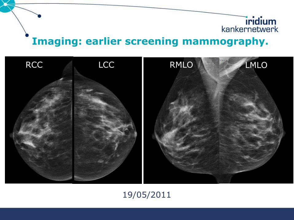
Imaging: earlier screening mammography.
19/05/2011
RCC LCC RMLO LMLO
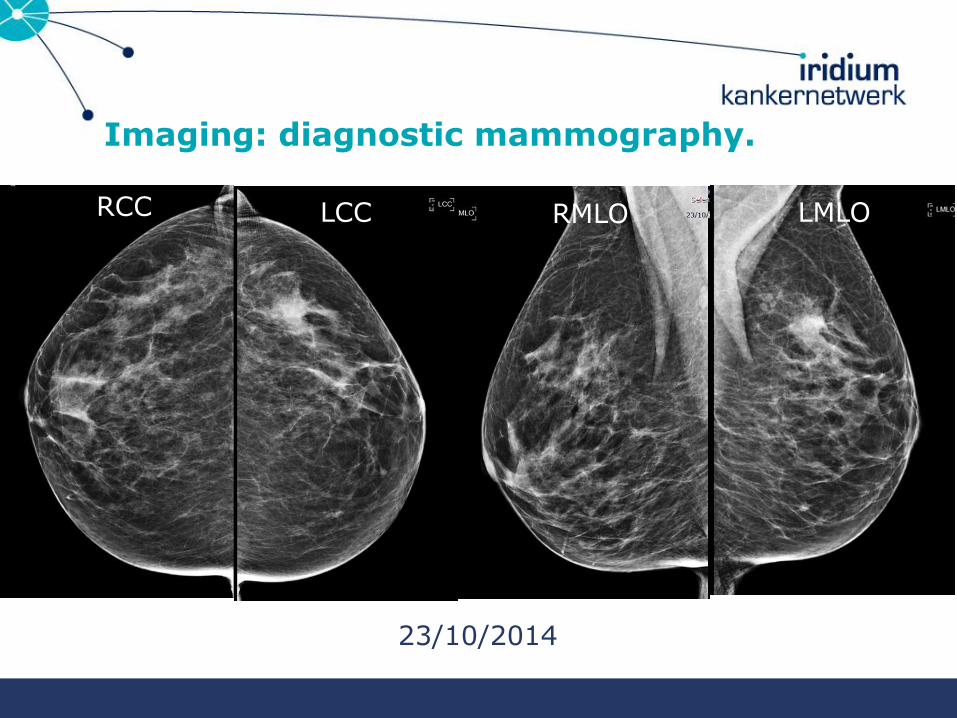
Imaging: diagnostic mammography.
23/10/2014
RCC LCC RMLO LMLO

Imaging: diagnostic US (1).

Imaging: diagnostic US (2).

Imaging: diagnostic MRI.

Imaging: US punction.

Pre-operative staging: conclusion.
• IDA grade 2, ER 8/8, PR 8/8, HER-2 2+ but SISH negative.
• cT1c cN0 cM0.

BCS + SLND on 04/11/2014.
• IDA grade 2, maximal diameter 1.8 cm.
• LVI+.
• ER 8/8, PR 8/8, HER-2 2+ but SISH negative.
• One of two SLNs positive with macroM+ of 3.5 mm.
• pT1c pN1a(sn).

• Further ALND or not?
• Adjuvant chemotherapy or not?
• Radiotherapy: • Whole-breast radiotherapy alone.
• Whole-breast + IM-MS radiotherapy.
• Whole-breast + axillary radiotherapy.
• Whole-breast + IM-MS + axillary radiotherapy.
• Hormonal therapy.
What now?
Multidisciplinary discussion.
• No further ALND.
• No adjuvant chemotherapy.
• Whole-breast radiotherapy alone, without explicit nodal irradiation. Not even what is called “high tangents”.
• Hormonal treatment: 5 yrs AI + 5 yrs Tam.

RT fields (1).
High tangets = cranial border ≤ 2 cm from the humeral head.ref
ML LM
refJagsi R. et al. J Clin Oncol 2014.

RT fields (2).
ML LM