Embed Size (px)
Citation preview
Cassandra GallatiRachel Harjes
Heather Hutchings
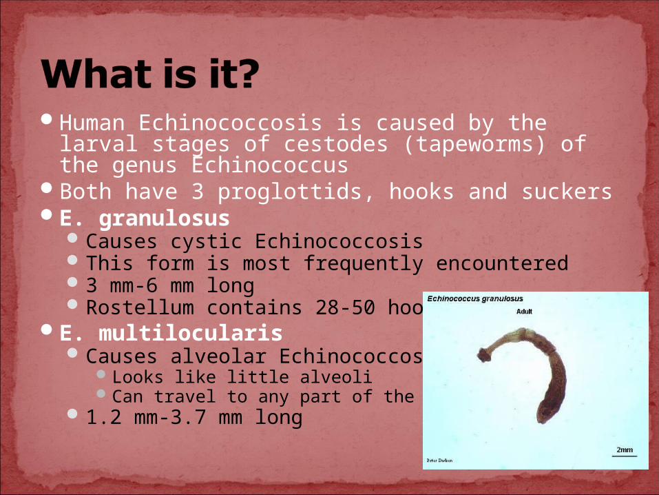
Human Echinococcosis is caused by the larval stages of cestodes (tapeworms) of the genus Echinococcus
Both have 3 proglottids, hooks and suckersE. granulosus
Causes cystic EchinococcosisThis form is most frequently encountered3 mm-6 mm longRostellum contains 28-50 hooks
E. multilocularisCauses alveolar Echinococcosis
Looks like little alveoliCan travel to any part of the body
1.2 mm-3.7 mm long
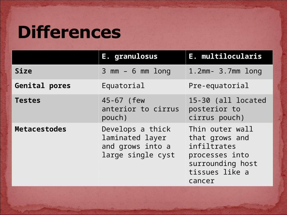
E. granulosus E. multilocularis
Size 3 mm – 6 mm long 1.2mm- 3.7mm long
Genital pores Equatorial Pre-equatorial
Testes 45-67 (few anterior to cirrus pouch)
15-30 (all located posterior to cirrus pouch)
Metacestodes Develops a thick laminated layer and grows into a large single cyst
Thin outer wall that grows and infiltrates processes into surrounding host tissues like a cancer
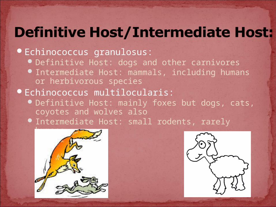
Echinococcus granulosus:Definitive Host: dogs and other carnivores Intermediate Host: mammals, including humans or
herbivorous speciesEchinococcus multilocularis:
Definitive Host: mainly foxes but dogs, cats, coyotes and wolves also
Intermediate Host: small rodents, rarely humans

E. granulosusWorldwideMore frequent in rural, sheep raising areas
E. multilocularis Occurs in northern hemisphereThis includes central and northern Europe, Asia,
and North AmericaMost frequently found in northern states

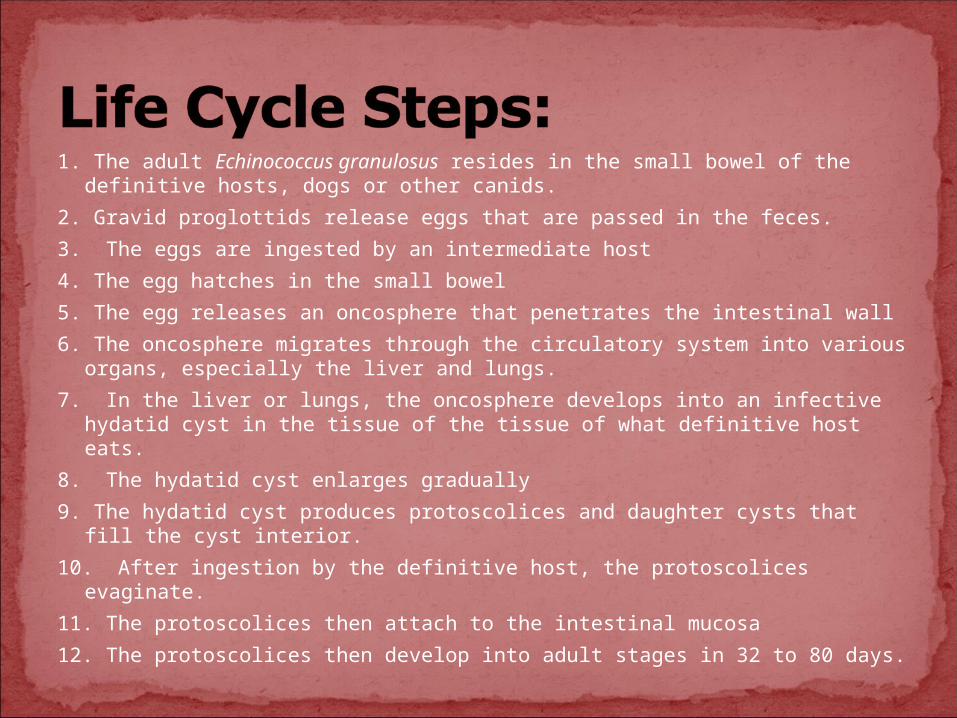
1. The adult Echinococcus granulosus resides in the small bowel of the definitive hosts, dogs or other canids.
2. Gravid proglottids release eggs that are passed in the feces.
3. The eggs are ingested by an intermediate host
4. The egg hatches in the small bowel
5. The egg releases an oncosphere that penetrates the intestinal wall
6. The oncosphere migrates through the circulatory system into various organs, especially the liver and lungs.
7. In the liver or lungs, the oncosphere develops into an infective hydatid cyst in the tissue of the tissue of what definitive host eats.
8. The hydatid cyst enlarges gradually
9. The hydatid cyst produces protoscolices and daughter cysts that fill the cyst interior.
10. After ingestion by the definitive host, the protoscolices evaginate.
11. The protoscolices then attach to the intestinal mucosa
12. The protoscolices then develop into adult stages in 32 to 80 days.
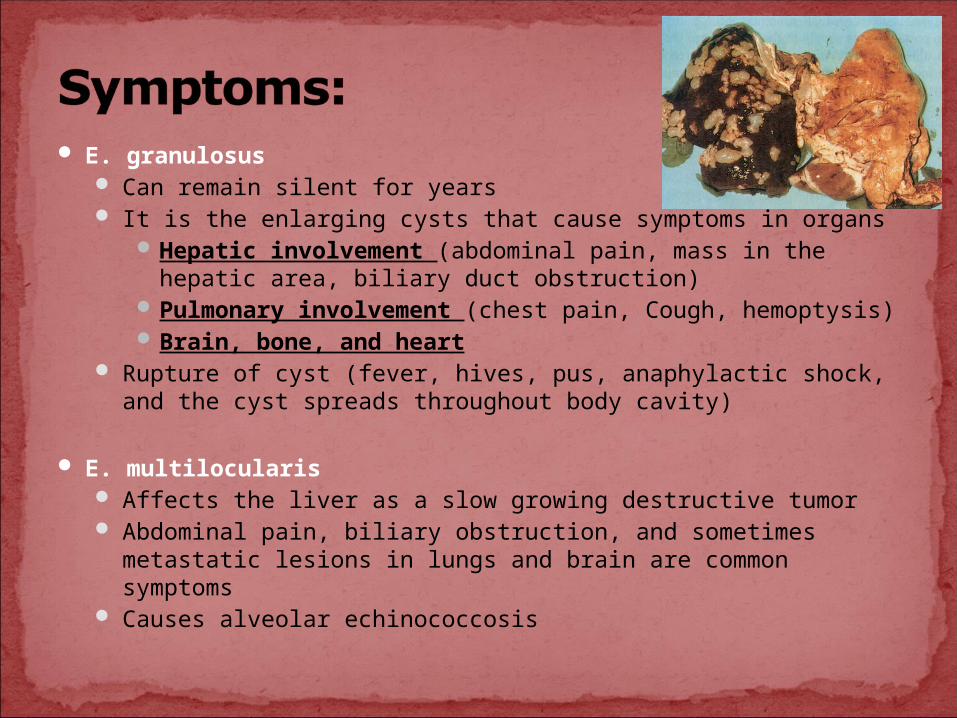
E. granulosus Can remain silent for years It is the enlarging cysts that cause symptoms in organs
Hepatic involvement (abdominal pain, mass in the hepatic area, biliary duct obstruction)
Pulmonary involvement (chest pain, Cough, hemoptysis)Brain, bone, and heart
Rupture of cyst (fever, hives, pus, anaphylactic shock, and the cyst spreads throughout body cavity)
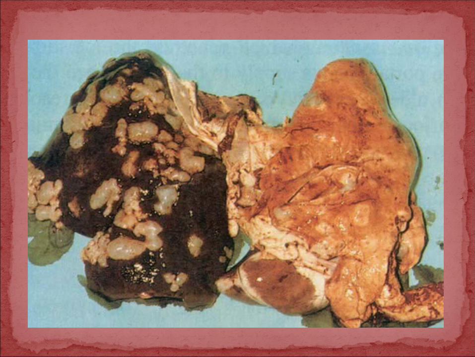
E. multilocularis Affects the liver as a slow growing destructive tumor Abdominal pain, biliary obstruction, and sometimes metastatic
lesions in lungs and brain are common symptoms Causes alveolar echinococcosis
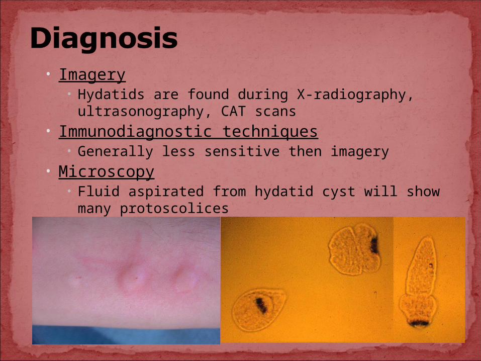
• Imagery• Hydatids are found during X-radiography,
ultrasonography, CAT scans• Immunodiagnostic techniques
• Generally less sensitive then imagery• Microscopy
• Fluid aspirated from hydatid cyst will show many protoscolices
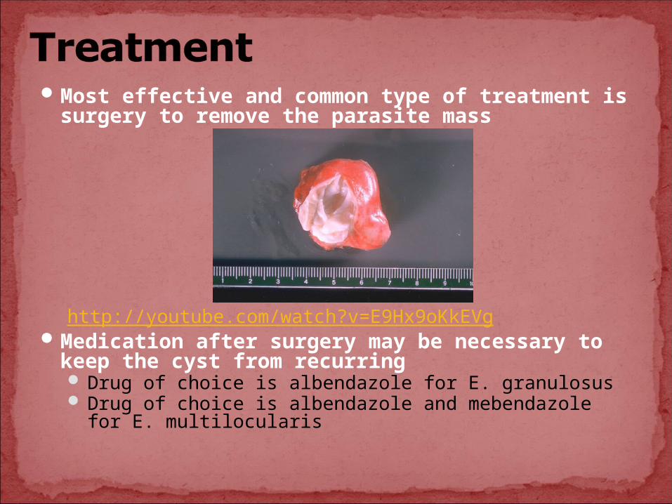
Most effective and common type of treatment is surgery to remove the parasite mass
http://youtube.com/watch?v=E9Hx9oKkEVgMedication after surgery may be necessary to
keep the cyst from recurring Drug of choice is albendazole for E. granulosus Drug of choice is albendazole and mebendazole for E.
multilocularis
Interrupt lifecycle by denying access of dogs to offal
Destroy stray dogs
General education program
Sheep herders should not live closely with their dogs
The drug that is so effective for most flatworm parasites, Praziquantel, may actually enhance growth of alveolar hydatids.
Center of Disease ControlFoundation of Parasitology (textbook)Compendium of Cystic Echinococcosis
(from Professor Bates)