Embed Size (px)
Citation preview
RESEARCH Open Access
CD8 positive T cells express IL-17 in patients withchronic obstructive pulmonary diseaseYing Chang1, Jessica Nadigel1, Nicholas Boulais1, Jean Bourbeau2, François Maltais3, David H Eidelman1 andQutayba Hamid1*
Abstract
Background: Chronic obstructive pulmonary disease (COPD) is a progressive and irreversible chronic inflammatorydisease of the lung. The nature of the immune reaction in COPD raises the possibility that IL-17 and relatedcytokines may contribute to this disorder. This study analyzed the expression of IL-17A and IL-17F as well as thephenotype of cells producing them in bronchial biopsies from COPD patients.
Methods: Bronchoscopic biopsies of the airway were obtained from 16 COPD subjects (GOLD stage 1-4) and 15control subjects. Paraffin sections were used for the investigation of IL-17A and IL-17F expression in the airways byimmunohistochemistry, and frozen sections were used for the immunofluorescence double staining of IL-17A or IL-17F paired with CD4 or CD8. In order to confirm the expression of IL-17A and IL-17F at the mRNA level, aquantitative RT-PCR was performed on the total mRNA extracted from entire section or CD8 positive cells selectedby laser capture microdissection.
Results: IL-17F immunoreactivity was significantly higher in the bronchial biopsies of COPD patients compared tocontrol subjects (P < 0.0001). In the submucosa, the absolute number of both IL-17A and IL-17F positive cells washigher in COPD patients (P < 0.0001). After adjusting for the total number of cells in the submucosa, we still foundthat more cells were positive for both IL-17A (P < 0.0001) and IL-17F (P < 0.0001) in COPD patients compared tocontrols. The mRNA expression of IL-17A and IL-17F in airways of COPD patients was confirmed by RT-PCR. Theexpression of IL-17A and IL-17F was co-localized with not only CD4 but also CD8, which was further confirmed byRT-PCR on laser capture microdissection selected CD8 positive cells.
Conclusion: These findings support the notion that Th17 cytokines could play important roles in the pathogenesisof COPD, raising the possibility of using this mechanism as the basis for novel therapeutic approaches.
Keywords: Chronic Obstructive Pulmonary Disease IL-17, Tc17 cells
IntroductionChronic obstructive pulmonary disease (COPD), a pro-gressive and irreversible chronic inflammatory disease ofthe lung caused predominantly by cigarette smoking, isone of the most important causes of mortality globally[1]. The inflammatory response in the lungs of COPDpatients has been found to be strongly linked to tissuedestruction and alveolar airspace enlargement, whichlead to disease progression [2].
The inflammatory response reflects both the innateimmune response to cigarette smoke exposure in theform of cellular infiltration by neutrophils and macro-phages, as well as the adaptive immune response invol-ving B and T cells, which is intimately linked withinnate immunity [3]. COPD is marked by the accumula-tion of both CD4+ and CD8+ T cells in the alveolarwalls, with CD8+ cells predominating [4]. Recent find-ings concerning the innate and acquired immuneresponses in COPD have led to the suggestion thatthere is an autoimmune component to its pathogenesis.This notion is supported by the similarity of pathophy-siological characteristics between COPD and severalautoimmune diseases, including rheumatoid arthritis
* Correspondence: [email protected] Laboratories and Respiratory Division, Department ofMedicine McGill University, 3626 rue St. Urbain, Montreal, QC, H2X 2P2CanadaFull list of author information is available at the end of the article
Chang et al. Respiratory Research 2011, 12:43http://respiratory-research.com/content/12/1/43
© 2011 Chang et al; licensee BioMed Central Ltd. This is an Open Access article distributed under the terms of the Creative CommonsAttribution License (http://creativecommons.org/licenses/by/2.0), which permits unrestricted use, distribution, and reproduction inany medium, provided the original work is properly cited.
(RA), defects in phagocytosis and other modes of clear-ance of necrotic cells and subcellular particles, a defi-ciency of regulatory T cells and the presence ofautoantibodies and autoreactive T cells [5].The nature of the immune reaction in COPD raises
the possibility that IL-17 and related cytokines maycontribute to this disorder. Th17, a newly describedsubset of T cells, were suggested to play a role in RAand psoriasis. To date six IL-17 family members(IL-17A, IL-17B, IL-17C, IL-17D, IL-17E/IL-25 and IL-17F) and five receptors (IL-17RA, IL-17RB, IL-17RC,IL-17RD and IL-17RE) have been identified, which areconserved in rodents and humans [6]. IL-17A and IL-17F display high sequence homology and can besecreted as homodimers, as well as IL-17A/F heterodi-mers, by both mouse and human cells [7,8]. AlthoughIL-17 has been closely associated with a subset of Thelper cells known as Th17 cells, gδ T cells, naturalkiller [9] T cells and neutrophils have also been shownto produce IL-17A in the lung [10]. IL-17 secretiontriggers production of numerous chemokines, resultingin neutrophil and macrophage recruitment and subse-quent pathogen clearance, thus IL-17 mediates cross-talk between the adaptive and innate immune systems,allowing for orchestration of an effective immuneresponse [10,11].Numerous studies demonstrated the importance of IL-
17 in the context of autoimmunity [10], however little isknown about IL-17 production in COPD. A recentstudy showed that IL-17A could induce production ofmucin (MUC)5AC in human bronchial epithelial cells[12], supporting the potential involvement of IL-17A inthe phenotypic manifestations of COPD. In addition,transgenic over expression of Il-17 in the alveoli of mur-ine lung induces lung inflammation with a COPD-likephenotype [13]. Aside from IL-17A, IL-17F mediatedpathways might also provide a link between local activa-tion of T cells and sustained accumulation of neutro-phils in inflamed airways [14]. A case-control studydemonstrates an association between an IL-17F genepolymorphism and chronic inflammatory lung diseases,including bronchial asthma and COPD, suggesting thatIL-17F may be critically involved in the pathogenesis ofchronic inflammatory lung diseases [15].A well-known hallmark of COPD is that it is rela-
tively unresponsive to treatment with steroids. Corti-costeroids alone have little impact on the cellularinflammation or increased protease burden observed inCOPD [16]. In addition, whereas exogenous steroidsare able to suppress cytokine production in cells col-lected from non-diseased airways, the same cell typesfrom patients with COPD are resistant to steroid treat-ment [17]. In this regard, it is of interest that IL-17expression has been associated with diminished steroid
responsiveness [15]. Moreover, there has been a recentsuggestion that autoimmunity plays a role in thepathogenesis of COPD [5] and given the increasedexpression of IL-17 in certain autoimmune diseases[10], this further raises the possibility of its involve-ment in the pathogenesis of COPD.In the present study, we analyzed the expression of IL-
17A and IL-17F as well as the phenotype of cells produ-cing them in the bronchial biopsies from COPD patientsusing immunohistochemistry, immunofluorescencestaining, laser capture microdissection and quantitativereverse transcription-PCR. For the first time, we demon-strated the IL-17A and IL-17F expression in CD4+ andespecially in CD8+ T cells in the airways of COPDpatients. We also showed higher expression of thesecytokines in COPD patients compared to control sub-jects. This study supports the notion that IL-17 is apathogenetic element of COPD and suggests the possi-bility that a strategy of targeting IL-17 as a therapeutictarget may be of value in this disease.
MethodsSubjectsBronchoscopic biopsies from the subsegmental bronchiwere obtained from 16 clinical diagnosis of COPDpatients (GOLD stage 1-4) and 15 control subjects usingpublished techniques [18] at the Montreal Chest Insti-tute of the McGill University Health Centre and LavalHospital, Canada. The COPD patients were eligible forthis study if they met the following criteria: age ≥ 40and ≤ 75 years; smoking history (≥ 10 pack-years); post-bronchodilator FEV1≥ 25% of predicted value and post-bronchodilator FEV1/forced vital capacity (FVC) ≤ 0.70;no history of asthma, atopy (as assessed by an allergyskin prick test during screening) or any other activelung disease. Patients on home oxygen or with raisedcarbon dioxide tension (>44 mmHg), a1-antitrypsin defi-ciency, recent exacerbation (in the last 4 weeks), uncon-trolled medical condition or hypersensitivity to inhaledcorticosteroids and bronchodilators were not eligible forthe study. The experimental procedures were performedwith ethical approval from the Research Ethics Boardsof the McGill University Health centre and Laval Uni-versity (Table 1).
Processing of airway biopsiesDuplicate biopsy specimens from each case were imme-diately fixed in 4% paraformaldehyde for 4 h, and thentreated in PBS/DEPC for overnight at 4°C. One speci-men was dehydrated in alcohol and xylol and embeddedin paraffin for immunohistochemistry, which was carriedout on 5 μm thick sections. The second was snap-frozenin liquid nitrogen-cooled isopentane for immunofluores-cence (6 μm thick), laser capture microdissection and
Chang et al. Respiratory Research 2011, 12:43http://respiratory-research.com/content/12/1/43
Page 2 of 10
quantitative reverse transcription-PCR studies (10 μmthick).
ImmunohistochemistryParaffin-embedded specimens were deparaffinized inxylene, rehydrated through a decreasing ethanol gradi-ent, and rinsed in PBS. Antigen unmasking was per-formed with 10 Mm citrate buffer pH 6 and followingwith 0.2% Triton X100 in PBS. Endogenous peroxidaseactivity was blocked with 6% hydrogen peroxide for 30min at room temperature. The slides were washed andpretreated with universal blocking solution (Dako, Car-pinteria, USA). Slides were incubated overnight at 4°Cusing diluted goat anti-human IL-17A (AF317-NA, R&DSystems) or IL-17F (AF1335, R&D Systems) polyclonalantibodies or relevant isotype controls (AB-108-C, R&DSystems). The slides were rinsed and incubated with abiotinylated secondary antibody for 30 min at roomtemperature. After washing in PBS, the complex Strepta-vidin/HorseRadish Peroxidase (Vector) was applied for30 min at room temperature. The reaction result wasvisualized with DAB/hydrogen peroxide (DAB Kit,Dako). The sections were finally rinsed in distilledwater, lightly stained with hematoxylin, dehydrated,cleared, and cover slipped. Sample processed the sameisotypes as primary antibody served as negative control.
Immunofluorescence double stainingAfter permeabilization in PBS-Triton X100 0.2% for 10min at room temperature, the sections were blocked
with the universal blocking solution (Dako) for 30 min.The sections for double labeling with IL-17A or IL-17Fpaired with CD4 and CD8 respectively were incubatedwith diluted goat anti-human IL-17A antibody (1:100)or IL-17F antibody (1:200) (R&D Systems) paired withmouse anti-human CD4 antibody (1:40) (VP-C319, Vec-tor), CD8 antibody (1:120) (M7103, Dako) or relevantisotype controls (MAB002, R&D Systems) for overnightat 4°C. After rinsing with PBS, the sections were thenreacted with Alexa 488-conjugated rabbit anti-goat IgGand Alexa 555-conjugated rabbit anti-mouse IgG (Mole-cular probes Inc., Eugene, OR), diluted together at 1:300in PBS for 30 min. The sections were then cover slippedwith PermaFluor Aqueous mounting medium (Thermo;Pittsburgh, PA). Fluorescence immunolabeling signalswere detected by a fluorescence microscope (OlympusBX51TF, Japan).
Laser Capture MicrodissectionLaser capture microdissection (LCM) was performedusing the PixCell II apparatus (Arcturus Biosciences,Moutain View, CA) in accordance with the manufac-turer ’s instructions. A fast immunohistochemistrystaining was performed on frozen tissue sections (10μm). Briefly, after treated with blocking solution(Dako) and 0.5% Triton-X100, the sections were incu-bated with mouse anti-human CD8 (Ced) for 10 minand following with biotinylated rabbit anti-mouse IgG(Dako) for 10 min. Then the sections were incubatedwith streptavidin-HRP for 8 min and visualized withDAB/hydrogen peroxide. After counterstaining withhaematoxyline, sections were dehydrated in increasingethanol gradient and 100% xylene immediately beforeperforming LCM. The labeled cells were captured byLCM. For each sample, LCM was performed on 8 to10 tissue sections yielding approximately 300 to 500cells per section. The sections were pooled to yieldapproximately 3000 to 5000 cells per sample. As CD4+
cells were already known to express IL-17 [17,19], thispart of study was done to confirm the expression ofIL-17 in CD8+ cells.
RNA Isolation and Quantitative Reverse Transcription-PCRTotal RNA was isolated using the RNeasy Micro RNAisolation kit (Qiagen) from LCM samples or RLT lysisbuffer (Qiagen) with 1% b-mercaptoethanol treated air-way tissues from entire frozen sections. ComplementaryDNA was synthesized by reverse transcription (RT) oftotal isolated RNA (Superscript II First Strand Synthesis,Invitrogen, Carlsbad, CA). Quantitative RT-PCR for IL-17A, IL-17F and glyceraldehyde-3-phosphate dehydro-genase (GAPDH) as performed using a Step One PlusThermal Cycler (Applied Biosystems, Foster City, CA)with Power SYBR Green PCR Master Mix (Applied
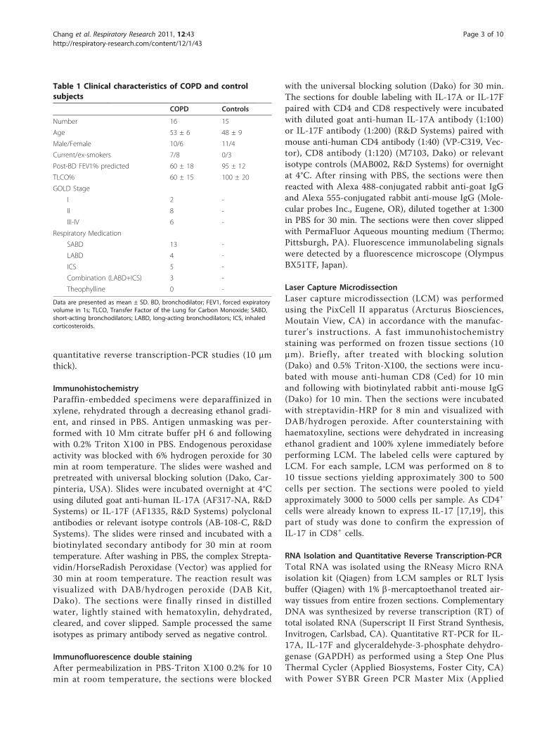
Table 1 Clinical characteristics of COPD and controlsubjects
COPD Controls
Number 16 15
Age 53 ± 6 48 ± 9
Male/Female 10/6 11/4
Current/ex-smokers 7/8 0/3
Post-BD FEV1% predicted 60 ± 18 95 ± 12
TLCO% 60 ± 15 100 ± 20
GOLD Stage
I 2 -
II 8 -
III-IV 6 -
Respiratory Medication
SABD 13 -
LABD 4 -
ICS 5 -
Combination (LABD+ICS) 3 -
Theophylline 0 -
Data are presented as mean ± SD. BD, bronchodilator; FEV1, forced expiratoryvolume in 1s; TLCO, Transfer Factor of the Lung for Carbon Monoxide; SABD,short-acting bronchodilators; LABD, long-acting bronchodilators; ICS, inhaledcorticosteroids.
Chang et al. Respiratory Research 2011, 12:43http://respiratory-research.com/content/12/1/43
Page 3 of 10

Biosystems). The primers used for the specific amplifiedgenes of IL-17A (174 bp), IL-17F (200 bp) and GAPDH(139 bp) are as follows:IL-17A forward: 5’-CATCCATAACCGGAATAC-
CAATA-3’; IL-17A reverse: 5’-TAGTCCACGTTCC-CATCAGC-3’; IL-17F forward: 5’-GTGCCAGGAGGTAGTATGAAGC-3’; IL-17F reverse: 5’-ATGTCTTCCTTTCCTTGAGCATT-3’; GAPDH forward: 5’-AGT-CAACGGATTTGGTCGTATT-3’; GAPDH reverse: 5’-ATGGGTGGAATCATATTGGAAC-3’;
Analysis for immunohistochemistryImmunostained cells in the airway submucosa werecounted at a magnification of 400. The area of submu-cosa was measured by using the software Image Pro 6.2(MediaCybernetics, Bethesda, USA). The final result wasexpressed as the number of positive cells/mm2. Thenumber of cells was corrected for the total number ofcells by counting the number of nuclei in the submu-cosa. The positive staining area of IL-17F in airwayepithelium was measured and the results were presentedas the percentage of positive area in total epitheliumarea.
Statistical analysisData were expressed as median (range). The mean valueof IL-17A (in positive cells/mm2) and IL-17F (in positivecells/mm2 or positive area percentage) in the COPDpatients and in the normal controls were analyzed usingMann-Whitney U test. Probability values of P < 0.05were considered significant. Data analysis was performedby using the Graphpad Instat 3 software (GraphPadSoftware, La Jolla, California).
ResultsIL-17A and IL-17F expression is increased in airways ofCOPD patientsWe observed IL-17A and IL-17F expression in the air-ways of control subjects and COPD patients by immu-nohistochemistry. We were only able to detectoccasional immunoreactivity of IL-17A expression inepithelium. In contrast, we observed considerablestaining for IL-17F in airway epithelium (Figure 1A),which was greater in the airways of COPD subjectscompared to controls (25 (5-65) % vs. 11 (0-32) %, P <0.0001) (Figure 1B). In the submucosa, both IL-17Aand IL-17F positive cells were observed (Figure 1A),and the absolute number of cells expressing both ofthese cytokines was higher in COPD subjects than incontrol subjects (IL-17A+: 199 (70-310) vs. 49 (0-150);IL-17F+: 287 (195-501) vs. 67 (0-203); P < 0.0001) (Fig-ure 1C). There was also some immunoreactivity of IL-17A and F in the endothelial site of few blood vesselsin some sections.
More submucosal cells expressed IL-17A and IL-17F inairways of COPD patientsAs expected, we found that the number of submucosalcells in the airways of COPD subjects was greater than incontrol subjects (2371 (509-5011) vs. 1025 (391-4087),P < 0.001) (Figure 2A). We therefore evaluated the rela-tive number of IL-17A+ and IL-17F+ cells taking inconsideration the total number of submucosal cells. Simi-larly there was greater number of cells positive for bothIL-17A and IL-17F in COPD subjects compared to con-trols (IL-17A: 8 (6-14) % vs. 3 (0-7) %, P < 0.0001; IL-17F: 10 (4-28) % vs. 4 (0-14) %, P < 0.0001) (Figure 2B).
IL-17A and IL-17F expression is not regulated ontranscriptional levelTo further investigate the expression of IL-17A and IL-17F in COPD, we performed quantitative RT-PCR onfrozen airways sections of COPD patients. As with pro-tein expression, the expression of IL-17A and IL-17FmRNA was also detected in airways of COPD patients(Figure 3A). Although there was trend for IL-17F to bemore increased in COPD patients compared to control,the quantification of IL-17A and IL-17F mRNA inCOPD patients was not statistically higher compared tocontrol (Figure 3B).
IL-17A and IL-17F expressed in CD4+ and CD8+ T cellsTo investigate the relationship of IL-17A&F to T cells,we used double immunofluorescence staining with anti-bodies to IL-17A or IL-17F and antibodies to CD4+ orCD8+ T cells. In the airways of COPD subjects, bothCD4+ and CD8+ T cells expressed IL-17A and IL-17F(Figure 4A). To our knowledge, this is the first demon-stration that CD8+ cells produce IL-17A and IL-17F inCOPD. Furthermore, we estimated the percentage ofCD4+ and CD8+ T cells that express IL-17A and IL-17Fas well as the percentage of IL-17A+ and IL-17F+ cellsthat co-express T cell markers. In COPD patients, simi-lar percentage of CD4+ and CD8+ T cells that expressIL-17A and IL-17F was observed (Figure 4B). While intotal IL-17A+ cells, the percentage of IL-17A positivecells that co-express CD8 immunoreactivity was signifi-cantly higher than that expressing CD4+ T cells (16.0 ±4.3% vs. 3.4 ± 2.0%, P < 0.05, Figure 4C). A similartrend was also observed in total IL-17F+ cells (15.8 ±8.8% vs. 6.6 ± 4.3%, Figure 4C). We further confirmedthis finding using LCM to select CD8+ T cells for thedetection of expression of IL-17 mRNA by RT-PCR(Figure 4D).
DiscussionThis study aimed to investigate the possibility that theTh17 cytokines including IL-17A and IL-17F areinvolved in the pathogenesis of COPD. Using bronchial
Chang et al. Respiratory Research 2011, 12:43http://respiratory-research.com/content/12/1/43
Page 4 of 10
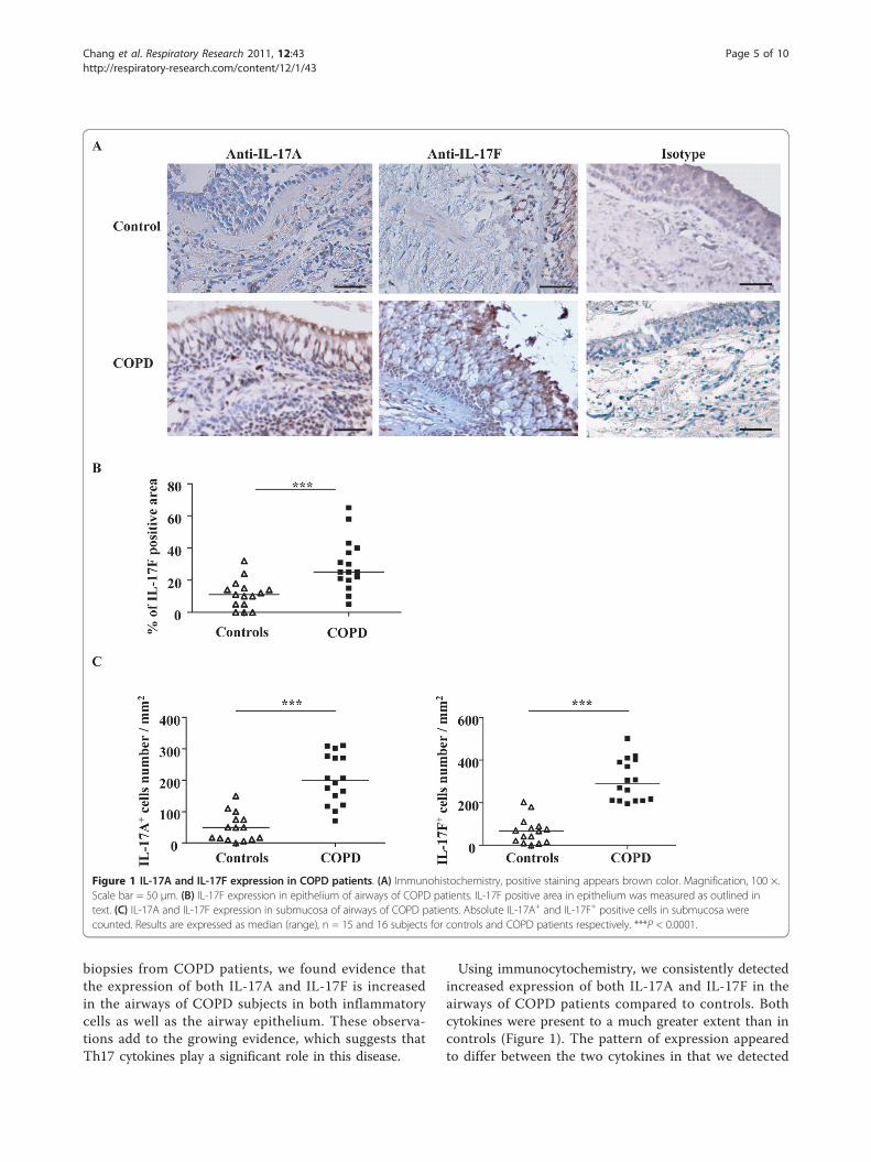
biopsies from COPD patients, we found evidence thatthe expression of both IL-17A and IL-17F is increasedin the airways of COPD subjects in both inflammatorycells as well as the airway epithelium. These observa-tions add to the growing evidence, which suggests thatTh17 cytokines play a significant role in this disease.
Using immunocytochemistry, we consistently detectedincreased expression of both IL-17A and IL-17F in theairways of COPD patients compared to controls. Bothcytokines were present to a much greater extent than incontrols (Figure 1). The pattern of expression appearedto differ between the two cytokines in that we detected
Figure 1 IL-17A and IL-17F expression in COPD patients. (A) Immunohistochemistry, positive staining appears brown color. Magnification, 100 ×.Scale bar = 50 μm. (B) IL-17F expression in epithelium of airways of COPD patients. IL-17F positive area in epithelium was measured as outlined intext. (C) IL-17A and IL-17F expression in submucosa of airways of COPD patients. Absolute IL-17A+ and IL-17F+ positive cells in submucosa werecounted. Results are expressed as median (range), n = 15 and 16 subjects for controls and COPD patients respectively. ***P < 0.0001.
Chang et al. Respiratory Research 2011, 12:43http://respiratory-research.com/content/12/1/43
Page 5 of 10

IL-17A and F in the epithelium of COPD patients butvery little in controls. However, the best control groupis smokers without COPD, but we were unable to obtainsuch a group.The detection of considerable level of IL-17F in the
epithelium is of interest given the potential importanceof the epithelium in the inflammatory process of COPD[20]. When stimulated with pro-inflammatory mediators,the airway epithelium releases chemoattractants CXCL1(GRO-a), CXCL5 (ENA-78), CXCL6 (GCP-2), CXCL8(IL-8) and CCL5 (RANTES) [21,22]. Overexpression ofIL-17F predominantly expressed in bronchial epithelialcells has also been reported in ovalbumin challengedmice [23]. In addition, overexpression of IL-17F in mur-ine lung epithelium leads to infiltration of lymphocytesand macrophages and mucus hyperplasia [24]. Taken
together, these observations suggest the possibility thatIL-17F contributes to amplification of the ongoinginflammatory processes not only through the recruit-ment and activation of specific subset of inflammatorycells, but by prolonging their survival in the airway.Our results contrast to some degree with the recent
report of Di Stefano et al [25] who found evidence ofincreased production of IL-17A but not IL-17F in thebronchial submucosa of COPD patients. Furthermore,they detected expression of both IL-17A and IL-17F inthe epithelium but failed to detect a difference betweencontrols and COPD patients. The discrepancy betweentheir results and ours may reflect differences in patientselection or technique. Notwithstanding these differ-ences, reports to date consistently support the notionthat there is increased expression of IL-17A and IL-17F
Figure 2 Percentage of IL-17A+ and IL-17F+ cells in airway submucosal cells of COPD patients. (A) Submucosal cells in airways of COPDpatients. (B) Percentage of IL-17A+ and IL-17F+ cells in airway submucosal cells of COPD patients. Results are expressed as median (range), n =15 and 16 subjects for controls and COPD patients respectively. **P < 0.001, ***P < 0.0001.
Chang et al. Respiratory Research 2011, 12:43http://respiratory-research.com/content/12/1/43
Page 6 of 10

in COPD patients, underscoring the potential impor-tance of Th17 cytokines in this disease.A potential explanation of increased expression of IL-
17 in COPD airways is that this may be simply a reflec-tion of the presence of greater numbers of submucosalcells. Indeed, consistent with previous studies, wedetected increased cell number in the airway submucosaof COPD patients (Figure 2). However, even afteraccounting for this, we still detected significant differ-ences between COPD and control, as the proportion ofsubmucosal cells expressing IL-17A and IL-17F inCOPD subjects was greater than that in controls. Tofurther explore the basis for this increased expression bysubmucosal cells, we undertook studies of cytokineexpression at the mRNA level. As expected, we wereable to consistently detect evidence of IL-17A and IL-17F mRNA in the airways of COPD subjects (Figure 3).However the quantification results showed that themRNA expression of IL-17A and F was not statisticallydifferent between COPD patients and controls, suggest-ing that there is a discrepancy between mRNA and pro-tein expression for IL-17A and F in COPD patients andthat increased IL-17A expression in COPD patients isregulated at translational level. To refine this
observation, we employed a combination of immunocy-tochemistry and laser capture microscopy. Doubleimmunostaining demonstrated detection of IL-17A andIL-17F not only in CD4+ cells as expected, but also inCD8+ cells (Figure 4). The high percentage of IL-17Aand IL-17F expressing CD immunoreactivity suggestedthat CD8+ T cells are major source of these cytokinesparticularly in COPD [4]. We then used laser capturemicroscopy to select regions of the airway that werepositive for either CD4 or CD8 by immunostaining fromwhich we extracted the RNA to confirm that both CD4+
and CD8+ cells express IL-17A and IL-17F mRNA (Fig-ure 4). To our knowledge, this is the first definitivedemonstration that both CD4+ and CD8+ cells are cap-able of expressing Th17 cytokines in COPD. COPD ismarked by increased number of T cells in lung parench-yma and both peripheral and central airways, with agreater increase in CD8+ cells relative to CD4+ T cells[4]. A number of studies have attempted to characterizethe pattern of lymphocyte cytokine production inCOPD, but the results are conflicting [18,26]. Neverthe-less, in the context of this observation it is noteworthythat a recent study has reported that CD8+ T cells areactivated in the presence of the cytokines IL-6 or IL-21
Figure 3 IL-17A and IL-17F mRNA expression in airways of COPD patients. (A) Quantitative RT-PCR was performed from frozen airwayssections of COPD patients. One representative example from 7 subjects with similar results is shown. (B) Quantification of IL-17A and IL-17FmRNA expression in airways of control subjects and COPD patients. Results are expressed as means ± SEM. N = 7 for both control subjects andCOPD patients.
Chang et al. Respiratory Research 2011, 12:43http://respiratory-research.com/content/12/1/43
Page 7 of 10

Figure 4 Double immunofluorescence staining for detection of IL-17A and IL-17F expression in CD4+ and CD8+ T cells in airways ofCOPD patients. (A) Double immunofluorescence staining was performed. Scale bar = 5 μm. (B) Percentage of CD4+ and CD8+ T cells thatexpress IL-17A and IL-17F. Results are expressed as means ± SEM. (C) Percentage of CD4+ and CD8+ T cells that express IL-17A and IL-17F intotal IL-17A+ and IL-17F+ cells. Results are expressed as means ± SEM. *P < 0.05. N = 3 COPD patients. (D) IL-17A and IL-17F mRNA expression inCD8+ T cells in airways of COPD patients. Immunohistochemistry determined CD8+ T cells were selected by LCM, and then RT-PCR wasperformed to detect the mRNA expression of IL-17A and IL-17F. One representative result from 3 subjects is shown.
Chang et al. Respiratory Research 2011, 12:43http://respiratory-research.com/content/12/1/43
Page 8 of 10

plus TGF-b, develop into IL-17-producing (Tc17) cells.Our findings also need to be taken seen in the contextof reports of Tc17 cells in a variety of immunologicaldiseases. For example, Tc17 have also been found incutaneous inflammatory diseases like psoriasis vulgaris[27] and allergic contact dermatitis [28]. Tc17 cells mayalso be important in defense against viruses [29,30].The observation that expression of Th17 cytokines is
increased in COPD raises questions as to how this maycome about. The combination of IL-6 and TGF-b isreported to skew the balance of T helper cells towardTh17 cell differentiation [31]. In this regard, it is ofinterest that increased production of IL-6 and TGF-bhas been reported in COPD patients [32], raising thepossibility that IL-6 and TGF-b may enable the promo-tion of Th17 cells differentiation in COPD. Regardlessof the mechanism, Th17 cytokines have the potential tocontribute to COPD in various ways. IL-17A actsdirectly on epithelial cells and on airway fibroblasts andsmooth muscle cells to induce the secretion of neutro-phil-recruiting chemokines, such as CXCL8 [31].Although a comprehensive comparative analysis of IL-17F and IL-17A has not been performed, IL-17F appearsto have biological actions similar to IL-17A both in vitroand in vivo [14]. Therefore it is possible that with acti-vation of IL-17A and IL-17F mediated pathways, acrosstalk between local activation of T cells and sus-tained accumulation of neutrophils in inflamed airwayscould be established. Zhu et al [33] have suggested thatbiopsies from patients with chronic bronchitis havemore inflammation compared to patients with COPDbut without chorionic bronchitis. This group of patientsmight have more IL-17 expression. However in ourstudy we did not group our subjects and presented thedata of our patients as one group according to GOLDclassification.In summary, in bronchial biopsies we detected clear
evidence that the expression of the cytokines IL-17Aand IL-17F is increased in COPD compared to control.In the case of IL-17F, this increased expression extendsto the epithelium and is not simply restricted to thesubmucosa. Most importantly, we detected increasedexpression of these cytokines in both CD4+ and CD8+
cells, suggesting that the inflammatory process in COPDmay resemble that in other disorders where Tc17 cellsare active. These findings contribute to the growingbody of information that supports the importance ofinvestigating the role of IL-17 and related cytokines inCOPD, potentially providing novel therapeutic targets inthis important chronic disease.
AcknowledgementsThis study was supported by a grant from the CIRF program.
Author details1Meakins-Christie Laboratories and Respiratory Division, Department ofMedicine McGill University, 3626 rue St. Urbain, Montreal, QC, H2X 2P2Canada. 2Respiratory Division, Research Institute of McGill University HealthCentre, 2155 Guy Street, Suite 900 Montreal, QC, H3H 2R9 Canada.3Respiratory Division, Laval University, 2325 rue de l’Université, Québec, QC,G1V0A6 Canada.
Authors’ contributionsYC carried out the cell counting and data analysis and drafted themanuscript. JN performed the RT-PCR. NB carried out theimmunohistochemistry staining and laser capture. JB and FM participated inthe sample collection and did the immunocytochemiostry. DHE participatedin the design of the study and corrected the manuscript. QH supervised ofthe study. All authors read and approved the final manuscript.
Competing interestsThe authors declare that they have no competing interests.
Received: 26 August 2010 Accepted: 10 April 2011Published: 10 April 2011
References1. Pauwels RA, Rabe KF: Burden and clinical features of chronic obstructive
pulmonary disease (COPD). Lancet 2004, 364(9434):613-620.2. Cosio MG, Saetta M, Agusti A: Immunologic aspects of chronic
obstructive pulmonary disease. N Engl J Med 2009, 360(23):2445-2454.3. Roth M: Pathogenesis of COPD. Part III. Inflammation in COPD. Int J
Tuberc Lung Dis 2008, 12(4):375-380.4. Cosio MG, Majo J: Inflammation of the airways and lung parenchyma in
COPD: role of T cells. Chest 2002, 121(5 Suppl):160S-165S.5. Agusti A, MacNee W, Donaldson K, Cosio M: Hypothesis: does COPD have
an autoimmune component? Thorax 2003, 58(10):832-834.6. Kolls JK, Linden A: Interleukin-17 family members and inflammation.
Immunity 2004, 21(4):467-476.7. Liang SC, Long AJ, Bennett F, Whitters MJ, Karim R, Collins M, Goldman SJ,
Dunussi-Joannopoulos K, Williams CM, Wright JF, Fouser LA: An IL-17F/Aheterodimer protein is produced by mouse Th17 cells and inducesairway neutrophil recruitment. J Immunol 2007, 179(11):7791-7799.
8. Wright JF, Guo Y, Quazi A, Luxenberg DP, Bennett F, Ross JF, Qiu Y,Whitters MJ, Tomkinson KN, Dunussi-Joannopoulos K, Carreno BM,Collins M, Wolfman NM: Identification of an interleukin 17F/17Aheterodimer in activated human CD4+ T cells. J Biol Chem 2007,282(18):13447-13455.
9. Huber M, Heink S, Grothe H, Guralnik A, Reinhard K, Elflein K, Hunig T,Mittrucker HW, Brustle A, Kamradt T, Lohoff M: A Th17-like developmentalprocess leads to CD8(+) Tc17 cells with reduced cytotoxic activity. Eur JImmunol 2009, 39(7):1716-1725.
10. Kramer JM, Gaffen SL: Interleukin-17: a new paradigm in inflammation,autoimmunity, and therapy. J Periodontol 2007, 78(6):1083-1093.
11. Miossec P, Korn T, Kuchroo VK: Interleukin-17 and type 17 helper T cells.N Engl J Med 2009, 361(9):888-898.
12. Fujisawa T, Velichko S, Thai P, Hung LY, Huang F, Wu R: Regulation ofairway MUC5AC expression by IL-1beta and IL-17A; the NF-kappaBparadigm. J Immunol 2009, 183(10):6236-6243.
13. Park H, Li Z, Yang XO, Chang SH, Nurieva R, Wang YH, Wang Y, Hood L,Zhu Z, Tian Q, Dong C: A distinct lineage of CD4 T cells regulates tissueinflammation by producing interleukin 17. Nat Immunol 2005,6(11):1133-1141.
14. Hizawa N, Kawaguchi M, Huang SK, Nishimura M: Role of interleukin-17Fin chronic inflammatory and allergic lung disease. Clin Exp Allergy 2006,36(9):1109-1114.
15. McKinley L, Alcorn JF, Peterson A, Dupont RB, Kapadia S, Logar A, Henry A,Irvin CG, Piganelli JD, Ray A, Kolls JK: TH17 cells mediate steroid-resistantairway inflammation and airway hyperresponsiveness in mice. J Immunol2008, 181(6):4089-4097.
16. Hattotuwa KL, Gizycki MJ, Ansari TW, Jeffery PK, Barnes NC: The effects ofinhaled fluticasone on airway inflammation in chronic obstructivepulmonary disease: a double-blind, placebo-controlled biopsy study. AmJ Respir Crit Care Med 2002, 165(12):1592-1596.
Chang et al. Respiratory Research 2011, 12:43http://respiratory-research.com/content/12/1/43
Page 9 of 10

17. Al-Ramli W, Prefontaine D, Chouiali F, Martin JG, Olivenstein R, Lemiere C,Hamid Q: T(H)17-associated cytokines (IL-17A and IL-17F) in severeasthma. J Allergy Clin Immunol 2009, 123(5):1185-1187.
18. Hodge G, Nairn J, Holmes M, Reynolds PN, Hodge S: Increased intracellularT helper 1 proinflammatory cytokine production in peripheral blood,bronchoalveolar lavage and intraepithelial T cells of COPD subjects. ClinExp Immunol 2007, 150(1):22-29.
19. Lane N, Robins RA, Corne J, Fairclough L: Regulation in chronicobstructive pulmonary disease: the role of regulatory T-cells and Th17cells. Clin Sci (Lond) 119(2):75-86.
20. Larsson K: Aspects on pathophysiological mechanisms in COPD. J InternMed 2007, 262(3):311-340.
21. Prause O, Laan M, Lotvall J, Linden A: Pharmacological modulation ofinterleukin-17-induced GCP-2-, GRO-alpha- and interleukin-8 release inhuman bronchial epithelial cells. Eur J Pharmacol 2003, 462(1-3):193-198.
22. Wang JH, Devalia JL, Xia C, Sapsford RJ, Davies RJ: Expression of RANTESby human bronchial epithelial cells in vitro and in vivo and the effect ofcorticosteroids. Am J Respir Cell Mol Biol 1996, 14(1):27-35.
23. Suzuki S, Kokubu F, Kawaguchi M, Homma T, Odaka M, Watanabe S, Ieki K,Matsukura S, Kurokawa M, Takeuchi H, Sasaki Y, Huang SK, Adachi M, Ota H:Expression of interleukin-17F in a mouse model of allergic asthma. IntArch Allergy Immunol 2007, 143(Suppl 1):89-94.
24. Yang XO, Chang SH, Park H, Nurieva R, Shah B, Acero L, Wang YH,Schluns KS, Broaddus RR, Zhu Z, Dong C: Regulation of inflammatoryresponses by IL-17F. J Exp Med 2008, 205(5):1063-1075.
25. Di Stefano A, Caramori G, Gnemmi I, Contoli M, Vicari C, Capelli A, Magno F,D’Anna SE, Zanini A, Brun P, Casolari P, Chung KF, Barnes PJ, Papi A,Adcock I, Balbi B: T helper type 17-related cytokine expression isincreased in the bronchial mucosa of stable chronic obstructivepulmonary disease patients. Clin Exp Immunol 2009, 157(2):316-324.
26. Zhu X, Gadgil AS, Givelber R, George MP, Stoner MW, Sciurba FC,Duncan SR: Peripheral T cell functions correlate with the severity ofchronic obstructive pulmonary disease. J Immunol 2009, 182(5):3270-3277.
27. Ortega C, Fernandez AS, Carrillo JM, Romero P, Molina IJ, Moreno JC,Santamaria M: IL-17-producing CD8+ T lymphocytes from psoriasis skinplaques are cytotoxic effector cells that secrete Th17-related cytokines.J Leukoc Biol 2009, 86(2):435-443.
28. Zhao Y, Balato A, Fishelevich R, Chapoval A, Mann DL, Gaspari AA: Th17/Tc17 infiltration and associated cytokine gene expression in elicitationphase of allergic contact dermatitis. Br J Dermatol 2009, 161(6):1301-1306.
29. Hamada H, Garcia-Hernandez Mde L, Reome JB, Misra SK, Strutt TM,McKinstry KK, Cooper AM, Swain SL, Dutton RW: Tc17, a unique subset ofCD8 T cells that can protect against lethal influenza challenge. JImmunol 2009, 182(6):3469-3481.
30. Kader M, Bixler S, Piatak M, Lifson J, Mattapallil JJ: Anti-retroviral therapyfails to restore the severe Th-17: Tc-17 imbalance observed in peripheralblood during simian immunodeficiency virus infection. J Med Primatol2009, 38(Suppl 1):32-38.
31. Nembrini C, Marsland BJ, Kopf M: IL-17-producing T cells in lungimmunity and inflammation. J Allergy Clin Immunol 2009, 123(5):986-994,quiz 995-986.
32. Kim V, Rogers TJ, Criner GJ: New concepts in the pathobiology of chronicobstructive pulmonary disease. Proc Am Thorac Soc 2008, 5(4):478-485.
33. Zhu J, Qiu Y, Valobra M, Qiu S, Majumdar S, Matin D, De Rose V, Jeffery PK:Plasma cells and IL-4 in chronic bronchitis and chronic obstructivepulmonary disease. Am J Respir Crit Care Med 2007, 175(11):1125-1133.
doi:10.1186/1465-9921-12-43Cite this article as: Chang et al.: CD8 positive T cells express IL-17 inpatients with chronic obstructive pulmonary disease. Respiratory Research2011 12:43.
Submit your next manuscript to BioMed Centraland take full advantage of:
• Convenient online submission
• Thorough peer review
• No space constraints or color figure charges
• Immediate publication on acceptance
• Inclusion in PubMed, CAS, Scopus and Google Scholar
• Research which is freely available for redistribution
Submit your manuscript at www.biomedcentral.com/submit
Chang et al. Respiratory Research 2011, 12:43http://respiratory-research.com/content/12/1/43
Page 10 of 10


![Chronic Obstructive Pulmonary Diseaseopenaccessebooks.com/chronic-obstructive-pulmonary...Chronic Obstructive Pulmonary Disease 5 a-MCI is made [32]. COPD patients without significant](https://img.pdfslide.net/doc/110x75/5f853ccf82a2412fd65b9e28/chronic-obstructive-pulmonary-dis-chronic-obstructive-pulmonary-disease-5-a-mci.jpg)