Embed Size (px)
Citation preview
Page 1/29
Generation of a Novel High-A�nity Antibody Binding toPCSK9 Catalytic Domain with Slow Dissociation Rate byCDR-Grafting, Alanine Scanning and Saturated Site-Directed MutagenesisZhengli Bai
China Pharmaceutical UniversityMenglong Xu
China Pharmaceutical UniversityYing Mei
China Pharmaceutical UniversityTuo Hu
China Pharmaceutical UniversityPanpan Zhang
China Pharmaceutical UniversityManman Chen
China Pharmaceutical UniversityWenxiu Lv
China Pharmaceutical UniversityChenchen Lu
China Pharmaceutical UniversityShuhua Tan ( [email protected] )
China Pharmaceutical University https://orcid.org/0000-0002-4992-8876
Original article
Keywords: single-chain variable fragment (scFv), Escherichia coli (E. coli), Alanine scanning, saturated site-directed mutagenesis, PCSK9, Molecular docking
Posted Date: October 21st, 2021
DOI: https://doi.org/10.21203/rs.3.rs-957578/v1
License: This work is licensed under a Creative Commons Attribution 4.0 International License. Read FullLicense
Page 2/29
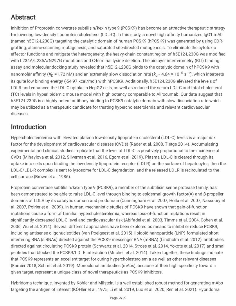
AbstractInhibition of Proprotein convertase subtilisin/kexin type 9 (PCSK9) has become an attractive therapeutic strategyfor lowering low-density lipoprotein cholesterol (LDL-C). In this study, a novel high a�nity humanized IgG1 mAb(named h5E12-L230G) targeting the catalytic domain of human PCSK9 (hPCSK9) was generated by using CDR-grafting, alanine-scanning mutagenesis, and saturated site-directed mutagenesis. To eliminate the cytotoxiceffector functions and mitigate the heterogeneity, the heavy-chain constant region of h5E12-L230G was modi�edwith L234A/L235A/N297G mutations and C-terminal lysine deletion. The biolayer interferometry (BLI) bindingassay and molecular docking study revealed that h5E12-L230G binds to the catalytic domain of hPCSK9 withnanomolar a�nity (KD =1.72 nM) and an extremely slow dissociation rate (koff, 4.84 × 10⁻⁵ s−1), which interpretsits quite low binding energy (-54.97 kcal/mol) with hPCSK9. Additionally, h5E12-L230G elevated the levels ofLDLR and enhanced the LDL-C uptake in HepG2 cells, as well as reduced the serum LDL-C and total cholesterol(TC) levels in hyperlipidemic mouse model with high potency comparable to Alirocumab. Our data suggest thath5E12-L230G is a highly potent antibody binding to PCSK9 catalytic domain with slow dissociation rate whichmay be utilized as a therapeutic candidate for treating hypercholesterolemia and relevant cardiovasculardiseases.
IntroductionHypercholesterolemia with elevated plasma low-density lipoprotein cholesterol (LDL-C) levels is a major riskfactor for the development of cardiovascular diseases (CVDs) (Rader et al. 2008, Tietge 2014). Accumulatingexperimental and clinical studies implicate that the level of LDL-C is positively proportional to the incidence ofCVDs (Mihaylova et al. 2012, Silverman et al. 2016, Egom et al. 2019). Plasma LDL-C is cleared through itsuptake into cells upon binding the low-density lipoprotein receptor (LDLR) on the surface of hepatocytes, then theLDL-C/LDL-R complex is sent to lysosome for LDL-C degradation, and the released LDLR is recirculated to thecell surface (Brown et al. 1986).
Proprotein convertase subtilisin/kexin type 9 (PCSK9), a member of the subtilisin serine protease family, hasbeen demonstrated to be able to raise LDL-C level through binding to epidermal growth factor(A) and β-propellerdomains of LDLR by its catalytic domain and prodomain (Cunningham et al. 2007, Holla et al. 2007, Nassoury etal. 2007, Poirier et al. 2009). In human, mechanistic studies of PCSK9 have shown that gain-of-functionmutations cause a form of familial hypercholesterolemia, whereas loss-of-function mutations result insigni�cantly decreased LDL-C level and cardiovascular risk (Abifadel et al. 2003, Timms et al. 2004, Cohen et al.2006, Wu et al. 2014). Several different approaches have been explored as means to inhibit or reduce PCSK9,including antisense oligonucleotides (van Poelgeest et al. 2015), lipidoid nanoparticle (LNP) formulated shortinterfering RNA (siRNAs) directed against the PCSK9 messenger RNA (mRNA) (Lindholm et al. 2012), antibodiesdirected against circulating PCSK9 protein (Schwartz et al. 2014, Stroes et al. 2014, Yokote et al. 2017) and smallpeptides that blocked the PCSK9/LDLR interaction (Mitchell et al. 2014). Taken together, these �ndings indicatethat PCSK9 represents an excellent target for curing hypercholesterolemia as well as other relevant diseases(Farnier 2018, Schmit et al. 2019). Monoclonal antibodies (mAbs), because of their high speci�city toward agiven target, represent a unique class of novel therapeutics as PCSK9 inhibitors.
Hybridoma technique, invented by Köhler and Milstein, is a well-established robust method for generating mAbstargeting the antigen of interest (KÖHler et al. 1975, Li et al. 2019, Luo et al. 2020, Ren et al. 2021). Hybridoma
Page 3/29
technology can screen hybridoma cell lines which not only have the immortality of myeloma cells but also havethe ability of splenocytes to secrete antibodies by fusing myeloma cells with splenocytes from immunized mice(Elgundi et al. 2017). However, murine mAbs prepared by the hybridoma technique may induce the human anti-mouse antibodies (HAMA) response which limits its utility and e�cacy in clinical treatment (Chan et al. 2001).
Humanization of murine mAbs is crucial to circumvent the problem. The �rst humanization strategy is toconstruct chimeric antibodies by substituting the murine constant region with an appropriate human constantregion to reduce the content of heterologous sequences (Morrison et al. 1984). Several chimeric antibodies havebeen approved by FDA, including ramucirumab, brentuximab, dinutuximab, etc. The variable domains of chimericantibodies that consist of framework regions (FRs) and complementarity determining regions (CDRs) arecompletely murine, while FRs are not necessarily required for antigen recognition (Chiu et al. 2016). Based on it,grafting murine CDRs onto human germline FRs and retaining the residues in murine FRs which may play a keyrole in maintaining the conformational integrity of CDRs to construct a CDR-grafted antibody can further reducethe immunogenicity (Haidar et al. 2012, Safdari et al. 2013). Humanization and a�nity maturation are the mostfrequently applied processes to develop a therapeutic antibody with high a�nity to a speci�c epitope (Inoue et al.2013, Ko et al. 2015). In vitro a�nity maturation of mAbs mimics the process of in vivo a�nity maturation whichrelies on somatic hypermutation of immunoglobulin genes and positive clonal selection (Ersching et al. 2017).The most commonly used in vitro a�nity maturation technologies are (1) site-directed mutagenesis and randommutagenesis consisting in the introduction of mutagenesis speci�cally or randomly throughout the gene, (2)chain shu�ing that recombines the heavy and light chains of different antibodies with high a�nity, (3) phagedisplay which allows screening the desired antibody from a large library with millions to trillions of variants(Levin et al. 2006, Sheedy et al. 2007, Yun et al. 2019).
In this work, we describe the generation, humanization and in vitro a�nity maturation of a hybridoma-derivedanti-PCSK9 antibody by using CDR-grafting, alanine-scanning mutagenesis, and saturated site-directedmutagenesis methods. Humanization and a�nity maturation of the generated murine antibody was conductedusing the single-chain variable fragment (scFv) format for the reason that scFvs are much more convenient tomodify and produce in E. coli host, then the selected optimized scFvs were reformatted into full-length IgG andexpressed transiently in CHO-3E7 mammalian cells for further identi�cation.
Material And Methods
MaterialsMEM, DMEM, Opti-MEM, and Pluronic-F68 were purchased from Thermo Fisher Scienti�c (Waltham, MA, USA).Fetal bovine serum (FBS), penicillin G sodium salt, streptomycin solution, and hypoxanthine-aminopterin-thymidine (HAT) were obtained from MilliporeSigma (Burlington, MA, USA). HyClone™ HyCell™ CHO Medium waspurchased from GE Healthcare (Piscataway, NJ, USA). 25 kDa Linear polyethyleneimine (LPEI) was obtainedfrom Polysciences (Warrington, Pennsylvania, USA). Quickantibody-Mouse 5W adjuvant (Cat# KX0210041) wasobtained from Biodragon Immunotechnologies (Beijing, China). Bovine serum albumin (BSA) was obtained fromBiofroxx (Einhausen, Hessen, Germany). Agarose Gel DNA Extraction Kit, RNAiso Plus, and PrimeScript RTReagent Kit with gDNA Eraser were bought from TaKaRa (Dalian, Liaoning, China). Rabbit anti-PCSK9 antibody(Cat# ab181142) and rabbit anti-LDLR antibody (Cat# ab52818) were obtained from Abcam (Cambridge, UK).Glutamine, TMB substrate, IPTG, rabbit anti-GAPDH antibody (Cat# D110016), HRP-conjugated goat anti-rabbit
Page 4/29
IgG (Cat# D110058), and Alexa Fluor 488®-conjugated goat anti-rabbit IgG (Cat# D110061) were bought fromBBI (Toronto, ON, Canada). LDL labeled with 1, 1’-dioctadecyl - 3, 3, 3’, 3’-tetramethyl-indocarbocyanineperchlorate (DiI-LDL) was obtained from Yiyuan Biotechnologies (Guangzhou, Guangdong, China). Commercialtest kits for LDL-C, TC, TG, and HDL-C were obtained from Nanjing Jiancheng Bioengineering Institute (Nanjing,Jiangsu, China).
Bacterial strains and cell linesEscherichia coli (E. coli) strains DH5α and BL21 (DE3) were used as hosts for plasmid preparation and single-chain variable fragment (scFv) prokaryotic expression, respectively. Chinese hamster ovary (CHO-3E7) cells wereobtained from Genscript Biotech (Nanjing, China), cultured in HyClone™ HyCell™ CHO Medium, and used as hostsfor IgG1 eukaryotic transient-expression. Mouse myeloma cell line SP2/0 was purchased from American TypeCulture Collection (ATCC, Manassas, VA, USA) and cultured in DMEM medium supplemented with penicillin (100U/ml), streptomycin (100 µg/ml) and 10% (v/v) FBS. Human hepatic HepG2 cells were obtained from ChinaInfrastructure of Cell Line Resources (Beijing, China) and maintained at 37°C, 5% CO2, in MEM supplementedwith penicillin (100 U/ml), streptomycin (100 µg/ml) and 10% (v/v) FBS. All cells were cultured in a humidi�edincubator at 37°C in an atmosphere of 5% CO2 in the air.
Antigen preparationTo produce human PCSK9 (hPCSK9) protein, the coding sequence of hPCSK9 (GenBank accession number:NM_174936.3) fused with a Kozak consensus sequence (GCCGCCACC) (Hernández et al. 2019) at the 5’-end anda 6×His-tag gene at the 3’-end was synthesized by GenScript Biotech (Nanjing, China) and subcloned into theeukaryotic expression vector pTT5 using the Hind III and EcoR I restriction enzyme sites. The yieldedrecombinant plasmids were further transiently transfected into suspension CHO-3E7 mammalian cells using PEItransfection reagent as described previously (Stuible et al. 2018). On day 7 post-transfection, the supernatantwas puri�ed with a Ni2+ Based immobilized metal ion a�nity chromatography (Ni-IMAC, GE Healthcare,Piscataway, NJ, USA), followed by SuperdexTM 200 HR 10/300GL size-exclusion chromatography (GEHealthcare) according to the manufacturer's instructions. Protein concentration was determined by using theBCA protein assay kit (Biomiga, San Diego, USA).
BALB/c mice immunizationPuri�ed hPCSK9 protein (20 µg per mouse) was emulsi�ed with an equal volume of Quickantibody-Mouse 5Wadjuvant and intramuscularly injected into the hind legs of female BALB/c mice (6-8-wk, QinglongshanExperimental Animal Breeding Farm, Nanjing, China) on day 1 and day 21. The �nal boost (50 µg of hPCSK9protein) was given intraperitoneally on day 35 without adjuvant. Three days after the �nal booster immunization,orbital blood of mice was collected for antibody titer detection. When the antibody titters attained 1:100000, thecell fusion was conducted.
Cell fusion and hybridoma screeningAccording to standard procedures(KÖHler 1975, Kim et al. 2014), splenocytes were harvested from theimmunized mice and fused with SP2/0mouse myeloma cells at a ratio of 5:1 using 50% (w/v) polyethyleneglycol (PEG) as a fusion regent, and the resulting hybridomas were then cultured in 96-well plates inhypoxanthine-aminopterin-thymidine (HAT) selective medium supplemented with 20% (w/v) FBS. Afterward, thepositive hybridoma cells were screened by indirect ELISA and subcloned three times by limiting dilution method.
Page 5/29
The ascites of identi�ed hybridoma were also prepared by injection of 1×10⁶ positive hybridoma cells into theperitoneal cavity of pristine-treated BALB/c mice, and the ascites containing speci�c mAbs were puri�ed byprotein A a�nity chromatography (Roche, Mannheim, Germany). The isotype of puri�ed mAb was determinedusing a mouse monoclonal subtype identi�cation kit (KMI-2, ProteinTech Group, Chicago, IL, USA) according tothe manufacturer’s instruction.
Enzyme-linked immunosorbent assayThe hybridoma cells producing antibodies against hPCSK9 were screened by Enzyme-linked immunosorbentassay (ELISA) using 96-well plates coated with hPCSK9 (1 µg/ml) in coating buffer (0.2 M Na2CO3/NaHCO3, pH9.6) overnight at 4℃. The plates were then blocked with PBS containing 3% (w/v) bovine serum albumin (BSA)for 2 h at 37℃ and incubated with 100 µl of hybridoma supernatants for 2 h at 37℃. Besides, non-competitivephage ELISA with the addition of increasing concentrations of mAb (10-1, 10, 102, 103, 104, 105 ng/ml) was alsoset up to further measure the a�nity constant (Kaff) of selected mAb as described previously (Beatty et al. 1987).After washing three times with 0.1% Tween in PBS (PBST), HRP-conjugated goat anti-mouse IgG antibody wasadded and incubated for 1 h at 37℃. Finally, the TMB substrate was added and allowed to develop for 15minutes at room temperature, and the absorbance at 450 nm was measured using a microplate reader (ThermoScienti�c, Waltham, MA, USA).
Western blot analysisWestern blot was performed to detect the protein expression levels of LDLR in HepG2 cells or liver tissues aspreviously described (Gu et al. 2019). Brie�y, the cells or tissues were lysed or homogenized in cold RIPA lysisbuffer (Solarbio, Beijing, China) containing 1 mM PMSF on ice for 0.5 h. After centrifugation at 12,000 g for 15min at 4°C, the cell lysates were collected and total protein concentrations were determined using BCA proteinassay. Equal amounts of protein from each sample were subjected to 12% (w/v) SDS-PAGE and transferred to0.22 µm polyvinylidene �uoride (PVDF) membrane (MerckMillipore, Darmstadt, Germany). After blocking with0.1% (v/v) TBS-Tween 20 (TBST) containing 5% (w/v) nonfat milk for 2 h at room temperature, the membranewas incubated with corresponding primary antibodies against GAPDH (Cat# D110016, 1:1000) or LDLR (Cat#ab52818, 1:1000) at 4°C overnight, followed by incubation with HRP-conjugated goat anti-rabbit IgG (Cat#D110058, 1:5000) at room temperature for 1 h. Protein bands were detected by enhanced chemiluminescence(ECL) solution (Thermo Scienti�c, Massachusetts, USA) and quanti�ed by ImageJ software (National Institutesof Health, Bethesda, MD, USA).
LDL-C uptake assayLDL-C uptake assay was conducted as previously described (Ly et al. 2014, Xu et al. 2020) with slightmodi�cation. In brief, HepG2 cells were seeded in black 96-well plates at a density of 3×10⁴ cells per well andcultured overnight. Then, cells were pretreated with opti-MEM for 12 h, followed by treatment with 20 µg/mlhPCSK9 protein alone or co-treatment with 50 µg/ml anti-PCSK9 antibodies for 8 h. Thereafter, 20 µg/ml DiI-LDLwas added each well and incubated for an additional 4 h. After washing 3 times with PBS in the dark, LDL-Cuptake was measured using a multimode microplate reader (Varioskan lux, Thermo scienti�c) at 520 nmexcitation/580 nm emission.
Cloning of VH and VL gene from hybridoma cells
Page 6/29
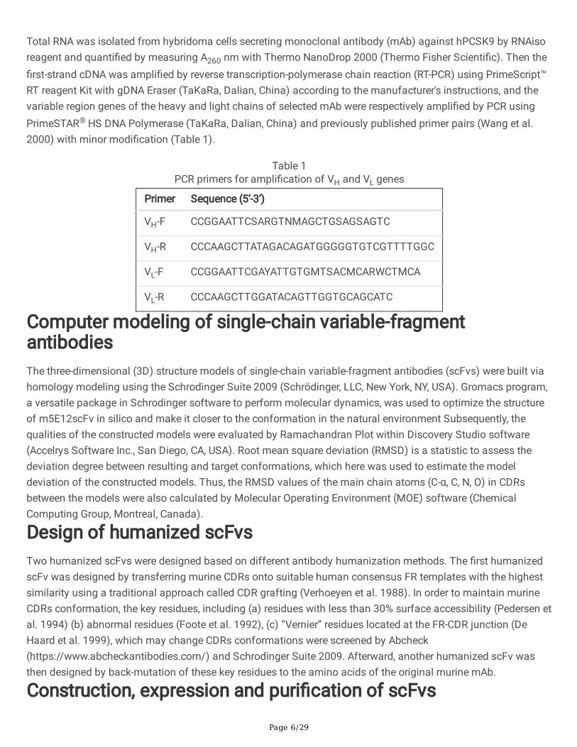
Total RNA was isolated from hybridoma cells secreting monoclonal antibody (mAb) against hPCSK9 by RNAisoreagent and quanti�ed by measuring A260 nm with Thermo NanoDrop 2000 (Thermo Fisher Scienti�c). Then the�rst-strand cDNA was ampli�ed by reverse transcription-polymerase chain reaction (RT-PCR) using PrimeScript™RT reagent Kit with gDNA Eraser (TaKaRa, Dalian, China) according to the manufacturer's instructions, and thevariable region genes of the heavy and light chains of selected mAb were respectively ampli�ed by PCR usingPrimeSTAR® HS DNA Polymerase (TaKaRa, Dalian, China) and previously published primer pairs (Wang et al.2000) with minor modi�cation (Table 1).
Table 1PCR primers for ampli�cation of VH and VL genes
Primer Sequence (5’-3’)
VH-F CCGGAATTCSARGTNMAGCTGSAGSAGTC
VH-R CCCAAGCTTATAGACAGATGGGGGTGTCGTTTTGGC
VL-F CCGGAATTCGAYATTGTGMTSACMCARWCTMCA
VL-R CCCAAGCTTGGATACAGTTGGTGCAGCATC
Computer modeling of single-chain variable-fragmentantibodiesThe three-dimensional (3D) structure models of single-chain variable-fragment antibodies (scFvs) were built viahomology modeling using the Schrodinger Suite 2009 (Schrödinger, LLC, New York, NY, USA). Gromacs program,a versatile package in Schrodinger software to perform molecular dynamics, was used to optimize the structureof m5E12scFv in silico and make it closer to the conformation in the natural environment Subsequently, thequalities of the constructed models were evaluated by Ramachandran Plot within Discovery Studio software(Accelrys Software Inc., San Diego, CA, USA). Root mean square deviation (RMSD) is a statistic to assess thedeviation degree between resulting and target conformations, which here was used to estimate the modeldeviation of the constructed models. Thus, the RMSD values of the main chain atoms (C-α, C, N, O) in CDRsbetween the models were also calculated by Molecular Operating Environment (MOE) software (ChemicalComputing Group, Montreal, Canada).
Design of humanized scFvsTwo humanized scFvs were designed based on different antibody humanization methods. The �rst humanizedscFv was designed by transferring murine CDRs onto suitable human consensus FR templates with the highestsimilarity using a traditional approach called CDR grafting (Verhoeyen et al. 1988). In order to maintain murineCDRs conformation, the key residues, including (a) residues with less than 30% surface accessibility (Pedersen etal. 1994) (b) abnormal residues (Foote et al. 1992), (c) “Vernier” residues located at the FR-CDR junction (DeHaard et al. 1999), which may change CDRs conformations were screened by Abcheck(https://www.abcheckantibodies.com/) and Schrodinger Suite 2009. Afterward, another humanized scFv wasthen designed by back-mutation of these key residues to the amino acids of the original murine mAb.
Construction, expression and puri�cation of scFvs
Page 7/29
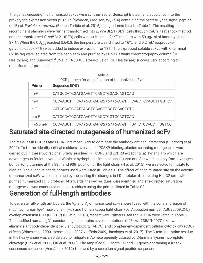
The genes encoding the humanized scFvs were synthesized at Genscript Biotech and subcloned into theprokaryotic expression vector pET-27b (Novagen, Madison, WI, USA) containing the pectate lyase signal peptide(pelB) of Erwinia carotovora (Blanco-Toribio et al. 2015) using primers listed in Table 2. The resultingrecombinant plasmids were further transformed into E. coli BL21 (DE3) cells through CaCl2 heat shock method,and the transformed E. coli BL21 (DE3) cells were cultured in 2×YT medium with 50 µg/ml of kanamycin at37℃. When the OD600 reached 0.6-0.8, the temperature was shifted to 16℃ and 0.2 mM isopropyl-β-galactosidase (IPTG) was added to induce expression for 18 h. The expressed soluble scFvs with C-terminal6×His-tag were isolated from the periplasm and puri�ed by Ni-NTA a�nity chromatography column (GEHealthcare) and SuperdexTM 75 HR 10/300GL size-exclusion (GE Healthcare) successively, according tomanufactures’ protocols.
Table 2PCR primers for ampli�cation of humanized scFvs
Primer Sequence (5’-3’)
m-F CATGCCATGGATGAAGTTCAGCTGGAGCAGTCAG
m-R CCCAAGCTTTCAATGGTGATGGTGATGGTGTTTCAGCTCCAGCTTGGTCC
h-F CATGCCATGGATCAGGTGCAGCTGGTGCAGTCTG
bm-F CATGCCATGGATGAAGTTCAGCTGGTGCAGTCAG
h-R/bm-R CCCAAGCTTTCAATGGTGATGGTGATGGTGTTTGATCTCCACCTTGGTCC
Saturated site-directed mutagenesis of humanized scFvThe residues in HCDR3 and LCDR3 are most likely to dominate the antibody-antigen interaction (Sundberg et al.2002). To further identify critical residues involved in hPCSK9 binding, alanine scanning mutagenesis wascarried out in these two regions. Brie�y, residues in HCDR3 and LCDR3 excepting (a) Tyr and Trp which areadvantageous for large van der Waals or hydrophobic interactions, (b) Asn and Ser which mainly form hydrogenbonds, (c) glutamine at the 89th and 90th position of the light chain (H et al. 2015), were selected to mutate toalanine. The oligonucleotide primers used were listed in Table S1. The effect of each mutated site on the activityof humanized scFv was determined by measuring the changes in LDL uptake after treating HepG2 cells withpuri�ed humanized scFv proteins. Afterwards, the key residues were identi�ed and site-directed saturationmutagenesis was conducted on these residues using the primers listed in Table S2.
Generation of full-length antibodiesTo generate full-length antibodies, the VH and VL of humanized scFvs were fused with the constant region ofmodi�ed human IgG1 heavy chain (HC) and human kappa light chain (LC, Accession number: ABU90709.2) byoverlap-extension PCR (OE-PCR) (Lu et al. 2018), respectively. Primers used for OE-PCR were listed in Table 3.The modi�ed human IgG1 constant region contains several mutations (L234A/L235A/N297G), known toeliminate antibody-dependent cellular cytotoxicity (ADCC) and complement-dependent cellular cytotoxicity (CDC)effects (Wines et al. 2000, Hessell et al. 2007, Jefferis 2009, Jacobsen et al. 2017). The C-terminal lysine residuein the heavy chain was also deleted to mitigate mAb heterogeneity caused by C-terminal lysine incompletecleavage (Dick et al. 2008, Liu et al. 2008). The ampli�ed full-length HC and LC genes containing a Kozakconsensus sequence (Hernández 2019) followed by a secretion signal peptide sequence
Page 8/29
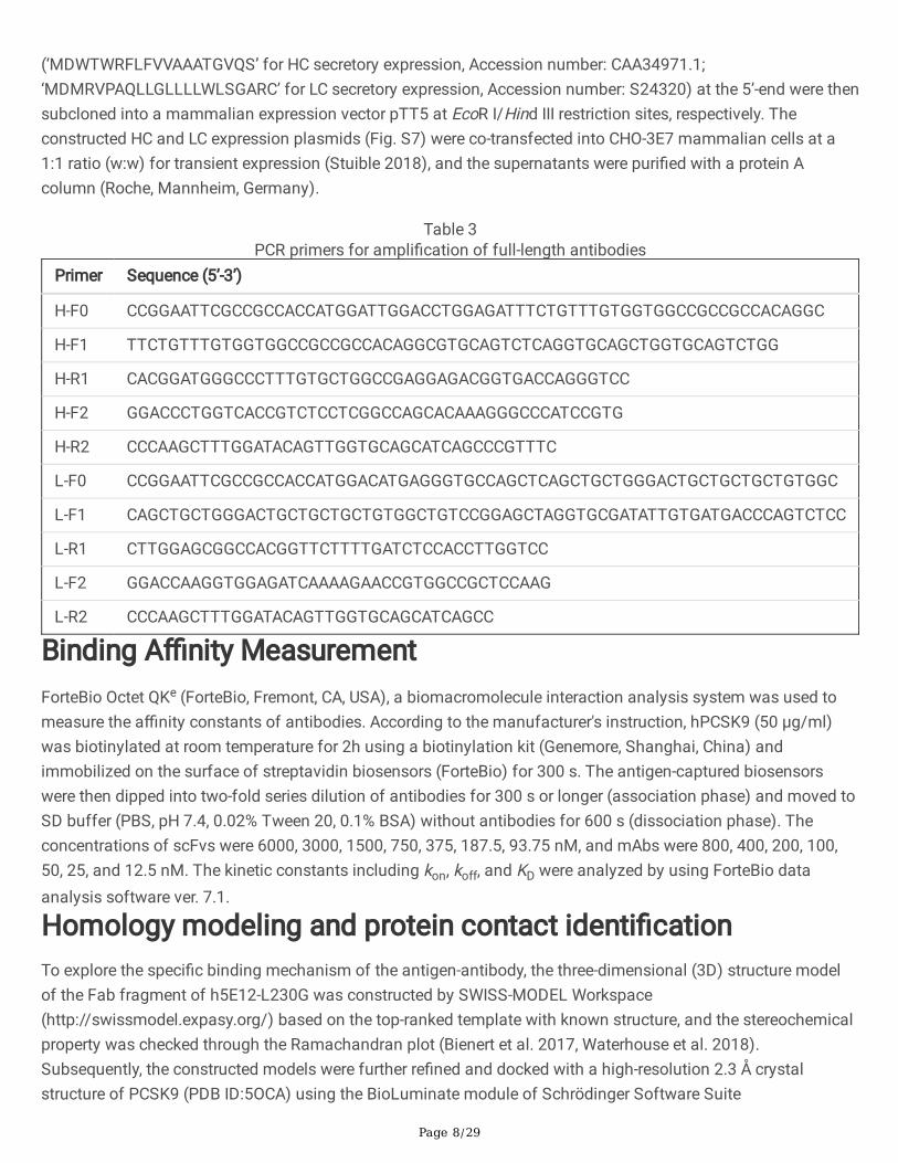
(‘MDWTWRFLFVVAAATGVQS’ for HC secretory expression, Accession number: CAA34971.1;‘MDMRVPAQLLGLLLLWLSGARC’ for LC secretory expression, Accession number: S24320) at the 5’-end were thensubcloned into a mammalian expression vector pTT5 at EcoR I/Hind III restriction sites, respectively. Theconstructed HC and LC expression plasmids (Fig. S7) were co-transfected into CHO-3E7 mammalian cells at a1:1 ratio (w:w) for transient expression (Stuible 2018), and the supernatants were puri�ed with a protein Acolumn (Roche, Mannheim, Germany).
Table 3PCR primers for ampli�cation of full-length antibodies
Primer Sequence (5’-3’)
H-F0 CCGGAATTCGCCGCCACCATGGATTGGACCTGGAGATTTCTGTTTGTGGTGGCCGCCGCCACAGGC
H-F1 TTCTGTTTGTGGTGGCCGCCGCCACAGGCGTGCAGTCTCAGGTGCAGCTGGTGCAGTCTGG
H-R1 CACGGATGGGCCCTTTGTGCTGGCCGAGGAGACGGTGACCAGGGTCC
H-F2 GGACCCTGGTCACCGTCTCCTCGGCCAGCACAAAGGGCCCATCCGTG
H-R2 CCCAAGCTTTGGATACAGTTGGTGCAGCATCAGCCCGTTTC
L-F0 CCGGAATTCGCCGCCACCATGGACATGAGGGTGCCAGCTCAGCTGCTGGGACTGCTGCTGCTGTGGC
L-F1 CAGCTGCTGGGACTGCTGCTGCTGTGGCTGTCCGGAGCTAGGTGCGATATTGTGATGACCCAGTCTCC
L-R1 CTTGGAGCGGCCACGGTTCTTTTGATCTCCACCTTGGTCC
L-F2 GGACCAAGGTGGAGATCAAAAGAACCGTGGCCGCTCCAAG
L-R2 CCCAAGCTTTGGATACAGTTGGTGCAGCATCAGCC
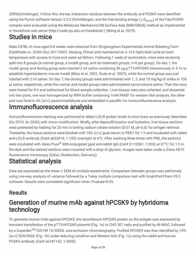
Binding A�nity MeasurementForteBio Octet QKe (ForteBio, Fremont, CA, USA), a biomacromolecule interaction analysis system was used tomeasure the a�nity constants of antibodies. According to the manufacturer's instruction, hPCSK9 (50 µg/ml)was biotinylated at room temperature for 2h using a biotinylation kit (Genemore, Shanghai, China) andimmobilized on the surface of streptavidin biosensors (ForteBio) for 300 s. The antigen-captured biosensorswere then dipped into two-fold series dilution of antibodies for 300 s or longer (association phase) and moved toSD buffer (PBS, pH 7.4, 0.02% Tween 20, 0.1% BSA) without antibodies for 600 s (dissociation phase). Theconcentrations of scFvs were 6000, 3000, 1500, 750, 375, 187.5, 93.75 nM, and mAbs were 800, 400, 200, 100,50, 25, and 12.5 nM. The kinetic constants including kon, koff, and KD were analyzed by using ForteBio dataanalysis software ver. 7.1.
Homology modeling and protein contact identi�cationTo explore the speci�c binding mechanism of the antigen-antibody, the three-dimensional (3D) structure modelof the Fab fragment of h5E12-L230G was constructed by SWISS-MODEL Workspace(http://swissmodel.expasy.org/) based on the top-ranked template with known structure, and the stereochemicalproperty was checked through the Ramachandran plot (Bienert et al. 2017, Waterhouse et al. 2018).Subsequently, the constructed models were further re�ned and docked with a high-resolution 2.3 Å crystalstructure of PCSK9 (PDB ID:5OCA) using the BioLuminate module of Schrödinger Software Suite
Page 9/29
2009(Schrödinger). Follow this, the key interaction residues between the antibody and PCSK9 were identi�edusing the Pymol software Version 2.3.0 (Schrödinger), and the free binding energy (△Gbinds) of the Fab-PCSK9-complex were evaluated using the Molecular Mechanics/GB Surface Area (MM/GBSA) method as implementedin HawkDock web server (http://cadd.zju.edu.cn/hawkdock/) (Weng et al. 2019).
Studies in miceMale C57BL/6 mice aged 6-8 weeks were obtained from Qinglongshan Experimental Animal Breeding Farm(Certi�cate no. SCXK (Su) 2017-0001; Nanjing, China) and maintained on a 12-h light/dark cycle at roomtemperature with access to food and water ad libitum. Following 1 week of acclimation, mice were randomlysplit into 8 groups (a normal group, a model group, and six treatment groups, n=6 per group). On day 1, themodel group and dosing group were injected 2 ml saline containing 50 µg pTT5-hPCSK9 intravenously in 5-7s toestablish hyperlipidemic mouse model (Miao et al. 2001, Suda et al. 2007), while the normal group was justinjected with 2 ml saline. On day 7, the dosing groups were administered with 1, 3, and 10 mg/kg of mAbs in 100µl saline, respectively, while the normal and model groups were administered same-volume saline. Then the micewere fasted for 8 h and euthanized for blood sample collection. Liver tissues were also collected, and dissectedinto two parts, one was homogenized by RIPA buffer containing 1mM PMSF for western blot analysis, the otherpart was �xed in 4% (w/v) paraformaldehyde and embedded in para�n for immuno�uorescence analysis.
Immuno�uorescence analysisImmuno�uorescence staining was performed to detect LDLR protein levels in mice livers as previously described(Gu 2019, Xu 2020) with minor modi�cation. Brie�y, after depara�nization and hydration, liver tissue sectionswere pretreated by heating for 20 min in boiling sodium citrate solution (0.01 M, pH 6.0) for antigen retrieval.Thereafter, the tissue sections were blocked with 10% (v/v) goat serum in PBST for 1 h and incubated with rabbitanti-LDLR antibody (Cat# ab52818, 1:100) overnight at 4°C. After washing three times with PBS, the sectionswere incubated with Alexa Fluor® 488-conjugated goat anti-rabbit IgG (Cat# D110061, 1:200) at 37℃ for 1 h inthe dark and the stained sections were mounted with a drop of glycerin. Images were taken under a Zeiss AX10�uorescence microscopy (Zeiss, Oberkochen, Germany).
Statistical analysisData are expressed as the mean ± SEM of multiple experiments. Comparison between groups was performedusing one-way analysis of variance followed by a Tukey multiple comparison test with GraphPad Prism V5.0software. Results were considered signi�cant when P-values<0.05.
Results
Generation of murine mAb against hPCSK9 by hybridomatechnologyTo generate murine mAb against hPCSK9, the recombinant hPCSK9 protein as the antigen was expressed bytransient transfection of the pTT5-hPCSK9 plasmid (Fig. 1a) to CHO 3E7 cells and puri�ed by Ni-IMAC, followedby a SuperdexTM 200 HR 10/300GL size-exclusion chromatography. Puri�ed hPCSK9 was then identi�ed by 10%(w/v) SDS-PAGE (Fig. 1b) under reducing condition and Western blot (Fig. 1c) using the rabbit anti-humanPCSK9 antibody (Cat# ab181142, 1:3000).
Page 10/29
Subsequently, BALB/c mice were immunized with puri�ed hPCSK9 protein. On day 38, the mice attainingantibody titer of 1:640000 (Fig. 1d) were sacri�ced, and its splenocytes were fused with SP2/0 cells forhybridoma production. A positive hybridoma clone, named 5E12, was identi�ed by ELISA (Fig. 1e) and subclonedthree times by limiting dilution. This hPCSK9-speci�c murine antibody (named m5E12) was then puri�ed frommouse ascites by protein A a�nity chromatography (Roche, Mannheim, Germany) and identi�ed by SDS-PAGEunder reducing and nonreducing conditions (Fig. 1f). The puri�ed m5E12 was analyzed by Shodex PROTEIN KW-802.5 (SHOWA DENKO K.K., Japan) showing a purity of 99% (Fig. S1).
Characterization of generated m5E12Firstly, we determined the isotype of m5E12 using a commercial murine antibody isotyping kit (KMI-2,ProteinTech Group, Chicago, IL, USA) according to the manufacturer’s instructions. The result showed thatm5E12 belonged to the subtype IgG1 and the light chain of the mAb was kappa (Fig. S2). Secondly, thespeci�city and a�nity (Kaff) of m5E12 to hPCSK9 were analyzed by Western blot (Fig. 2A) and ELISA (Fig. 2b).The results revealed that hPCSK9 protein could be speci�cally recognized by m5E12 (Fig. 2a) and the a�nityconstant (Kaff) of m5E12 to hPCSK9 protein was 1.04×10⁹ M−1 (Fig. 2b). Finally, we tested the effects of m5E12on the expression levels of LDLR and LDL-C uptake in HepG2 cells. As shown in Fig. 2c and 2d, m5E12effectively elevated the levels of LDLR and promoted the LDL-C uptake in HepG2 cells as compared to thehPCSK9 treated group.
Humanization of murine 5E12 scFv (m5E12scFv)For antibody humanization, the VH (Fig. 3a) and VL (Fig. 3b) amino acid sequences of m5E12 were determinedby RT-PCR and gene sequencing. The VH and VL of m5E12 were then linked in a format of VH-(Gly4Ser)3-VL,named m5E12scFv. Afterward, humanization of m5E12scFv was accomplished by CDR grafting without (namedh5E12scFv) or with back mutation (h5E12scFv-bm) by modeling.
In detail, one of the humanized variable fragments, named h5E12scFv, was designed by grafting the CDRs ofm5E12 onto the heavy chain (GenBank accession number: AMK70123.1) and light chain (GenBank accessionnumber: APZ85158.1) of human antibodies (Fig. 3a and 3b). Back-mutation was then performed on h5E12scFv.On the one hand, N97 at the heavy chain and E45, R63, V78 at the light chain of the two human templates wererare residues and were mutated to corresponding conserved residues. On the other hand, the three-dimensional(3D) structures of m5E12scFv (Fig. 3c), h5E12scFv (Fig. 3d), and h5E12scFv (Fig. 3e) were modeled bySchrodinger software based on the highest identity template crystal structure and veri�ed by Ramachandran Plot(Fig. S3), and nine key residues (E1, K38, I48, K67, A68, V72, A79, L81, and S92) and seven key residues (I4, T8,I46, H59, E85, F87, and E100) in FRs of murine VH and VL, which may change CDRs conformations, were alsoback-mutated (named h5E12scFv-bm).
The RMSD value for the three modeled entire structures (Fig. 3c-e) was 0.724 Å. The RMSDs for �ve non-HCDR3loops ranged from 0.255 Å to 0.523 Å, and the HCDR3 RMSD was 1.150 Å. Since all RMSDs were less than 1.5 Å,the three structures were considered to share the same conformation at the computer level.
Preparation and selection of humanized 5E12 scFv
Page 11/29
To prepare humanized 5E12 scFv proteins for selection, the genes encoding m5E12scFv, h5E12scFv, andh5E12scFv-bm fragments were synthesized at Genscript Biotech (Nanjing, China), ampli�ed by PCR (Fig. S4) andthen inserted into T7 promoter driven expression vector pET-27b between Nco I and Hind III sites (Fig. 4a). Theconstruct was transformed in E. coli BL21 (DE3) cells as described. After induction with 0.2 mM IPTG at 16℃ for18 h, humanized 5E12 scFv proteins (Fig. S5) were puri�ed by Ni-NTA a�nity chromatography column.
The speci�city and kinetic parameters of puri�ed humanized 5E12 scFvs binding to hPCSK9 were furtherdetermined by Competitive ELISA and Bio-Layer Interferometry (BLI) using a ForteBio Octet QKe System. Asshown in Fig. 4b, both h5E12scFv and h5E12scFv-bm could competitively react with hPCSK9, but the bindingability of h5E12scFv-bm was relatively weaker than h5E12scFv. It was further observed by BLI (Table S3) thath5E12scFv exhibited the highest a�nity (KD =1.71×10⁻⁷ M) to hPCSK9 with slower dissociation rate (koff =
1.08×10⁻³ s−1) than that of m5E12scFv (koff = 7.44×10⁻³ s−1) and h5E12scFv-bm (koff = 1.00×10⁻² s−1), which isclosely related with the lifetime of the drug-target complex (Copeland 2010, Vauquelin et al. 2010).
Additionally, we tested the effects of humanized 5E12 scFvs on the expression levels of LDLR and LDL-C uptakein HepG2 cells. As shown in Fig. 4c and 4d, h5E12scFv potently elevated the levels of LDLR and enhanced theLDL-C uptake in HepG2 cells as compared to the hPCSK9 group, but there is still a certain gap (Fig. 4d)compared with the scFv form of Alirocumab (named Ali-scFv).
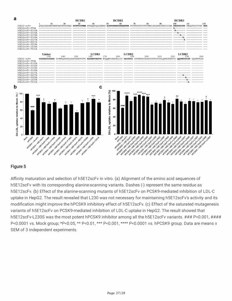
A�nity maturation of h5E12scFv in vitroTo further enhance the a�nity and bioactivity of h5E12scFv, we �rstly identi�ed the critical residues by alanine-scanning mutagenesis of several residues including F99, H100, D102, D104, F106, D107, R227, P229, L230,T231, respectively, and 10 mutants (Fig. 5a) named h5E12scFv-F99A, h5E12scFv-H100A, h5E12scFv-D102A,h5E12scFv-D104A, h5E12scFv-F106A, h5E12scFv-D107A, h5E12scFv-R227A, h5E12scFv-P229A, h5E12scFv-L230A and h5E12scFv-T231A were puri�ed (data not shown). The biological activity of these mutants wascompared by measuring the changes of LDL uptake using HepG2 cell-based assay. The results (Fig. 5b) showedthat h5E12scFv-D102A and h5E12scFv-D107A exhibited scarcely any PCSK9 inhibitory effect, indicating thatD102 and D107 was an essential residue for maintaining h5E12scFv’ activity and should be retained. Besides,h5E12scFv-L230A displayed stronger biological activity than parental antibody h5E12scFv, suggesting that themutation of L230 to other residues might improve the hPCSK9 inhibitory effect of h5E12scFv.
Secondly, site-saturated mutagenesis experiments were carried out on the L230 residue of h5E12scFv, and thecorresponding mutants (Fig. S6) were puri�ed and screened by LDL uptake assay (Fig. 5c). It was shown that theLDL uptake levels were effectively enhanced by h5E12scFv-L230A, h5E12scFv-L230S and h5E12scFv-L230G,and the LDL uptake levels in these three groups were restored to the comparable levels as in the Ali-scFv group.Thus, we chose h5E12scFv-L230A, h5E12scFv-L230S, and h5E12scFv-L230G for further construction of the full-length antibodies and in vivo functional studies.
Generation and characterization of full-length anti-PCSK9antibodiesThe full-length format of anti-PCSK9 antibodies was constructed by fusing the VH and VL with a modi�ed humanIgG1 heavy-chain constant region and kappa light chain constant region, respectively. The heavy (GenBankaccession number: MW715631) and light chain DNA sequence (GenBank accession number: MW715632,
Page 12/29
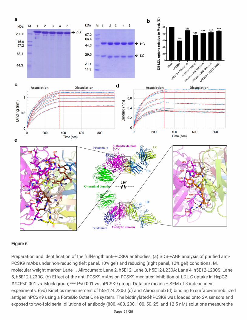
MW725291, MW725292, MW725293) of full-length antibodies were then inserted into the pTT5 vector (Fig. S7)and co-transfected into CHO-3E7 cells for transient expression. After puri�cation by protein A a�nitychromatography columns, the obtained mAbs were veri�ed by SDS-PAGE under non-reducing and reducingconditions (Fig. 6a).
Subsequently, Dil-LDL uptake assay was performed as described above to test the hPCSK9 inhibitory effect ofpuri�ed mAbs. The results (Fig. 6b) showed that all the generated mAbs, including h5E12, h5E12-L230A, h5E12-L230S, h5E12-L230G, could signi�cantly inhibit PCSK9-induced decrease in Dil-LDL uptake of HepG2 cells(P<0.001). Among them, h5E12-L230G exhibited the most potent activity in enhancing the LDL-C uptake levels,which was comparable to the positive control Alirocumab.
We further detect and compare the a�nity constant of h5E12-L230G and Alirocumab to hPCSK9 using theForteBio Octet QKe system. As shown in Fig. 6c, d and Table S4, h5E12-L230G displayed a moderate slowerassociation rate (kon= 2.81 × 10⁴ M-1s-1) and a slightly slower dissociation rate (koff = 4.84 × 10⁻⁵ s-1) than
Alirocumab (kon= 8.04 × 10⁴ M-1s-1, koff = 6.87 × 10⁻⁵ s-1), thus yielding a ~2-fold lower a�nity (KD = 1.72 × 10⁻⁹ M
vs. 8.54 × 10⁻¹⁰ M) as compared to Alirocumab. It can be concluded that the slower dissociation rate (koff)between h5E12-L230G and hPCSK9 results in a longer binding period, which may enhance the hPCSK9 inhibitoryof h5E12-L230G to the comparable level of Alirocumab.
In addition, to further elucidate the epitope–paratope interaction details, the 3D structure of Fab region of h5E12-L230G (Fig. S8a) was built based on the top-ranked template crystal structure (PDB ID:6DW2). Ramachandranplot (Fig. S8b) of the modeled h5E12-L230G revealed that 97.91% of the residues were in the most favorable andallowed regions, indicating that the modeled structure was suitable for the molecular docking analysis. Thefollowing docking results (Fig. 6e) suggested that h5E12-L230G binds to the catalytic domain of hPCSK9, andboth the heavy-chain and light-chain variable domain contributed to protein-ligand interaction. It appeared thatas many as fourteen residues (S25, T28, W33, N55, H100, D102, Y103, D107, Y108 in heavy chain and Y49, S50,Y53, R54, S56 in light chain) in h5E12-L230G and up to fourteen residues (A168, L179, E181, E197, G198, R199,V200, V202, R237, D238, K243, S246, P279, S401 in catalytic domain) in hPCSK9 involved in the interactions,forming �fteen hydrogen bonds and two ionic bonds in the binding pocket (Table S5). Finally, the binding freeenergy (ΔGbind) of the illustrated docking mode calculated using the MM-GBSA method was as low as -54.97kcal/mol, indicating the h5E12-L230G showed high binding strength with hPCSK9.
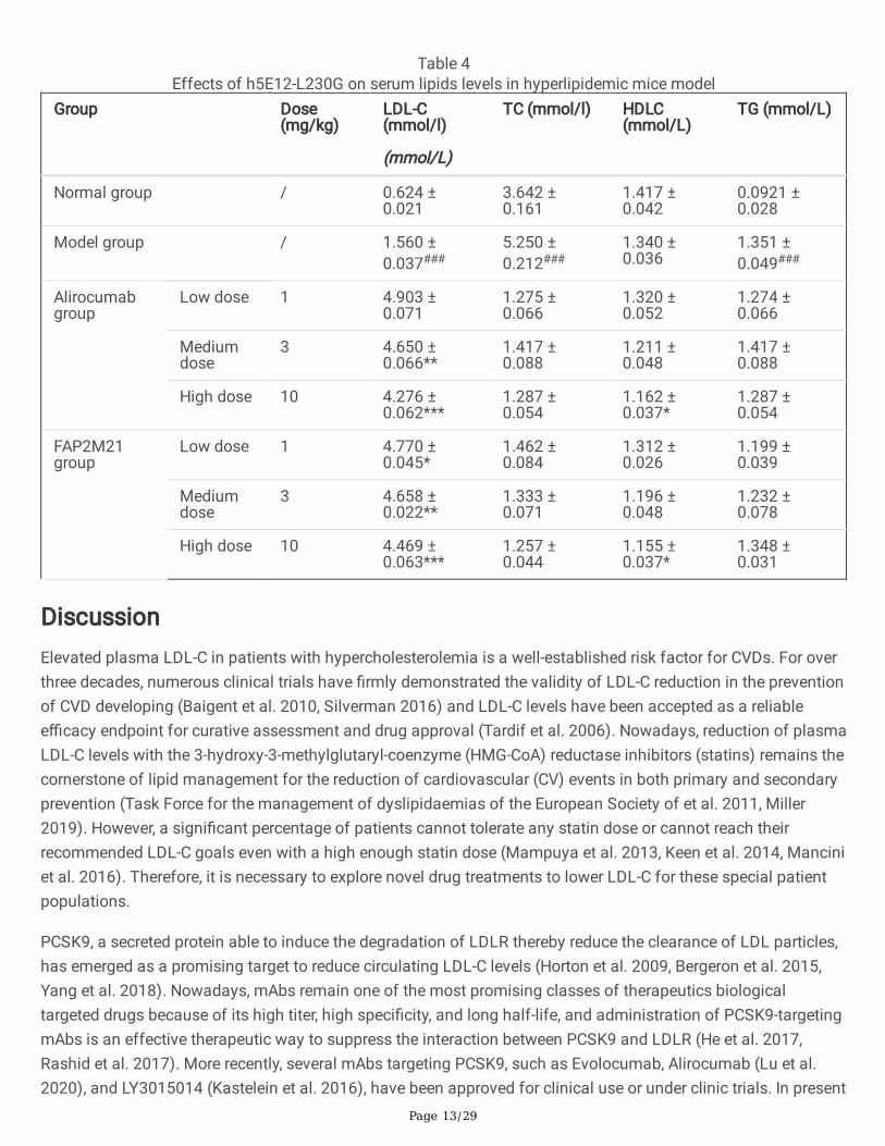
Hypolipidemic effect of h5E12-L230G in mice over-expressinghPCSK9We next evaluated the lipid-lowering e�cacy of h5E12-L230G in vivo. The mice over-expressing hPCSK9 wasestablished through hydrodynamic delivery (HDD) of 50 µg naked plasmid DNA (pTT5-hPCSK9) in 2 ml normalsaline. On day 6 after HDD, the mice in the treatment groups were given a single tail i.v. injection of h5E12-L230Gwith various doses. 18 hours after administration, the levels of LDLR in liver tissues were dose-dependently up-regulated as assessed by Western blot (Fig. 7b) and Immuno�uorescence (Fig. 7c). Besides, it was shown thattreatment with h5E12-L230G at 1, 3, and 10 mg/kg resulted in a 5.8% (P>0.05), 30.1% (P<0.01), and 36.2%(P<0.001) decrease in serum LDL-C relative to the model group, respectively (Fig. 7a and Table 4), and h5E12-L230G treatment could also signi�cantly lowered the levels of serum total cholesterol (TC) and triglyceride (TG),but did not signi�cantly affect serum High-density lipoprotein (HDL-C) levels (Table 4).
Page 13/29
Table 4Effects of h5E12-L230G on serum lipids levels in hyperlipidemic mice model
Group Dose(mg/kg)
LDL-C(mmol/l)
(mmol/L)
TC (mmol/l) HDLC(mmol/L)
TG (mmol/L)
Normal group / 0.624 ±0.021
3.642 ±0.161
1.417 ±0.042
0.0921 ±0.028
Model group / 1.560 ±0.037###
5.250 ±0.212###
1.340 ±0.036
1.351 ±0.049###
Alirocumabgroup
Low dose 1 4.903 ±0.071
1.275 ±0.066
1.320 ±0.052
1.274 ±0.066
Mediumdose
3 4.650 ±0.066**
1.417 ±0.088
1.211 ±0.048
1.417 ±0.088
High dose 10 4.276 ±0.062***
1.287 ±0.054
1.162 ±0.037*
1.287 ±0.054
FAP2M21group
Low dose 1 4.770 ±0.045*
1.462 ±0.084
1.312 ±0.026
1.199 ±0.039
Mediumdose
3 4.658 ±0.022**
1.333 ±0.071
1.196 ±0.048
1.232 ±0.078
High dose 10 4.469 ±0.063***
1.257 ±0.044
1.155 ±0.037*
1.348 ±0.031
DiscussionElevated plasma LDL-C in patients with hypercholesterolemia is a well-established risk factor for CVDs. For overthree decades, numerous clinical trials have �rmly demonstrated the validity of LDL-C reduction in the preventionof CVD developing (Baigent et al. 2010, Silverman 2016) and LDL-C levels have been accepted as a reliablee�cacy endpoint for curative assessment and drug approval (Tardif et al. 2006). Nowadays, reduction of plasmaLDL-C levels with the 3-hydroxy-3-methylglutaryl-coenzyme (HMG-CoA) reductase inhibitors (statins) remains thecornerstone of lipid management for the reduction of cardiovascular (CV) events in both primary and secondaryprevention (Task Force for the management of dyslipidaemias of the European Society of et al. 2011, Miller2019). However, a signi�cant percentage of patients cannot tolerate any statin dose or cannot reach theirrecommended LDL-C goals even with a high enough statin dose (Mampuya et al. 2013, Keen et al. 2014, Manciniet al. 2016). Therefore, it is necessary to explore novel drug treatments to lower LDL-C for these special patientpopulations.
PCSK9, a secreted protein able to induce the degradation of LDLR thereby reduce the clearance of LDL particles,has emerged as a promising target to reduce circulating LDL-C levels (Horton et al. 2009, Bergeron et al. 2015,Yang et al. 2018). Nowadays, mAbs remain one of the most promising classes of therapeutics biologicaltargeted drugs because of its high titer, high speci�city, and long half-life, and administration of PCSK9-targetingmAbs is an effective therapeutic way to suppress the interaction between PCSK9 and LDLR (He et al. 2017,Rashid et al. 2017). More recently, several mAbs targeting PCSK9, such as Evolocumab, Alirocumab (Lu et al.2020), and LY3015014 (Kastelein et al. 2016), have been approved for clinical use or under clinic trials. In present
Page 14/29
study, we chose Alirocumab as the positive control and expected to generate a potent anti-hPCSK9 mAb withgood druggability utilizing hybridoma-based methods.
Using the hybridoma technique, we initially generated a murine mAb targeting hPCSK9 (named m5E12), whichcould restore PCSK9-induced LDLR degradation and inhibit LDL uptake in HepG2 cells (Fig. 2c and 2d). Whatneeds illustration is that the humanization and a�nity maturation of m5E12 here were performed using the scFvformat instead of using the full-length antibody format, this is because scFv proteins can be easily obtainedfrom E. coli expression system, which has the advantage of fast-growing, inexpensive and easy manipulation(Ahmad et al. 2012, Agha Amiri et al. 2017).
Speci�cally, to reduce the immunogenicity of mouse antibody, m5E12 was humanized by means of CDR graftingand back-mutation methods (Verhoeyen 1988, Foote 1992, Pedersen 1994, De Haard 1999), generating twohumanized m5E12 (h5E12scFv and h5E12scFv-bm) in the respective scFv format. Intriguingly, although theRMSD analysis showed that the humanized scFvs converged to the same conformation (Fig. 3c-e), their a�nity(KD) and biological activity were different. It was revealed that h5E12scFv displayed a higher a�nity (KD) andhPCSK9 inhibitory effect than h5E12scFv-bm (Fig. 4b-d). Thus, we chose h5E12scFv for further study. To furtherimprove the antibody’s a�nity and bioactivity, alanine scanning mutagenesis was �rstly conducted on theHCDR3 and LCDR3 of h5E12scFv by one-step PCR technology (Zheng 2004). Ten residues in HCDR3 and LCDR3were selected to mutated to alanine, and we found that the L230A variant, termed h5E12scFv-L230A, was able torestore LDL uptake to the level slightly higher than that of parental antibody h5E12scFv (Fig. 5b). Therefore, weassumed that the alteration of L230 residues in h5E12scFv might help to improve its activity, and fortunately, thefollowing site-saturated mutagenesis experiment proved this hypothesis (Fig. 5c).
To date, the majority of therapeutic antibodies developed are full-length IgG, and the full-length IgG molecules areconsidered one of the most suitable formats for clinical applications (Yang et al. 2017). Therefore, the selectedoptimized scFv mutants (h5E12scFv-L230A, h5E12scFv-L230G, h5E12scFv-L230S) were then reformatted intofull-length Fc-silenced IgG1 format (Wines 2000, Hessell 2007, Dick 2008, Liu 2008, Jefferis 2009, Jacobsen2017), expressed transiently in CHO mammalian cells and puri�ed from culture supernatant. Of these full-lengthantibodies, h5E12-L230G could reverse LDL uptake to the similar levels of Alirocumab (Fig. 6b) and bind tohPCSK9 with a 1.41-fold slower dissociation rate (koff = 4.84 × 10⁻⁵ s−1) than Alirocumab (koff = 6.87 × 10⁻⁵ s−1).
Molecular Modeling and docking techniques are wildly used to predict the binding mode of an antibody with itsprotein target (Kosztyu et al. 2019, Vahed et al. 2020). In this work, the docking results (Fig. 6e and Table S5)showed that up to fourteen amino acid residues in h5E12-L230G form the paratope that interacts with fourteenamino acid residues in hPCSK9’s epitope, creating as many as �fteen hydrogen bonds and two ionic bonds onthe interaction site. Hence, the multiple interactions between the CDRs of h5E12-L230G and the catalytic domainof PCSK9 resulted in a tight binding of h5E12-L230G to hPCSK9, which might well explain the excellent PCSK9inhibitory activity of h5E12-L230G.
It is also remarkable that a targeted therapeutic drug requires a long period on the target to be effective, hencethe fairly slow dissociation rate of h5E12-L230G to hPCSK9 is desirable to facilitate prolonged duration ofantibody-antigen interaction and is especially important for its in vivo targeting applications (Axworthy et al.2000, Chmura et al. 2001, Bottermann et al. 2016). As expected, h5E12-L230G showed considerable potency inraising LDLR expression in mice liver (Fig. 7b) and effectively reduced serum LDL-C and TC levels in
Page 15/29
hyperlipidemic C57BL/6 mice in a dose-dependent manner (Table 4), with high potency comparable to thepositive control Alirocumab.
In conclusion, h5E12-L230G is a humanized high-a�nity hPCSK9 blocking antibody which binds to the catalyticdomain of hPCSK9 with a slow dissociation rate and effectively inhibits PCSK9-mediated LDLR degradation,thereby signi�cantly promotes LDL-C uptake in HepG2 cells and reduces the serum LDL-C and total cholesterol(TC) levels in hyperlipidemic mouse model. The data demonstrate that h5E12-L230G has the potential to serveas a therapeutic antibody targeting PCSK9 for treating hypercholesterolemia and relevant cardiovasculardiseases.
AbbreviationsCDR, complementarity-determining region; CHO, Chinese hamster ovary cells; CVDs, cardiovascular diseases;DMEM, Dulbecco’s modi�ed Eagle’s medium; Escherichia coli, E. Coli. ELISA, Enzyme-linked immunosorbentassay; FBS, fetal bovine serum. HC, heavy chain; HDL-C, High-density lipoprotein receptor; IPTG, isopropyl-β-galactosidase. LDLR, low-density lipoprotein receptor; LDL-C, low-density lipoprotein cholesterol; LC, light chain;MM/GBSA, Molecular Mechanics/GB Surface Area; mAb, Monoclonal antibody; PCSK9, Proprotein convertasesubtilisin/kexin type 9; RMSD, Root mean square deviation; scFv, single-chain variable fragment; TC, totalcholesterol; TG, triglyceride; VH, variable region of heavy chain; VL, variable region of light chain.
DeclarationsEthics approval and consent to participate
Experiments were performed under a project license (NO.: 201601179) granted by the Animal Ethics Committeesof China Pharmaceutical University, in compliance with national guidelines for the care and use of animals.
Consent to participate
Not applicable.
Consent for publication
Authors consent to publish with no con�ict of interest.
Availability of data and materials
All data generated or analysed during this study are included in this manuscript and supplementary information.
Competing interests
The authors have no con�icts of interest to declare.
Funding
This work was funded by National Fund for Major Projects of China (2009ZX09103-653; 2013ZX09301303-006;2018ZX09301035), The Priority Academic Program Development of Jiangsu Higher Education Institutions
Page 16/29
(PAPD), National Fund for Fostering Talents of Basic Science (NFFTBS, 3050040016), China PharmaceuticalUniversity “Double First-Class” project (CPU2018GY15).
Authors’ contributions
S. Tan conceptualized the project and reviewing the manuscript. Z. Bai planned for the project, performed thehybridoma experiment and wrote the �rst draft of the manuscript. M. Xu designed research and edited themanuscript. Y. Mei performed the humanization experiment and participated in drafting the article. T. Hu puri�edthe antibodies, analyzed the data and co-wrote the manuscript. P. Zhang helped Y. Mei and T. Hu with analysis ofmolecular modeling and docking studies. M. Chen expressed and puri�ed the antigen. W. Lv helped M. Chen inantigen preparation. C. Lu preparation the antigen.
Acknowledgements
Not applicable.
References1. Abifadel, M., M. Varret, J. P. Rabes, D. Allard, K. Ouguerram, M. Devillers, C. Cruaud, S. Benjannet, L. Wickham,
D. Erlich, A. Derre, L. Villeger, M. Farnier, I. Beucler, E. Bruckert, J. Chambaz, B. Chanu, J. M. Lecerf, G. Luc, P.Moulin, J. Weissenbach, A. Prat, M. Krempf, C. Junien, N. G. Seidah and C. Boileau (2003). "Mutations inPCSK9 cause autosomal dominant hypercholesterolemia." Nat Genet 34(2): 154-156. DOI:10.1038/ng1161
2. Agha Amiri, S., S. Shahhosseini, N. Zarei, D. Khorasanizadeh, E. Aminollahi, F. Rezaie, M. Zargari, M. Aziziand V. Khalaj (2017). "A novel anti-CD22 scFv-apoptin fusion protein induces apoptosis in malignant B-cells."AMB Express 7(1): 112. DOI:10.1186/s13568-017-0410-5
3. Ahmad, Z. A., S. K. Yeap, A. M. Ali, W. Y. Ho, N. B. Alitheen and M. Hamid (2012). "scFv antibody: principlesand clinical application." Clin Dev Immunol 2012: 980250. DOI:10.1155/2012/980250
4. Axworthy, D. B., J. M. Reno, M. D. Hylarides, R. W. Mallett, L. J. Theodore, L. M. Gustavson, F. M. Su, L. J.Hobson, P. L. Beaumier and A. R. Fritzberg (2000). "Cure of human carcinoma xenografts by a single dose ofpretargeted yttrium-90 with negligible toxicity." Proceedings of the National Academy of Sciences 97(4):1802. DOI:10.1073/pnas.97.4.1802
5. Baigent, C., L. Blackwell, J. Emberson, L. E. Holland, C. Reith, Bhala, N., , R. Peto, Barnes, E., K. H., A., , J.Simes and R. Collins (2010). "E�cacy and safety of more intensive lowering of LDL cholesterol: a meta-analysis of data from 170 000 participants in 26 randomised trials." The Lancet 376(9753): 1670-1681.DOI:10.1016/s0140-6736(10)61350-5
�. Beatty, J. D., B. G. Beatty and W. G. Vlahos (1987). "Measurement of monoclonal antibody a�nity by non-competitive enzyme immunoassay." J Immunol Methods 100(1): 173-179.DOI:https://doi.org/10.1016/0022-1759(87)90187-6
7. Bergeron, N., B. A. Phan, Y. Ding, A. Fong and R. M. Krauss (2015). "Proprotein convertase subtilisin/kexintype 9 inhibition: a new therapeutic mechanism for reducing cardiovascular disease risk." Circulation132(17): 1648-1666. DOI:10.1161/CIRCULATIONAHA.115.016080
�. Bienert, S., A. Waterhouse, T. A. de Beer, G. Tauriello, G. Studer, L. Bordoli and T. Schwede (2017). "TheSWISS-MODEL Repository-new features and functionality." Nucleic Acids Res 45(D1): D313-D319.
Page 17/29
DOI:10.1093/nar/gkw1132
9. Blanco-Toribio, A., A. Alvarez-Cienfuegos, N. Sainz-Pastor, N. Merino, M. Compte, L. Sanz, F. J. Blanco and L.Alvarez-Vallina (2015). "Bacterial secretion of soluble and functional trivalent scFv-based N-terminaltrimerbodies." AMB Express 5(1): 137. DOI:10.1186/s13568-015-0137-0
10. Bottermann, M., H. E. Lode, R. E. Watkinson, S. Foss, I. Sandlie, J. T. Andersen and L. C. James (2016)."Antibody-antigen kinetics constrain intracellular humoral immunity." Sci Rep 6: 37457.DOI:10.1038/srep37457
11. Brown, M. S. and J. L. Goldstein (1986). "A receptor-mediated pathway for cholesterol homeostasis." Science232(4746): 34-47. DOI:10.1126/science.3513311
12. Chan, K. T., S. C. Cheng, H. Xie and Y. Xie (2001). "A humanized monoclonal antibody constructed fromintronless expression vectors targets human hepatocellular carcinoma cells." Biochem Biophys ResCommun 284(1): 157-167. DOI:10.1006/bbrc.2001.4837
13. Chiu, M. L. and G. L. Gilliland (2016). "Engineering antibody therapeutics." Curr Opin Struct Biol 38: 163-173.DOI:10.1016/j.sbi.2016.07.012
14. Chmura, A. J., M. S. Orton and C. F. Meares (2001). "Antibodies with in�nite a�nity." Proc Natl Acad Sci U S A98(15): 8480-8484. DOI:10.1073/pnas.151260298
15. Cohen, J. C., E. Boerwinkle, T. H. Mosley, Jr. and H. H. Hobbs (2006). "Sequence variations in PCSK9, lowLDL, and protection against coronary heart disease." N Engl J Med 354(12): 1264-1272.DOI:10.1056/NEJMoa054013
1�. Copeland, R. A. (2010). "The dynamics of drug-target interactions: drug-target residence time and its impacton e�cacy and safety." Expert Opinion on Drug Discovery 5(4): 305-310. DOI:10.1517/17460441003677725
17. Cunningham, D., D. E. Danley, K. F. Geoghegan, M. C. Griffor, J. L. Hawkins, T. A. Subashi, A. H. Varghese, M.J. Ammirati, J. S. Culp, L. R. Hoth, M. N. Mansour, K. M. McGrath, A. P. Seddon, S. Shenolikar, K. J. Stutzman-Engwall, L. C. Warren, D. Xia and X. Qiu (2007). "Structural and biophysical studies of PCSK9 and itsmutants linked to familial hypercholesterolemia." Nat Struct Mol Biol 14(5): 413-419.DOI:10.1038/nsmb1235
1�. De Haard, H., B. Kazemier, V. D. B. Arie, P. Oudshoorn, P. Boender, J. W. Arends and B. J. I. Van Gemen (1999)."Vernier zone residue 4 of mouse subgroup II kappa light chains is a critical determinant for antigenrecognition." Immunotechnology 4(3-4): 203-215.
19. Dick, L. W., Jr., D. Qiu, D. Mahon, M. Adamo and K. C. Cheng (2008). "C-terminal lysine variants in fullyhuman monoclonal antibodies: investigation of test methods and possible causes." Biotechnology andBioengineering 100(6): 1132-1143. DOI:10.1002/bit.21855
20. Egom, E. E., R. B. Pharithi, S. Hesse, N. Starr, R. Armstrong, H. M. Sulaiman, K. Gazdikova, I. Mozos, M.Caprnda, P. Kubatka, P. Kruzliak, B. Khan, L. Gaspar and V. M. G. Maher (2019). "Latest Updates on LipidManagement." High Blood Press Cardiovasc Prev 26(2): 85-100. DOI:10.1007/s40292-019-00306-8
21. Elgundi, Z., M. Reslan, E. Cruz, V. Sifniotis and V. Kayser (2017). "The state-of-play and future of antibodytherapeutics." Adv Drug Deliv Rev 122: 2-19. DOI:10.1016/j.addr.2016.11.004
22. Ersching, J., A. Efeyan, L. Mesin, J. T. Jacobsen, G. Pasqual, B. C. Grabiner, D. Dominguez-Sola, D. M.Sabatini and G. D. Victora (2017). "Germinal Center Selection and A�nity Maturation Require DynamicRegulation of mTORC1 Kinase." Immunity 46(6): 1045-1058 e1046. DOI:10.1016/j.immuni.2017.06.005
Page 18/29
23. Farnier, M. (2018). "Lowering low-density lipoprotein cholesterol by PCSK9 inhibition in patients withdiabetes on insulin therapy: is it e�cacious and safe?" Ann Transl Med 6(3): 60.DOI:10.21037/atm.2018.01.02
24. Foote, J. and G. J. J. o. M. B. Winter (1992). "Antibody framework residues affecting the conformation of thehypervariable loops." J Mol Biol 224(2): 0-499.
25. Gu, L., P. Ye, H. Li, Y. Wang, Y. Xu, Q. Tian, G. Lei, C. Zhao, Z. Gao, W. Zhao and S. Tan (2019). "Lunasinattenuates oxidant-induced endothelial injury and inhibits atherosclerotic plaque progression in ApoE(-/-)mice by up-regulating heme oxygenase-1 via PI3K/Akt/Nrf2/ARE pathway." FASEB J 33(4): 4836-4850.DOI:10.1096/fj.201802251R
2�. H, A. and T. K (2015). "Thermodynamics of antibody-antigen interaction revealed by mutation analysis ofantibody variable regions." Journal of Biochemistry 158(1): 1-13. DOI:10.1093/jb/mvv049
27. Haidar, J. N., Q. A. Yuan, L. Zeng, M. Snavely, X. Luna, H. Zhang, W. Zhu, D. L. Ludwig and Z. Zhu (2012). "Auniversal combinatorial design of antibody framework to graft distinct CDR sequences: a bioinformaticsapproach." Proteins 80(3): 896-912. DOI:10.1002/prot.23246
2�. He, N. Y., Q. Li, C. Y. Wu, Z. Ren, Y. Gao, L. H. Pan, M. M. Wang, H. Y. Wen, Z. S. Jiang, Z. H. Tang and L. S. Liu(2017). "Lowering serum lipids via PCSK9-targeting drugs: current advances and future perspectives." ActaPharmacol Sin 38(3): 301-311. DOI:10.1038/aps.2016.134
29. Hernández, G., V. G. Osnaya and X. Pérez-Martínez (2019). "Conservation and Variability of the AUG InitiationCodon Context in Eukaryotes." Trends in Biochemical Sciences 44(12): 1009-1021.DOI:https://doi.org/10.1016/j.tibs.2019.07.001
30. Hessell, A. J., L. Hangartner, M. Hunter, C. E. Havenith, F. J. Beurskens, J. M. Bakker, C. M. Lanigan, G.Landucci, D. N. Forthal, P. W. Parren, P. A. Marx and D. R. Burton (2007). "Fc receptor but not complementbinding is important in antibody protection against HIV." Nature 449(7158): 101-104.DOI:10.1038/nature06106
31. Holla, O. L., J. Cameron, K. E. Berge, T. Ranheim and T. P. Leren (2007). "Degradation of the LDL receptors byPCSK9 is not mediated by a secreted protein acted upon by PCSK9 extracellularly." BMC Cell Biol 8: 9.DOI:10.1186/1471-2121-8-9
32. Horton, J. D., J. C. Cohen and H. H. Hobbs (2009). "PCSK9: a convertase that coordinates LDL catabolism." JLipid Res 50 Suppl: S172-177. DOI:10.1194/jlr.R800091-JLR200
33. Inoue, H., A. Suganami, I. Ishida, Y. Tamura and Y. Maeda (2013). "A�nity maturation of a CDR3-grafted VHHusing in silico analysis and surface plasmon resonance." J Biochem 154(4): 325-332.DOI:10.1093/jb/mvt058
34. Jacobsen, F. W., R. Stevenson, C. Li, H. Salimi-Moosavi, L. Liu, J. Wen, Q. Luo, K. Daris, L. Buck, S. Miller, S. Y.Ho, W. Wang, Q. Chen, K. Walker, J. Wypych, L. Narhi and K. Gunasekaran (2017). "Engineering an IgGScaffold Lacking Effector Function with Optimized Developability." J Biol Chem 292(5): 1865-1875.DOI:10.1074/jbc.M116.748525
35. Jefferis, R. (2009). "Recombinant antibody therapeutics: the impact of glycosylation on mechanisms ofaction." Trends Pharmacol Sci 30(7): 356-362. DOI:10.1016/j.tips.2009.04.007
3�. Kastelein, J. J., S. E. Nissen, D. J. Rader, G. K. Hovingh, M. D. Wang, T. Shen and K. A. Krueger (2016). "Safetyand e�cacy of LY3015014, a monoclonal antibody to proprotein convertase subtilisin/kexin type 9 (PCSK9):
Page 19/29
a randomized, placebo-controlled Phase 2 study." Eur Heart J 37(17): 1360-1369.DOI:10.1093/eurheartj/ehv707
37. Keen, H. I., J. Krishnarajah, T. R. Bates and G. F. Watts (2014). "Statin myopathy: the �y in the ointment forthe prevention of cardiovascular disease in the 21st century?" Expert Opin Drug Saf 13(9): 1227-1239.DOI:10.1517/14740338.2014.937422
3�. Kim, H. Y., A. Stojadinovic and M. J. Izadjoo (2014). "Immunization, hybridoma generation, and selection formonoclonal antibody production." Methods Mol Biol 1131: 33-45. DOI:10.1007/978-1-62703-992-5_3
39. Ko, B. K., S. Choi, L. G. Cui, Y. H. Lee, I. S. Hwang, K. T. Kim, H. Shim and J. S. Lee (2015). "A�nity Maturationof Monoclonal Antibody 1E11 by Targeted Randomization in CDR3 Regions Optimizes Therapeutic AntibodyTargeting of HER2-Positive Gastric Cancer." Plos One 10(7): e0134600.
40. KÖHler, G. and C. Milstein (1975). "Continuous cultures of fused cells secreting antibody of prede�nedspeci�city." Nature 256(5517): 495-497. DOI:10.1038/256495a0
41. Kosztyu, P., M. Kuchar, J. Cerny, L. Barkocziova, M. Maly, H. Petrokova, L. Czernekova, V. Liskova, L. RaskovaKafkova, P. Knotigova, J. Masek, J. Turanek, P. Maly and M. Raska (2019). "Proteins mimicking epitope ofHIV-1 virus neutralizing antibody induce virus-neutralizing sera in mice." EBioMedicine 47: 247-256.DOI:10.1016/j.ebiom.2019.07.015
42. Levin, A. M. and G. A. Weiss (2006). "Optimizing the a�nity and speci�city of proteins with moleculardisplay." Mol Biosyst 2(1): 49-57. DOI:10.1039/b511782h
43. Li, S., H. Shen and Q. Shu (2019). "S3Ab, a novel antibody targeting B lymphocytes, is a potential therapeuticagent for B-lineage malignancies." J Drug Target 27(10): 1053-1060. DOI:10.1080/1061186X.2019.1584809
44. Lindholm, M. W., J. Elmen, N. Fisker, H. F. Hansen, R. Persson, M. R. Moller, C. Rosenbohm, H. Orum, E. M.Straarup and T. Koch (2012). "PCSK9 LNA antisense oligonucleotides induce sustained reduction of LDLcholesterol in nonhuman primates." Mol Ther 20(2): 376-381. DOI:10.1038/mt.2011.260
45. Liu, H., G. Gaza-Bulseco, D. Faldu, C. Chumsae and J. Sun (2008). "Heterogeneity of monoclonal antibodies."J Pharm Sci 97(7): 2426-2447. DOI:10.1002/jps.21180
4�. Lu, R. M., Y. C. Hwang, I. J. Liu, C. C. Lee, H. Z. Tsai, H. J. Li and H. C. Wu (2020). "Development of therapeuticantibodies for the treatment of diseases." J Biomed Sci 27(1): 1. DOI:10.1186/s12929-019-0592-z
47. Lu, Y., S. Xiao, M. Yuan, Y. Gao, J. Sun and C. Xue (2018). "Using overlap-extension PCR technique to fusinggenes for constructing recombinant plasmids." J Basic Microbiol 58(3): 273-276.DOI:10.1002/jobm.201700455
4�. Luo, S., X. Deng, Z. Xie, J. Huang, M. Zhang, M. Li, L. Xie, D. Li, Q. Fan, S. Wang, T. Zeng, Y. Zhang and Z. Xie(2020). "Production and identi�cation of monoclonal antibodies and development of a sandwich ELISA fordetection of the H3-subtype avian in�uenza virus antigen." AMB Express 10(1): 49. DOI:10.1186/s13568-020-00988-7
49. Ly, K., Y. G. Saavedra, M. Canuel, S. Routhier, R. Desjardins, J. Hamelin, J. Mayne, C. Lazure, N. G. Seidah andR. Day (2014). "Annexin A2 reduces PCSK9 protein levels via a translational mechanism and interacts withthe M1 and M2 domains of PCSK9." J Biol Chem 289(25): 17732-17746. DOI:10.1074/jbc.M113.541094
50. Mampuya, W. M., D. Frid, M. Rocco, J. Huang, D. M. Brennan, S. L. Hazen and L. Cho (2013). "Treatmentstrategies in patients with statin intolerance: the Cleveland Clinic experience." Am Heart J 166(3): 597-603.DOI:10.1016/j.ahj.2013.06.004
Page 20/29
51. Mancini, G. B., S. Baker, J. Bergeron, D. Fitchett, J. Frohlich, J. Genest, M. Gupta, R. A. Hegele, D. Ng, G. J.Pearson, J. Pope and A. Y. Tashakkor (2016). "Diagnosis, Prevention, and Management of Statin AdverseEffects and Intolerance: Canadian Consensus Working Group Update (2016)." Can J Cardiol 32(7 Suppl):S35-65. DOI:10.1016/j.cjca.2016.01.003
52. Miao, C. H., A. R. Thompson, K. Loeb and X. Ye (2001). "Long-term and therapeutic-level hepatic geneexpression of human factor IX after naked plasmid transfer in vivo." Mol Ther 3(6): 947-957.DOI:10.1006/mthe.2001.0333
53. Mihaylova, B., J. Emberson, L. Blackwell, A. Keech, J. Simes, E. Barnes, M. Voysey, A. Gray, R. Collins and C.Baigent (2012). "The effects of lowering LDL cholesterol with statin therapy in people at low risk of vasculardisease: meta-analysis of individual data from 27 randomised trials." The Lancet 380(9841): 581-590.DOI:10.1016/s0140-6736(12)60367-5
54. Miller, M. (2019). "ACC/AHA lipids & ASCVD guidelines: 2018 update." Metabolism 99: 116-118.DOI:10.1016/j.metabol.2019.03.008
55. Mitchell, T., G. Chao, D. Sitkoff, F. Lo, H. Monshizadegan, D. Meyers, S. Low, K. Russo, R. DiBella, F. Denhez,M. Gao, J. Myers, G. Duke, M. Witmer, B. Miao, S. P. Ho, J. Khan and R. A. Parker (2014). "Pharmacologicpro�le of the Adnectin BMS-962476, a small protein biologic alternative to PCSK9 antibodies for low-densitylipoprotein lowering." J Pharmacol Exp Ther 350(2): 412-424. DOI:10.1124/jpet.114.214221
5�. Morrison, S. L., M. J. Johnson, L. A. Herzenberg and V. T. Oi (1984). "Chimeric human antibody molecules:mouse antigen-binding domains with human constant region domains." Proc Natl Acad Sci U S A 81(21):6851-6855.
57. Nassoury, N., D. A. Blasiole, A. Tebon Oler, S. Benjannet, J. Hamelin, V. Poupon, P. S. McPherson, A. D. Attie, A.Prat and N. G. Seidah (2007). "The cellular tra�cking of the secretory proprotein convertase PCSK9 and itsdependence on the LDLR." Tra�c 8(6): 718-732. DOI:10.1111/j.1600-0854.2007.00562.x
5�. Pedersen, J. T., A. H. Henry, S. J. Searle, B. C. Guild, M. Roguska and A. R. Rees (1994). "Comparison ofSurface Accessible Residues in Human and Murine Immunoglobulin Fv Domains." J Mol Biol 235(3): 959-973.
59. Poirier, S., G. Mayer, V. Poupon, P. S. McPherson, R. Desjardins, K. Ly, M. C. Asselin, R. Day, F. J. Duclos, M.Witmer, R. Parker, A. Prat and N. G. Seidah (2009). "Dissection of the endogenous cellular pathways ofPCSK9-induced low density lipoprotein receptor degradation: evidence for an intracellular route." J Biol Chem284(42): 28856-28864. DOI:10.1074/jbc.M109.037085
�0. Rader, D. J. and A. Daugherty (2008). "Translating molecular discoveries into new therapies foratherosclerosis." Nature 451(7181): 904-913. DOI:10.1038/nature06796
�1. nature06796 [pii]
�2. Rashid, H., I. T. Meredith and A. Nasis (2017). "PCSK9 Monoclonal Antibodies in 2016: Current Status andFuture Challenges." Heart Lung Circ 26(8): 786-798. DOI:10.1016/j.hlc.2016.12.005
�3. Ren, K., B. Wang and Q. Qi (2021). "Development of a new EGFR antibody antagonist which exhibitspotential biological effects against laryngeal cancer." Ann Transl Med 9(12): 964. DOI:10.21037/atm-21-1839
�4. Safdari, Y., S. Farajnia, M. Asgharzadeh and M. Khalili (2013). "Antibody humanization methods – a reviewand update." Biotechnology and Genetic Engineering Reviews 29(2): 175-186.DOI:10.1080/02648725.2013.801235
Page 21/29
�5. Schmit, D., D. Fliser and T. Speer (2019). "Proprotein convertase subtilisin/kexin type 9 in kidney disease."Nephrol Dial Transplant. DOI:10.1093/ndt/gfz122
��. Schwartz, G. G., L. Bessac, L. G. Berdan, D. L. Bhatt, V. Bittner, R. Diaz, S. G. Goodman, C. Hanotin, R. A.Harrington, J. W. Jukema, K. W. Mahaffey, A. Moryusef, R. Pordy, M. T. Roe, T. Rorick, W. J. Sasiela, C.Shirodaria, M. Szarek, J. F. Tamby, P. Tricoci, H. White, A. Zeiher and P. G. Steg (2014). "Effect of alirocumab,a monoclonal antibody to PCSK9, on long-term cardiovascular outcomes following acute coronarysyndromes: rationale and design of the ODYSSEY outcomes trial." Am Heart J 168(5): 682-689.DOI:10.1016/j.ahj.2014.07.028
�7. Sheedy, C., C. R. MacKenzie and J. C. Hall (2007). "Isolation and a�nity maturation of hapten-speci�cantibodies." Biotechnol Adv 25(4): 333-352. DOI:10.1016/j.biotechadv.2007.02.003
��. Silverman, M. G., B. A. Ference, K. Im, S. D. Wiviott, R. P. Giugliano, S. M. Grundy, E. Braunwald and M. S.Sabatine (2016). "Association Between Lowering LDL-C and Cardiovascular Risk Reduction Among DifferentTherapeutic Interventions: A Systematic Review and Meta-analysis." JAMA 316(12): 1289-1297.DOI:10.1001/jama.2016.13985
�9. Stroes, E., D. Colquhoun, D. Sullivan, F. Civeira, R. S. Rosenson, G. F. Watts, E. Bruckert, L. Cho, R. Dent, B.Knusel, A. Xue, R. Scott, S. M. Wasserman and M. Rocco (2014). "Anti-PCSK9 antibody effectively lowerscholesterol in patients with statin intolerance: the GAUSS-2 randomized, placebo-controlled phase 3 clinicaltrial of evolocumab." J Am Coll Cardiol 63(23): 2541-2548. DOI:10.1016/j.jacc.2014.03.019
70. Stuible, M., A. Burlacu, S. Perret, D. Brochu, B. Paul-Roc, J. Baardsnes, M. Loignon, E. Grazzini and Y.Durocher (2018). "Optimization of a high-cell-density polyethylenimine transfection method for rapid proteinproduction in CHO-EBNA1 cells." J Biotechnol 281: 39-47. DOI:10.1016/j.jbiotec.2018.06.307
71. Suda, T. and D. Liu (2007). "Hydrodynamic gene delivery: its principles and applications." Mol Ther 15(12):2063-2069. DOI:10.1038/sj.mt.6300314
72. Sundberg, E. J. and R. A. Mariuzza (2002). "Molecular recognition in antibody-antigen complexes." AdvProtein Chem 61: 119-160. DOI:10.1016/s0065-3233(02)61004-6
73. Tardif, J. C., T. Heinonen, D. Orloff and P. Libby (2006). "Vascular biomarkers and surrogates incardiovascular disease." Circulation 113(25): 2936-2942. DOI:10.1161/CIRCULATIONAHA.105.598987
74. Task Force for the management of dyslipidaemias of the European Society of, C., S. the EuropeanAtherosclerosis, A. L. Catapano, Z. Reiner, G. De Backer, I. Graham, M. R. Taskinen, O. Wiklund, S. Agewall, E.Alegria, M. J. Chapman, P. Durrington, S. Erdine, J. Halcox, R. Hobbs, J. Kjekshus, P. Perrone Filardi, G.Riccardi, R. F. Storey, D. Wood, E. S. C. C. f. P. Guidelines and Committees (2011). "ESC/EAS Guidelines forthe management of dyslipidaemias: the Task Force for the management of dyslipidaemias of the EuropeanSociety of Cardiology (ESC) and the European Atherosclerosis Society (EAS)." Atherosclerosis 217 Suppl 1:S1-44. DOI:10.1016/j.atherosclerosis.2011.06.012
75. Tietge, U. J. (2014). "Hyperlipidemia and cardiovascular disease: in�ammation, dyslipidemia, andatherosclerosis." Curr Opin Lipidol 25(1): 94-95. DOI:10.1097/MOL.0000000000000051
7�. 00041433-201402000-00016 [pii]
77. Timms, K. M., S. Wagner, M. E. Samuels, K. Forbey, H. Gold�ne, S. Jammulapati, M. H. Skolnick, P. N.Hopkins, S. C. Hunt and D. M. Shattuck (2004). "A mutation in PCSK9 causing autosomal-dominanthypercholesterolemia in a Utah pedigree." Hum Genet 114(4): 349-353. DOI:10.1007/s00439-003-1071-9
Page 22/29
7�. Vahed, M., F. Ramezani, V. Tafakori, V. S. Mirbagheri, A. Naja� and G. Ahmadian (2020). "Moleculardynamics simulation and experimental study of the surface-display of SPA protein via Lpp-OmpA system forscreening of IgG." AMB Express 10(1): 161. DOI:10.1186/s13568-020-01097-1
79. van Poelgeest, E. P., M. R. Hodges, M. Moerland, Y. Tessier, A. A. Levin, R. Persson, M. W. Lindholm, K.Dumong Erichsen, H. Orum, A. F. Cohen and J. Burggraaf (2015). "Antisense-mediated reduction ofproprotein convertase subtilisin/kexin type 9 (PCSK9): a �rst-in-human randomized, placebo-controlled trial."Br J Clin Pharmacol 80(6): 1350-1361. DOI:10.1111/bcp.12738
�0. Vauquelin, G. and S. J. Charlton (2010). "Long-lasting target binding and rebinding as mechanisms toprolong in vivo drug action." Br J Pharmacol 161(3): 488-508. DOI:10.1111/j.1476-5381.2010.00936.x
�1. Verhoeyen, M., C. Milstein and G. Winter (1988). "Reshaping human antibodies: grafting an antilysozymeactivity." Science 239(4847): 1534-1536. DOI:10.1126/science.2451287
�2. Wang, Z., M. Raifu, M. Howard, L. Smith, D. Hansen, R. Goldsby and D. Ratner (2000). "Universal PCRampli�cation of mouse immunoglobulin gene variable regions: the design of degenerate primers and anassessment of the effect of DNA polymerase 3' to 5' exonuclease activity." J Immunol Methods 233(1-2):167-177. DOI:10.1016/s0022-1759(99)00184-2
�3. Waterhouse, A., M. Bertoni, S. Bienert, G. Studer, G. Tauriello, R. Gumienny, F. T. Heer, T. A. P. de Beer, C.Rempfer, L. Bordoli, R. Lepore and T. Schwede (2018). "SWISS-MODEL: homology modelling of proteinstructures and complexes." Nucleic Acids Res 46(W1): W296-W303. DOI:10.1093/nar/gky427
�4. Weng, G., E. Wang, Z. Wang, H. Liu, F. Zhu, D. Li and T. Hou (2019). "HawkDock: a web server to predict andanalyze the protein-protein complex based on computational docking and MM/GBSA." Nucleic Acids Res47(W1): W322-W330. DOI:10.1093/nar/gkz397
�5. Wines, B. D., M. S. Powell, P. W. Parren, N. Barnes and P. M. Hogarth (2000). "The IgG Fc contains distinct Fcreceptor (FcR) binding sites: the leukocyte receptors Fc gamma RI and Fc gamma RIIa bind to a region in theFc distinct from that recognized by neonatal FcR and protein A." J Immunol 164(10): 5313-5318.DOI:10.4049/jimmunol.164.10.5313
��. Wu, N. Q. and J. J. Li (2014). "PCSK9 gene mutations and low-density lipoprotein cholesterol." Clin ChimActa 431(3): 148-153. DOI:10.1016/j.cca.2014.01.043
�7. Xu, Y., J. Gao, Y. Gong, M. Chen, J. Chen, W. Zhao and S. Tan (2020). "Hsa-miR-140-5p down-regulates LDLreceptor and attenuates LDL-C uptake in human hepatocytes." Atherosclerosis 297: 111-119.DOI:10.1016/j.atherosclerosis.2020.02.004
��. Yang, C., X. Gao and R. Gong (2017). "Engineering of Fc Fragments with Optimized PhysicochemicalProperties Implying Improvement of Clinical Potentials for Fc-Based Therapeutics." Front Immunol 8: 1860.DOI:10.3389/�mmu.2017.01860
�9. Yang, X., J. Zhang, L. Chen, Q. Wu and C. Yu (2018). "Chitosan oligosaccharides enhance lipid droplets viadown-regulation of PCSK9 gene expression in HepG2 cells." Exp Cell Res 366(2): 152-160.DOI:10.1016/j.yexcr.2018.03.013
90. Yokote, K., S. Kanada, O. Matsuoka, H. Sekino, K. Imai, J. Tabira, N. Matsuoka, S. Chaudhuri and T. Teramoto(2017). "E�cacy and Safety of Bococizumab (RN316/PF-04950615), a Monoclonal Antibody AgainstProprotein Convertase Subtilisin/Kexin Type 9, in Hypercholesterolemic Japanese Subjects Receiving aStable Dose of Atorvastatin or Treatment-Naive- Results From a Randomized, Placebo-Controlled, Dose-Ranging Study." Circ J 81(10): 1496-1505. DOI:10.1253/circj.CJ-16-1310

Page 23/29
91. Yun, S., S. Lee, J. P. Park, J. Choo and E. K. Lee (2019). "Modi�cation of phage display technique forimproved screening of high-a�nity binding peptides." J Biotechnol 289: 88-92.DOI:10.1016/j.jbiotec.2018.11.020
92. Zheng, L. (2004). "An e�cient one-step site-directed and site-saturation mutagenesis protocol." Nucleic AcidsRes 32(14): e115. DOI:10.1093/nar/gnh110
Figures
Figure 1
Generation of murine mAb against hPCSK9 protein. (a) Schematic representation of plasmid expressinghPCSK9. Kozak, Kozak consensus sequence; hPCSK9, the full-length sequence of human PCSK9 (GenBankaccession number: NM_174936.3). (b) 10% (w/v) SDS-PAGE analysis of purified hPCSK9. M, molecular weightmarker; Lane 1, puri�ed hPCSK9 protein. There was a major band of 62 kDa corresponding to the catalytic and C-terminal domains of hPCSK9. (c) Puri�ed hPCSK9 was identi�ed by Western blot using the rabbit anti-humanPCSK9 antibody (Cat# ab181142, 1:3000). (d) Serum titration after immunization with hPCSK9. The immuneserum (antiserum) from mice immunized with hPCSK9 was titrated in dilutions from 1:200 to 1:640,000 andtested for antigen speci�city by ELISA. Native mouse serum (pre-immune serum) was used as a negative control.(e) Screening of positive hybridoma clone secreting mAbs against hPCSK9 by ELISA. Cell culture medium was
Page 24/29
used as the negative control (NC). (f) 10% (w/v) SDS-PAGE analysis of the puri�ed m5E12. The puri�ed antibodym5E12 was analyzed under nonreducing condition (lane 1) and reducing condition (lane 2).
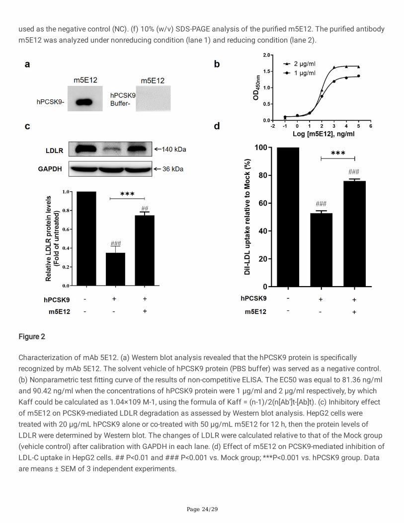
Figure 2
Characterization of mAb 5E12. (a) Western blot analysis revealed that the hPCSK9 protein is speci�callyrecognized by mAb 5E12. The solvent vehicle of hPCSK9 protein (PBS buffer) was served as a negative control.(b) Nonparametric test �tting curve of the results of non-competitive ELISA. The EC50 was equal to 81.36 ng/mland 90.42 ng/ml when the concentrations of hPCSK9 protein were 1 μg/ml and 2 μg/ml respectively, by whichKaff could be calculated as 1.04×10⁹ M-1, using the formula of Kaff = (n-1)/2(n[Ab’]t-[Ab]t). (c) Inhibitory effectof m5E12 on PCSK9-mediated LDLR degradation as assessed by Western blot analysis. HepG2 cells weretreated with 20 μg/mL hPCSK9 alone or co-treated with 50 μg/mL m5E12 for 12 h, then the protein levels ofLDLR were determined by Western blot. The changes of LDLR were calculated relative to that of the Mock group(vehicle control) after calibration with GAPDH in each lane. (d) Effect of m5E12 on PCSK9-mediated inhibition ofLDL-C uptake in HepG2 cells. ## P<0.01 and ### P<0.001 vs. Mock group; ***P<0.001 vs. hPCSK9 group. Dataare means ± SEM of 3 independent experiments.
Page 25/29
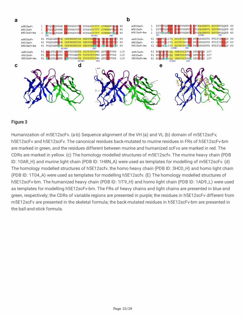
Figure 3
Humanization of m5E12scFv. (a-b) Sequence alignment of the VH (a) and VL (b) domain of m5E12scFv,h5E12scFv and h5E12scFv. The canonical residues back-mutated to murine residues in FRs of h5E12scFv-bmare marked in green, and the residues different between murine and humanized scFvs are marked in red. TheCDRs are marked in yellow. (c) The homology modelled structures of m5E12scfv. The murine heavy chain (PDBID: 10AR_H) and murine light chain (PDB ID: 1H8N_A) were used as templates for modelling of m5E12scFv. (d)The homology modelled structures of h5E12scfv. the homo heavy chain (PDB ID: 3HC0_H) and homo light chain(PDB ID: 1T04_A) were used as templates for modelling h5E12scfv. (E) The homology modelled structures ofh5E12scFv-bm. The humanized heavy chain (PDB ID: 1IT9_H) and homo light chain (PDB ID: 1AD9_L) were usedas templates for modelling h5E12scFv-bm. The FRs of heavy chains and light chains are presented in blue andgreen, respectively; the CDRs of variable regions are presented in purple; the residues in h5E12scFv different fromm5E12scFv are presented in the skeletal formula; the back-mutated residues in h5E12scFv-bm are presented inthe ball-and-stick formula.
Page 26/29
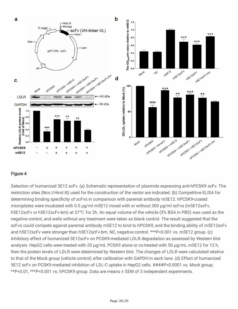
Figure 4
Selection of humanized 5E12 scFv. (a) Schematic representation of plasmids expressing anti-hPCSK9 scFv. Therestriction sites (Nco I/Hind III) used for the construction of the vector are indicated. (b) Competitive ELISA fordetermining binding speci�city of scFvs in comparison with parental antibody m5E12. hPCSK9-coatedmicroplates were incubated with 0.5 μg/ml m5E12 mixed with or without 300 μg/ml scFvs (m5E12scFv,h5E12scFv or h5E12scFv-bm) at 37℃ for 2h. An equal volume of the vehicle (3% BSA in PBS) was used as thenegative control, and wells without any treatment were taken as blank control. The result suggested that thescFvs could compete against parental antibody m5E12 to bind to hPCSK9, and the binding ability of m5E12scFvand h5E12scFv were stronger than h5E12scFv-bm. NC, negative control. ***P<0.001 vs. m5E12 group. (c)Inhibitory effect of humanized 5E12scFv on PCSK9-mediated LDLR degradation as assessed by Western blotanalysis. HepG2 cells were treated with 20 μg/mL PCSK9 alone or co-treated with 50 μg/mL m5E12 for 12 h,then the protein levels of LDLR were determined by Western blot. The changes of LDLR were calculated relativeto that of the Mock group (vehicle control) after calibration with GAPDH in each lane. (d) Effect of humanized5E12 scFv on PCSK9-mediated inhibition of LDL-C uptake in HepG2 cells. ####P<0.0001 vs. Mock group;**P<0.01, ***P<0.001 vs. hPCSK9 group. Data are means ± SEM of 3 independent experiments.
Page 27/29
Figure 5
A�nity maturation and selection of h5E12scFv in vitro. (a) Alignment of the amino acid sequences ofh5E12scFv with its corresponding alanine-scanning variants. Dashes (-) represent the same residue ash5E12scFv. (b) Effect of the alanine-scanning mutants of h5E12scFv on PCSK9-mediated inhibition of LDL-Cuptake in HepG2. The result revealed that L230 was not necessary for maintaining h5E12scFv’s activity and itsmodi�cation might improve the hPCSK9 inhibitory effect of h5E12scFv. (c) Effect of the saturated mutagenesisvariants of h5E12scFv on PCSK9-mediated inhibition of LDL-C uptake in HepG2. The result showed thath5E12scFv-L230S was the most potent hPCSK9 inhibitor among all the h5E12scFv variants. ### P<0.001, ####P<0.0001 vs. Mock group; *P<0.05, ** P<0.01, *** P<0.001, **** P<0.0001 vs. hPCSK9 group. Data are means ±SEM of 3 independent experiments.
Page 28/29
Figure 6
Preparation and identi�cation of the full-length anti-PCSK9 antibodies. (a) SDS-PAGE analysis of puri�ed anti-PCSK9 mAbs under non-reducing (left panel, 10% gel) and reducing (right panel, 12% gel) conditions. M,molecular weight marker; Lane 1, Alirocumab; Lane 2, h5E12; Lane 3, h5E12-L230A; Lane 4, h5E12-L230S; Lane5, h5E12-L230G. (b) Effect of the anti-PCSK9 mAbs on PCSK9-mediated inhibition of LDL-C uptake in HepG2.###P<0.001 vs. Mock group; *** P<0.001 vs. hPCSK9 group. Data are means ± SEM of 3 independentexperiments. (c-d) Kinetics measurement of h5E12-L230G (c) and Alirocumab (d) binding to surface-immobilizedantigen hPCSK9 using a ForteBio Octet QKe system. The biotinylated-hPCSK9 was loaded onto SA sensors andexposed to two-fold serial dilutions of antibody (800, 400, 200, 100, 50, 25, and 12.5 nM) solutions measure the
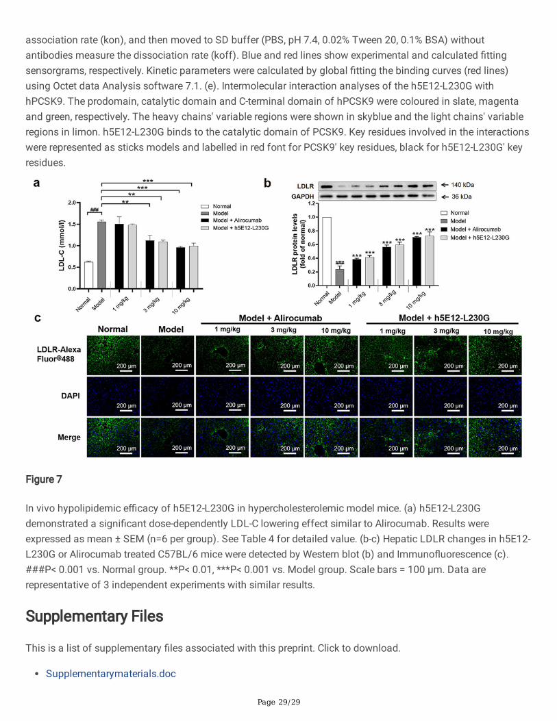
Page 29/29
association rate (kon), and then moved to SD buffer (PBS, pH 7.4, 0.02% Tween 20, 0.1% BSA) withoutantibodies measure the dissociation rate (koff). Blue and red lines show experimental and calculated �ttingsensorgrams, respectively. Kinetic parameters were calculated by global �tting the binding curves (red lines)using Octet data Analysis software 7.1. (e). Intermolecular interaction analyses of the h5E12-L230G withhPCSK9. The prodomain, catalytic domain and C-terminal domain of hPCSK9 were coloured in slate, magentaand green, respectively. The heavy chains' variable regions were shown in skyblue and the light chains' variableregions in limon. h5E12-L230G binds to the catalytic domain of PCSK9. Key residues involved in the interactionswere represented as sticks models and labelled in red font for PCSK9' key residues, black for h5E12-L230G' keyresidues.
Figure 7
In vivo hypolipidemic e�cacy of h5E12-L230G in hypercholesterolemic model mice. (a) h5E12-L230Gdemonstrated a signi�cant dose-dependently LDL-C lowering effect similar to Alirocumab. Results wereexpressed as mean ± SEM (n=6 per group). See Table 4 for detailed value. (b-c) Hepatic LDLR changes in h5E12-L230G or Alirocumab treated C57BL/6 mice were detected by Western blot (b) and Immuno�uorescence (c).###P< 0.001 vs. Normal group. **P< 0.01, ***P< 0.001 vs. Model group. Scale bars = 100 μm. Data arerepresentative of 3 independent experiments with similar results.
Supplementary Files
This is a list of supplementary �les associated with this preprint. Click to download.
Supplementarymaterials.doc