Embed Size (px)
Citation preview

ORIGINAL ARTICLE
CDX-2 Homeobox Gene Product Expression inNeuroendocrine Tumors
Its Role as a Marker of Intestinal Neuroendocrine Tumors
Mattia Barbareschi, MD,* Claudia Roldo, MD,† Giuseppe Zamboni, MD,‡ Paola Capelli, MD,†Alberto Cavazza, MD,§ Ettore Macri, MD,¶ M. Giulia Cangi, PhD,� Marco Chilosi, MD,†
and Claudio Doglioni, MD�
Abstract: CDX-2 is a homeobox gene product essential for intesti-nal development and differentiation. It can be used as a specificmarker of colorectal adenocarcinomas and other tumors with intesti-nal differentiation, but little is known about its expression in endo-crine and neuroendocrine (NE) cells and NE primary and metastatictumors. Using the Cdx-2-88 monoclonal antibody, we evaluatedCDX-2 expression in routine samples of 20 normal endocrine/NE tis-sues and of 299 samples of well-differentiated NE tumors (WDNET)and high-grade NE carcinomas (NEC) from different sites. For 17cases, we examined primary and corresponding metastatic lesions.We also examined 8 cytologic samples of liver metastases derivedfrom 4 ileal WDNETs, 1 lung WDNET, and 3 pancreatic endocrinetumors. CDX-2 mRNA expression with RT-PCR technique on frozenmaterial was evaluated in 5 WDNETs. CDX-2 was expressed in nor-mal NE cells of the intestine and gastric fundus. High CDX-2 expres-sion was seen in all ileal and appendiceal WDNET, while low levelswere seen in WDNETs from stomach, duodenum, and rectum; noreactivity was seen in other WDNETs. Low levels of CDX-2 expres-sion were seen in one third of nonfunctioning pancreatic WDNETwhere it was more frequently observed in cases with metastatic dis-ease (P = 0.002). CDX-2 was identified in all cytologic specimens ofmetastatic ileal WDNETs. CDX-2 mRNA analysis confirmed immu-nohistochemical results. CDX-2 was expressed at high levels in 81%of intestinal NEC. Unexpectedly, variable levels of expression ofCDX-2 were seen also in 39% of NEC of other sites, without anyrelation with the site of origin. This reactivity frequently overlapped
TTF-1 expression, suggesting deregulated expression of homeoboxgenes in NEC. The restricted pattern of CDX-2 expression may havediagnostic value in the identification of the primary site of a meta-static WDNET. Conversely, a limited diagnostic role is suggested forCDX-2 in NEC because of its frequent expression in nongastrointes-tinal tumors.
Key Words: CDX-2, immunohistochemistry, neuroendocrine, hu-man neoplasms, diagnostic marker
(Am J Surg Pathol 2004;28:1169–1176)
CDX-2 is the product of the Cdx-2 homeobox gene relatedto the Drosophila melanogaster gene caudal which is es-
sential for the axial patterning and intestinal development ofthe fruit fly. Three Cdx homeobox genes have been identifiedin mice (Cdx-1, Cdx-2, and Cdx4), and two have been identi-fied so far in humans (CDX-14 and CDX-28). These genes en-code for transcription factors that play an essential role as regu-latory proteins for proliferation and differentiation of intestinalepithelial cells in fetal as well as in adult tissues.5 CDX-1 andCDX-2 are lineage specific: in normal adult human tissues,they are expressed only in colonic and small intestinal epithe-lium and regulate the expression of several different genes in-volved in intestinal cell differentiation and metabolism.14,22
CDX-2 is a very specific and sensitive marker of colorectaladenocarcinomas,12 which can be used in the differential diag-nosis of metastatic adenocarcinomas of unknown primarysites.2,23
Well-differentiated neuroendocrine (NE) tumors(WDNET) are relatively common neoplasms arising mainly inthe lung, gastrointestinal tract, and pancreas. WDNETs areable to metastasize to liver, lymph nodes, or other organs.Since WDNETs share similar morphology and overlappingimmunohistochemical profiles, these features are not usefulfor determining the site of origin of a metastatic lesion. Iden-tifying the primary site of WDNET has significant clinical andtherapeutic relevance. Similar considerations apply also for
From the *Department of Histopathology, S. Chiara Hospital, Trento; †De-partment of Pathology, University of Verona Verona; ‡Department of Pa-thology, University of Verona and S. Cuore-Negrar; §Department of His-topathology, Arcispedale S. Maria Nuova, Reggio Emilia; ¶Department ofHistopathology, S. Martino Hospital, Belluno; and �Department of Histo-pathology, S. Raffaele Hospital, Milano, Italy.
Supported in part by grants of Italian Ministry of Health, Ricerca Finalizzata2002-2004, ORSA Association, MIUR, Rome, and Associazione ItalianaRicerca Cancro, Milan, Italy, and Fondazione Cariverona (Bando 2001),CNR-MIUR Diagnostica Molecolare in Oncologia, and European Com-munity QLG1-CT-2002-01196.
Reprints: Claudio Doglioni, MD, Department of Histopathology, S. RaffaeleHospital, 20132 Milano, Italy (e-mail: [email protected]).
Copyright © 2004 by Lippincott Williams & Wilkins
Am J Surg Pathol • Volume 28, Number 9, September 2004 1169
high-grade NE carcinomas (NECs). In this context, TTF-1, an-other homeobox gene, has been proposed for the identificationof metastatic lung NE tumors.6,16 The possible role of CDX-2in promoting endocrine differentiation in intestinal epithelialcells and its expression in intestinal NE cells has not been in-vestigated. CDX-2 expression has indeed been described in alimited series of NE intestinal neoplasms, but little informationis available about its expression in other NE tumors.15,23
In the present study, we evaluated CDX-2 expression ina large series of common and uncommon NE human tumors ofdifferent origin, both primary and metastatic as well in normalendocrine cells in different tissues. Our aims were: 1) to evalu-ate CDX-2 expression in normal intestinal endocrine cells, 2)to delineate the pattern of CDX-2 expression in NE tumorsfrom different sites, and 3) to verify if CDX-2 can be used as amarker in the differential diagnosis of metastatic NE tumors.
MATERIALS AND METHODS
CasesTwenty samples of formalin-fixed, paraffin-embedded
normal endocrine/NE tissues, including hypophysis, thyroid,
parathyroid, gastrointestinal and lung tissue, pancreas, para-ganglia, adrenal gland, ovary, and testis, were evaluated. Atotal of 299 cases representative of most NE tumors of differ-ent sites, including primary and metastatic lesions, were re-trieved from the files of the Departments of Pathology of Bel-luno, Reggio Emilia, Verona, and Trento. All cases had beenpreviously characterized for NE phenotype and, for pancreaticlesions, for hormone production.
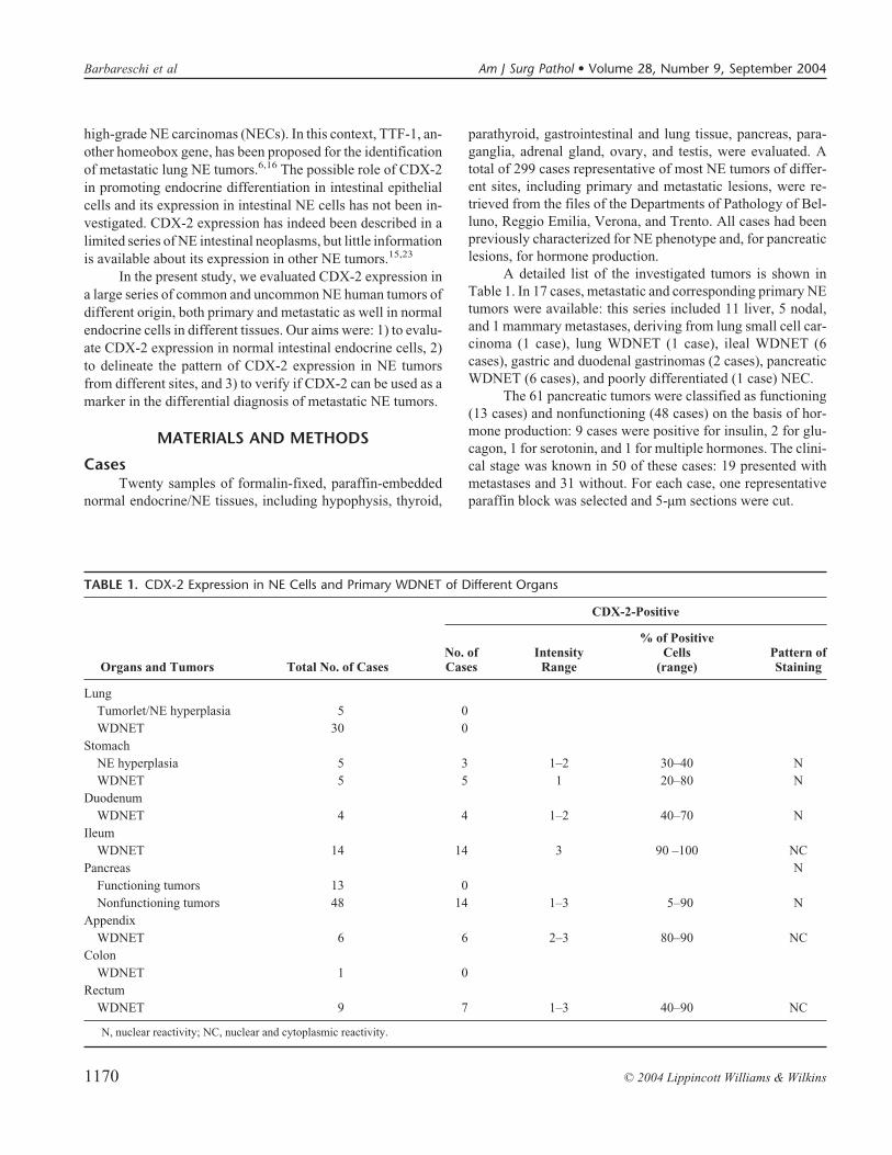
A detailed list of the investigated tumors is shown inTable 1. In 17 cases, metastatic and corresponding primary NEtumors were available: this series included 11 liver, 5 nodal,and 1 mammary metastases, deriving from lung small cell car-cinoma (1 case), lung WDNET (1 case), ileal WDNET (6cases), gastric and duodenal gastrinomas (2 cases), pancreaticWDNET (6 cases), and poorly differentiated (1 case) NEC.
The 61 pancreatic tumors were classified as functioning(13 cases) and nonfunctioning (48 cases) on the basis of hor-mone production: 9 cases were positive for insulin, 2 for glu-cagon, 1 for serotonin, and 1 for multiple hormones. The clini-cal stage was known in 50 of these cases: 19 presented withmetastases and 31 without. For each case, one representativeparaffin block was selected and 5-µm sections were cut.
TABLE 1. CDX-2 Expression in NE Cells and Primary WDNET of Different Organs
Organs and Tumors Total No. of Cases
CDX-2-Positive
No. ofCases
IntensityRange
% of PositiveCells
(range)Pattern ofStaining
LungTumorlet/NE hyperplasia 5 0WDNET 30 0
StomachNE hyperplasia 5 3 1–2 30–40 NWDNET 5 5 1 20–80 N
DuodenumWDNET 4 4 1–2 40–70 N
IleumWDNET 14 14 3 90 –100 NC
Pancreas NFunctioning tumors 13 0Nonfunctioning tumors 48 14 1–3 5–90 N
AppendixWDNET 6 6 2–3 80–90 NC
ColonWDNET 1 0
RectumWDNET 9 7 1–3 40–90 NC
N, nuclear reactivity; NC, nuclear and cytoplasmic reactivity.
Barbareschi et al Am J Surg Pathol • Volume 28, Number 9, September 2004
1170 © 2004 Lippincott Williams & Wilkins

We also examined a series of eight cytologic samplesobtained by fine needle aspiration of liver nodules, for whichsurgical resection specimens were available and for which anunequivocal diagnosis was established. The liver metastaseswere derived from ileal WDNETs (4 cases), lung WDNET (1case), and pancreatic endocrine tumors (3 cases).
Five cases of WDNET (2 ileal, 2 pancreatic, and 1 pul-monary) for which frozen material was available were alsoevaluated for CDX-2 mRNA expression, with an RT-PCRtechnique. One case of colonic carcinoma was used as positivecontrol.
ImmunostainingAll cases were immunostained with a StreptABC tech-
nique using a monoclonal antibody against CDX-2 (Cdx-2-88,Biogenex, San Ramon, CA, 1:200 dilution).2 Samples of nor-mal ileal and colonic mucosa were submitted to double immu-nostainings for chomogranin A (LH2H10, Labvision, Fre-mont, CA) and CDX-2, using a previously described proce-dure.3 Selected cases of high-grade NE tumors were alsoimmunostained for TTF-1 (8G8G3/1, Labvision, Fremont,CA). Heat-induced antigen retrieval was performed using ci-trate buffer in a microwave oven. Primary antibody was de-tected using a sensitive Strept-ABC technique with diamino-benzidine development.3 All stainings were performed with anautomatic immunostainer (Biogenex Optimax). Appropriatepositive and negative controls were run simultaneously. Papa-nicolaou stained cytologic samples were bleached, subjectedto heat-induced antigen retrieval, and immunostained. Immu-nostaining was scored semiquantitatively according to the es-
timated percentage of positive tumor cells, ie, 0 (no staining),1 (1%–10% reactive cells), 2 (11%–50%), and 3 (51%–100%).Intensity (weak, +; moderate, ++; intense, +++) and pattern(cytoplasmic/nuclear) of staining were also recorded.
RNA Extraction and RT-PCR for CDX-2Frozen samples were ground by a tissue dismembrator
(Mikro-Dismembrator S, B. Braun International, Germany)and RNA were extracted using a guanidine isothiocyanate pro-tocol (TRIzol Reagent GIBCO BRL, Life Technologies, Gai-thersburg, MD). Following DNAse treatment, equal amountsof total RNA (1 µg) were reverse transcribed into cDNA usingrandom primers and Superscript First-Strand Synthesis Sys-tem (GIBCO BRL, Life Technologies) according to manufac-turer’s instructions. The human Gs� gene was used as endog-
FIGURE 1. Double immunostaining for CDX-2 (brown) andchromogranin A (red) in normal small intestine. NE cells showstrong chromogranin stain in the cytoplasm and CDX-2 im-munoreactivity in the nucleus. All other enterocytes presentCDX-2 immunoreactivity in the nucleus.
FIGURE 2. CDX-2 immunoreactivity in ileal WDNETs. In mostcases, ileal WDNETs show strong nuclear staining with minordegrees of cytoplasmic staining (A); rare cases may also showa dot-like paranuclear reactivity pattern (B).
Am J Surg Pathol • Volume 28, Number 9, September 2004 CDX-2 Expression in Intestinal NE Tumors
© 2004 Lippincott Williams & Wilkins 1171

enous control. PCR for CDX-2 and Gs� was performed in 25µL reaction mixture containing DNA template, PCR buffer (50mM KCl, 10 mM Tris-HCl, pH 8.3), 1.5 mM MgCl2, 200 mMof each dNTP, 0.4 mM of each primer (human CDX-2 up-stream primer 5�-TGGAGCTGGAGAAGGAGTTTCA-3�,downstream primer 5�-ACAGAGCCAGACACTGAGGCTT-3�; human Gs� upstream primer 5�-GTGATCAAGCAGGCTGACTAT-3�, downstream primer 5�-GCTGCTGGCCAC-CACGAAGATGAT-3�), and 0.5 unit TaKaRa Taq DNApolymerase (Takara Shuzo CO, LTD, Japan). TouchdownPCR 65°C to 55°C consisting of one cycle at 95°C (5 minutes),
10 touchdown cycles at 94°C (30 seconds), 65°C (30 seconds)with a decrease of 1°C each cycle, and at 72°C for 30 seconds,and followed by 20 cycles of 94°C (30 seconds), 55°C (30seconds), and 72°C (30 seconds), with a final extension at72°C for 7 minutes was performed. The resulting amplificationproducts were then analyzed by agarose gel electrophoresis.
RESULTS
Normal TissuesIn normal tissues, CDX-2 was expressed in the nuclei of
almost all intestinal epithelial cells from the duodenum to therectum, including intestinal endocrine cells as demonstratedby double immunohistochemical staining (Fig. 1). Normalgastric mucosa was devoid of CDX-2 immunoreactive cells,with the exception of single NE cells in the fundic glands. Noreactivity was observed in all other tested endocrine/NE tis-sues, including pancreatic insulae.
WDNETIn WDNET, CDX-2 was expressed at high levels in all
ileal and appendiceal lesions with intense nuclear and moder-ate cytoplasmic reactivity (Fig. 2A). In some cases, dot-likeparanuclear staining was also observed (Fig. 2B). CDX-2 im-munoreactivity in NE cell hyperplasias of the stomach (Fig. 3),WDNETs from the stomach, duodenum, and rectum was usu-ally weak and restricted to a limited percentage of cells.Among pancreatic WDNETs, CDX-2 was seen in one third ofnonfunctioning tumors, usually with a low percentage of im-munoreactive nuclei and faint staining intensity (Fig. 4); noreactivity was seen in functioning tumors. CDX-2 immunore-activity was more frequently observed in pancreatic WDNETwith metastatic disease (P = 0.002, Table 2).
No CDX-2 reactivity was seen in NE cell hyperplasias ofbronchial mucosa and in lung tumorlets, as well as in all lungWDNETs. No reactivity was observed in paragangliomas (6cases), pheochromocytomas (4 cases), cortical adenoma (1case) of the adrenal gland, thyroid (3 follicular, 7 papillary, and
FIGURE 3. Gastric NE cell hyperplasia. In this case hyperplasticNE cells show a distinctive nuclear CDX-2 immunoreactivity.
FIGURE 4. Nonfunctioning WDNET of the pancreas. CDX-2immunoreactivity is seen in a limited percentage of nuclei withmoderate to faint staining intensity.
TABLE 2. CDX-2 Expression in Pancreatic NE Tumors,According to the Metastatic Behavior of the Lesions (50Cases With Complete Clinicopathologic Data)
CDX-2-Positive
CDX-2-NegativeN
IntensityRange Range
Nonmetastatic tumors (31) 2 2 90 29Metastatic tumors (19) 9 1–3 5–90 10
P 0.002
Barbareschi et al Am J Surg Pathol • Volume 28, Number 9, September 2004
1172 © 2004 Lippincott Williams & Wilkins

2 medullary carcinomas), and parathyroid (2 adenomas) neo-plasms.
High-grade NE TumorsCDX-2 was expressed at high levels in the majority (13
of 16 cases, 81%) of intestinal poorly differentiated NECs. Avariable percentage of CDX-2 reacting cells was seen also in16 of 41 (39%) NECs of other sites, without an apparent rela-tion with the site of origin (Table 3; Fig. 5). Fifty-one NECcases were also evaluated for TTF-1 expression (Table 4).TTF-1 was observed in 3 of 13 (23%) gastrointestinal tumors,in 12 of 23 (52%) nongastrointestinal, nonpulmonary tumors,and in 9 of 13 (69%) lung NECs. Inappropriate expression ofthese gene products, ie, expression in tumors deriving from
organs normally devoid of CDX-2 and/or TTF-1, was ob-served in 25 of 51 (49%) cases (Table 3).
Metastatic NE TumorsIn the 17 paired samples of primary and metastatic le-
sions analyzed, the pattern of immunoreactivity was similar inboth sites. Strong and diffuse nuclear and cytoplasmic CDX-2immunoreactivity was limited to metastatic ileal WDNETs(100% of cases); intermediate/low levels of staining were seenin one of six pancreatic well-differentiated carcinomas, in thesingle case of pancreatic poorly differentiated carcinoma (2+in 30%), and in two gastrinomas from stomach and duodenum(1+ in 20% of the cells).
TABLE 3. CDX-2 Expression in NEC of Different Organs
Organs and Tumors Total No. of Cases
CDX-2-Positive
No. ofCases
IntensityRange
% of PositiveCells
(range)Pattern ofStaining
LungLarge cell NE carcinoma 8 5 1–3 20–60 NSmall cell lung carcinoma 8 1 3 30 NC
StomachNEC 7 6 2–3 10–90 N/NC
DuodenumNEC 1 1 1 10 N
IleumNEC 1 1 3 90 NC
ColonNEC 5 5 2–3 60–80 NC
RectumNEC 2 0
LiverNEC 1 1 2 10 N
BladderNEC 10 4 1–3 5–70 N
UterusNEC 3 2 2–3 20–90 N
Salivary glandNEC 3 1 2 20 N
ProstateNEC 5 2 1–2 20–40 N
BreastNEC 3 1 2 60 NEndocrine ductal carcinoma 1 0
SkinMerkel carcinoma 15 1 2 30 N
N, nuclear reactivity; NC, nuclear and cytoplasmic reactivity.
Am J Surg Pathol • Volume 28, Number 9, September 2004 CDX-2 Expression in Intestinal NE Tumors
© 2004 Lippincott Williams & Wilkins 1173

Fine Needle Aspiration Samples ofHepatic Metastases
CDX-2 immunoreactivity was readily identified in allfine needle aspiration biopsies of ileal WDNETs (Fig. 6).Weak and focal staining was present in a single case of pan-creatic endocrine carcinoma. No reactivity was seen in the re-maining samples.
CDX-2 mRNA ExpressionCDX-2 mRNA was expressed in the two samples of ileal
WDNETs and in a colon adenocarcinoma used as positive con-
trol (Fig. 7). CDX-2 mRNA was not detectable in two pancre-atic and one lung WDNET (Fig. 7). Gs� mRNA levels wereconstant in all tumors analyzed. mRNA results correlated withthe CDX-2 expression at the immunohistochemical level.
DISCUSSIONOur study shows that CDX-2 is expressed in normal en-
docrine cells of the intestinal tract. We also demonstrate thatCDX-2 is expressed in all primary and metastatic WDNET ofthe ileum and appendix, and in a subset of gastric, colorectal,and pancreatic WDNETs. The absence of CDX-2 in primaryand metastatic WDNET of other sites suggests that CDX-2expression may be used as a marker when the origin of a meta-static WDNET has to be determined. In addition, we observedCDX-2 expression in most NECs of the gastrointestinal tractbut, surprisingly, also in a significant subset of NEC of othersites.
CDX-2 plays an important role in proliferation and dif-ferentiation of intestinal epithelial cells, where it is normallyexpressed throughout embryonic and postnatal life. The ex-pression of CDX-2 in enterocytes, goblet, and Paneth cells aswell as in NE cells of the human intestine may be interpreted inthe light of their hypothesized origin from a common totipotentstem cell.7,20 Moreover, the expression of CDX-2 in all NEcells of the gastrointestinal tract in contrast to the lack of ex-pression in NE cells of other organs (eg, bronchial tree) is inkeeping with the hypothesis that cells of the dispersed NE sys-tem originate from pluripotent cells that differentiate locallyunder the control of factors unique to a specific site or organ.18
Data on CDX-2 expression in human tumors are stilllimited: we and others recently showed that CDX-2 is ex-
TABLE 4. Combined CDX-2 and TTF-1 Expression Patterns in 51 High-Grade NE Tumors
SiteNo.
of CasesCDX-2+TTF-1+
CDX-2+TTF-1−
CDX-2−TTF-1+
CDX-2−TTF-1−
InappropriateExpression
Small intestine 1 0 1 0 0Colon/rectum 7 0 5 1 1 1Stomach 4 1 3 0 0 1Duodenum 1 1 0 0 0 1Liver 1 1 0 0 0 1Bladder 10 2 2 0 6 4Breast 3 0 1 1 1 2Uterus 3 2 0 0 1 2Salivary gland 3 0 1 1 1 2Prostate 5 2 0 3 0 5Lung small cell carcinoma 5 0 1 3 1 1Lung large cell NE carcinoma 8 4 1 2 1 5
Total 51 13 15 11 12 25/51
Note: Cases are categorized as positive if the markers are expressed in at least 5% of tumor cells.
FIGURE 5. High-grade NE carcinoma of the bladder withprominent CDX-2 immunoreactivity.
Barbareschi et al Am J Surg Pathol • Volume 28, Number 9, September 2004
1174 © 2004 Lippincott Williams & Wilkins

pressed in almost all colorectal adenocarcinomas and in a sub-set of gastric, pancreatic, and ovarian cancers, while it was notexpressed in a large series of human tumors, including lungneoplasms.2,11,15,19,23 This restricted pattern of expression,which is a characteristic of several other homeobox gene prod-ucts, such as TTF-1, is very useful in differential diagnosis in avariety of settings, including the evaluation of cytologic speci-mens.
In the present study, we expand the knowledge about thepossible use of CDX-2 as a diagnostic marker in the setting ofWDNET. Our results demonstrate that CDX-2 strong expres-sion is a highly sensitive marker for the identification ofWDNET originating from the small intestine and appendix.CDX-2 is highly effective both on histologic and cytologicspecimens. CDX-2 is expressed at low levels also in a sig-nificant percentage of pancreatic WDNETs. This phenomenon
must be kept in mind when dealing with a metastatic WDNETof unknown primary site. However, CDX-2 immunoreactivityin pancreatic WDNET tumors is usually weak and heteroge-neous, limited to the nucleus, thus making this pattern of reac-tivity quite different from that observed in ileal and appendi-ceal WDNETs.
Interestingly, CDX-2 expression correlated with thefunctional and clinical behavior of pancreatic WDNET: it wasnot expressed in functioning and in the majority of nonmeta-static tumors, whereas it was observed in one third of nonfunc-tioning and in almost half of tumors with metastatic behavior.The possible biologic and clinical relevance of these findingsis unknown and deserves further investigation.
CDX-2 can be added to the limited list of markers thatcan be used in the differential diagnosis of WDNET of un-known primary origin. This list includes, beside specific hor-mones, the product of another homeobox gene, TTF-1. TTF-1has been considered specific for lung WDNET, although thereare major discrepancies in the literature regarding its sensitiv-ity.9,10,13,16,21 CDX-2 in combination with TTF-1 may help tocorrectly differentiate intestinal from lung WDNET.
CDX-2 expression was observed also in the majority ofgastric and intestinal NECs, but, unexpectedly, also in a signifi-cant percentage of NEC of other sites. CDX-2 expression in thesenongastrointestinal tumors was considered inappropriate be-cause the organs where these tumor originate do not normallyexpress CDX-2, nor do these tumors have any morphologic orbiologic known feature of intestinal differentiation. This inap-propriate expression limits its diagnostic utility in this setting.This pattern of CDX-2 expression parallels the similar abnor-mal expression of TTF-1 in non-lung NECs.13,17 The biologicsignificance of this inappropriate expression of both homeodo-main proteins in NEC is at present unclear but suggests a com-plex deregulated expression of homeobox genes in NEC.1
FIGURE 6. Fine needle aspiration sample of a metastatic ilealWDNET in the liver. The cells show moderate pleomorphysm,an ill-defined cytoplasm (A), and intense nuclear CDX-2 im-munoreactivity (B).
FIGURE 7. CDX-2 and Gs� mRNA expression in NE tumors.CDX-2 mRNA was expressed in the two samples of ilealWDNETs (lanes 2 and 3) and in the colon adenocarcinomaused as positive control (lane 1). CDX-2 mRNA was not de-tectable in two pancreatic and one lung WDNET (lanes 4–6).Gs� mRNA levels were constant in all tumors analyzed.
Am J Surg Pathol • Volume 28, Number 9, September 2004 CDX-2 Expression in Intestinal NE Tumors
© 2004 Lippincott Williams & Wilkins 1175

In summary, our study expands the knowledge about thedistribution of CDX-2 immunoreactivity in human neoplasms.In the setting of WDNET, strong nucleocytoplasmic reactivityis limited to tumors of the intestinal tract, while weak expres-sion can be observed in subsets of aggressive pancreatic tu-mors. This restricted pattern of expression may have diagnos-tic value in the identification of the primary site of a metastaticWDNET. Conversely, a limited diagnostic role for CDX-2 issuggested in NEC because of its frequent inappropriate expres-sion in NECs originating outside the gastrointestinal tract.
NOTE ADDED IN PROOFSimilar results on CDX-2 expression in NE cells and Tu-
mors have been reported by La Rosa et al. (Virchows Archive,in press 2004). These Authors also underscore the relation be-tween CDX-2 expression and the type of hormonal secretion ofthe NE cells and Tumors.
ACKNOWLEDGMENTSWe thank Lucia Tamis, Silvia Funes, Enzo Meggiolaro
and Roberto Cairella for their skillful technical assistance.
REFERENCES1. Abate-Shen C. Deregulated homeobox gene expression in cancer: cause
or consequence? Natl Rev Cancer. 2002;2:777–785.2. Barbareschi M, Murer B, Colby TV, et al. CDX-2 Homeobox gene ex-
pression is a reliable marker of colorectal adenocarcinoma metastases tothe lungs. Am J Surg Pathol. 2003;27:141–149.
3. Barbareschi M, Pecciarini L, Cangi MG, et al. p63, a p53 homologue, is aselective nuclear marker of myoepithelial cells of the human breast. Am JSurg Pathol. 2001;25:1054–1060.
4. Bonner CA, Loftus SK, Wasmuth JJ. Isolation, characterization, and pre-cise physical localization of human CDX1, a caudal-type homeobox gene.Genomics. 1995;28:206–211.
5. Burglin TR. A Comprehensive Classification of Homeobox Genes. NewYork: Sambrook and Tooze, 1994.
6. Cai Y, Banner B, Glickman J, et al. Cytokeratin 7 and 20 and thyroidtranscription factor 1 can help distinguish pulmonary from gastrointesti-nal carcinoid and pancreatic endocrine tumors. Hum Pathol. 2002;32:1087–1093.
7. Cheng H, Leblond CP. Origin, differentiation and renewal of the fourmain epithelial cell types in the mouse small intestine: V. Unitarian theoryof the origin of the four epithelial cell types. Am J Anat. 1974;141:537–561.
8. Drummond F, Putt W, Fox M, et al. Cloning and chromosome assignmentof the human CDX-2 gene. Ann Hum Genet. 1997;61:393–400.
9. Fabbro D, di Loreto C, Stamerra O, et al. TTF-1 gene expression in humanlung tumours. Eur J Cancer. 1996;32A:512–517.
10. Folpe AL, Gown AM, Lamps LW, et al. Thyroid transcription factor-1:immunohistochemical evaluation in pulmonary neuroendocrine tumors.Mod Pathol. 1999;12:5–8.
11. Fraggetta F, Pelosi G, Cafici A, et al. CDX-2 immunoreactivity in primaryand metastatic ovarian mucinous tumours. Virchows Arch. 2003;443:7826.
12. Giordano TJ, Shedden KA, Schwartz DR, et al. Organ-specific molecularclassification of primary lung, colon, and ovarian adenocarcinomas usinggene expression profiles. Am J Pathol. 2001;159:1231–1238.
13. Kaufmann O, Dietel M. Expression of thyroid transcription factor-1 inpulmonary and extrapulmonary small cell carcinomas and other neuroen-docrine carcinomas of various primary sites. Histopathology. 2000;36:415–420.
14. Lui VC, Li L, Sham MH, et al. CDX-1 and CDX-2 are expressed in humancolonic mucosa and are down-regulated in patients with Hirschsprung’sdisease associated enterocolitis. Biochim Biophys Acta. 2001;1537:89–100.
15. Moskaluk CA, Zhang H, Powell SM, et al. Cdx2 protein expression innormal and malignant human tissues: an immunohistochemical surveyusing tissue microarrays. Mod Pathol. 2003;16:913–919.
16. Oliveira AM, Tazelaar HD, Myers JL, et al. Thyroid transcription factor-1distinguishes metastatic pulmonary from well-differentiated neuroendo-crine tumors of other sites. Am J Surg Pathol. 2001;25:815–819.
17. Ordonez NG. Value of thyroid transcription factor-1 immunostaining indistinguishing small cell lung carcinomas from other small cell carcino-mas. Am J Surg Pathol. 2000;24:1217–1223.
18. Pearse AG, Takor T. Embryology of the diffuse neuroendocrine systemand its relationship to the common peptides. Fed Proc. 1979;38:2288–2294.
19. Phillips RW, Frierson JH, Moskaluk CA. Cdx2 as a marker of epithelialintestinal differentiation in the esophagus. Am J Surg Pathol. 2003;27:1442–1447.
20. Rindi G, Ratineau C, Ronco A, et al. Targeted ablation of secretin-producing cells in transgenic mice reveals a common differentiation path-way with multiple enteroendocrine cell lineages in the small intestine.Development. 1999;126:4149–4156.
21. Sturm N, Lantuejoul S, Laverriere MH, et al. Thyroid transcription factor1 and cytokeratins 1, 5, 10, 14 (34betaE12) expression in basaloid andlarge-cell neuroendocrine carcinomas of the lung. Hum Pathol. 2001;32:918–925.
22. Walters JR, Howard A, Rumble HE, et al. Differences in expression ofhomeobox transcription factors in proximal and distal human small intes-tine. Gastroenterology. 1997;113:472–477.
23. Werling RW, Yaziji H, Bacchi CE, et al. CDX-2, a highly sensitive andspecific marker of adenocarcinomas of intestinal origin: an immunohis-tochemical survey of 476 primary and metastatic carcinomas. Am J SurgPathol. 2003;27:303–310.
Barbareschi et al Am J Surg Pathol • Volume 28, Number 9, September 2004
1176 © 2004 Lippincott Williams & Wilkins