Embed Size (px)
Citation preview
BACTERIAL CONJUNCTIVITIS
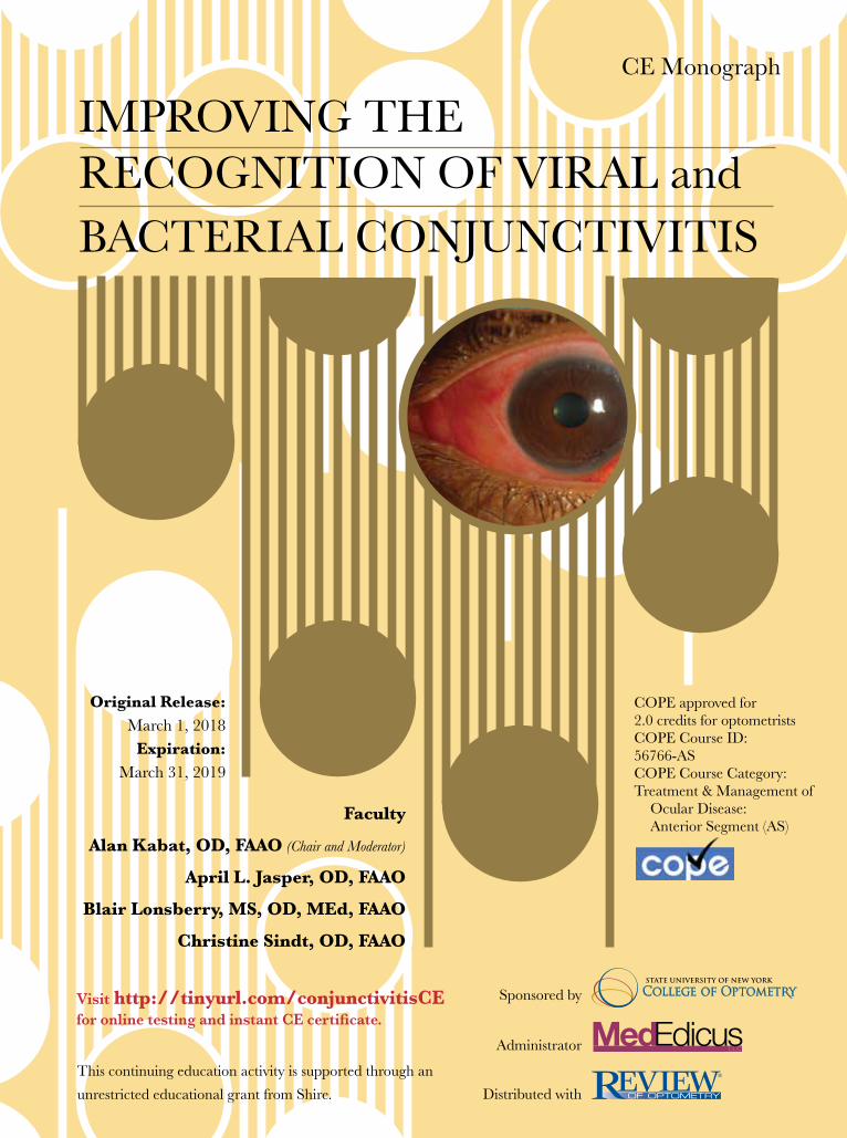
IMPROVING THE RECOGNITION OF VIRAL and
CE Monograph
Faculty
Alan Kabat, OD, FAAO (Chair and Moderator)
April L. Jasper, OD, FAAO
Blair Lonsberry, MS, OD, MEd, FAAO
Christine Sindt, OD, FAAO
Original Release: March 1, 2018
Expiration: March 31, 2019
This continuing education activity is supported through an unrestricted educational grant from Shire.
Administrator
Distributed with
COPE approved for 2.0 credits for optometristsCOPE Course ID: 56766-ASCOPE Course Category: Treatment & Management of Ocular Disease: Anterior Segment (AS)
Visit http://tinyurl.com/conjunctivitisCE for online testing and instant CE certificate.
Sponsored by
2
LEARNING METHOD AND MEDIUMThis educational activity consists of a supplement and twenty (20) study questions. The participant should, in order, read the learning objectives contained at the beginning of this supplement, read the supplement, answer all questions in the post test, and complete the Activity Evaluation/Credit Request form. To receive credit for this activity, please follow the instructions provided on the post test and Activity Evaluation/Credit Request form. This educational activity should take a maximum of 2.0 hours to complete.
CONTENT SOURCEThis continuing education (CE) activity captures content from a roundtable discussion held on November 1, 2017.
ACTIVITY DESCRIPTIONAcute infectious conjunctivitis is estimated to affect 6 million people annually in the United States. Although viral conjunctivitis is much more commonly seen than bacterial conjunctivitis, it is important for optometrists to consider both etiologies from the time patients enter the clinic and to use state-of-the-art assessments to confirm infectious etiology. This is important because of the economic and practical implications of missed school and work days due to infectious conjunctivitis. The purpose of this activity is to provide practical information according to clinical best practices and available evidence to improve outcomes for patients with infectious conjunctivitis.
TARGET AUDIENCEThis educational activity is intended for optometrists.
LEARNING OBJECTIVESUpon completion of this activity, participants will be better able to:• Compare the clinical presentations of viral and bacterial conjunctivitis • Describe how clinical presentation and laboratory testing can be used to diagnose infectious conjunctivitis • Identify the types of health and economic costs of infectious conjunctivitis
ACCREDITATION STATEMENTCOPE approved for 2.0 CE credits for optometrists.COPE Course ID: 56766-ASCOPE Course Category: Treatment & Management of Ocular Disease: Anterior Segment (AS) Administrator:
DISCLOSURESApril L. Jasper, OD, had a financial agreement or affiliation during the past year with the following commercial interests in the form of Consultant/Advisory Board: Alcon; Allergan; CIBA VISION; Konan Medical USA, Inc; and MARCO; Contracted Research: Alcon; CIBA VISION; and Essilor of America, Inc; Honoraria from promotional, advertising or non-CME services received directly from commercial interests or their Agents (eg, Speakers Bureaus): Alcon; Allergan; Carl Zeiss AG; CIBA VISION; Essilor of America, Inc; Konan Medical USA, Inc; MARCO; OCULUS, Inc; OCuSOFT; and Optovue, Incorporated.
Alan Kabat, OD, had a financial agreement or affiliation during the past year with the following commercial interests in the form of Consultant/Advisory Board: Bio-Tissue; Bruder Healthcare; Lacrivera; OCuSOFT; Shire; and Vmax Vision; Contracted Research: Bio-Tissue; Shire; and Vmax Vision; Honoraria from promotional, advertising or non-CME services received directly from commercial interests or their Agents (eg, Speakers Bureaus): OCuSOFT; and Shire.
Blair Lonsberry, MS, OD, MEd, had a financial agreement or affiliation during the past year with the following commercial interests in the form of Consultant/Advisory Board: Shire; and Sun Pharmaceutical Industries Ltd; Honoraria from promotional, advertising or non-CME services received directly from commercial interests or their Agents (eg, Speakers Bureaus): Alcon; Carl Zeiss Meditec, Inc; Optovue, Incorporated; and Shire.
Christine Sindt, OD, had a financial agreement or affiliation during the past year with the following commercial interests in the form of Consultant/Advisory Board: Alcon; and NovaBay; Ownership Interest (Stock options, or other holdings, excluding diversified mutual funds): Eyeprint Prosthetics.
EDITORIAL SUPPORT DISCLOSURESTony Realini, MD, had a financial agreement or affiliation during the past year with the following commercial interests in the form of Consultant/Advisory Board: Aerie Pharmaceuticals, Inc; Alcon; Bausch & Lomb Incorporated; Inotek Pharmaceuticals Corporation; Intelligent Retinal Imaging Systems; New World Medical, Inc; and Smith & Nephew.
Diane McArdle, PhD; Cynthia Tornallyay, RD, MBA, CHCP; and Michelle Ong have no relevant commercial relationships to disclose.
DISCLOSURE ATTESTATIONThe contributing physicians listed above have attested to the following:1) that the relationships/affiliations noted will not bias or otherwise influence their involvement in this activity;2) that practice recommendations given relevant to the companies with whom they have relationships/affiliations will be supported by the best available evidence or, absent evidence, will be consistent with generally accepted medical practice; and3) that all reasonable clinical alternatives will be discussed when making practice recommendations.
PRODUCT USAGE IN ACCORDANCE WITH LABELINGPlease refer to the official prescribing information for each drug discussed in this activity for approved indications, contraindications, and warnings.
GRANTOR STATEMENTThis continuing education activity is supported through an unrestricted educational grant from Shire.
TO OBTAIN CE CREDITWe offer instant certificate processing and support Green CE. Please take this post test and evaluation online by going to http://tinyurl.com/conjunctivitisCE. Upon passing, you will receive your certificate immediately. You must answer 14 out of 20 questions correctly in order to pass, and may take the test up to 2 times. Upon registering and successfully completing the post test, your certificate will be made available online and you can print it or file it. Please make sure you take the online post test and evaluation on a device that has printing capabilities. There are no fees for participating in and receiving CE credit for this activity. DISCLAIMERThe views and opinions expressed in this educational activity are those of the faculty and do not necessarily represent the views of the State University of New York College of Optometry, MedEdicus LLC, Shire, or Review of Optometry.
This CE activity is copyrighted to MedEdicus LLC ©2018. All rights reserved.
3For instant processing, complete the CE Post Test online
http://tinyurl.com/conjunctivitisCE
INTRODUCTION
Optometrists provide more than two-thirds of primary eye care visits in the United States, and managing patients with acute infectious conjunctivitis constitutes a significant portion of their practice.1 The most common causes of infectious conjunctivitis, nonherpetic viruses and bacteria, cannot consistently be distinguished on the basis of signs and symptoms alone,2 demonstrating a role for testing in some cases. Infectious conjunctivitis causes both social and economic burdens for patients and for parents of pediatric patients.3 Current practice patterns and public health policies may not be entirely evidence-based. Herein, we review the prevalence, burden of disease, and differential diagnosis of acute viral and bacterial conjunctivitis.
CAUSES OF CONJUNCTIVITIS
Dr Kabat: The term conjunctivitis is nonspecific. Its literal definition is inflammation of the conjunctiva, with no indication of a specific etiology. What are the various etiologies of conjunctivitis that our colleagues need to consider?
Dr Jasper: When I see a patient who presents with a red eye, the first thing I want to do is determine if the cause is infectious or not. Infectious conjunctivitis can be caused by bacteria or viruses. Among these possibilities, my primary concern is the possibility of herpetic infection. As we will discuss later, this can be a serious infection and I do not want to miss it.
Dr Lonsberry: I also focus first on ruling out infectious causes of the red eye. Viral conjunctivitis is a common reason for patients to seek same-day care in my practice. I agree that herpetic infections can be serious and sight-threatening, but fortunately they are relatively uncommon. The most common cause of viral conjunctivitis is adenovirus, which accounts for 65% to 90% of all viral conjunctival infections.4 I almost never see bacterial conjunctivitis. Also common in my practice are red eyes associated with contact lens wear. Wearers of contact lenses are at an increased risk for a number of very serious causes of red eye, so I approach these patients differently, as we will discuss later. In addition, allergic conjunctivitis is common. Fortunately, these patients tend to know that they have allergies and are familiar with the itching and watering that accompany allergic conjunctivitis.
Dr Sindt: In our practice, we assume that every red eye is an infectious red eye until proven otherwise. Given how contagious some forms of infectious conjunctivitis can be, we treat patients with red eye differently from the moment they arrive. They do not sit in our general waiting room; instead,
FACULTYALAN KABAT, OD, FAAO (Chair and Moderator)
Professor Southern College of Optometry Memphis, Tennessee
APRIL L. JASPER, OD, FAAO Advanced Eyecare SpecialistsWest Palm Beach, Florida
BLAIR LONSBERRY, MS, OD, MED, FAAO Professor of Optometry Pacific University College of Optometry Portland, Oregon
CHRISTINE SINDT, OD, FAAO Director, Contact Lens Service Professor of Clinical Ophthalmology and Visual SciencesUniversity of Iowa Carver College of Medicine Iowa City, Iowa
“The most common cause of viral conjunctivitis is adenovirus, which accounts for 65% to 90% of all viral conjunctival infections.4” - Blair Lonsberry, MS, OD, MEd, FAAO
BACTERIAL CONJUNCTIVITIS
IMPROVING THE RECOGNITION OF VIRAL and
4
we place them directly into an examination room, essentially isolating them until the diagnosis is clear.
Dr Kabat: To summarize, conjunctivitis generally falls into 1 of 3 broad categories: infectious, allergic, and secondary to other factors or conditions (Table 1).5,6 Infectious etiologies include viral—of which adenovirus is the most common4—as well as less common causes such as bacterial infections.5 Allergic conjunctivitis is also common and can take many forms, including seasonal, atopic, and vernal, to name a few.6 Conjunctivitis can also arise secondarily. One important secondary form of conjunctivitis was mentioned by Dr Lonsberry: contact lens wear. The differential diagnosis for red eye in a patient who wears contact lenses is different from that for a non–contact lens wearer. What etiologies do we consider for red eye in the contact lens wearer?
Dr Lonsberry: Probably the most common cause of red eye in a contact lens wearer is overwear. This produces a relatively nonspecific red eye, with no preponderance of papillae or follicles. The diagnosis is typically made by history, which will often be significant for extended wear without proper lens hygiene or timely replacement. This condition typically responds well to a short period without lens wear coupled with a mild steroid. Among the allergic forms of conjunctivitis, giant papillary conjunctivitis is common among contact lens wearers.5 This clinical entity is characterized by redness and itching, and the pathognomonic finding is the presence of giant papillae on the superior palpebral conjunctiva visible with lid eversion. Although giant papillary conjunctivitis is classified as an allergic form of conjunctivitis, its true etiology is likely more related to repetitive trauma of the palpebral conjunctiva against the superior edge of the contact lens associated with blink. Finally, contact lens wear also predisposes to a higher risk of infectious conjunctivitis. Ensuring that there are no corneal infiltrates is critical in these eyes because these infiltrates can be significantly more serious than isolated conjunctivitis.
EPIDEMIOLOGY OF CONJUNCTIVITIS
Dr Kabat: Conjunctivitis is common. Every year approximately 6 million people in the United States experience some form of conjunctivitis.3 It has been estimated that approximately 1% of all visits to primary care physicians (PCPs) are for the evaluation of red eye.7 Among all causes of conjunctivitis, allergic conjunctivitis is the most common, with a prevalence of 15% to 40% in various studies,8 and occurs most often in the spring and summer.9 Among infectious causes of conjunctivitis, viral is the most common overall10,11 and is more prevalent in the summer.9 Bacterial is the second most common cause overall,10 the most common cause in children,9 and is more prevalent in the winter.9 How common overall is conjunctivitis in your practice? How often to do you see all the various types, and which are the most common?
Dr Lonsberry: Among the patients we see on an emergency basis, either same day or after hours, I estimate that approximately two-thirds present for evaluation of a red eye. Of these, the most common cause I see in my practice is related to contact lens wear, and viral conjunctivitis is the second most common. Depending on the time of year, as previously mentioned, allergic conjunctivitis is also common. In Portland, Oregon, where I practice, we have a second allergy season in the fall, likely arising from both ragweed and molds associated with damp or rotting leaves that have fallen from the trees.12,13 In more southern locations, seasonal allergies may be a year-round issue.
Dr Sindt: By far, contact lens–related red eyes are the majority of what I see, followed by viral conjunctivitis. A fair number of our contact lens–related red eyes progress to keratitis.
Dr Jasper: The type of conjunctivitis I see most often in clinical practice is allergic. In Florida, allergy season extends year-round, and on any given day, I see approximately 5 patients who present for evaluation of a red eye. I have a large contact lens practice as well, so I see a number of cases of giant papillary conjunctivitis. Viral conjunctivitis is less common, but represents the majority of red eyes that I see on an urgent basis.
Dr Kabat: It seems that our experiences with conjunctivitis in our practices closely mirror the known epidemiology of conjunctivitis described in the literature.5 I would like to follow up on 1 key point raised earlier by Dr Sindt, and that is the issue of contagion. Viral conjunctivitis, in particular, is highly contagious, and Dr Sindt described her practice’s effort to prevent the spread of this infection by quickly isolating patients who might be infected. Whenever I see a case of adenoviral conjunctivitis, I know to expect more cases because they tend to present in clusters. Once the virus is in the community, it spreads rapidly. We have to take special care to avoid spreading it from patient to patient in our practices. The adenovirus can survive and maintain its infectivity for up to 7 weeks on nonporous surfaces.14 The most important steps we can take are to scrupulously wash our hands between patients and to carefully disinfect the examination room after seeing a suspected or confirmed case of adenovirus. Special attention should be paid to the tonometer tip, which has been implicated in cases of nosocomial infections.14 In acute red eyes, it might be wise to defer tonometry entirely or to employ a tonometer
Etiology Specific Causes
Infectious ViralBacterial
Allergic
Simple allergic conjunctivitisSeasonal conjunctivitis
Atopic keratoconjunctivitisVernal keratoconjunctivitis
Giant papillary conjunctivitis
Secondary
Contact lens relatedMechanicalTraumatic
ToxicNeonatal
Parinaud oculoglandular syndromePhlyctenular keratoconjunctivitisOther ocular surface conditions
Table 1. Causes of Conjunctivitis5,6
“In our practice, we assume that every red eye is an infectious red eye until proven otherwise.” - Christine Sindt, OD, FAAO
5For instant processing, complete the CE Post Test online
http://tinyurl.com/conjunctivitisCE
that uses a disposable tip or tip cover, such as the iCare or Tonopen. Goldmann tonometry should be avoided to prevent contamination of the tip, and noncontact tonometry should be avoided to prevent dispersing the virus by aerosolization. These measures can help prevent a major outbreak, several of which have been reported and can number into the dozens or even hundreds of cases.15
VIRAL CONJUNCTIVITIS
Dr Kabat: When faced with a red eye that we suspect may be viral conjunctivitis, what are the key elements of the history that might help us make the correct diagnosis?
Dr Jasper: Duration is important. Is it acute or chronic? I ask if the eye has been red for 2 weeks or less. If the red eye persists longer than 2 weeks, it is unlikely to be acute viral conjunctivitis. Is it unilateral or bilateral? Adenovirus is typically bilateral, whereas herpetic conjunctivitis is typically unilateral. Has the patient recently had or been in contact with someone who had red eyes or an acute upper respiratory infection? If so, this favors adenoviral conjunctivitis.
Dr Kabat: What clinical findings on examination support the diagnosis of viral conjunctivitis?
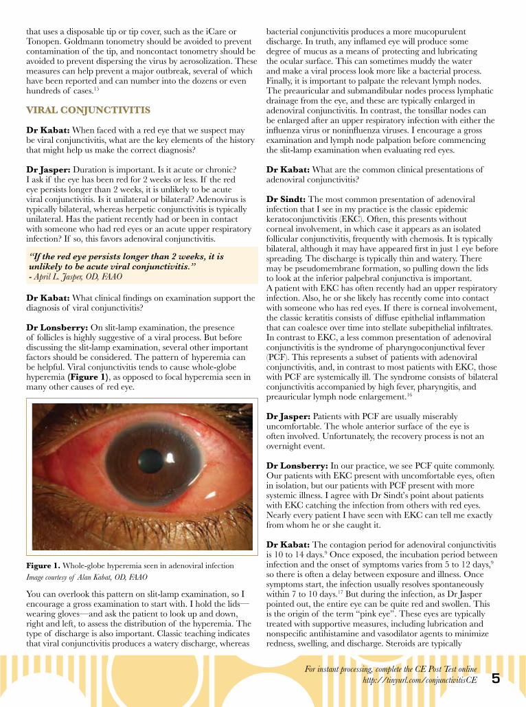
Dr Lonsberry: On slit-lamp examination, the presence of follicles is highly suggestive of a viral process. But before discussing the slit-lamp examination, several other important factors should be considered. The pattern of hyperemia can be helpful. Viral conjunctivitis tends to cause whole-globe hyperemia (Figure 1), as opposed to focal hyperemia seen in many other causes of red eye.
You can overlook this pattern on slit-lamp examination, so I encourage a gross examination to start with. I hold the lids—wearing gloves—and ask the patient to look up and down, right and left, to assess the distribution of the hyperemia. The type of discharge is also important. Classic teaching indicates that viral conjunctivitis produces a watery discharge, whereas
bacterial conjunctivitis produces a more mucopurulent discharge. In truth, any inflamed eye will produce some degree of mucus as a means of protecting and lubricating the ocular surface. This can sometimes muddy the water and make a viral process look more like a bacterial process. Finally, it is important to palpate the relevant lymph nodes. The preauricular and submandibular nodes process lymphatic drainage from the eye, and these are typically enlarged in adenoviral conjunctivitis. In contrast, the tonsillar nodes can be enlarged after an upper respiratory infection with either the influenza virus or noninfluenza viruses. I encourage a gross examination and lymph node palpation before commencing the slit-lamp examination when evaluating red eyes.
Dr Kabat: What are the common clinical presentations of adenoviral conjunctivitis?
Dr Sindt: The most common presentation of adenoviral infection that I see in my practice is the classic epidemic keratoconjunctivitis (EKC). Often, this presents without corneal involvement, in which case it appears as an isolated follicular conjunctivitis, frequently with chemosis. It is typically bilateral, although it may have appeared first in just 1 eye before spreading. The discharge is typically thin and watery. There may be pseudomembrane formation, so pulling down the lids to look at the inferior palpebral conjunctiva is important. A patient with EKC has often recently had an upper respiratory infection. Also, he or she likely has recently come into contact with someone who has red eyes. If there is corneal involvement, the classic keratitis consists of diffuse epithelial inflammation that can coalesce over time into stellate subepithelial infiltrates. In contrast to EKC, a less common presentation of adenoviral conjunctivitis is the syndrome of pharyngoconjunctival fever (PCF). This represents a subset of patients with adenoviral conjunctivitis, and, in contrast to most patients with EKC, those with PCF are systemically ill. The syndrome consists of bilateral conjunctivitis accompanied by high fever, pharyngitis, and preauricular lymph node enlargement.16
Dr Jasper: Patients with PCF are usually miserably uncomfortable. The whole anterior surface of the eye is often involved. Unfortunately, the recovery process is not an overnight event.
Dr Lonsberry: In our practice, we see PCF quite commonly. Our patients with EKC present with uncomfortable eyes, often in isolation, but our patients with PCF present with more systemic illness. I agree with Dr Sindt’s point about patients with EKC catching the infection from others with red eyes. Nearly every patient I have seen with EKC can tell me exactly from whom he or she caught it.
Dr Kabat: The contagion period for adenoviral conjunctivitis is 10 to 14 days.9 Once exposed, the incubation period between infection and the onset of symptoms varies from 5 to 12 days,9 so there is often a delay between exposure and illness. Once symptoms start, the infection usually resolves spontaneously within 7 to 10 days.17 But during the infection, as Dr Jasper pointed out, the entire eye can be quite red and swollen. This is the origin of the term “pink eye”. These eyes are typically treated with supportive measures, including lubrication and nonspecific antihistamine and vasodilator agents to minimize redness, swelling, and discharge. Steroids are typically
Figure 1. Whole-globe hyperemia seen in adenoviral infection Image courtesy of Alan Kabat, OD, FAAO
“If the red eye persists longer than 2 weeks, it is unlikely to be acute viral conjunctivitis.” - April L. Jasper, OD, FAAO
6
contraindicated to prevent the infection from becoming chronic. If corneal involvement occurs, and if, over time, the patient begins to develop subepithelial infiltrates, it may be of value to consider consultation with a cornea specialist at a tertiary care center in some cases.
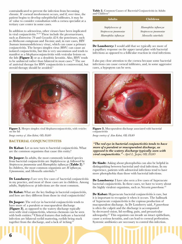
In addition to adenovirus, other viruses have been implicated in viral conjunctivitis.18,19 These include the picornaviruses, such as Enterovirus 70 and Coxsackie A24; the poxviruses, such as Molluscum contagiosum and Vaccinia; and, in rare cases, HIV (human immunodeficiency virus), which can cause a primary conjunctivitis. The herpes simplex virus (HSV) can cause an isolated conjunctivitis, but this is very uncommon and tends to manifest as a blepharoconjunctivitis with vesicular lesions on the lids (Figure 2) or as a dendritic keratitis. Also, HSV tends to be unilateral rather than bilateral in most cases.20 The use of antiviral therapy for HSV conjunctivitis is controversial, but steroid therapy should be avoided.6
BACTERIAL CONJUNCTIVITIS
Dr Kabat: Let us now turn to bacterial conjunctivitis. What are the common organisms that cause this entity?
Dr Jasper: In adults, the most commonly isolated species from bacterial conjunctivitis are Staphylococcus sp, followed by Streptococcus pneumoniae and Haemophilus influenzae (Table 2).21 In children, the most common organisms are H influenzae, S pneumoniae, and Moraxella catarrhalis.21
Dr Lonsberry: I see very few cases of bacterial conjunctivitis in my practice, and most of those cases are in children. Among adults, Staphylococcus sp infections are the most common.
Dr Kabat: What are the key findings in bacterial conjunctivitis, and how are they different from those in viral conjunctivitis?
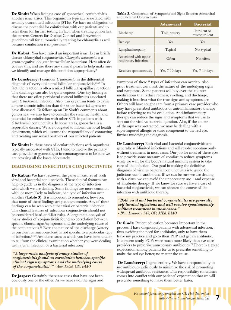
Dr Jasper: The red eye in bacterial conjunctivitis tends to have more of a purulent or mucopurulent discharge (Figure 3), as opposed to the watery discharge typically seen with viral conjunctivitis, whereas chemosis can be seen with both entities.22 Clinical features that indicate a bacterial infection are bilateral eyelid mattering, eyelids being stuck together from the discharge, and a lack of itching.23
Dr Lonsberry: I would add that we typically see more of a papillary response on the upper tarsal plate with bacterial infections as opposed to a follicular reaction in viral infections.
I also pay close attention to the cornea because some bacterial infections can cause corneal infiltrates, and, in some aggressive cases, a hypopyon can be seen.
Dr Sindt: Asking about photophobia can also be helpful in distinguishing between bacterial and viral infections. In my experience, patients with adenoviral infections tend to have more photophobia than those with bacterial infections.
Dr Lonsberry: I have also seen a few cases of hyperacute bacterial conjunctivitis. In these cases, we have to worry about the highly virulent organisms, such as Neisseria gonorrhoeae.22
Dr Kabat: Hyperacute bacterial conjunctivitis is rare, but it is important to recognize it when it occurs. The hallmark of hyperacute conjunctivitis is the copious production of mucopurulent discharge. As Dr Lonsberry said, N gonorrhoeae is often the culprit and the discharge is accompanied by decreased vision, lid swelling, pain, and preauricular adenopathy.22 This organism can invade an intact epithelium, cause a serious keratitis, and can lead to corneal perforation. Systemic antibiotics are necessary to control this infection.
Figure 2. Herpes simplex viral blepharoconjunctivitis, with vesicles on the lid Image courtesy of Alan Kabat, OD, FAAO
Figure 3. Mucopurulent discharge associated with bacterial conjunctivitisImage courtesy of Alan Kabat, OD, FAAO
Adults Children
Staphylococcus spStreptococcus pneumoniaeHaemophilus influenzae
Haemophilus influenzaeStreptococcus pneumoniae
Moraxella catarrhalis
Table 2. Common Causes of Bacterial Conjunctivitis in Adults and Children21
“The red eye in bacterial conjunctivitis tends to have more of a purulent or mucopurulent discharge, as opposed to the watery discharge typically seen with viral conjunctivitis.” - April L. Jasper, OD, FAAO
7For instant processing, complete the CE Post Test online
http://tinyurl.com/conjunctivitisCE
Dr Sindt: When facing a case of gonorrheal conjunctivitis, another issue arises. This organism is typically associated with sexually transmitted infections (STIs). We have an obligation to discuss the potential for coinfections with our patients and to refer them for further testing. In fact, when treating gonorrhea, the current Centers for Disease Control and Prevention guidelines call for automatically treating for chlamydia as well because coinfection is so prevalent.24
Dr Kabat: You have raised an important issue. Let us briefly discuss chlamydial conjunctivitis. Chlamydia trachomatis is a gram-negative, obligate intracellular bacterium. How often do you see this, and are there any clinical pearls to help make sure we identify and manage this condition appropriately?
Dr Lonsberry: I consider C trachomatis in the differential diagnosis of every unilateral follicular conjunctivitis.25,26 In fact, the reaction is often a mixed follicular-papillary reaction. The discharge can also be quite copious. One key finding is that there are often peripheral corneal infiltrates associated with C trachomatis infection. Also, this organism tends to cause a more chronic infection than the other bacterial agents we have discussed. To follow up on Dr Sindt’s comments about gonorrhea, we also have to consider the systemic health and potential for coinfection with other STIs in patients with C trachomatis conjunctivitis. In some areas, gonorrhea is a reportable disease. We are obligated to inform the local health department, which will assume the responsibility of contacting and treating any sexual partners of our infected patients.
Dr Sindt: In these cases of ocular infections with organisms typically associated with STIs, I tend to involve the primary care provider or gynecologist in comanagement to be sure we are covering all the bases adequately.
DIAGNOSING INFECTIOUS CONJUNCTIVITIS
Dr Kabat: We have reviewed the general features of both viral and bacterial conjunctivitis. These clinical features can help to guide us in the diagnosis of the type of infection with which we are dealing. Some findings are more common with, or more likely to indicate, one type of infection over another (Table 3). It is important to remember, however, that none of these findings are pathognomonic. Any of these findings can be seen with either viral or bacterial infection. The clinical features of infectious conjunctivitis should not be considered hard-and-fast rules. A large meta-analysis of many studies of conjunctivitis found no correlation between specific clinical signs/symptoms and the underlying cause of the conjunctivitis.27 Even the nature of the discharge (watery vs purulent vs mucopurulent) is not specific to a particular type of infection.23,28 Are there cases in which you have been unable to tell from the clinical examination whether you were dealing with a viral infection or a bacterial infection?
Dr Jasper: Certainly, there are cases that have not been obviously one or the other. As we have said, the signs and
symptoms of these 2 types of infections can overlap. Also, prior treatment can mask the nature of the underlying signs and symptoms. Some patients will buy over-the-counter medications that reduce redness, swelling, and discharge, making it less clear what the true signs and symptoms are. Others will have sought care from a primary care provider who may have prescribed antibiotics or anti-inflammatory therapy before referring to us for evaluation. Anti-inflammatory therapy can reduce the signs and symptoms that we use to sort out the viral vs bacterial question. Also, if the course of therapy was protracted, we may be dealing with a superimposed allergic or toxic component to the red eye, further muddying the diagnosis.
Dr Lonsberry: Both viral and bacterial conjunctivitis are generally self-limited infections and will resolve spontaneously without treatment in most cases. Our job for most of these eyes is to provide some measure of comfort to reduce symptoms while we wait for the body’s natural immune system to take care of the infection. Our goal in making the definitive diagnosis of viral vs bacterial conjunctivitis is to guide the judicious use of antibiotics. If we can be sure we are dealing with a virus, we can avoid the unnecessary use—and expense—of antibiotic therapy. If we know for sure we have a case of bacterial conjunctivitis, we can shorten the course of the infection with antibiotic therapy.
Dr Sindt: Patient education becomes important in the process. I have diagnosed patients with adenoviral infection, thus avoiding the need for antibiotics, only to have them leave my practice and go to their PCP and get an antibiotic. In a recent study, PCPs were much more likely than eye care providers to prescribe unnecessary antibiotics.29 There is a great expectation among patients for us to prescribe something to make the red eye better, no matter the cause.
Dr Lonsberry: I agree entirely. We have a responsibility to use antibiotics judiciously to minimize the risk of promoting widespread antibiotic resistance. This responsibility sometimes comes into conflict with our patients’ expectation that we will prescribe something to make them better faster.
Adenoviral Bacterial
Discharge Thin, watery Purulent or mucopurulent
Red eye Yes Yes
Lymphadenopathy Typical Not typical
Associated with upper respiratory infection Often Not often
Resolves spontaneously Yes, 7-10 days Yes, 7-14 days
Table 3. Comparison of Symptoms and Signs Between Adenoviral and Bacterial Conjunctivitis
“Both viral and bacterial conjunctivitis are generally self-limited infections and will resolve spontaneously without treatment in most cases.” - Blair Lonsberry, MS, OD, MEd, FAAO
“A large meta-analysis of many studies of conjunctivitis found no correlation between specific clinical signs/symptoms and the underlying cause of the conjunctivitis.27” - Alan Kabat, OD, FAAO

8
Dr Sindt: AdenoPlus tests for all 53 serotypes of the adenovirus by focusing on a region of a protein common to all of them.31 We use it in our practice, but I only use it selectively. If the clinical case is typical and I am confident in my diagnosis, I see little value in the extra time and expense of testing. I reserve this test for less obvious cases, those in which the test result is more likely to influence my management plan.
Dr Kabat: I do not use it in all cases, either. I tend to reserve it for cases that are of unclear etiology based on clinical examination alone and in cases in which my students and I disagree on the etiology. As we have said, AdenoPlus has a high specificity,32,33 so we can be confident that a positive test result accurately diagnoses adenoviral conjunctivitis. We must be aware, however, that the sensitivity is lower,32,33 so a negative test result does not rule out the possibility of an adenoviral infection.
Let us turn to testing for bacterial conjunctivitis. What is the role of culturing the conjunctiva in eyes with presumed bacterial infections?
Dr Sindt: The biggest limitation of cultures is that many, if not most, of the tests end up positive for Staphylococcus and Streptococcus, which may be true infections but are just as likely to be normal lid flora.
Dr Lonsberry: I do not typically culture for conjunctivitis, but I will for a corneal infection. I agree with Dr Sindt that the false-positive rate can be high, but if you get an unusual species in the culture—such as N gonorrhoeae or Pseudomonas aeruginosa—that may be of value because these species are not part of normal eyelid flora.
Dr Sindt: I agree. I might consider a culture in a hyperacute bacterial infection. I also have a lower threshold for culturing the eyes of children with suspected bacterial conjunctivitis.
Dr Jasper: We do not culture for bacterial conjunctivitis in my practice.
Dr Kabat: To summarize and add a few more points, I would say that routine culturing of bacterial conjunctivitis is not necessary. We should consider it, however, in some cases, including neonatal conjunctivitis, hyperacute conjunctivitis, recurrent conjunctivitis, cases recalcitrant to therapy, those with severe purulent discharge, in suspected cases of gonococcal or chlamydial infections, and in eyes with associated corneal infiltrates.6
HEALTH AND ECONOMIC COSTS OF INFECTIOUS CONJUNCTIVITIS
Dr Kabat: What are the financial costs to our society from this disorder and, as a secondary consideration, how does the potential for misdiagnosis affect these costs?
Dr Sindt: There are many types of costs. There are the direct costs of an office visit for evaluation, possible testing costs, and possible medication costs. There are indirect costs: productivity lost at work and transportation costs to the doctor and the pharmacy, among others. There are societal costs as well. The
Dr Sindt: It is not just our patients who have this expectation. In many cases, schools or employers require that a person with a red eye be treated with an antibiotic for 24 hours before he or she can return to school or work, regardless of the reason for the red eye. This policy is problematic for 2 reasons. First, it unnecessarily exposes patients with viral infections to medications that will not help them get better; second, and perhaps more importantly, it assumes that antibiotics will render viral infections noncontagious after 24 hours of antibiotic therapy, which then puts people with infectious conjunctivitis back into schools and workplaces with a false sense of security that they are no longer contagious. We have an opportunity, and perhaps a responsibility, to reach out to our communities to help educate them on the common misconceptions about red eyes in the interest of improving community health.
Dr Lonsberry: We also have a responsibility to educate our patients on the role that antibiotics play in treating eye infections, specifically that they do no good in viral infections and may even cause harm. This approach can reduce the expectation to receive an antibiotic prescription for viral infections.30
Dr Kabat: We have raised the important point that there are societal benefits to distinguishing between viral and bacterial conjunctivitis. We can avoid antibiotics when not indicated, and we can better counsel patients and community partners on the infectivity of patients on the basis of their diagnosis. We have also discussed the reality that we cannot always distinguish between viral and bacterial conjunctival infections on the basis of signs and symptoms alone.
Let us now discuss the role of testing in the setting of infectious red eyes. The AdenoPlus point-of-care test can detect viral particles in the tear film in as little as 10 minutes.31 This rapid antigen test has high specificity—approximately 96%32,33—and, if positive, can identify infectious individuals and prevent unnecessary antibiotic use. The sensitivity of the test is less clear, with published reports providing estimates ranging from 39.5% to 90%,32,33 which means there can be a high rate of false-negative outcomes. What is the role of this test in your practice?
Dr Jasper: I have no experience with this test so far. I have not yet been convinced that it would significantly change my treatment strategy. I am eager to hear if my colleagues have a different perspective. Dr Lonsberry: We do use AdenoPlus in our practice. The test is easy to use, but must be performed correctly to achieve the expected accuracy. I find comfort in knowing for sure that I am dealing with an adenovirus infection in positive cases. I use the test as a means to educate my patients on the nature of their infection and to justify not prescribing antibiotics for viral infections.
“We cannot always distinguish between viral and bacterial conjunctival infections on the basis of signs and symptoms alone.” - Alan Kabat, OD, FAAO
“Routine culturing of bacterial conjunctivitis is not necessary.” - Alan Kabat, OD, FAAO
“There is a great expectation among patients for us to prescribe something to make the red eye better, no matter the cause.” - Christine Sindt, OD, FAAO
9For instant processing, complete the CE Post Test online
http://tinyurl.com/conjunctivitisCE
infection can spread rapidly through a community. There are social considerations related to overuse of antibiotics and the issue of promoting widespread resistance. There can also be costs to your practice if you are unfortunate enough to be the epicenter of an EKC outbreak. To reiterate, there are many types of costs, and they all add up.
Dr Jasper: In school-aged children, there are costs associated with missing school and difficulties that can arise from falling behind in class work. There are also costs to parents—time off from work to take the child to the doctor and to stay home or to arrange for child care if the youngster must be isolated for some time.
Dr Lonsberry: Additionally, unnecessary costs accrue when patients seek care from non–eye care providers. Visits to emergency departments, PCPs, and free-standing ambulatory care clinics often result in misdiagnoses and inappropriate treatments.29
Dr Kabat: As Dr Sindt so eloquently stated, these costs add up! According to a 2009 publication, the direct and indirect costs of diagnosing and treating bacterial conjunctivitis can be estimated at $589 million per year in the United States.34 When considering all types of conjunctivitis, the direct costs alone approach $800 million.35 Although we may not think that we play a large role in the overall health care costs in the United States, we really cannot afford to be cavalier with our diagnoses and treatments. We have a societal obligation to help keep the financial burden of this disease to a minimum whenever possible. We can test more judiciously and refrain from prescribing antibiotics when they are not indicated. We can better educate our patients so they understand that we are providing them with the most appropriate and most economically sound therapy. As optometrists—the primary eye care providers in the country—we can do a better job at cutting these costs.
TAKE-HOME POINTS• Conjunctivitis is common in the United States and can be caused by a variety of etiologies• The diagnosis and treatment of all types of conjunctivitis cost up to $800 million in the United States annually• Infectious conjunctivitis is common, and adenovirus represents the most common cause of infectious conjunctivitis• A careful history of exposure to affected individuals can often aid in the diagnosis of conjunctivitis• Clinical signs such as redness, discharge, pain, and itching can be suggestive of viral vs bacterial causes, but none of these findings are pathognomonic for a specific etiology• Gross examination of the patient, the eye, and the regional lymph nodes can be helpful adjuncts to the slit-lamp examination for diagnosis• Judicious use of rapid adenovirus testing and bacterial cultures can aid in making the diagnosis when the clinical findings are inconclusive• Resist the impulse to prescribe antibiotics to placate patients who expect a prescription to treat their red eyes• When infectious conjunctivitis persists beyond 2 weeks or is unresponsive to therapy, consider less common causes, such as herpes, chlamydia, or molluscum• The economic burden of bacterial conjunctivitis in the United States amounts to over $500 million a year in the form of office visits, medications, lost productivity, lost wages, and other intangible factors
REFERENCES1. Jobson Medical Information LLC. The State of the Optometric Profession: 2013. New York, NY: Jobson Medical Information LLC; 2013. 2. Uchio E, Takeuchi S, Itoh N, Matsuura N, Ohno S, Aoki K. Clinical and epidemiological features of acute follicular conjunctivitis with special reference to that caused by herpes simplex virus type 1. Br J Ophthalmol. 2000;84(9):968-972.3. Udeh BL, Schneider JE, Ohsfeldt RL. Cost effectiveness of a point-of-care test for adenoviral conjunctivitis. Am J Med Sci. 2008;336(3):254-264.4. O’Brien TP, Jeng BH, McDonald M, Raizman MB. Acute conjunctivitis: truth and misconceptions. Curr Med Res Opin. 2009;25(8):1953-1961.5. American Optometric Association. Optometric Clinical Practice Guideline: Care of the Patient With Conjunctivitis. St Louis, MO: American Optometric Association; 2002.6. American Academy of Ophthalmology. Cornea/External Disease Panel. Preferred Practice Pattern® Guidelines. Conjunctivitis. San Francisco, CA: American Academy of Ophthalmology; 2013.7. Shields T, Sloane PD. A comparison of eye problems in primary care and ophthalmology practices. Fam Med. 1991;23(7):544-546.8. Bielory BP, O’Brien TP, Bielory L. Management of seasonal allergic conjunctivitis: guide to therapy. Acta Ophthalmol. 2012;90(5):399-407.9. Høvding G. Acute bacterial conjunctivitis. Acta Ophthalmol. 2008;86(1):5-17.10. Fitch CP, Rapoza PA, Owens S, et al. Epidemiology and diagnosis of acute conjunctivitis at an inner-city hospital. Ophthalmology. 1989;96(8):1215-1220.11. Azari AA, Barney NP. Conjunctivitis: a systematic review of diagnosis and treatment. JAMA. 2013;310(16):1721-1729.12. American College of Allergy, Asthma & Immunology. Mold allergy. http://acaai.org/ allergies/types/mold-allergy. Accessed January 16, 2018.13. American College of Allergy, Asthma & Immunology. Ragweed allergy. http://acaai.org/allergies/types/ragweed-allergy. Accessed January 16, 2018.14. Gordon YJ, Gordon RY, Romanowski E, Araullo-Cruz TP. Prolonged recovery of desiccated adenoviral serotypes 5, 8, and 19 from plastic and metal surfaces in vitro. Ophthalmology. 1993;100(12):1835-1839.15. Centers for Disease Control and Prevention. Adenovirus-associated epidemic keratoconjunctivitis outbreaks—four states, 2008-2010. MMWR Morb Mortal Wkly Rep. 2013;62(32):637-641.16. Jhanji V, Chan TC, Li EY, Agarwal K, Vajpayee RB. Adenoviral keratoconjunctivitis. Surv Ophthalmol. 2015;60(5):435-443.17. Leibowitz HM. The red eye. N Engl J Med. 2000;343(5):345-351.18. Newman H, Gooding C. Viral ocular manifestations: a broad overview. Rev Med Virol. 2013;23(5):281-294.19. Pavan-Langston D. Ocular viral infections. Med Clin North Am. 1983;67(5):973-990.20. American Academy of Ophthalmology. Herpes Simplex Virus Keratitis: A Treatment Guideline. San Francisco, CA: American Academy of Ophthalmology; 2014.21. Epling J, Smucny J. Bacterial conjunctivitis. Clin Evid. 2005;(14):756-761.22. Mannis MJ, Plotnik RD. Bacterial conjunctivitis. In: Tasman W, Jaeger EA, eds. Duane’s Ophthalmology [CD-ROM]. Philadelphia, PA: Lippincott Williams & Wilkins; 2006.23. Rietveld RP, ter Riet G, Bindels PJ, Sloos JH, van Weert HC. Predicting bacterial cause in infectious conjunctivitis: cohort study on informativeness of combinations of signs and symptoms. BMJ. 2004;329(7459):206-210.24. Centers for Disease Control and Prevention. 2015 sexually transmitted diseases treatment guidelines. Gonococcal infections. https://www.cdc.gov/std/tg2015/ gonorrhea.htm. Accessed November 16, 2017.25. Katusic D, Petricek I, Mandic Z, et al. Azithromycin vs doxycycline in the treatment of inclusion conjunctivitis. Am J Ophthalmol. 2003;135(4):447-451.26. Cronau H, Kankanala RR, Mauger T. Diagnosis and management of red eye in primary care. Am Fam Physician. 2010;81(2):137-144.27. Rietveld RP, van Weert HC, ter Riet G, Bindels PJ. Diagnostic impact of signs and symptoms in acute infectious conjunctivitis: systematic literature search. BMJ. 2003;327(7418):789.28. Tarabishy AB, Jeng BH. Bacterial conjunctivitis: a review for internists. Cleve Clin J Med. 2008;75(7):507-512.29. Shekhawat NS, Shtein RM, Blachley TS, Stein JD. Antibiotic prescription fills for acute conjunctivitis among enrollees in a large United States managed care network. Ophthalmology. 2017;124(8):1099-1107.30. Everitt H, Kumar S, Little P. A qualitative study of patients’ perceptions of acute infective conjunctivitis. Br J Gen Pract. 2003;53(486):36-41.31. AdenoPlus [package insert]. Sarasota, FL: Rapid Pathogen Screening, Inc; 2014.32. Kam KY, Ong HS, Bunce C, Ogunbowale L, Verma S. Sensitivity and specificity of the AdenoPlus point-of-care system in detecting adenovirus in conjunctivitis patients at an ophthalmic emergency department: a diagnostic accuracy study. Br J Ophthalmol. 2015;99(9):1186-1189.33. Sambursky R, Trattler W, Tauber S, et al. Sensitivity and specificity of the AdenoPlus test for diagnosing adenoviral conjunctivitis. JAMA Ophthalmol. 2013;131(1):17-22.34. Smith AF, Waycaster C. Estimate of the direct and indirect annual cost of bacterial conjunctivitis in the United States. BMC Ophthalmol. 2009;9:13.35. Schneider JE, Scheibling CM, Segall D, Sambursky R, Ohsfeldt RL, Lovejoy L. Epidemiology and economic burden of conjunctivitis: a managed care perspective. J Manag Care Med. 2014;17(1):78-83.
1. Conjunctivitis literally means _________ of the conjunctiva. a. Infection b. Swelling c. Inflammation d. Redness
2. The most common cause of viral conjunctivitis is: a. HSV b. Molluscum c. Chlamydia d. Adenovirus
3. ____________ is a form of allergic conjunctivitis. a. Vernal keratoconjunctivitis b. EKC c. Pharyngoconjunctival fever d. Phlyctenular keratoconjunctivitis
4. Among contact lens wearers, ________ is a common form of conjunctivitis. a. Parinaud oculoglandular disorder b. Chlamydia conjunctivitis c. Giant papillary conjunctivitis d. Herpes conjunctivitis
5. Approximately how many people in the United States seek care for conjunctivitis per year? a. 1,500,000 b. 6,000,000 c. 10,000,000 d. 15,000,000
6. In which season is adenoviral conjunctivitis more common? a. Spring b. Summer c. Winter d. Fall
7. The adenovirus can survive on nonporous surfaces for up to: a. 7 minutes b. 7 days c. 7 weeks d. 7 months
8. Which piece of equipment has been implicated in clinic- based adenovirus outbreaks? a. Tonometer tip b. Phoropter c. Penlight d. Direct ophthalmoscope
9. On examination, which of the following findings is highly suggestive of a viral infection? a. Giant papillae b. Conjunctival hemorrhage c. Blepharitis d. Follicles
10. A thin, watery discharge is least likely to be seen in: a. Adenoviral conjunctivitis b. Seasonal allergic conjunctivitis c. Contact lens overwear d. Hyperacute N gonorrhoeae conjunctivitis
CE POST TEST QUESTIONSTo obtain COPE CE Credit for this activity, read the material in its entirety and consult referenced sources as necessary.
We offer instant certificate processing and support Green CE. Please take this post test and evaluation online by going to http://tinyurl.com/conjunctivitisCE. Upon passing, you will receive your certificate immediately. You must score 70% or higher to receive credit for this activity, and may take the test up to 2 times.
For instant processing, complete the CE Post Test onlinehttp://tinyurl.com/conjunctivitisCE

11. ________ is the most common bacterial genus responsible for conjunctivitis in adults. a. Staphylococcus b. Pseudomonas c. Moraxella d. Chlamydia
12. Regarding hyperacute conjunctivitis: a. The causative agent is usually a virus b. A thin, watery discharge is common c. Corneal complications can be severe d. Topical antibiotics are usually effective
13. With which of the following forms of conjunctivitis is systemic illness most common? a. EKC b. Bacterial conjunctivitis c. Atopic conjunctivitis d. Pharyngoconjunctival fever
14. A person with adenoviral conjunctivitis is typically contagious for: a. 1 to 3 days b. 5 to 7 days c. 10 to 14 days d. 12 to 24 days
15. Which of the following does not suggest a bacterial cause for conjunctivitis? a. Bilateral eyelid mattering b. Conjunctival hemorrhage c. Eyelids stuck together from discharge d. Lack of itching
16. The high specificity of the rapid adenovirus test (AdenoPlus) means that: a. A negative test is likely to indicate that the patient does not have adenovirus infection b. It should be used only for cases in which adenovirus infection is specifically suspected c. A positive test is likely to indicate that the patient does have adenovirus infection d. It detects only approximately half of the known serotypes of adenovirus
17. The relatively low sensitivity of the rapid adenovirus test (AdenoPlus) means that: a. It also detects viruses other than adenovirus b. The patient may have an adenovirus infection even if the test is negative c. A positive test is likely to be a false-positive result d. It does not test for all serotypes of the adenovirus
18. Conjunctival cultures are indicated in: a. All eyes with suspected bacterial conjunctivitis b. Eyes with suspected viral conjunctivitis to rule out a bacterial superinfection c. Eyes with hyperacute conjunctivitis d. Eyes with itching as the primary symptom
19. Which of the following is true regarding antibiotics for infectious conjunctivitis? a. They should be prescribed for all cases of infectious conjunctivitis to be on the safe side b. They are safe for everyone and have no side effects c. Overprescribing can contribute to antibiotic resistance d. They can cure most cases of infectious conjunctivitis within 24 hours so people can return to work/school
20. The direct and indirect costs of diagnosis and treatment of bacterial conjunctivitis in the United States are approximately _______ annually. a. $89 million b. $198 million c. $589 million d. $958 million

150 BACTERIAL CONJUNCTIVITIS
IMPROVING THE RECOGNITION OF VIRAL and