Embed Size (px)
Citation preview
From Department of Clinical Science, Intervention and
Technology, CLINTEC, Division of Surgery Karolinska Institutet, Stockholm, Sweden
ASPECTS OF TREATMENT OF COMMON BILE DUCT
STONES
Cecilia Strömberg
Stockholm 2011


All previously published papers were reproduced with permission from the publisher. Published by Karolinska Institutet. Printed by Universitetsservice AB. © Cecilia Strömberg, 2011 ISBN 978-91-7457-207-0

Those are my principles. If you don’t like them I have others. Groucho Marx
ABSTRACT
Background: Gallstone disease is a major cause of morbidity and at least 10 000 cholecystectomies are performed
annually in Sweden. At the time of surgery about 5-15% of the patients also have common bile duct stones (CBDS).
Introduction of new techniques like Endoscopic Retrograde CholangioPancreatography (ERCP) and laparoscopy
have changed the treatment for these patients.
Aims: To analyze how CBDS has been treated in Sweden 1965-2009 and to calculate mortality connected to the
different procedures, to assess the risk of malignancy after ERCP in benign disease and identify risk factors for death
within 90 days. To evaluate the short term clinical outcome and identify risk-factors for failure in laparoscopic
transcystic common bile duct exploration (LTCE).
Methods: Data on all patients with an in-patient procedure code of common bile duct exploration or ERCP 1965-
2009 were collected from the Swedish Hospital Discharge Register and those with a diagnosis of malignancy in the
bile ducts, liver or pancreas were excluded. The outcome death was identified by cross-linkage to the Registry of
Causes of Death and readmission in the Swedish Hospital Discharge Register. For assessment of cancer risk a cohort
study of all patients in Sweden having had an ERCP before the end of 2003 without a diagnosis of malignancy at the
time of the procedure or within two years after it was performed. To calculate mortality and analyze risk-factors for
death after ERCP a case-control study based on the population of Stockholm 1990-2003 was performed. Cases were
defined as patients having died within 90 days of the procedure and controls were randomly chosen among those who
did not die. Data were collected prospectively on patients having a cholecystectomy at S:t Göran’s Hospital 1994-
2002, in 155 patients a LTCE were attempted and the outcome analyzed.
Results: The Swedish Hospital Discharge Registry contained records of 126 885 procedures for treatment of
common bile duct stones in 110 119 individuals, without a diagnosis of malignancy at the time of the procedure,
during 1965-2009. The 90-day mortality was 0.24 % after open surgery, 0.90% after ERCP, 0.67% after combined
procedures and 0% after laparoscopic surgery. After adjustment for confounding factors, mainly age and comorbidity,
in the multivariate analyses there was no significant difference in mortality between open surgery and ERCP. The risk
of malignancy in the bile ducts alone and in the bile ducts, liver and pancreas together was significantly elevated in
the cohort of individuals having had an ERCP before 2003, irrespective of if an ES was performed or not. The risk of
malignancy diminished with increasing follow-up time. Patients ever having had a cholecystectomy had a
significantly lower risk of the studied malignancies. In Stockholm County during 1990-2003 the 90-day mortality
after ERCP was 1.6%. Advanced age, severe comorbidity, high complexity of the procedure and the occurrence of a
complication were associated to death within 90 days, whereas a previous cholecystectomy or the simultaneous
performance of an endoscopic sphincterotomy reduced the risk. An attempt of transcystic CBD exploration with
complete stone clearance in the 155 patients at S:t Göran’s Hospital could be fulfilled in 85 %. The median operating
time was 184 minutes (range 89-384 minutes) and the median postoperative hospital stay was one day. There was a
significant 3-fold increase in risk of failure of clearance of the bile ducts among patients with stones of >5 mm
compared to patients with stones 5 mm.
Conclusions: Common bile duct stones were mainly treated endoscopically. ERCP and open surgery were
associated with a similar mortality after adjustment for confounding factors. Laparoscopic treatment was chosen in
younger and healthier patients, probably with a less severe disease, and no 90-day mortality was recorded. The risk of
malignancy in the bile ducts, liver or pancreas was elevated after ERCP in benign disease. However, ES did not seem
to affect this risk. Old age and comorbidity were the main risk factors for death after ERCP but a complex procedure
or the occurrence of a complication also seemed to increase short term mortality. The performance of a
sphincterotomy may decrease the risk of death, possibly by facilitating adequate drainage. Previous cholecystectomy
may also decrease the risk of dying after ERCP. Laparoscopic transcystic exploration of the CBD had a high
frequency of stone clearance and low morbidity in the present study. Moreover, large stones were a risk factor for
failure in stone clearance.
LIST OF PUBLICATIONS
I. Strömberg, C, Nilsson M Treatment of Common Bile Duct Stones in Sweden 1965-2009, a Nation-wide Population-based Study. In manuscript.
II. Strömberg C, Luo J, Enochsson L, Arnelo U , Nilsson M Endoscopic Sphincterotomy and Risk of Malignancy in the Bile Ducts, Liver and Pancreas. Clin Gastroenterol Hepatol. 2008 Sep; 6(9):1049-53. Epub 2008 Jun 30.
III. Strömberg C, Arnelo U, Enochsson L, Löhr M, Nilsson M Possible Mortality Reduction of Endoscopic Sphincterotomy during ERCP; a Population Based Case-control Study. Manuscript submitted.
IV. Strömberg C, Nilsson M, Leijonmarck CE Stone Clearance and Risk Factors for Failure in Laparoscopic Transcystic Exploration of the Common Bile Duct. Surg Endosc. 2008 May; 22(5):1194-9.
CONTENTS 1 Introduction...................................................................................................1
1.1 History of gallstone disease and treatment ........................................1 1.2 About Gallstone Disease ....................................................................4
1.2.1 Prevalence...............................................................................4 1.2.2 Pathogenesis ...........................................................................4 1.2.3 Symptoms ...............................................................................5 1.2.4 Radiology in Diagnosis of Gallstone Disease .......................6 1.2.5 Indications for Treatment in Gallstone Disease ....................6 1.2.6 Complications.........................................................................7
1.3 Common Bile Duct Stones.................................................................9 1.3.1 Prevalence...............................................................................9 1.3.2 Pathogenesis ...........................................................................9 1.3.3 Symptoms .............................................................................10 1.3.4 Radiology in Diagnosis of Common Bile Duct Stones.......11 1.3.5 Indications for Treatment of Common Bile Duct Stones ...12
1.4 Treatment ..........................................................................................13 1.4.1 Surgery..................................................................................13 1.4.2 ERCP ....................................................................................15 1.4.3 Laparoscopic Exploration of the Common Bile Duct.........16
2 Aims............................................................................................................18 3 Material and Methods.................................................................................19
3.1 Paper I, II and III...............................................................................19 3.2 Paper I ...............................................................................................19
3.2.1 Study Base ............................................................................19 3.2.2 Registry Data ........................................................................20 3.2.3 Statistical Analyses...............................................................20
3.3 Paper II..............................................................................................20 3.3.1 Study Base ............................................................................20 3.3.2 Statistical Analyses...............................................................21
3.4 Paper III.............................................................................................22 3.4.1 Study Base ............................................................................22 3.4.2 Exposures..............................................................................22 3.4.3 Statistical Analyses...............................................................23
3.5 Paper IV ............................................................................................23 3.5.1 Study Base ............................................................................23 3.5.2 Statistical Analyses...............................................................24
4 Results.........................................................................................................25 4.1 Paper I ...............................................................................................25
4.1.1 Factors Influencing Survival................................................28 4.2 Paper II..............................................................................................30 4.3 Paper III.............................................................................................32
4.3.1 Age and sex ..........................................................................33 4.3.2 Comorbidity..........................................................................34 4.3.3 Indications ............................................................................34 4.3.4 Procedure-related factors .....................................................34
4.4 Paper IV............................................................................................ 35 5 Discussion................................................................................................... 38
5.1 How Have Common Bile Duct Stones Been Treated in Sweden? . 38 5.1.1 What Risk Factors for Death Could Be Identified? ............ 39 5.1.2 Did Mortality Differ? ........................................................... 39
5.2 Is the Risk of Development of Malignancy Elevated After Endoscopic Sphincterotomy?......................................................................................... 40 5.3 Does Endoscopic Sphincterotomy Increase Short-term Complications after ERCP? ........................................................................................................ 40 5.4 What is the Short-term Outcome of Laparoscopic Transcystic Common Bile Duct Exploration and When Does it Fail?................................................. 41 5.5 General Discussion........................................................................... 42
6 Conclusions ................................................................................................ 43 7 Populärvetenskaplig sammanfattning........................................................ 44 8 Acknowledgements .................................................................................... 47 9 References .................................................................................................. 49
LIST OF ABBREVIATIONS CBDS Common bile duct stones ERCP Endoscopic retrograde cholangiopancreatography LTCE Laparoscopic transcystic common bile duct exploration CBD Common bile duct ES Endoscopic sphincterotomy BC Before Christ IAP International Association of Pancreatology MRCP Magnetic resonance cholangiopancreatography CT Computed tomography MR Magnetic resonance EUS Endoscopic ultrasound ESWL Extra-corporeal shock-wave lithotripsy SILS Single incision laparoscopic surgery NOTES Natural orifice transluminal endoscopic surgery IOC Intraoperative cholangiography ICD International classification of disease HR Hazard ratio CI Confidence interval SIR Standardized incidence ratio OR Odds ratio E Expected O Observed ERC Endoscopic retrograde cholangiography BFF Best friends forever

1 INTRODUCTION Gallstone disease is a major cause of morbidity world-wide. About 10-15 % of
Europeans have gallstones and though many of them are asymptomatic and need no
treatment at least 10 000 cholecystectomies are performed annually in Sweden. About
5-15 % of the patients also have stones in the common bile duct at the time of surgery.
When open surgery was performed these stones were removed at the same time
through a choledochotomy. Treatment of common bile duct stones has changed, first by
the introduction of endoscopic retrograde cholangiography with endoscopic
sphincterotomy in the 1970s and later by the revolution of laparoscopic cholecyst-
ectomy. The endoscopic treatment was initially used in patients who had had a previous
cholecystectomy or when surgery was considered too risky. When laparoscopic
cholecystectomy was introduced several alternative techniques were used to treat
patients with simultaneous stones in the common bile duct: conversion to open surgery,
combinations of laparoscopic and endoscopic methods in one or two stages and finally,
laparoscopic treatment of the common bile duct stones as well, either by laparoscopic
transcystic exploration or by a laparoscopic choledochotomy. Presently all techniques
are being used and which one to choose is probably often decided by local tradition.
1.1 HISTORY OF GALLSTONE DISEASE AND TREATMENT
Concerning the liver and the biliary tract there are early records of observations by
man. About 2000 B.C. the Babylonians described the gallbladder and the extrahepatic
biliary tree in sacrificial animals and made a clay model of them, currently on display at
the British Museum. The Babylonian and Assyrian priests examined organs to interpret
omens and the model is believed to have been used to instruct their students [1].
The liver was believed to be the centre of the soul, which gives light to the extreme
gravity of Prometheus’ fate, that was to have his liver plucked by vultures in eternity, a
punishment for defying Zeus and bringing fire to mankind [2].
Biliary tract stone disease has tormented man since ages. The oldest gallstone known
was found in Gotland, in the remnants of a tomb dating back to the Stone Age,
approximately 4000 years old [3].
2
The mummy of a priestess of the 21st dynasty in ancient Egypt (about 1000 B.C.) was
found with a preserved liver with a gallbladder containing 30 gallstones. The mummy
was presented at the Royal College of Surgeons in London in 1909, but destroyed in
the bombings during World War II, now only photographs remains [1, 4].
Alexander the Great died at the age of 34. The cause of his death is believed to have
been malaria or an overdose of Hellebore (Christmas rose) but the course of his disease
was also compatible with perforation of the gallbladder or associated pancreatitis. After
a party of excess alcohol intake and overeating he deceased after eleven days of fever
and abdominal pain [1].
The earliest description of biliary stones and colic is probably to be found in the works
of Alexander of Tralles, a Greek physician of the fifth century, however neglected in
favour of the theories of Galen, who thought that the yellow bile (one of the four body
fluids in humoral pathology) was produced in the gallbladder and disorders of it was
held responsible for diseases like cholera [1].
In 1556 an autopsy was performed on Saint Ignatius of Loyola, the founder of the Jesuit
Order, by Realdo Colombo, an anatomist from Padua. Numerous gallstones were found
in the gallbladder and also a large stone impacted in the common bile duct which had
eroded into the portal vein. This was one of the earliest descriptions of adverse effects
of stones in the biliary tree but successful treatment lacked for a long time. The famous
English anatomist Francois Glisson stated in 1678 that “only death was the solution for
biliary colic” [4].
Elective surgery of gallstone disease was first proposed by J L W Thudichum in 1859,
a description of which was published in the British Medical Journal the same year [5].
His method of choice was the forming of a biliary fistula by fixing the gallbladder in an
abdominal wound and then removing gallstones after crushing them. J L W Thudichum
didn’t use the method himself but it was adopted by several other surgeons, including
Theodor Kocher [6, 7].
The first cholecystectomy was performed in 1882 by Carl Langenbuch in Berlin. A 43-
year old man with recurrent attacks of biliary colic and obstructive jaundice was purged
for five days prior to surgery. The operation was performed through a “macroinvasive”
3
incision, a “T” with one limb of 10-15 cm below the right costal margin and the other
of the same length along the lateral margin of the rectus abdominis muscle. The cystic
duct was ligated with silk and the gallbladder removed. The patient had a cigar the
following morning and was strictly forbidden to leave his bed for twelve days. He
survived and was discharged from hospital seven weeks later [8]. The new method was
ignored and even received with contempt by Langenbuch’s contemporary colleagues
and not adopted until the end of the century.
The first common bile duct explorations were done in London in 1889 by Knowsley
Thornton and in Basel in 1890 by Ludwig Courvoisier who introduced the use of a T-
tube for safer closure of the common bile duct [4, 9, 10]. However, surgery of the
common bile duct remained to be a “risky business” illustrated by the fact that William
Halsted, the famous American surgeon, watched his mother die of complications of
common bile duct stones even though she had been operated on by her legendary son.
Ironically he himself died of the same disease in 1922 in spite of the fact that his former
students and colleagues at Johns Hopkins University in Baltimore performed three
operations on him. The autopsy showed a large common bile duct stone, impacted in
the papilla [4, 11].
The surgeons blamed the gastroenterologists for referring the patients too late thereby
causing an overrepresentation of hopeless cases and a high mortality. The mortality
after surgery was indeed very high, in the first decades of the 20th century reports were
published with a mortality of 8-9 % after cholecystectomy with exploration of the
common bile duct and 6 % after simple cholecystectomies [3].
In 1974 gastroenterologists from Japan and Germany described an endoscopic
alternative to treat common bile duct stones by Endoscopic Retrograde
Cholangiopancreatography (ERCP). The papilla of Vater where the common bile duct
and the pancreatic duct enter the duodenum was localized, the common bile duct
cannulated and if common bile duct stones were diagnosed they could be removed after
the performance of an endoscopic sphinterotomy (ES) of the biliary sphincter of Oddi.
The technique was recommended to be used in patients with stones impacted in the
ampulla of Vater and used exclusively in patients who had had a prior cholecystectomy
or in those considered unfit for surgery [12, 13].
4
A major change in the treatment of gallstones occurred in the late 1980s when
laparoscopic cholecystectomy was introduced. The French surgeon and gynaecologist
Philippe Mouret performed a laparoscopic cholecystectomy in 1987 starting a
revolution in surgery [14, 15]. The technique was not new, in fact a German surgeon,
Eric Mühe, from Böblingen had performed a similar operation a few years earlier [16]
and the Swedish physician Hans Christian Jakobaeus had described laparo- and
thoracoscopy as early as 1912 [17]. It was however the invention of a video computer
chip that allowed the image to be shown on a television screen that started the
laparoscopic era [18].
1.2 ABOUT GALLSTONE DISEASE
1.2.1 Prevalence
The prevalence of gallstone disease varies around the world. Ultrasound studies reveal
gallstone disease in about 10-15% of adult individuals in Western countries and in 3-
5% of African and Asian populations [19]. A very high prevalence is seen among adult
Pima Indian women in south Arizona (73 %) [20] while in Sweden the frequency is
estimated to be 11% in women [21]. It rises with age from 11% in 40 year old women,
25% in 60 year women and in 77-78 year old women 51 % have gall stone disease or
have had a cholecystectomy. Gallstone disease is not so common in Swedish men, it is
found in 4% of 40 year old men and 15 % in the 60 year old [22, 23]. The difference
between men and women concerning prevalence of gallstone disease has been
explained by Jorgensen to be related to estrogen therapy and child birth [24]. About 60-
80 % of gallstones are silent, giving no symptoms, and need no treatment [25]. With the
high prevalence of gallstone disease the remaining symptomatic gallstones will raise a
demand on national health care. In the Scandinavian countries the cholecystectomy rate
annually is 6-12/10 000 inhabitants [26] and presently in Sweden 10 000-11 000
cholecystectomies are performed every year [27].
1.2.2 Pathogenesis
Gallstones are divided into three major categories: Cholesterol, brown pigment and
black pigment stones depending of their composition and pathogenesis. They can also
be divided into two categories depending of their origin; the gallbladder or the
intrahepatic bile ducts.
5
In Western countries about 75-80% of the stones are cholesterol-based [28] and female
gender, fecundity and a family history of gallstone disease are strong risk factors as
well as the metabolic syndrome for development of cholesterol gallstones [29]. Dietary
constituents are more questionable, Cuevas et al stress the fact that high energy simple
sugar and saturated fat favours cholesterol gallstone formation while fibres and alcohol
consumption reduces the risk [30] but legume intake is also identified as a risk factor
for gallstone formation [31].
Cholesterol is made soluble in bile by micelle formation with bile salts and
phospholipids and precipitation occurs when the bile is hypersaturated with cholesterol
or hyposaturated with phospholipids. The supersaturated cholesterol nucleates into
crystals and the crystals form into stones [29, 32] in the gallbladder [33] from where
they can migrate through the cystic duct or through the wall of the gallbladder into the
bile ducts [34]. Impaired gallbladder motility is a risk factor for gallstone formation
though it may be a secondary effect of the biliary cholesterol supersaturation [35].
Pigment stones have an estimated prevalence rate of about 20-25% among patients
undergoing cholecystectomy [36] and while black pigment stones form in the
gallbladder and is associated with biliary hypersecretion of bilirubin or impairment of
the enterohepatic recycling of bilirubin [37], brown pigment stones form in the
intrahepatic bile ducts as a result of bacterial infection and biliary stasis [33]. Brown
pigment stones are more common in Asia while in western countries cholesterol stones
are predominant.
1.2.3 Symptoms
In many cases biliary stones are asymptomatic [25] and otherwise non-specific.
Symptoms of gallbladder stones such as biliary colic are considered to be caused by the
impaction of one or several stones in the gallbladder neck. The most common symptom
is abdominal pain with radiation to the upper back with onset more than an hour after a
meal. However, abdominal pain for other reasons is common in the population and
biliary stones are often silent [38, 39]. Muhrbeck found abdominal symptoms to be as
common in individuals with gallstones as in those without [40].
In a Swedish study of asymptomatic patients with gallstones about 10% develop
symptoms or complications that require treatment within five years [41] which is
6
compatible with the literature review made by Friedman [42] but higher incidences
have also been reported [43].
Food intolerance is commonly believed to be a symptom of gallstone disease and is
typically described as a tendency of abdominal pain related to intake of fatty or fried
food and fruit and vegetables with thin skins. Festi et al found abdominal pain related to
intake of fatty food in a significantly larger extent in gallstone patients [44], in
opposition to other studies [38, 45]. It has been argued that the food intolerance is not
related to the gallstones themselves and could persist after surgery [46]. In summary,
symptoms of uncomplicated biliary stones are often vague and hard to interpret.
1.2.4 Radiology in Diagnosis of Gallstone Disease
Since gallstone disease gives non-specific symptoms the establishment of a correct
diagnosis could not have been easy before the introduction of radiological methods. In
1890 plain x-ray was introduced and gallstones could be detected if they were calcified
which occurs in only about 10-15%. In 1924 Evarts Graham and Warren Cole
performed a cholecystography, the gallbladder was visualized after intravenous
injection of tetrabromphenolphthalein and failure to obtain a shadow was interpreted as
cholecystitis [47]. Several roentgenograms were taken during a period of 32 hours.
Today the diagnosis of gallstone disease is quite easily made by ultrasound, introduced
in the 1970s, no preparations other than six hours of fasting is needed and a correct
diagnosis is set in >90% [48, 49] of gallbladder stones.
1.2.5 Indications for Treatment in Gallstone Disease
Gallbladder stones are asymptomatic in many cases and no treatment is necessary [50]
[51], however some authors have recommended prophylactic surgery in children, due
to the unknown natural course of gallstone disease among pediatric patients [52]. There
have been suggestions that asymptomatic stones should be treated in diabetic patients,
due to a higher incidence of infectious complications and that cholecystitis presents
unexpectedly and proceeds quickly in diabetics [53], but that strategy is refrained from
since later studies showed no benefit. Prophylactic treatment is no longer recommended
[54, 55]. On the other hand, in symptomatic gallbladder stones treatment is
recommended [56, 57], since the risk of development of a complication is higher. The
importance of a proper assessment before surgery was illustrated by Halldestam et al
7
who found that patients who had typical pain location and specific food intolerance had
a lower risk of persistent abdominal pain after elective cholecystectomy [58].
1.2.6 Complications
Simple gallstone disease could be complicated by cholecystitis, acute pancreatitis,
gallstone ileus, gallbladder carcinoma and finally common bile duct stones the latter
being the scope of this thesis. The risk of complications has been correlated to the
patient’s age at the time of onset of biliary symptoms [59] and also to the severity of the
symptoms [42, 60, 61], older age and more severe symptoms being risk factors for the
occurrence of a complication.
1.2.6.1 Acute Cholecystitis
Acute cholecystitis is caused by an obstruction of the cystic duct by gallstones or
sludge impacted in Hartman’s pouch, leading to an increased pressure in the
gallbladder in presence of bile hypersaturated with cholesterol [62]. The distension
enhances prostaglandin formation giving a vicious circle since the prostaglandins
further activates the epithelial cells to secrete fluid [63], also explaining why
prostaglandin inhibitors relieve pain [64]. The inflammation is often sterile and the
bacterial growth demonstrated in 40-60% of cases is believed to be secondary [65, 66].
Acute cholecystitis is generally considered to be an indication for surgery, either in the
acute stage or as a delayed procedure. In high-risk patients needing treatment a
cholecystostomy could be performed as a temporary solution [67].
1.2.6.2 Gallstone Pancreatitis
Biliary pancreatitis is the most frequent form of acute pancreatitis in western countries
[68] and it occurs in about 3-8% of patients with symptomatic gallstones [69].
Microlithiasis has been detected in a substantial part of patients previously classified as
having idiopathic pancreatitis [70, 71]. The pathogenesis of biliary pancreatitis may be
multifactorial but two mechanisms could be of importance: reflux of bile into the
pancreatic duct, as proposed by Claude Bernard already in 1856 and transient
ampullary obstruction, caused by a stone or sludge passing through the ampulla of
Vater into the duodenum or impacted in the ampulla [72]. The obstruction gives a raise
of the pressure in the pancreatic duct and thereby an activation of digestive enzymes
[73, 74].
8
In Denmark a 30-day mortality of 5% and one-year mortality of 11% has been reported
[75] and in a literary review a decreasing mortality (<10%) is reported in later studies
compared to 15-21% in the earlier from 1960-1985 [76]. Mortality rises with age and
by the severity of the pancreatitis. Many scoring systems have been used for predicting
the subsequent occurrence of multiple organ failure causing death since the severity is
not always clinically apparent at admission [77-79].
In biliary pancreatitis indication for definite treatment of gallstones is very strong since
recurrence rate otherwise is as high as 25-61% [80, 81]. However, the timing of the
procedure has been a topic of debate. During the days of open surgery Kelly and
Wagner did a prospective study of mortality and morbidity in early and delayed surgery
and found it to be much higher in early surgery. On the other hand Burch et al reported
similar mortality but a higher incidence of recurrent biliary attacks in the delayed group
[82]. More recent reports during the laparoscopic era have shown an advantage of early
surgery. In a Swiss study of 112 patients no difference in mortality or morbidity could
be shown, but early cholecystectomy gave a lower risk of new biliary attacks [83]. Uhl
et al recommend early (within 7 days) surgery in cases of mild pancreatitis, while in
severe cases surgery should be postponed at least three weeks, due to increased risk of
infection [84]. In the IAP guidelines for surgical management of acute pancreatitis
three grade B guidelines concerning surgery to prevent future attacks of biliary
pancreatitis are stated: cholecystectomy should be performed to prevent further attacks,
in mild biliary pancreatitis surgery should be performed as soon as the patients have
recovered, ideally during the same admission, and in severe gallstone-pancreatitis
cholecystectomy should be delayed until the patient has recovered with sufficient
resolution of the inflammatory response [85]. The indication for early ERCP in
gallstone pancreatitis has also been much debated. Fan et al reported beneficial effects
of ERCP performed within 24 hours of admission in terms of lower incidence of biliary
sepsis [86] and Neoptolemos found benefits concerning morbidity but not mortality,
[87] while Petrov in a metaanalysis found no reduction of mortality or complications of
early ERCP in patients with predicted mild or severe gallstone pancreatitis but without
cholangitis [88].
1.2.6.3 Gallstone Ileus
Gallstone ileus is a rare complication of gallstone disease. It arises mostly in the elderly
and accounts for about 1-4% of all cases of mechanical bowel obstruction [89]. It
9
occurs when a gallstone erodes the gallbladder wall into the intestinal lumen and gets
impacted in the valvula of Bauhini although impaction in other locations of the
gastrointestinal tract has been described [90, 91]. The management is associated with
high morbidity and also mortality and consists of enterotomy with retrieval of the
gallstone. A laparoscopic approach has also been described [92]. Whether a biliary
procedure should be added and the timing of such a procedure remains a controversy.
The literature consists mainly of single centre experiences of small numbers of patients
collected over many years and in a recent review Ravikumar and Williams argued that
future case series would hardly help decision making [93].
1.2.6.4 Gallbladder Carcinoma
There is a strong association between gallstone disease and gallbladder cancer since
gallstones are present in a vast majority of gallbladder cancer patients [94, 95] and the
size of gallstones is a major risk factor, the bigger the stones the bigger the risk of
development of a carcinoma [96]. However, the causal relationship between stones and
development of carcinoma remains unclear [97].
1.3 COMMON BILE DUCT STONES
1.3.1 Prevalence
About 5-15 % of patients with symptomatic gallstones have common bile duct stones
(CBDS) at the time of surgery [43, 98, 99], the portion grows with increasing age and
duration of symptoms. CBDS that appear after common bile duct surgery or endoscopy
are defined as retained if they are diagnosed less than six months postoperatively,
otherwise as recurrent. Retained stones, mostly due to incomplete clearance, are
reported in a frequency of 4-10% after both surgery and ERCP [100-102]. The rate of
recurrence after ERCP is estimated to be 10-20% and as risk-factors for recurrence a
gall-bladder left in situ (if it contains gallstones), a bile duct with a diameter > 15mm
and the existence of a periampullary diverticula have been identified [103-105] . The
risk of recurrence after ERCP is about the same as after open surgery [100] but it may
be lower after laparoscopic common bile duct surgery [106, 107].
1.3.2 Pathogenesis
Most common bile duct stones form in the gallbladder and migrate through the cystic
duct to the common bile duct and are composed as gallbladder stones, i.e. in Europe
predominately of cholesterol, but they may also form primarily in the common bile
10
duct. Brown pigment stones, however uncommon in the Western world, form in the
intrahepatic bile ducts in patients with infections and obstruction.
Mirizzi syndrome was originally described in 1948 as jaundice caused by a gallstone
impacted in the gallbladder neck compressing the bile duct and thereby causing
obstruction. It has later been classified by Csendes et al into four categories. In type I
obstruction is caused by the external compression originally described, in type II a
cholecystobiliary fistula is present with destruction of less than one-third of the
diameter of the bile duct. In type III the fistula involves up to two thirds of the duct
diameter while in type IV there is a complete destruction of the bile duct [108]. Mirizzi
syndrome is known to be difficult to diagnose. In a British retrospective study of 33
patients it was found that the diagnosis was most easily set with MRCP [109].
1.3.3 Symptoms
The natural history of common bile duct stones is unpredictable, varying from no
symptoms at all to life-threatening conditions. Rosseland found asymptomatic CBDS in
up to 10% of patients scheduled for laparoscopic cholecystectomy [110], making the
prevalence of common bile duct stones at the time of cholecystectomy hard to predict
in spite of the use of scoring systems [111-113]. CBDS may also pass spontaneously
into the duodenum without causing symptoms or reside in the bile duct for a long time
and still be asymptomatic, in a literature review Metcalfe et al found that to be the case
in 85% of patients with stones discovered unexpectedly during surgery [114].
The most common symptom is biliary colic while jaundice, cholangitis and pancreatitis
would rather count as complications of common bile duct stones.
When a bile duct stone gets impacted in the papilla it will cause obstructive jaundice,
often fluctuating, when jaundice clears the stone may have passed into the duodenum
but it may also have floated up in the bile duct.
Cholangitis is a result of an infection complicating obstructive jaundice and has been
reported with a mortality of 13-88% if left untreated [115]. The classical symptoms of
Charcots triad (jaundice, fever and pain) is encountered in about 75% of the patients
but in some this life-threatening condition is presented with vague symptoms [116].
Stones passing the papilla may also induce a pancreatitis by obstructing the pancreatic
duct (se above).
11
In longstanding obstruction secondary biliary cirrhosis and portal hypertension may
develop [102].
Patients with symptomatic common bile duct stones could have a higher risk of
developing a complication, studies have shown a risk of 25-50% of jaundice,
cholangitis or pancreatitis if the stones were left untreated [116] and in a Dutch study of
175 patients with complicated gallstone disease half of the patients had had “warning”
colic attacks five months before the complication occurred [116, 117].
1.3.4 Radiology in Diagnosis of Common Bile Duct Stones
Ultrasound is frequently used in the diagnosis of gallbladder stones but is less
trustworthy in terms of sensitivity in diagnosing common bile duct stones, especially if
they don’t cause dilatation of the bile duct [116] which is the case in about half of the
patients [118]. The sensitivity for detection of intraductal stones is 10-63% [119-121]
but since the specificity is high ultrasound remains an important tool and is often used
as primary imaging modality.
At computed tomography (CT) biliary stones are heterogenic in appearance from
heavily calcified too less radiopaque than bile when containing mainly cholesterol.
They can also have gas attenuation due to nodules of nitrogen gas [118]. The
sensitivity for detection is reported to be 69-88% and the specificity 83-97% [122-124].
Contrast material excreted in the bile can be injected intravenously for better sensitivity
but the use is not wide-spread, perhaps because the excretion is variable and reduced
when serum bilirubin is high. It is also believed to cause more severe allergic reactions
than conventional intravenous contrast material [118] and no water-soluble iodine-
based contrast material with affinity to the liver is available for use in Sweden [125].
Magnetic Resonance (MR) imaging is generally better than CT in diagnosing common
bile duct stones [118] and with special techniques it has the advantage of permitting
imaging of the whole biliary tract within one breath-hold [126]. T2 weighted MR
cholangiography is highly sensitive and specific in the detection of CBDS while T1
weighted images give a more variable appearance [118]. Sensitivity is reported to be
88-100% and specificity 72-97% [127-129]. However, when the stones are small (< 3
mm) sensitivity of detection by MR is low, maybe as low as 50% [130].
12
Endoscopic ultrasound (EUS), where an ultrasonic probe is placed in the duodenum
under endoscopic guidance, gives a detailed imaging especially of the distal common
bile duct [131]. EUS has been used the last twenty years for diagnosing CBDS and in
two meta-analyses the pooled sensitivity and specificity were 89-94% and 94-95%
respectively [132, 133], comparable to the sensitivity and specificity of MR. EUS is
useful for diagnosing microlithiasis (stones < 3 mm) in pancreatitis and in “idiopathic
pancreatitis” the cause can be identified in up to two-thirds of the patients [70] where
other modalities have failed but otherwise the choice between MR and EUS is decided
by local availability [102].
The evolvement of new less invasive modalities has gradually replaced ERCP as a
diagnostic tool and it is nowadays used mainly as a therapeutic procedure [102].
Routine use of preoperative mapping of the bile ducts, such as intravenous
cholangiography, MRCP or ERCP, to detect asymptomatic common bile duct stones
and to reduce the risk of a bile duct injury by identifying anatomic anomalies, has been
a topic of much debate [134]. Scoring systems containing a history of colic pain and/or
jaundice, dyspepsia, cholecystitis, ultrasound describing number and size of stones in
the gallbladder and the level of transaminases and/or alkaline phosphatase have been
used to try to predict the existence of silent common bile duct stones prior to
laparoscopic cholecystectomy thereby reducing the need of preoperative radiology
[111, 112]. In patients with no history of jaundice or pancreatitis, normal liver function
tests and a normal sized common bile duct (≤ 5 mm) Majeed et al reports a 6% risk of
common bile duct stone [135]. Preoperative evaluation of the bile ducts is now much
abandoned and in a Swedish study Järhult found it unnecessary since it had no impact
on preventing bile duct injury or on the frequency of retained common bile duct stones
[136].
1.3.5 Indications for Treatment of Common Bile Duct Stones
Ammori et al has suggested a policy of leaving small stones found unexpectedly in bile
ducts of normal width at surgery and wait and see if symptoms occur before removing
them [137], on the other hand Collins found no correlation between the number or size
of silent stones or size of ducts and spontaneous passage [138]. However, it is generally
agreed that all symptomatic bile duct stones should be removed [102, 111, 139].
13
Obstructive jaundice should be treated as an emergency due to potentially dangerous
complications such as cholangitis, renal failure, cardiovascular dysfunction or
coagulopathy [52, 102].
1.4 TREATMENT
Several attempts have been made to dissolve gallstones. In traditional Chinese medicine
different herbs have been used and in Europe artichoke and hawthorn in wine were
believed to have a dissolving effect. Aeter is also known to be able to dissolve
gallstones and was used for many years before the era of surgery. It was probably one
of the ingredients in “ Liquor anodynus mineralis Hoffmanni” [140, 141] used from the
18th century in many conditions like colic and hysteria [142].
Gallstones consist mainly of cholesterol in individuals of industrialised countries and
are thereby susceptible to dissolution by bile acids, first described in 1937. A high
success rate (80 to 90% in one year) has been reported in small calculi, but the
recurrence rate is high [143]. Extracorporeal shock-wave lithotripsy (ESWL) has been
used to disintegrate gallstones but it also has the disadvantage of a high recurrence rate
[144] to some extent prevented by post- ESWL medication with bile acids. However
Nicholl et all found that ESWL did not relieve symptoms and that it was not cost-
effective [145] and surgery was recommended as first line treatment of symptomatic
gallstones. ESWL has also been used to fragment big common bile duct stones [146]
but a randomized study of 60 patients with difficult common bile duct stones by
Neuhaus et al showed intracorporal laser lithotripsy to be more efficient in clearing the
bile ducts [147]. ESWL is still recommended as treatment for intrahepatic stones [148].
1.4.1 Surgery
Cholecystectomy was first performed in 1882 and exploration of the common bile duct
in 1889, initially and in the first decades of the last century with high mortality and
morbidity, however with growing experience and the introduction of antibiotics in the
1940s the mortality and morbidity decreased [3, 85, 87, 149, 150]. In the 1970s small-
incision cholecystectomy was introduced and morbidity and complications seemed to
decline [151]. Laparoscopic cholecystectomy was introduced in 1987 and became
quickly the method of choice even though scientific evidence of its superiority were
lacking [152]. A recent Cochrane report concluded a low mortality (< 0.09%)
regardless of the method of access to the abdominal cavity chosen, no difference in
14
complications or risk of bile duct injury, but a shorter hospital stay and sick-leave for
both mini-invasive procedures. Small-incision cholecystectomy had a shorter operating
time and a lower cost than laparoscopic cholecystectomy [153] but the latter is by far
the predominant procedure. In recent years new techniques have been introduced,
single incision laparoscopic surgery (SILS) and natural orifice transluminal endoscopic
surgery (NOTES), to reduce the number of incisions and thereby morbidity [154-156].
These methods need to be evaluated.
The most feared complication of surgery is a bile duct injury, a lesion that has been
reported to be more common in laparoscopic surgery, especially when the technique
was introduced [157, 158]. The risk was increased in hospitals doing less than 100
procedures yearly [159], to some extent similar to the findings of Andrén-Sandberg et
al during the era of open surgery, that most bile duct injuries are caused by surgeons
under training [160]. The increase has been explained to be a result of the learning
curve but Waage et al found that not to be the case [161].
Whatever access is chosen to the abdominal cavity the rest of procedure is more or less
the same. The gallbladder in dissected either from above (fundus-first), a technique
often used in open surgery and also recommended to be used in laparoscopic surgery
[162] , though Dolan et al point out that using fundus-first technique could lead to a
higher incidence of retained common bile duct stones[163], or from below. The critical
point is the dissection of the triangle of Calot and the correct identification of the ducts,
in laparoscopic cholecystectomy made easier by lateral traction of the infundibulum of
the gallbladder [164] and unintentional thermal damage could be avoided by careful
use of electro-cautery in this area [165].
The use of intraoperative cholangiography (IOC) was introduced by PL Mirizzi in 1931
[166] and in Sweden by Hulten in Uppsala in 1937 [167] and since then there has been
a debate of whether a routine use of peroperative cholangiography should be
recommended or not [161, 168-170]. The main advantage would be that it would lower
the risk of serious bile duct injuries [158, 161] while other studies find no such
correlation [171, 172]. The main disadvantage would be that it is time-consuming and
sometimes difficult to perform [173, 174]. It is however accepted that it should be done
prior to an exploration of the bile ducts, thereby reducing the number of unnecessary
explorations [170, 175].
15
During the era of open cholecystectomy surgical removal of CBDS found under
operation was considered gold standard and in a randomized trial between surgery
alone and preoperative ERCP followed by biliary surgery Neoptolemos et al found no
advantage of the two-stage procedure [176]. The introduction of laparoscopic surgery
caused a change of therapeutic strategy since, at least in the beginning, no method of
treating CBDS laparoscopically existed, instead a variety of options developed. The
laparoscopic cholecystectomy could be converted to open surgery when CBDS were
revealed or laparoscopic cholecystectomy could be combined with ERCP and ES either
as a one-stage procedure or as a two-stage one where the endoscopy could be
performed either before or after surgery. Laparoscopic techniques of common bile duct
exploration have later evolved and CBDS can be removed either by a transcystic
approach or by a choledochotomy (see below).
1.4.2 ERCP
When ERCP with endoscopic sphincterotomy (ES) was introduced in the 1970s [12,
13] as treatment of common bile duct stones (CBDS) it was reserved for patients who
had had a previous cholecystectomy or were considered unfit for surgery. After the
introduction of laparoscopic cholecystectomy treatment of CBDS by ERCP and ES of
the biliary sphincter of Oddi has become a routine procedure world-wide [102] even in
young patients without previous cholecystectomy, a fact that could be alarming, since
ERCP is known to cause complications in a substantial extent. Short-term
consequences as perforation of the bowel, hemorrhage, cholangitis and pancreatitis are
reported in 3-23% of cases with a mortality of 0-6% [177-180]. Late complications
include cholangitis and stone recurrence reported in 10-15% of cases [179-181]. The
splitting of the sphincter of Oddi has been reported to increase the risk of
cholangiocarcinoma in the long run, thought to be an effect of long-standing bacterial
overgrowth in the bile ducts due to an ascendant infection from the duodenum [181].
In patients scheduled for laparoscopic cholecystectomy a policy of performing
preoperative ERCP will result in a number of unnecessary potentially dangerous
procedures [182, 183] since only 27-54% of the patients believed to have CBDS
according to screening tests actually have so [184]. If on the other hand a postoperative
ERCP is chosen a problem could be that the ERCP will sometimes fail, the papilla
might be impossible to cannulate or clearance of the bile ducts may be difficult, Rhodes
et al found a 93% clearance of the bile ducts when using postoperative ERCP [185].
16
Randomized trials have however found that a two-stage procedure has the same
efficacy in treating CBDS compared to a single-stage one but implies a longer hospital
stay and higher costs [185-187].
During laparoscopic cholecystectomy insertion of a soft-tipped guide-wire through the
cystic duct and sphincter of Oddi out in the duodenum could facilitate cannulation
[187-189]. A single-stage rendez-vous procedure has been described by Enochsson et
al, if the intraoperative cholangiography showed CBDS the guide-wire was introduced
to duodenum through the IOC catheter by the surgeon while waiting for the endoscopy
team to arrive. The endoscopist caught the guide-wire with a polypectomy snare and
after pulling it through the working channel of the endoscope a sphincterotomy could
be introduced. After ES the stones could be removed using a balloon or a basket and
after the endoscopic intervention the surgeon could complete the laparoscopic
cholecystectomy [188]. With this rendez-vous technique Rabago et al found morbidity
as well as post-procedure pancreatitis to be reduced, probably because injection of
contrast material in the pancreatic duct could be avoided [187]. If intraoperative ERCP
for some reason is refrained from, the guide-wire can be left in place after completed
surgery an used to facilitate postoperative cannulation as a two-stage procedure [190].
ERCP and ES are still, as in the beginning, used also in patients who have had a prior
cholecystectomy and as only treatment in biliary stone disease in elderly with a severe
comorbidity. However, a recent Cochrane report concluded that there was a higher
mortality if cholecystectomy was deferred from after ERCP and endoscopic clearance
of common bile duct stones [191] in contrast to what earlier was recommended [192-
194].
1.4.3 Laparoscopic Exploration of the Common Bile Duct
Laparoscopic exploration of the common bile duct could be done either by a transcystic
approach or by a choledochotomy [195-197]. In the transcystic technique a guide-wire
is placed through the cystic duct into the CBD. After balloon-dilatation of the cystic
duct a thin choledochoscope is placed over the guide-wire and the stones are retrieved
one by one. Good results have been published with a clearance of 85% or more [198,
199] but the technique has limitations, big stones are not possible to pull through the
cystic duct, intrahepatic stones may be impossible to reach and when there are
numerous stones in the biliary tract the technique can be time-consuming.
17
The laparoscopic choledochotomy has no limitations in size of stones [195, 200] but
carries a higher morbidity that could be due to the use of a T-tube when closing the
incision in the common bile duct [201]. Alternative techniques have been described, the
common bile duct could be closed either by a direct suture or over a stent instead of T-
drain [202]. In a Cochrane review dated 2007 Gurusamy and Samray found no
evidence of benefit of the use of a T-tube or not [203] but in 2008 Leida et al published
a randomized study of 80 patients and found a primary closure of the choledochotomy
to be superior to the use of a T-tube by a shorter hospital stay, lower costs and less
postoperative and biliary complications [201].
18
2 AIMS
Paper I aimed to analyze how CBDS has been treated in Sweden 1965-2009 and to
calculate mortality and morbidity related to the different procedures.
Paper II aimed to evaluate the relationship between ES for benign disease and
subsequent development of malignancy in the biliary tract. A secondary aim was to
study the relation between severe CBDS exposure and malignancy in the biliary tract.
Paper III aimed to identify risk factors for mortality within 90 days of the procedure in
patients who have had ERCP in non-malignant disease. The main hypothesis was that a
potentially dangerous procedure like ES would be one such risk factor.
Paper IV aimed to evaluate the short term clinical outcome, especially the stone
clearance rate, of LTCE as well as to examine potential risk factors for failure in stone
clearance in the use of this technique.
19
3 MATERIAL AND METHODS
3.1 PAPER I, II AND III
We used data from the Swedish Hospital Discharge registry (“slutenvårdsregistret”),
where the Swedish National Board of Health and Welfare has been collecting data on
individual hospital discharges since 1965. The coverage of the Hospital Discharge
Register was 60% in 1969, 85% in 1983, and included all Swedish hospitals from 1987
and thereafter [204]. By cross-linkage to the Swedish National Cancer Registry
(“cancerregistret”) we identified individuals with a diagnosis of malignancy in the bile
ducts, liver or pancreas. Cross-linkage to the registry of Causes of Death was
performed to ascertain death as an outcome or censoring in the event of death and
Domestic and International Relocations for censoring in the event of emigration of a
cohort member. The Swedish National Cancer Registry is 98% complete [205] and the
registry of Causes of Death (“dödsorsaksregistret”) is also essentially complete [206,
207]. A detailed description of the methods used in these studies has been described
elsewhere [208].
3.2 PAPER I
3.2.1 Study Base
Using the national registration numbers we identified all individuals who had had at
least one in-hospital episode with a discharge procedure code of open or laparoscopic
exploration of the common bile duct, ERCP or ES (JKB00, JKB01, JKB11, JKB20,
JKB21, JKE00, JKE02, JKE12, JKE15, JKE18, JKE25, UJK02, UJK05, UJK12,
UJK15, 5300, 5302, 5304, 5351, 5352, 5356, 5357, 5388, 5394 and 9014 [209, 210])
during the years 1965-2009. This total cohort was divided into four sub-groups in the
further analyses, open surgery (JKB00, JKB20, 5300, 5302, 5351 and 5352), ERCP
(JKE02, JKE12, JKE15, JKE18, JKE25, JKE96, JKW96, UJK02, UJK05, 5388, 5394
and 9014), laparoscopic common bile duct exploration (JKB01, JKB11, JKB21) and
finally surgery combined with ERCP (any ERCP-code and any code for open or
laparoscopic common bile duct exploration during the same hospital stay).
The outcome death was identified by cross-linkage to the Causes of Death Registry. All
individuals with discharge code or a code in the Swedish National Cancer Registry of
20
malignancy in the bile ducts, liver and pancreas (ICD 7 155-157 or corresponding
codes in the later classification) within 90 days of the procedure were excluded.
3.2.2 Registry Data
From the Swedish Hospital Discharge registry we collected information on the age,
sex, time-period, co-morbidity, complications and length of hospital stay, readmissions
and redo procedures of the cohort members.
.
3.2.3 Statistical Analyses
Survival among patients who have undergone treatment of common bile duct stones by
open surgery, ERCP, laparoscopic exploration or combination of endoscopic and
surgical methods was assessed by the Kaplan-Meier method.
Cox proportional Hazard ratios (HR) and their 95% confidence intervals (CI) were used
for univariate and multivariate assessment of the association between potential risk
factors and the hazard of death within 90 days of the procedure. The potential risk
factors used in the regression modeling were categorized in order to facilitate the
analyses. Potential confounding effects were tested by introducing the variables under
study stepwise into the multivariate regression model, and the risk factors were also
tested for possible interactions. Statistical analyses were performed using SPSS version
15.0 for Windows (SPSS, Inc., Chicago, IL). The level of statistical significance was
specified to be 0.05.
3.3 PAPER II
3.3.1 Study Base
From 1965 to 2003, we identified all individuals with at least one in-hospital episode
with a discharge procedure code for ERCP or endoscopic sphincterotomy (Swedish
Classification of Operations and Major Procedures, codes 9014, UJK02, UJK05,
UJK12, UJK15 for ERCP or 5388, 5394, JKE 02, JKE 12, JKE 15, JKE 18, JKE 25,
JKE 98 for ES or procedures for which ES normally is a prerequisite). This total ERCP
cohort was in the further analyses divided into two subgroups: 1. Patients having at
least one procedure code registration for ES or any other endoscopic biliary procedures
for which an ES normally is a prerequisite (Swedish Classification of Operations and
Major Procedures, codes 5388, 5394, JKE 02, JKE 12, JKE 15, JKE 18, JKE 25, JKE
21
98). 2. Patients in the cohort without any procedure code registration for ES or any
other endoscopic biliary procedure implying ES.
Those patients who had a diagnosis of malignant or benign tumour in the bile ducts,
liver or pancreas at the time of the procedure or within two years after it were excluded
from further analyses to avoid bias, since the registered ERCP in these cases may have
been performed because of the tumour or due to symptoms caused by a tumour that
was still undiagnosed. Considering the poor prognosis of malignancies in the biliary
tract, liver and pancreas it is highly unlikely that a tumour causing symptoms would be
diagnosed more than two years later. The cohort was then followed from entry until
diagnosis of an outcome malignancy (primary malignant tumours in the liver, bile ducts
including ampullary region and pancreas, but excluding gallbladder malignancy, ICD7
codes: 155 and 157, but excluding 1551), death, emigration or end of follow-up
(December 31st 2003), whichever occurred first.
3.3.2 Statistical Analyses
Several patients had ERCP or ES procedures registered at more than one point in time.
As index procedure for cancer relative risk analyses were, first of all, every first time
procedure for every patient. If the first time procedure included or implied ES the
patient’s person-time was only included in the ES subgroup. If, on the contrary, a
patient’s first time procedure was non-ES or ES-implying, and followed by a
subsequent procedure including a code for, or implying, ES, this patient had two index
procedures. One without ES, with person-time counted in the non-ES subgroup from
two years after this procedure until the subsequent, second index procedure ES, after
which person-time was counted in the ES subgroup from two years after that procedure
and on.
The standardized incidence ratio (SIR), the ratio of the observed to the expected
number of malignancies, was used to calculate relative risk. The expected number of
cancers occurring in the entire Swedish population was calculated by multiplying the
observed person-time by age- (in 5-year groups), sex- and year of entry-specific cancer
incidence rates. The standardized incidence ratios are inherently adjusted for
confounding by age at follow-up, sex and year of entry. The 95% confidence intervals
(CI) of standardized incidence ratios were calculated assuming that the observed
numbers followed a Poisson distribution [211].
22
To assess the general risk of malignancy in the cohort the SIR for all malignancies
(ICD 7: 140 – 209) was performed. Moreover, the SIR for lung malignancy (ICD 7:
162 – 163) was calculated as an indirect estimate of tobacco smoking exposure in the
cohort, since smoking may be a relevant confounder, especially concerning pancreatic
cancer risk analyses.
3.4 PAPER III
3.4.1 Study Base
In the cohort used in paper II we identified all individuals of the population in
Stockholm County with one or more in-patient episodes with a discharge code of any
ERCP-procedure but without a diagnosis of malignancy at the time of the procedure
from a Stockholm County hospital during 1990-2003.
3.4.1.1 Case Identification
Cases were defined as individuals in the study base who had died within 90 days of the
procedure. For those registered as having undergone several procedures, the last one
counted as index procedure. Of the 323 patients initially identified 90 were eligible to
study.
3.4.1.2 Selection of Controls
The controls were randomly chosen from the study base among individuals not
identified as cases and, as in the cases, when several procedures were registered, the
last one counted as index procedure. Written informed consent was obtained from the
control subjects who were still alive.
3.4.2 Exposures
The exposures studied were age, sex, hospital volume, time-period, co-morbidity,
previous cholecystectomy, indication for the procedure, complexity of the procedure,
pancreatic duct contrast injection, ES and finally the occurrence of complications. For
age, sex, hospital volume, time-period, co-morbidity and previous cholecystectomy,
registry data were used. Concerning indication for the procedure, complexity of the
procedure, pancreatic duct contrast injection, ES and complications, data were collected
by reviewing the medical records. The part of the medical record concerning the ERCP
23
procedure, including indications and complications, was coded with a number for each
study subject and reviewed blinded for case or control status.
3.4.3 Statistical Analyses
Means and frequencies of exposures were calculated according to the outcome variable
death or not. Data were presented as means ± standard deviations for continuous
variables and proportions for dichotomized variables. Significant differences between
groups were determined using Student’s t-test for comparisons of means and Chi-
square test for comparisons of proportions.
Odds ratios (OR) and their 95% confidence intervals (CI), derived from unconditional
logistic regression, were used for univariate and multivariate assessment of the
association between potential risk factors and the outcome death within 90 days of the
procedure. The potential risk factors used in the logistic regression modeling were
categorized in order to facilitate the analyses. Potential confounding effects were tested
by introducing the variables under study stepwise into the multiple logistic regression
model, and the exposures shown to be significant in the univariate analysis were tested
for possible statistical interactions. Statistical analyses were performed using SPSS
version 15.0 for Windows (SPSS, Inc., Chicago, IL). The level of statistical
significance was specified to be 0.05 and the fit of the model was estimated by the
Hosmer and Lemeshow goodness-of-fit test [212].
3.5 PAPER IV
3.5.1 Study Base
Laparoscopic transcystic common bile duct exploration (LTCE) was attempted in 155
patients at S: t Göran’s Hospital during the years 1994-2002. Data on all patients
having cholecystectomy was collected prospectively and registered in a database.
During the time period a total of 3106 patients underwent cholecystectomy. In 273
patients the intraoperative cholangiogram suggested CBD stones and in 118 of them the
surgeon chose other methods of stone clearance (conversion to open surgery, ERCP or
laparoscopic choledochotomy). The 155 patients in whom LTCE was attempted were
included in this study. The patients were followed up six week postoperatively
including analysis of liver enzymes. If the tests were normal and the patient was well,
no further follow-up was done.
24
3.5.2 Statistical Analyses
All LTCE procedures registered were classified as either successful or failed with
regard to stone clearance. Stone clearance was considered successful if a second
intraoperative or postoperative cholangiogram after the attempted clearance showed no
stones.
The success rate was then analyzed with regard to age, sex, procedure priority, common
bile duct diameter, number of stones, stone size, intrahepatic stones and length of
hospital stay. In significance testing Fischer’s exact test was used for dicotomized
discrete variables and the nonparametric Wilcoxon method for comparisons between
means.
Odds ratios (OR) and their 95% confidence intervals (CI), derived from unconditional
logistic regression were used for univariate and multivariate assessment of the
association between studied potential risk factors and the risk of failure in stone
clearance. Linear trends of the associations were tested by treating categorical variables
as continuous in the logistic regression model. Potential confounding effects were
tested by introducing the variables under study one by one into the model.
25
4 RESULTS
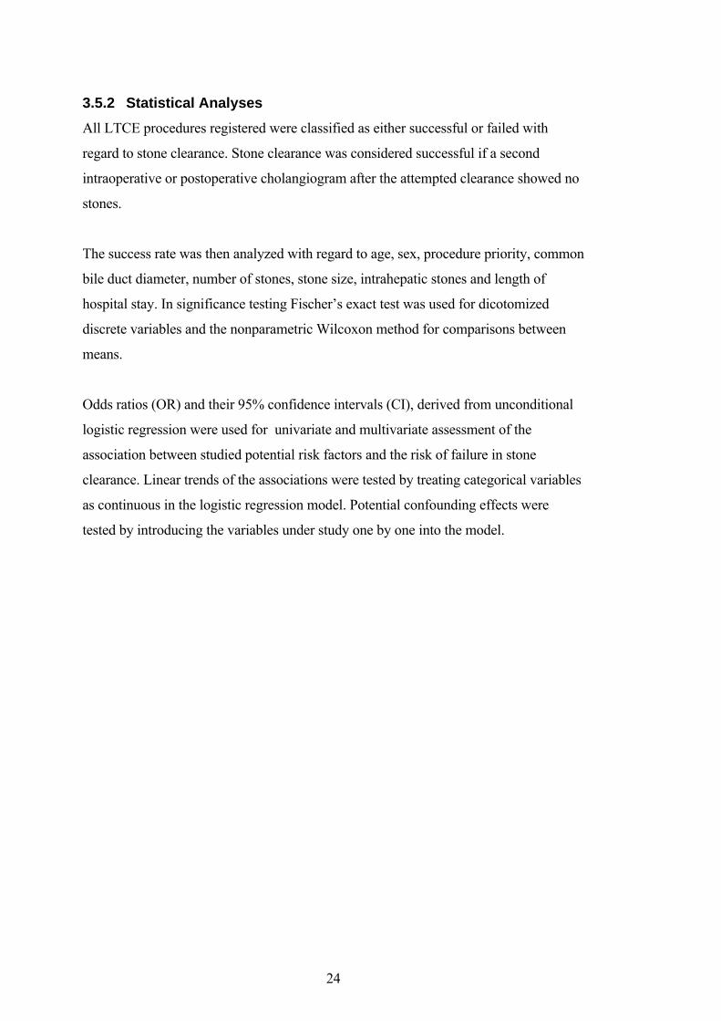
4.1 PAPER I
During the period 1965-2009 a total of 126 885 procedures of open or laparoscopic
common bile duct exploration or ERCP were performed in 110 119 patients without a
diagnosis of malignancy in the bile ducts, liver or pancreas at the time of the procedure
or within 90 days after it. The distribution of the different procedures by five-year terms
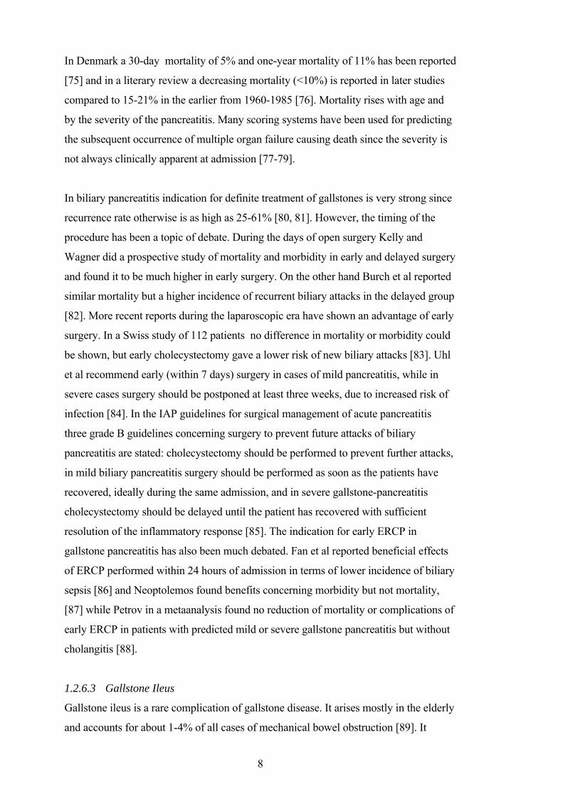
is shown in Figure 1. Endoscopic procedures have gradually replaced open surgery as
treatment for common bile duct stones, only to a small extent challenged by
laparoscopic methods.
Figure 1. Treatment of Common Bile Duct Stones in Sweden 1965-2009.
2005-
2009
2000-
2004
1995-
1999
1990-
1994
1985-
1989
1980-
1984
1975-
1979
1970-
1974
1965-
1969
Count
20 000
15 000
10 000
5 000
0
Open surgery
ERCP
Laparoscopic surgery
Surgery and ERCP
26
The profile of age, sex and co-morbidity differs between the cohorts where the ERCP
patients tend to be older, to a larger extent of male sex and to have more severe co-
morbidity according to the Charlson index. On the contrary, patients treated with
laparoscopic CBD exploration are significantly younger, more often women, and tend
to have less comorbidity. Pancreatitis, which may be either a complication of the biliary
stone disease or a complication of the procedure, is reported in 0.6% of patients after
open surgery, in 5.9% after ERCP and in 3.4% after laparoscopic exploration (Table 1).
Both the 30- and 90-day mortality is around 3-fold higher after ERCP than after open
CBD exploration. Also in this respect laparoscopic CBD exploration differs markedly
from the other interventions as 90-day mortality is 0 in this cohort. The proportion that
has a reintervention within 90 days after ERCP is also significantly, i.e. up to 5-fold
higher, than in the laparoscopic and open CBD exploration cohorts.
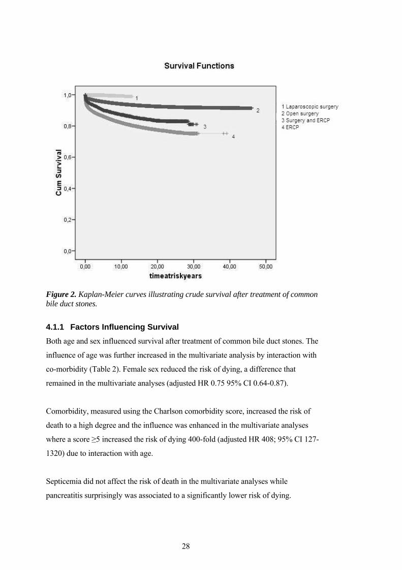
The Kaplan-Meier survival curves (Figure 2) confirm that crude survival is higher after
laparoscopic common bile duct exploration and then gradually decreases for open
surgery, surgery and ERCP and is lowest after ERCP alone.
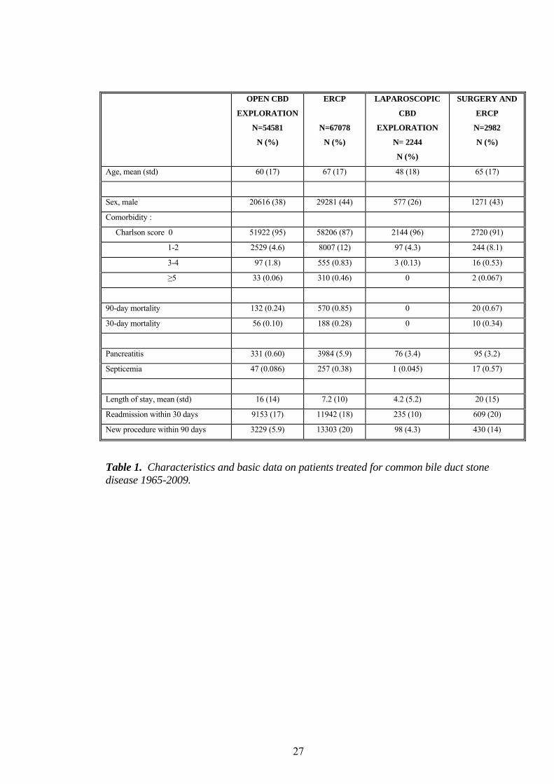
27
OPEN CBD
EXPLORATION
N=54581
N (%)
ERCP
N=67078
N (%)
LAPAROSCOPIC
CBD
EXPLORATION
N= 2244
N (%)
SURGERY AND
ERCP
N=2982
N (%)
Age, mean (std) 60 (17) 67 (17) 48 (18) 65 (17)
Sex, male 20616 (38) 29281 (44) 577 (26) 1271 (43)
Comorbidity :
Charlson score 0 51922 (95) 58206 (87) 2144 (96) 2720 (91)
1-2 2529 (4.6) 8007 (12) 97 (4.3) 244 (8.1)
3-4 97 (1.8) 555 (0.83) 3 (0.13) 16 (0.53)
≥5 33 (0.06) 310 (0.46) 0 2 (0.067)
90-day mortality 132 (0.24) 570 (0.85) 0 20 (0.67)
30-day mortality 56 (0.10) 188 (0.28) 0 10 (0.34)
Pancreatitis 331 (0.60) 3984 (5.9) 76 (3.4) 95 (3.2)
Septicemia 47 (0.086) 257 (0.38) 1 (0.045) 17 (0.57)
Length of stay, mean (std) 16 (14) 7.2 (10) 4.2 (5.2) 20 (15)
Readmission within 30 days 9153 (17) 11942 (18) 235 (10) 609 (20)
New procedure within 90 days 3229 (5.9) 13303 (20) 98 (4.3) 430 (14)
Table 1. Characteristics and basic data on patients treated for common bile duct stone disease 1965-2009.
28
Figure 2. Kaplan-Meier curves illustrating crude survival after treatment of common bile duct stones.
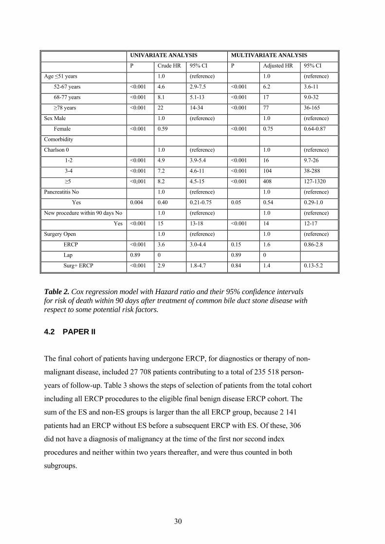
4.1.1 Factors Influencing Survival
Both age and sex influenced survival after treatment of common bile duct stones. The
influence of age was further increased in the multivariate analysis by interaction with
co-morbidity (Table 2). Female sex reduced the risk of dying, a difference that
remained in the multivariate analyses (adjusted HR 0.75 95% CI 0.64-0.87).
Comorbidity, measured using the Charlson comorbidity score, increased the risk of
death to a high degree and the influence was enhanced in the multivariate analyses
where a score ≥5 increased the risk of dying 400-fold (adjusted HR 408; 95% CI 127-
1320) due to interaction with age.
Septicemia did not affect the risk of death in the multivariate analyses while
pancreatitis surprisingly was associated to a significantly lower risk of dying.
29
The length of hospital stay had no impact on the risk of death and neither had a
readmission. On the contrary the occurrence of a biliary reintervention, either
endoscopic or transabdominal, within 90 days was associated to a 14-fold increased
risk of dying (adjusted HR 14; 95% CI 12-17).
The type of procedure seemed to affect 90-day mortality where ERCP had a crude HR
of 3.6 (95% CI 3.0-4.4) and 2.9 (95% CI 1.98-4.7) if combined with surgery compared
to open surgery. However, in the multivariate analyses the effect was reduced to a non-
significant trend (HR 1.6; 95% CI 0.86 – 2.8). As survival in the laparoscopic cohort
was close to complete even at long term follow-up the statistical power was not
sufficient for Cox regression for this exposure. It is however clear that survival in this
cohort is many-fold higher than in the other intervention cohorts.
30
UNIVARIATE ANALYSIS MULTIVARIATE ANALYSIS
P Crude HR 95% CI P Adjusted HR 95% CI
Age ≤51 years 1.0 (reference) 1.0 (reference)
52-67 years <0.001 4.6 2.9-7.5 <0.001 6.2 3.6-11
68-77 years <0.001 8.1 5.1-13 <0.001 17 9.0-32
≥78 years <0.001 22 14-34 <0.001 77 36-165
Sex Male 1.0 (reference) 1.0 (reference)
Female <0.001 0.59 <0.001 0.75 0.64-0.87
Comorbidity
Charlson 0 1.0 (reference) 1.0 (reference)
1-2 <0.001 4.9 3.9-5.4 <0.001 16 9.7-26
3-4 <0.001 7.2 4.6-11 <0.001 104 38-288
≥5 <0,001 8.2 4.5-15 <0.001 408 127-1320
Pancreatitis No 1.0 (reference) 1.0 (reference)
Yes 0.004 0.40 0.21-0.75 0.05 0.54 0.29-1.0
New procedure within 90 days No 1.0 (reference) 1.0 (reference)
Yes <0.001 15 13-18 <0.001 14 12-17
Surgery Open 1.0 (reference) 1.0 (reference)
ERCP <0.001 3.6 3.0-4.4 0.15 1.6 0.86-2.8
Lap 0.89 0 0.89 0
Surg+ ERCP <0.001 2.9 1.8-4.7 0.84 1.4 0.13-5.2
Table 2. Cox regression model with Hazard ratio and their 95% confidence intervals for risk of death within 90 days after treatment of common bile duct stone disease with respect to some potential risk factors.
4.2 PAPER II
The final cohort of patients having undergone ERCP, for diagnostics or therapy of non-
malignant disease, included 27 708 patients contributing to a total of 235 518 person-
years of follow-up. Table 3 shows the steps of selection of patients from the total cohort
including all ERCP procedures to the eligible final benign disease ERCP cohort. The
sum of the ES and non-ES groups is larger than the all ERCP group, because 2 141
patients had an ERCP without ES before a subsequent ERCP with ES. Of these, 306
did not have a diagnosis of malignancy at the time of the first nor second index
procedures and neither within two years thereafter, and were thus counted in both
subgroups.
31
ALL ERCP
ES ERCP WITHOUT ES
Total 54 135 30 431 25 846 Cancer date before entry 10 085 5 734 4 764 Error registration 3 192 3 000 329
Follow-up less than two years
13 150 9 068 5 368
Eligible 27 708 12 629 15 385
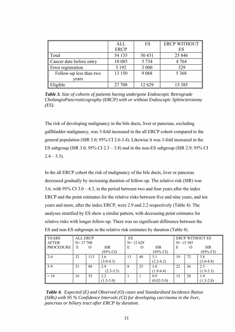
Table 3. Size of cohorts of patients having undergone Endoscopic Retrograde CholangioPancreaticography (ERCP) with or without Endoscopic Sphincterotomy (ES).
The risk of developing malignancy in the bile ducts, liver or pancreas, excluding
gallbladder malignancy, was 3-fold increased in the all ERCP cohort compared to the
general population (SIR 3.0; 95% CI 2.6-3.4). Likewise it was 3-fold increased in the
ES subgroup (SIR 3.0; 95% CI 2.3 – 3.8) and in the non-ES subgroup (SIR 2.9; 95% CI
2.4 – 3.3).
In the all ERCP cohort the risk of malignancy of the bile ducts, liver or pancreas
decreased gradually by increasing duration of follow-up. The relative risk (SIR) was
3.6, with 95% CI 3.0 – 4.3, in the period between two and four years after the index
ERCP and the point estimates for the relative risks between five and nine years, and ten
years and more, after the index ERCP, were 2.9 and 2.2 respectively (Table 4). The
analyses stratified by ES show a similar pattern, with decreasing point estimates for
relative risks with longer follow-up. There was no significant difference between the
ES and non-ES subgroups in the relative risk estimates by duration (Table 4).
YEARS AFTER PROCEDURE
ALL ERCP N= 27 708 E O SIR (95% CI)
ES N= 12 629 E O SIR (95% CI)
ERCP WITHOUT ES N= 15 385 E O SIR (95% CI)
2-4 32 113 3.6 (3.0-4.3)
13 40 3.1 (2.2-4.2)
19 72 3.8 (3.0-4.8)
5-9 31 88 2.9 (2.3-3.5)
8 25 3.0 (1.9-4.4)
22 56 2.5 (1.9-3.3)
> 10 16 35 2.2 (1.5-3.0)
1 1 0.9 (0.02-5.0)
15 28 1.9 (1.3-2.8)
Table 4. Expected (E) and Observed (O) cases and Standardized Incidence Ratios (SIRs) with 95 % Confidence Intervals (CI) for developing carcinoma in the liver, pancreas or biliary tract after ERCP by duration.
32
The risk of malignancy in the bile ducts, liver and pancreas two years or more after
ERCP was significantly lower among patients who ever had had a cholecystectomy
(SIR 2.3; 95% CI 1.8 – 2.9 in the all ERCP-group) compared to patients who had not
(SIR 3.4; 95% CI 3.0 – 4.0). This finding was not at all affected by ES exposure.
4.3 PAPER III
During the time period a total of 5750 ERCP procedures were performed in Stockholm
County in patients without a diagnosis of malignancy in the liver, pancreas or bile ducts
and 90 patients died within 90 days, giving a total mortality of 1.6%. In seven cases the
cause of death could be directly related to the procedure (one case of hemorrhage, two
of pancreatitis and four perforations), giving a procedure-caused mortality of 0.1%.
Characteristics of patients who had undergone ERCP for non-malignant disease are
shown in Table 5 together with basic information on their procedures.
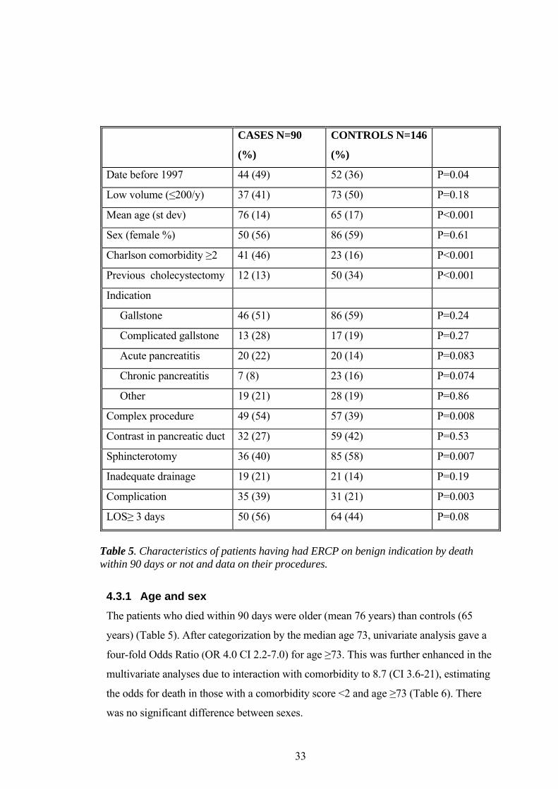
33
Table 5. Characteristics of patients having had ERCP on benign indication by death within 90 days or not and data on their procedures.
4.3.1 Age and sex
The patients who died within 90 days were older (mean 76 years) than controls (65
years) (Table 5). After categorization by the median age 73, univariate analysis gave a
four-fold Odds Ratio (OR 4.0 CI 2.2-7.0) for age ≥73. This was further enhanced in the
multivariate analyses due to interaction with comorbidity to 8.7 (CI 3.6-21), estimating
the odds for death in those with a comorbidity score <2 and age ≥73 (Table 6). There
was no significant difference between sexes.
CASES N=90
(%)
CONTROLS N=146
(%)
Date before 1997 44 (49) 52 (36) P=0.04
Low volume (≤200/y) 37 (41) 73 (50) P=0.18
Mean age (st dev) 76 (14) 65 (17) P<0.001
Sex (female %) 50 (56) 86 (59) P=0.61
Charlson comorbidity ≥2 41 (46) 23 (16) P<0.001
Previous cholecystectomy 12 (13) 50 (34) P<0.001
Indication
Gallstone 46 (51) 86 (59) P=0.24
Complicated gallstone 13 (28) 17 (19) P=0.27
Acute pancreatitis 20 (22) 20 (14) P=0.083
Chronic pancreatitis 7 (8) 23 (16) P=0.074
Other 19 (21) 28 (19) P=0.86
Complex procedure 49 (54) 57 (39) P=0.008
Contrast in pancreatic duct 32 (27) 59 (42) P=0.53
Sphincterotomy 36 (40) 85 (58) P=0.007
Inadequate drainage 19 (21) 21 (14) P=0.19
Complication 35 (39) 31 (21) P=0.003
LOS≥ 3 days 50 (56) 64 (44) P=0.08
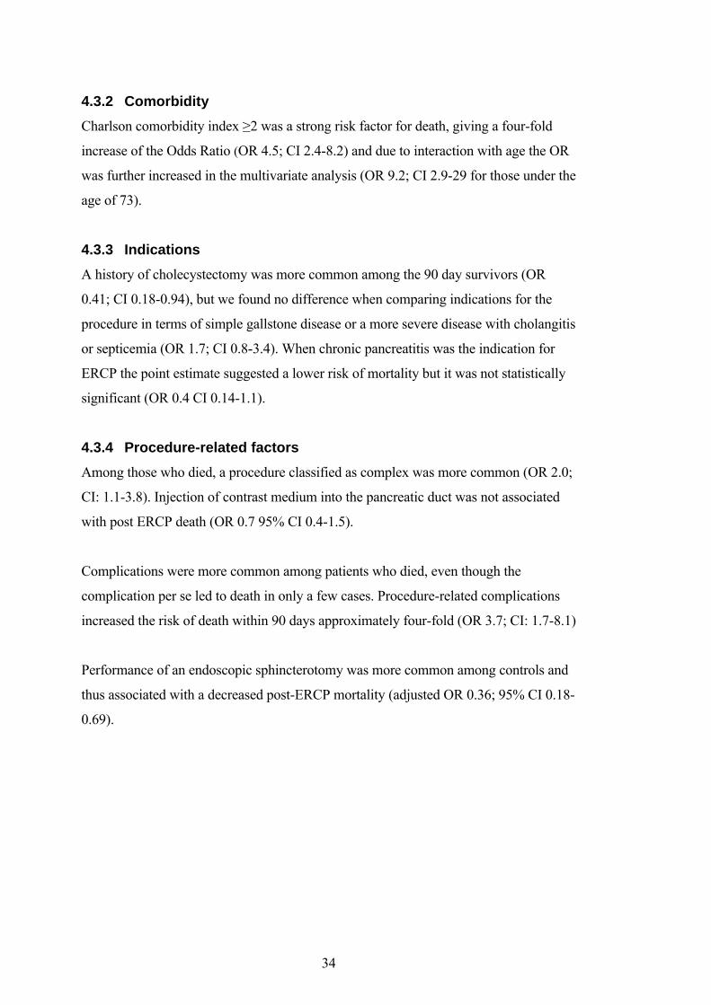
34
4.3.2 Comorbidity
Charlson comorbidity index ≥2 was a strong risk factor for death, giving a four-fold
increase of the Odds Ratio (OR 4.5; CI 2.4-8.2) and due to interaction with age the OR
was further increased in the multivariate analysis (OR 9.2; CI 2.9-29 for those under the
age of 73).
4.3.3 Indications
A history of cholecystectomy was more common among the 90 day survivors (OR
0.41; CI 0.18-0.94), but we found no difference when comparing indications for the
procedure in terms of simple gallstone disease or a more severe disease with cholangitis
or septicemia (OR 1.7; CI 0.8-3.4). When chronic pancreatitis was the indication for
ERCP the point estimate suggested a lower risk of mortality but it was not statistically
significant (OR 0.4 CI 0.14-1.1).
4.3.4 Procedure-related factors
Among those who died, a procedure classified as complex was more common (OR 2.0;
CI: 1.1-3.8). Injection of contrast medium into the pancreatic duct was not associated
with post ERCP death (OR 0.7 95% CI 0.4-1.5).
Complications were more common among patients who died, even though the
complication per se led to death in only a few cases. Procedure-related complications
increased the risk of death within 90 days approximately four-fold (OR 3.7; CI: 1.7-8.1)
Performance of an endoscopic sphincterotomy was more common among controls and
thus associated with a decreased post-ERCP mortality (adjusted OR 0.36; 95% CI 0.18-
0.69).
35
UNIVARIATE ANALYSIS
P OR 95% CI
MULTIVARIATE
ANALYSIS
P OR 95% CI
Age<73 years 1.0 (reference) 1.0 (reference)
Age≥73 years <0.001 4.0 2.2-7.0 <0.001 8.7 3.6-21
Gender (male) 1.0 (reference) 1.0 (reference)
Gender (female) 0.61 0.87 0.5-1.5 0.73 0.89 0.46-1.7
Charlson comorbidity<2 1.0 (reference) 1.0 (reference)
Charlson comorbidity≥2 <0.001 4.5 2.4-8.2 <0.001 9.2 2.9-29
Previous cholecystectomy
no 1.0 (reference) 1.0 (reference)
yes 0.001 0.30 0.15-0.60 0.035 0.41 0.18-0.94
Complex procedure
no 1.0 (reference) 1.0 (reference)
yes 0.009 2.1 1.2-3.7 0.036 2.0 1.1-3.8
Sphincterotomy
no 1.0 (reference) 1.0 (reference)
yes 0.007 0.5 0.3-0.8 0.002 0.36 0.18-0.69
Drainage
good 1.0 (reference) 1.0 (reference)
not good 0.20 0.64 0.32-1.3 0.85 0.92 0.38-2.2
Complication
no 1.0 (reference) 1.0 (reference)
yes 0.004 2.4 1.3-4.2 0.001 3.7 1.7-8.1
Table 6. Risk of death after ERCP with respect to some potential risk factors.
4.4 PAPER IV
Laparoscopic transcystic common bile duct exploration was commenced in 155
patients and was completed in 132 (85 %), without remaining CBD stones on the
second cholangiogram. Hospital stay and mean operating time were significantly longer
among patients where stone clearance failed. Stone size and common bile duct diameter
were both significantly larger in patients where stone clearance failed, compared to
36
those where it was successful, using conventional significance testing. There were no
significant differences between patients with successful and failed stone clearance
regarding age, sex, procedure priority, number of stones and intrahepatic stone location.
In 123 out of 132 stone free LTCE patients one or more stones were extracted. In the
remaining nine, no stone could be found at cholangioscopy, or in the second
cholangiogram, which could either be because of false positive first cholangiograms or
because of stone clearance between the cholangiogram and cholangioscopy, perhaps
due to flushing of saline through the bile duct at the onset of cholangioscopy. In 24% of
the patients an intrahepatic cholangioscopy was performed.
There were eight postoperative complications among the LTCE patients. Three patients
had haemorrhage requiring transfusion, two had pneumonia, one had pancreatitis, one
had pulmonary embolism and finally one had bile leakage needing percutaneous
drainage but no further intervention. One of the patients who had haemorrhage
requiring transfusion was reoperated because of an infection in the hematoma.
Otherwise no reoperations were needed. There was no mortality during the hospital
stay or at the follow-up six week postoperatively. Follow-up at six weeks was
completed in 91% of the patients and all of them were clinically well, but one had
elevated liver enzymes. A postoperative normal ERC was performed and an ES was
made as a precaution, although no retained CBD stone was found. An additional patient
had a postoperative ERC done due to a history of repeated biliary colic two years after
surgery. The ERC showed a CBD stone that was removed after ES. Apart from that no
patient was shown to have retained or recurrent stones.
In the logistic regression analyses there were no significant effects of age, sex or
number of stones on the risk of failure in stone clearance (Table 7).
Univariately, there was an almost 11-fold significant increase in the risk of failure in
stone clearance among patients with a common bile duct diameter of >6 mm compared
with those with a diameter of 6 mm (OR: 10.62; 95% CI: 1.39 – 81.48). However in
the multivariate analyses this effect decreased markedly and was no longer statistically
significant (OR: 6.90; 95% CI: 0.87 – 54.61). This difference was due to confounding
from the effect of stone size (Table 7).
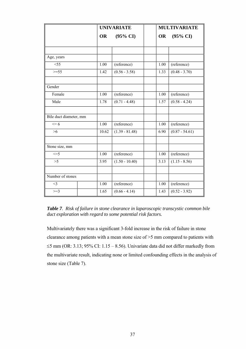
37
UNIVARIATE
OR (95% CI)
MULTIVARIATE
OR (95% CI)
Age, years
<55 1.00 (reference) 1.00 (reference)
>=55 1.42 (0.56 - 3.58) 1.33 (0.48 - 3.70)
Gender
Female 1.00 (reference) 1.00 (reference)
Male 1.78 (0.71 - 4.48) 1.57 (0.58 - 4.24)
Bile duct diameter, mm
<= 6 1.00 (reference) 1.00 (reference)
>6 10.62 (1.39 - 81.48) 6.90 (0.87 - 54.61)
Stone size, mm
<=5 1.00 (reference) 1.00 (reference)
>5 3.95 (1.50 - 10.40) 3.13 (1.15 - 8.56)
Number of stones
<3 1.00 (reference) 1.00 (reference)
>=3 1.65 (0.66 - 4.14) 1.43 (0.52 - 3.92)
Table 7. Risk of failure in stone clearance in laparoscopic transcystic common bile duct exploration with regard to some potential risk factors.
Multivariately there was a significant 3-fold increase in the risk of failure in stone
clearance among patients with a mean stone size of >5 mm compared to patients with
5 mm (OR: 3.13; 95% CI: 1.15 – 8.56). Univariate data did not differ markedly from
the multivariate result, indicating none or limited confounding effects in the analysis of
stone size (Table 7).
38
5 DISCUSSION During the last decades treatment of common bile duct stone disease has changed.
Open surgery has gradually been replaced by endoscopic surgery and in the later years
also by laparoscopic surgery to some extent. The central issue in this thesis was to try to
clarify how common bile duct stone disease may be treated in these times of changes.
Our hypothesis when starting the work was that laparoscopic treatment would be
superior to other methods of treatment, based on the reports on short- and long-term
side-effects of ERCP, especially ES. Reports on short-term side-effects included
pancreatitis, perforations, hemorrhage and cholangitis causing a substantial morbidity
and mortality [213] while long-term side-effects included reports on an elevated risk of
cholangiocarcinoma after endoscopic sphincterotomy. The under-lying mechanism
could be that the interruption of the sphincter would cause an over-growth of bacteria
with longstanding carcinogenic inflammation in the bile ducts that normally are sterile
[178, 181]. This could be alarming since endoscopic surgery in recent years has
become a routine procedure even in young and healthy individuals and not only in the
old and fragile as in the early days.
5.1 HOW HAVE COMMON BILE DUCT STONES BEEN TREATED IN
SWEDEN?
To address this question we conducted a population-based retrospective cohort study
(study I) which comprised all patients in Sweden 1965-2009 with an in-patient
procedure code of open or laparoscopic exploration of the common bile duct or ERCP
but without a diagnosis of malignancy in the bile ducts, liver or pancreas by the time of
the procedure or within 90 days after it. In the majority of these patients the indication
for the procedure would have been common bile duct stone disease. The cohort was
divided into four subgroups: open surgery, ERCP, laparoscopic surgery and finally
open and/or laparoscopic surgery combined with ERCP.
The study reflects treatment in a vast majority of the Swedish population and the
registries from which the data are collected have been validated as mentioned in the
methods-section giving an essentially complete follow-up. The Hospital Discharge
Registry was established in 1964 and included all Swedish Hospitals from 1987 and
onwards. It has been validated with a 94% agreement for surgical procedure codes
[214] and 87-95% for discharge diagnoses [215, 216], however the codes and
39
diagnoses in this study have not been validated specifically. Misclassification could
introduce information bias, although it seems unlikely that misclassification would
differ between the subgroups or regarding outcome status, meaning that introduced bias
would be non-differential and thereby diluting the strength of true associations. It was
however not possible to analyze the procedures on an intention to treat basis and
converted laparoscopic procedures are registered in the cohorts of open or combined
surgery. This could imply an over-representation of severe cases in these cohorts and a
corresponding under-representation in the laparoscopic cohort.
In the beginning of the studied time period open surgery was the only available method
for treatment of common bile duct stone disease but from the early 1990s and on
endoscopic treatment has become predominant. Laparoscopic treatment was introduced
in the early 1990s and has been used in a small proportion of cases.
5.1.1 What Risk Factors for Death Could Be Identified?
Age and comorbidity were identified as main risk factors for death. There were
differences between the subgroups in the way that patients treated with ERCP were
older and had a more severe comorbidity than those treated with open surgery, not
surprisingly, since ERCP was used in patients considered too old and frail to have open
surgery, at least in the beginning of the time period. Patients treated with laparoscopic
surgery were younger and healthier than the others and also more often of female sex, a
circumstance connected to a significant decreased risk of dying after surgery in this
study.
A new biliary procedure within 90 days, probably implying that clearance of the bile
duct was not achieved at the first attempt, was a significant risk factor for death while a
discharge code of acute pancreatitis was associated with a better survival. Both a redo
procedure and a diagnosis of pancreatitis were more common among ERCP-patients.
5.1.2 Did Mortality Differ?
The crude 90-day mortality was significantly higher after ERCP compared to after open
surgery but after adjusting for age and comorbidity this result was reduced to a trend.
The results after laparoscopic surgery were excellent with no 90-day mortality at all
recorded, but that must be interpreted with caution, since it to some extent could be
explained by a case-selection described above. The laparoscopy subgroup consisted of
40
members of lower age and comorbidity and the fact that the procedure could be
completed laparoscopically could imply a less severe disease.
5.2 IS THE RISK OF DEVELOPMENT OF MALIGNANCY ELEVATED
AFTER ENDOSCOPIC SPHINCTEROTOMY?
A cohort study (study II) was designed to evaluate whether the longstanding
inflammation caused by ascending infection after interruption of the sphincter of Oddi
would lead to an increased risk of cholangiocarcinoma. The population-based cohort
comprised all individuals in Sweden with a discharge code of an in-patient ERCP with
or without ES and without a diagnosis of malignancy in the bile ducts, liver or pancreas
at the time of the procedure or within two years after it up to the year 2003.
Since cholangiocarcinoma is a rare disease and there is a risk of missclassification
between extrahepatic cholangiocarcinoma and pancreatic cancer as well as intrahepatic
cholangiocarcinoma and liver cancer both cholangiocarcinoma alone and all three
tumours were analyzed.
We found a three-fold elevated risk of malignancy in the whole cohort, i e disregarding
if an ES had been performed or not, compared to the general population. The risk
declined with time. These results imply that the elevated risk of malignancy was not a
result of the ES per se but rather of some other exposure common for the entire cohort,
the most likely would be gallstone disease, a notion that to some extent could support
the finding that a previous cholecystectomy in this study was shown to be associated to
a lower risk of malignancy.
5.3 DOES ENDOSCOPIC SPHINCTEROTOMY INCREASE SHORT-TERM
COMPLICATIONS AFTER ERCP?
A case-control study (study III) was designed to evaluate short-term outcome after
ERCP, especially mortality, and to study risk factors for death. The main hypothesis
was that a potentially dangerous procedure like sphincterotomy would be such a risk
factor. It was performed on a subgroup of patients included in paper II. Cases were
defined as all individuals who had had an ERCP in Stockholm County during 1990-
2003 and who died within 90 days of the procedure and as controls served a random
sample from the same subgroup. The population-based design was chosen to reduce the
41
risk of bias in case and control selection. Risk factors for death were discerned from the
medical records, to reduce information bias the parts of the medical records regarding
the ERCP procedure were coded and reviewed blinded for case and control status.
The all cause mortality was 1.6% but only seven deaths (0.1%) were directly
procedure-related. Advanced age and severe co-morbidity were identified as the main
risk factors for death. A complex procedure, here defined as the presence of a duodenal
diverticulum, multiple attempts of cannulation, difficult position of the papilla or the
performance of a pre-cut sphincterotomy, was also identified as a risk factor as well as
the occurrence of a post-procedural complication. A previous cholecystectomy served
protective for death, possibly by reducing the severity of the common bile duct stone
disease by the time of the ERCP, even though that could not be confirmed in the
medical records.
In contrast to our prior hypothesis the performance of a sphincterotomy could not be
identified as a risk factor for death, on the contrary it reduced the risk, a condition that
remained significant in the multivariate analyses (OR 0.36, 95% CI 0.18-0.69), thereby
not explained by the factors we adjusted for (age, comorbidity, date of procedure,
previous cholecystectomy, complex procedure or the occurrence of a complication). A
possible explanation could be that a completed sphincterotomy could reduce the risk of
cholangitis by facilitating a good drainage from the bile ducts.
5.4 WHAT IS THE SHORT-TERM OUTCOME OF LAPAROSCOPIC
TRANSCYSTIC COMMON BILE DUCT EXPLORATION AND WHEN
DOES IT FAIL?
To address these issues we conducted a study of patients in whom LTCE were
attempted at S: t Göran’s Hospital during the years 1994- 2002. The study included all
patients, thereby reducing the risk of selection bias. Data had been prospectively
collected in a data-base and analyses showed that stone clearance was achieved in 85%.
There was no recorded 30-day mortality (in agreement with the findings in study I), a
low mortality and a short postoperative hospital stay. As risk factors for mortality stone
size >5 mm could be identified and probably also a wide bile duct (>6mm).
42
5.5 GENERAL DISCUSSION
Our main hypothesis, that laparoscopic common bile duct exploration would be
superior to other methods of treatment of common bile duct stones, could not be proven
by this thesis. Indeed, laparoscopic treatment was safe and no 90-day mortality was
reported in Sweden in cases where it could be fulfilled, yet it was applied in a minority
of patients. We could identify large stones as a risk factor for failure in laparoscopic
transcystic exploration, in laparoscopic choledochotomy stone size is not regarded as an
obstacle [195, 200] and the reason why laparoscopic treatment was refrained from in a
majority of cases is not clear. We can only speculate- perhaps the methods are regarded
as complicated or the instruments fragile or difficult to handle. Laparoscopic treatment
was chosen mainly in young and otherwise healthy subjects and the complexity of the
bile duct stone disease could have had impact, patients with less severe disease could
have been selected to laparoscopy. When laparoscopic treatment was chosen stone
clearance could be achieved in a majority of cases, but not in all, and there is a need for
alternative methods.
The by far most common way to treat gallstones and common bile duct stones in later
years is by a combination of laparoscopic cholecystectomy and endoscopic treatment of
the bile duct stones. Since ERCP and especially ES have been reported with a
substantial morbidity and mortality [177-180, 217] and also with an increased risk of
malignancy in the long run [181] this has been a matter of some concern. Our study
showed an increased risk of development of a malignancy in all patients having had an
ERCP on benign indication but the risk was elevated regardless of if an ES was
performed or not and diminished with time thereby contradicting that the ES per se
would be a causal factor. The 90-day mortality after ERCP was substantial in our study
and as risk factors advanced age and severe comorbidity could be identified while the
performance of an ES reduced the risk of dying. Our studies do not support the reports
that the performance of a sphincterotomy would be a causal factor of short- or long-
term mortality and morbidity after ERCP.
43
6 CONCLUSIONS Treatment of patients with gallbladder stones and simultaneous stones in the common
bile duct has changed. Earlier all patients had open surgery while in the recent years a
combination of laparoscopic cholecystectomy and ERCP is predominant even though
open surgery and laparoscopic treatment are also used.
The mortality after ERCP in benign disease was higher than after open surgery but
when adjusting for age and comorbidity the difference was reduced to an insignificant
trend.
Laparoscopic treatment of common bile duct stones was safe but could not be used in
all patients.
The risk of malignancy in the bile ducts, liver and pancreas after ERCP on benign
indication was elevated in all patients regardless of if an ES was performed or not and
diminished with time contradicting that ES would be a causal factor.
The main risk factors for death after ERCP in benign disease were old age and
advanced co-morbidity, while a procedure considered complex or causing a
complication contributed to the risk. The performance of an ES reduced the risk of
dying within 90 days after the procedure and the risk was also reduced in patients
having had a previous cholecystectomy.
Stone size > 5 mm was a risk factor for failure in laparoscopic transcystic exploration
of the common bile duct.
44
7 POPULÄRVETENSKAPLIG SAMMANFATTNING
Gallstensjukdom är vanlig och årligen utförs drygt 10 000 galloperationer i Sverige.
Mellan 5-15 % av patienterna har även sten i de djupa gallvägarna (koledokussten) vid
operationen. Dessa patienter behandlades under större delen av 1900-talet i en seans
med öppen kirurgi. Under 1970-talet kom endoskopiska metoder där man med ett
flexibelt endoskop via munnen kunde genomföra kontraströntgen och avlägsna stenar i
gallvägarna efter klyvning av sfinkter Oddi, ringmuskeln runt gallvägens mynning i
tolvfingertarmen (endoskopisk retrograd cholangiopankreatografi, ERCP, med
endoskopisk sfinkterotomi). Sfinkterotomi visade sig vara förenad med risk för
allvarliga komplikationer och reserverades först för patienter som ansågs vara för
gamla eller sjuka för att klara öppen kirurgi. Farhågor framfördes också att delningen
av sfinktern skulle kunna ge en kronisk infektion i gallvägarna genom tillträde för
bakterier från tolvfingertarmen och att denna kroniska infektion/inflammation skulle ge
en ökad risk för cancer på lång sikt.
Senare, under 1990-talet, introducerades galloperation via titthål (laparoskopisk
kolecystektomi) och denna metod blev snart helt dominerande. Behandlingen av
samtidig sten i gallgången blev mer varierande och öppen kirurgi, kombination av
endoskopisk och laparoskopisk teknik i en eller två seanser har använts olika på olika
sjukhus sannolikt beroende på lokal behandlingstradition.
Syftet med denna avhandling var att kartlägga hur patienter med samtidig sten i
gallgång och gallblåsa behandlats i Sverige under denna period av förändringar (1965-
2009), att beräkna dödlighet efter olika behandlingsmetoder och att identifiera
riskfaktorer för död. Svängningen i behandlingstradition har inneburit att även unga
patienter genomgår ERCP och sfinkterotomi vilket skulle kunna vara alarmerande om
cancerrisken ökar på lång sikt, detta ville vi undersöka närmare. En ökad användning
av ett potentiellt farligt ingrepp som sfinkterotomi skulle kunna vara farligt även på kort
sikt varför vi ville identifiera riskfaktorer för död efter ERCP, och särskilt studera om
sfinkterotomi var en sådan riskfaktor. På S: t Görans sjukhus har sedan 1994 den
dominerande tekniken varit att behandla samtidig sten i gallblåsa och gallgång i en
seans, där gallblåsan och stenarna i gallgången avlägsnas med titthålskirurgi och vi ville
utforska resultatet samt om metoden har några begränsningar.
45
Fyra delarbeten ingår:
1. Behandling av koledokussten i Sverige 1965-2009, en landsomfattande
populationsbaserad studie.
2. Endoskopisk sfinkterotomi och risk för malignitet i gallvägar, lever och
bukspottskörtel.
3. Endoskopisk sfinkterotomi kan minska risken för död efter ERCP- en
populationsbaserad fall-kontroll studie.
4. Stenfrihet och riskfaktorer för misslyckande att åstadkomma detta vid
titthålsoperation av gallsten i gallvägarna.
Delarbete 1 omfattar alla personer i Sverige som vårdats inneliggande för en operation
av sten i gallvägarna eller ERCP under åren 1965-2009. ERCP görs också vid
cancersjukdom i gallvägar, lever och bukspottskörtel och efter samkörning med
cancerregistret kunde dessa individer identifieras och exkluderas ur studien. Totalt
110 119 personer identifierades och dessa hade genomgått totalt 126 889 ingrepp.
Dödligheten efter olika ingrepp skiljde sig, efter öppen operation dog 0,24 %, efter
ERCP 0,90 % och efter titthålskirurgi dog ingen person inom 90 dagar. Vid närmare
analys visade sig skillnaden i dödlighet efter öppen kirurgi och ERCP förklaras av att
patienterna som genomgått ERCP var äldre och sjukare.
Delarbete 2 omfattar alla personer i Sverige som genomgått ERCP under åren 1965-
2003 utan att ha en diagnos av malignitet i gallvägar, lever eller bukspottskörtel vid
ingreppet eller inom två år därefter. I cancerregistret identifierades de som senare
utvecklade malignitet. Det visade sig att risken att utveckla cancer i gallvägar, lever
eller bukspottskörtel var större hos alla dessa patienter jämfört med
normalbefolkningen men att risken minskade med ökande uppföljningstid och inte
påverkades om sfinkter Oddi delades eller inte.
Delarbete 3 omfattar en del av personerna i delarbete 2, de som genomgått ERCP i
Stockholms län 1990-2003. De som dött inom 90 dagar efter ingreppet jämfördes med
slumpvis utvalda kontroller bland dem som inte dött. Som riskfaktorer för att dö kunde
46
hög ålder, sjuklighet, ett komplicerat ingrepp och att en komplikation tillstötte
identifieras medan om personen ifråga tidigare genomgått galloperation eller om
klyvning av sfinkter Oddi utfördes vid det aktuella ingreppet minskade risken för död.
Delarbete 4 omfattar alla patienter som opererats på S: t Görans sjukhus under perioden
1994-2002 där man försökt ta bort stenar i gallgången med en typ av titthålskirurgi.
Den metod som användes var transcystisk exploration där gången mellan gallblåsan
och djupa gallvägen öppnas och vidgas. Stenarna i gallgången fångas med hjälp av ett
tunt endoskop som förs in denna väg och de kan sedan avlägsnas en efter en. Av de 155
patienter där man inlett denna operation kunde stenfrihet uppnås hos 85 % och som
riskfaktor för misslyckande identifierades stenstorlek, hos patienter med
koledokusstenar större än 5 mm ökade risken för misslyckande trefaldigt.
Slutsatser: Dödlighet efter behandling av choledochussten skiljer sig mellan olika
behandlingsmetoder. Dödligheten är högre efter ERCP än efter öppen kirurgi men detta
förklaras av att patienterna som genomgår ERCP är äldre och sjukare. Dödligheten
efter laparoskopisk gallvägskirurgi är noll inom 90 dagar, dessa patienter är dock yngre,
friskare och har möjligen en mindre avancerad gallstenssjukdom.
Risken för att utveckla cancer i gallvägar, lever och bukspottskörtel är högre hos alla
patienter som genomgått ERCP än hos normalbefolkningen. Risken avtar med ökande
uppföljningstid och påverkas inte av om sfinkter Oddi klyvs eller inte, utan orsakas av
någon annan faktor som dessa patienter har gemensamt, sannolikt gallstenssjukdom.
Hög ålder, sjuklighet, svårt och komplext ingrepp ökar risken för död efter ERCP
liksom om en komplikation uppträder efter ingreppet. Den potentiellt farliga
klyvningen av sfinkter Oddi minskar däremot risken sannolikt p.g.a av att dränaget från
gallgången förbättras och risken för uppåtstigande infektion i gallvägarna minskar.
Laparoskopisk behandling av sten i gallvägarna genom en transcystisk exploration är
en metod som ger få komplikationer och kort vårdtid när den är genomförbar. Den kan
dock inte användas hos alla patienter och i denna studie kunde stenstorlek över 5 mm
identifieras som en riskfaktor för misslyckande.
47
8 ACKNOWLEDGEMENTS
I would like to express my sincere gratitude to all those who have contributed and
helped me in different ways in completing this thesis, especially to
Magnus Nilsson, main supervisor, head of upper GI surgery, friend and champagne-
advisor, for your never-ending enthusiasm, patience, scientific know-how and your
rapidness to respond. Without you it would not have been possible to teach the old dog
to sit!
Urban Arnelo, co-supervisor and ERCP-guru, for knowledge and constructive ideas
Lars Enochsson, co-supervisor, for guidance, support and help to defeat the computers
Carl-Eric Leijonmarck co-supervisor, head of the upper GI-team at S: t Göran’s
Hospital, friend and former colleague, for patience when introducing me to the world of
laparoscopic surgery, ERCP and science
Susanna Axelsson external mentor and old friend from high-school, always
encouraging even though this project seemed to be never-ending.
Jörgen Larsson, for support, encouragement and strength when dealing with
bureaucracy
Johan Permert, for giving me the opportunity to finish this work
Matthias Löhr, co-author, for valuable work and advice with Paper III and for help to
fund several weeks of research time
Juhua Luo, for priceless help with the statistics of paper II
Helene Jansson, for support and the final finish of the manuscript
Bengt Isaksson, Lars Lundell, Thorhallur Agustsson, Anders Jansson, Mats Savlid,
Gunnar Söderdahl, Henrik Gjertsen, Rafal Dlugosz and Greg Nowak, colleagues at the
liver team, for your dedicated work for a continuous improvement of liver surgery and
sharing with
Åke Andrén Sandberg, Mikael Wirén, Ralf Segersvärd, Mari Hult, Cristoph Ansorge,
John Blomberg, Ann Kjellin, Mats Lindblad, Per Lundquist, Lisa Strömmer, Fredrik
Swahn, Jon Tsai and Björn Törnqvist and all other colleagues of the team of Upper GI
surgery, your enthusiasm creating a warm, open and exciting atmosphere to work in
Lotta Orre, bff
Anne Waage, for letting me steal your wonderful ideas

48
My sisters Anna, Antonia and Gabriella and your families for good laughs and long
discussions in times of better or worse
My father Anders and Trudi for being exceedingly helpful, always welcoming and
hospitable in your home in “Festerås”
My mother Eva for your support and belief in me and to Ove who tragically died way
too early
Linnea, for your sence of humour, knowledge of English, for being a wonderful person
and my daughter and to Ivar for being the dog that you are
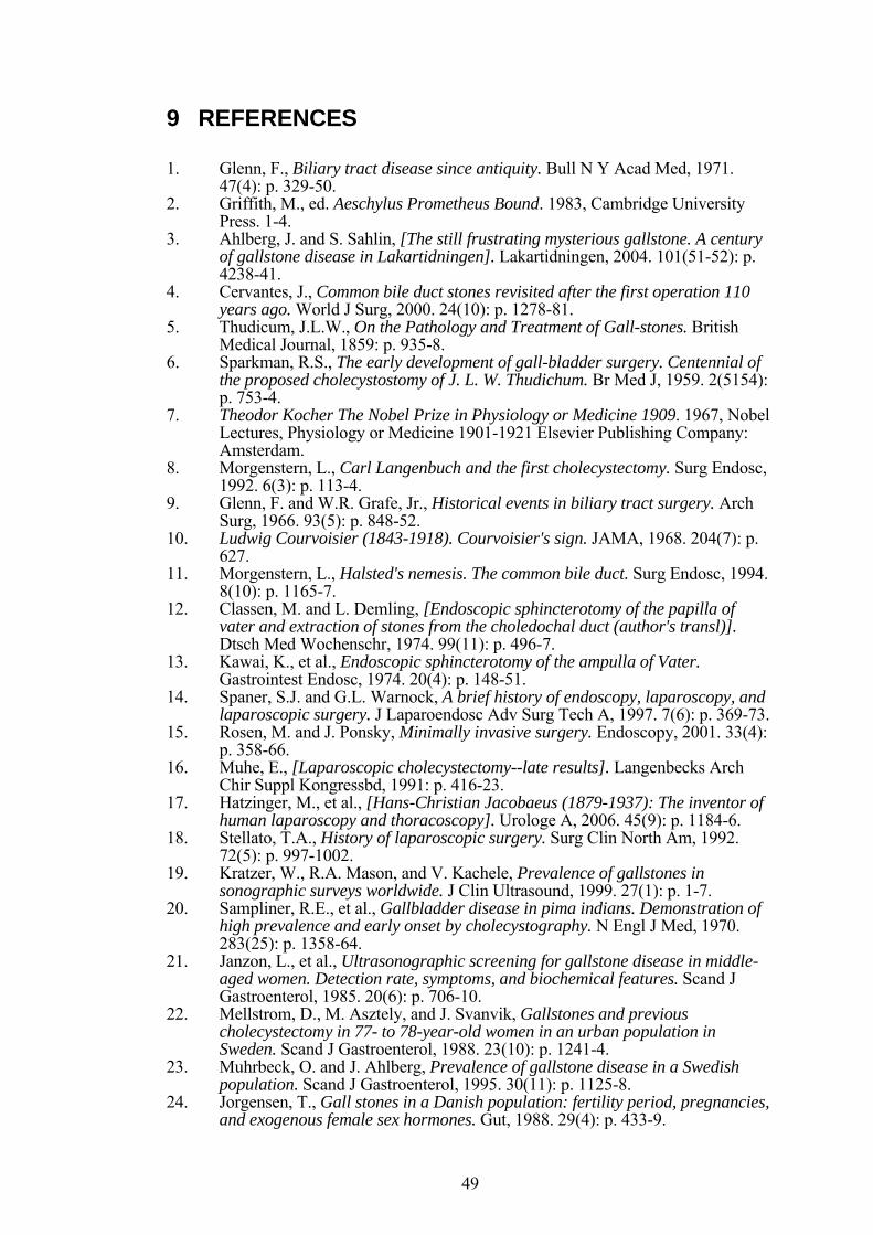
49
9 REFERENCES 1. Glenn, F., Biliary tract disease since antiquity. Bull N Y Acad Med, 1971.
47(4): p. 329-50. 2. Griffith, M., ed. Aeschylus Prometheus Bound. 1983, Cambridge University
Press. 1-4. 3. Ahlberg, J. and S. Sahlin, [The still frustrating mysterious gallstone. A century
of gallstone disease in Lakartidningen]. Lakartidningen, 2004. 101(51-52): p. 4238-41.
4. Cervantes, J., Common bile duct stones revisited after the first operation 110 years ago. World J Surg, 2000. 24(10): p. 1278-81.
5. Thudicum, J.L.W., On the Pathology and Treatment of Gall-stones. British Medical Journal, 1859: p. 935-8.
6. Sparkman, R.S., The early development of gall-bladder surgery. Centennial of the proposed cholecystostomy of J. L. W. Thudichum. Br Med J, 1959. 2(5154): p. 753-4.
7. Theodor Kocher The Nobel Prize in Physiology or Medicine 1909. 1967, Nobel Lectures, Physiology or Medicine 1901-1921 Elsevier Publishing Company: Amsterdam.
8. Morgenstern, L., Carl Langenbuch and the first cholecystectomy. Surg Endosc, 1992. 6(3): p. 113-4.
9. Glenn, F. and W.R. Grafe, Jr., Historical events in biliary tract surgery. Arch Surg, 1966. 93(5): p. 848-52.
10. Ludwig Courvoisier (1843-1918). Courvoisier's sign. JAMA, 1968. 204(7): p. 627.
11. Morgenstern, L., Halsted's nemesis. The common bile duct. Surg Endosc, 1994. 8(10): p. 1165-7.
12. Classen, M. and L. Demling, [Endoscopic sphincterotomy of the papilla of vater and extraction of stones from the choledochal duct (author's transl)]. Dtsch Med Wochenschr, 1974. 99(11): p. 496-7.
13. Kawai, K., et al., Endoscopic sphincterotomy of the ampulla of Vater. Gastrointest Endosc, 1974. 20(4): p. 148-51.
14. Spaner, S.J. and G.L. Warnock, A brief history of endoscopy, laparoscopy, and laparoscopic surgery. J Laparoendosc Adv Surg Tech A, 1997. 7(6): p. 369-73.
15. Rosen, M. and J. Ponsky, Minimally invasive surgery. Endoscopy, 2001. 33(4): p. 358-66.
16. Muhe, E., [Laparoscopic cholecystectomy--late results]. Langenbecks Arch Chir Suppl Kongressbd, 1991: p. 416-23.
17. Hatzinger, M., et al., [Hans-Christian Jacobaeus (1879-1937): The inventor of human laparoscopy and thoracoscopy]. Urologe A, 2006. 45(9): p. 1184-6.
18. Stellato, T.A., History of laparoscopic surgery. Surg Clin North Am, 1992. 72(5): p. 997-1002.
19. Kratzer, W., R.A. Mason, and V. Kachele, Prevalence of gallstones in sonographic surveys worldwide. J Clin Ultrasound, 1999. 27(1): p. 1-7.
20. Sampliner, R.E., et al., Gallbladder disease in pima indians. Demonstration of high prevalence and early onset by cholecystography. N Engl J Med, 1970. 283(25): p. 1358-64.
21. Janzon, L., et al., Ultrasonographic screening for gallstone disease in middle-aged women. Detection rate, symptoms, and biochemical features. Scand J Gastroenterol, 1985. 20(6): p. 706-10.
22. Mellstrom, D., M. Asztely, and J. Svanvik, Gallstones and previous cholecystectomy in 77- to 78-year-old women in an urban population in Sweden. Scand J Gastroenterol, 1988. 23(10): p. 1241-4.
23. Muhrbeck, O. and J. Ahlberg, Prevalence of gallstone disease in a Swedish population. Scand J Gastroenterol, 1995. 30(11): p. 1125-8.
24. Jorgensen, T., Gall stones in a Danish population: fertility period, pregnancies, and exogenous female sex hormones. Gut, 1988. 29(4): p. 433-9.
50
25. Sauerbruch, T. and G. Paumgartner, Gallbladder stones: management. Lancet, 1991. 338(8775): p. 1121-4.
26. Mjaland, O., et al., Cholecystectomy rates, gallstone prevalence, and handling of bile duct injuries in Scandinavia. A comparative audit. Surg Endosc, 1998. 12(12): p. 1386-9.
27. Rosenmuller, M., et al., Cholecystectomy in Sweden 2000-2003: a nationwide study on procedures, patient characteristics, and mortality. BMC Gastroenterol, 2007. 7: p. 35.
28. Whiting, M.J., B.M. Bradley, and J.M. Watts, Chemical and physical properties of gall stones in South Australia: implications for dissolution treatment. Gut, 1983. 24(1): p. 11-5.
29. Marschall, H.U. and C. Einarsson, Gallstone disease. J Intern Med, 2007. 261(6): p. 529-42.
30. Cuevas, A., et al., Diet as a risk factor for cholesterol gallstone disease. J Am Coll Nutr, 2004. 23(3): p. 187-96.
31. Nervi, F., et al., Influence of legume intake on biliary lipids and cholesterol saturation in young Chilean men. Identification of a dietary risk factor for cholesterol gallstone formation in a highly prevalent area. Gastroenterology, 1989. 96(3): p. 825-30.
32. Donovan, J.M., Physical and metabolic factors in gallstone pathogenesis. Gastroenterol Clin North Am, 1999. 28(1): p. 75-97.
33. Tazuma, S., Gallstone disease: Epidemiology, pathogenesis, and classification of biliary stones (common bile duct and intrahepatic). Best Pract Res Clin Gastroenterol, 2006. 20(6): p. 1075-83.
34. Mirizzi, P.L., [Internal spontaneous bilio-biliary fistulas.]. J Chir (Paris), 1952. 68(1): p. 32-46.
35. Venneman, N.G. and K.J. van Erpecum, Pathogenesis of gallstones. Gastroenterol Clin North Am. 39(2): p. 171-83, vii.
36. Trotman, B.W., Pigment gallstone disease. Gastroenterol Clin North Am, 1991. 20(1): p. 111-26.
37. Vitek, L. and M.C. Carey, Enterohepatic cycling of bilirubin as a cause of 'black' pigment gallstones in adult life. Eur J Clin Invest, 2003. 33(9): p. 799-810.
38. Kraag, N., C. Thijs, and P. Knipschild, Dyspepsia--how noisy are gallstones? A meta-analysis of epidemiologic studies of biliary pain, dyspeptic symptoms, and food intolerance. Scand J Gastroenterol, 1995. 30(5): p. 411-21.
39. Berger, M.Y., et al., Abdominal symptoms: do they predict gallstones? A systematic review. Scand J Gastroenterol, 2000. 35(1): p. 70-6.
40. Muhrbeck, O., Symptoms of gallstone disease in a Swedish population. Eur J Gastroenterol Hepatol, 1995. 7(12): p. 1209-14.
41. Halldestam, I., et al., Development of symptoms and complications in individuals with asymptomatic gallstones. Br J Surg, 2004. 91(6): p. 734-8.
42. Friedman, G.D., Natural history of asymptomatic and symptomatic gallstones. Am J Surg, 1993. 165(4): p. 399-404.
43. Schirmer, B.D., K.L. Winters, and R.F. Edlich, Cholelithiasis and cholecystitis. J Long Term Eff Med Implants, 2005. 15(3): p. 329-38.
44. Festi, D., et al., Clinical manifestations of gallstone disease: evidence from the multicenter Italian study on cholelithiasis (MICOL). Hepatology, 1999. 30(4): p. 839-46.
45. Schoenfield, L.J., et al., Gallstones. West J Med, 1976. 124(4): p. 299-315. 46. Diehl, A.K., Symptoms of gallstone disease. Baillieres Clin Gastroenterol, 1992.
6(4): p. 635-57. 47. Graham, E.A., W.H. Cole, and G.H. Copher, Rontgenological Visualization of
the Gall-Bladder by the Intravenous Injection of Tetrabromphenolphthalein. Ann Surg, 1924. 80(3): p. 473-7.
48. Anderson, J.C. and R.K. Harned, Gray scale ultrasonography of the gallbladder: an evaluation of accuracy and report of additional ultrasound signs. AJR Am J Roentgenol, 1977. 129(6): p. 975-7.
49. Leopold, G.R., et al., Gray scale ultrasonic cholecystography: a comparison with conventional radiographic techniques. Radiology, 1976. 121(2): p. 445-8.
51
50. Fendrick, A.M., et al., Asymptomatic gallstones revisited. Is there a role for laparoscopic cholecystectomy? Arch Fam Med, 1993. 2(9): p. 959-68.
51. Gurusamy, K.S. and K. Samraj, Cholecystectomy versus no cholecystectomy in patients with silent gallstones. Cochrane Database Syst Rev, 2007(1): p. CD006230.
52. Gurusamy, K.S. and B.R. Davidson, Surgical treatment of gallstones. Gastroenterol Clin North Am. 39(2): p. 229-44.
53. Turrill, F.L., C.M. Mc, and W.P. Mikkelsen, Gallstones and diabetes: an ominous association. Am J Surg, 1961. 102: p. 184-90.
54. Friedman, L.S., et al., Management of asymptomatic gallstones in the diabetic patient. A decision analysis. Ann Intern Med, 1988. 109(11): p. 913-9.
55. Aucott, J.N., et al., Management of gallstones in diabetic patients. Arch Intern Med, 1993. 153(9): p. 1053-8.
56. Toouli, J., Symptomatic gallstones: management options for the 1990s. HPB Surg, 1991. 4(4): p. 255-60.
57. NIH Consensus conference. Gallstones and laparoscopic cholecystectomy. JAMA, 1993. 269(8): p. 1018-24.
58. Halldestam, I., E. Kullman, and K. Borch, Defined indications for elective cholecystectomy for gallstone disease. Br J Surg, 2008. 95(5): p. 620-6.
59. Ransohoff, D.F. and W.A. Gracie, Treatment of gallstones. Ann Intern Med, 1993. 119(7 Pt 1): p. 606-19.
60. McSherry, C.K., et al., The natural history of diagnosed gallstone disease in symptomatic and asymptomatic patients. Ann Surg, 1985. 202(1): p. 59-63.
61. Gracie, W.A. and D.F. Ransohoff, The natural history of silent gallstones: the innocent gallstone is not a myth. N Engl J Med, 1982. 307(13): p. 798-800.
62. Roslyn, J.J., et al., Roles of lithogenic bile and cystic duct occlusion in the pathogenesis of acute cholecystitis. Am J Surg, 1980. 140(1): p. 126-30.
63. Jivegard, L., E. Thornell, and J. Svanvik, Pathophysiology of acute obstructive cholecystitis: implications for non-operative management. Br J Surg, 1987. 74(12): p. 1084-6.
64. Thornell, E., et al., Effect of short-term indomethacin treatment on the clinical course of acute obstructive cholecystitis. Eur J Surg, 1991. 157(2): p. 127-30.
65. Truedson, H., T. Elmros, and S. Holm, The incidence of bacteria in gallbladder bile at acute and elective cholecystectomy. Acta Chir Scand, 1983. 149(3): p. 307-13.
66. Bergan, T., I. Dobloug, and I. Liavag, Bacterial isolates in cholecystitis and cholelithiasis. Scand J Gastroenterol, 1979. 14(5): p. 625-31.
67. Keus, F., I.A. Broeders, and C.J. van Laarhoven, Gallstone disease: Surgical aspects of symptomatic cholecystolithiasis and acute cholecystitis. Best Pract Res Clin Gastroenterol, 2006. 20(6): p. 1031-51.
68. Lankisch, P.G., et al., Acute pancreatitis: does gender matter? Dig Dis Sci, 2001. 46(11): p. 2470-4.
69. Armstrong, C.P., et al., The biliary tract in patients with acute gallstone pancreatitis. Br J Surg, 1985. 72(7): p. 551-5.
70. Tandon, M. and M. Topazian, Endoscopic ultrasound in idiopathic acute pancreatitis. Am J Gastroenterol, 2001. 96(3): p. 705-9.
71. Lee, S.P., J.F. Nicholls, and H.Z. Park, Biliary sludge as a cause of acute pancreatitis. N Engl J Med, 1992. 326(9): p. 589-93.
72. Acosta, J.M. and C.L. Ledesma, Gallstone migration as a cause of acute pancreatitis. N Engl J Med, 1974. 290(9): p. 484-7.
73. Wang, G.J., et al., Acute pancreatitis: etiology and common pathogenesis. World J Gastroenterol, 2009. 15(12): p. 1427-30.
74. Frossard, J.L., M.L. Steer, and C.M. Pastor, Acute pancreatitis. Lancet, 2008. 371(9607): p. 143-52.
75. Andersen, A.M., et al., Mortality in alcohol and biliary acute pancreatitis. Pancreas, 2008. 36(4): p. 432-4.
76. Yadav, D. and A.B. Lowenfels, Trends in the epidemiology of the first attack of acute pancreatitis: a systematic review. Pancreas, 2006. 33(4): p. 323-30.
52
77. Garcea, G., et al., Predictors of severity and survival in acute pancreatitis: validation of the efficacy of early warning scores. Pancreas, 2008. 37(3): p. e54-61.
78. Taylor, S.L., et al., A comparison of the Ranson, Glasgow, and APACHE II scoring systems to a multiple organ system score in predicting patient outcome in pancreatitis. Am J Surg, 2005. 189(2): p. 219-22.
79. McKay, C.J. and C.W. Imrie, Staging of acute pancreatitis. Is it important? Surg Clin North Am, 1999. 79(4): p. 733-43.
80. Cameron, D.R. and A.J. Goodman, Delayed cholecystectomy for gallstone pancreatitis: re-admissions and outcomes. Ann R Coll Surg Engl, 2004. 86(5): p. 358-62.
81. Alimoglu, O., et al., Timing of cholecystectomy for acute biliary pancreatitis: outcomes of cholecystectomy on first admission and after recurrent biliary pancreatitis. World J Surg, 2003. 27(3): p. 256-9.
82. Burch, J.M., et al., Gallstone pancreatitis. The question of time. Arch Surg, 1990. 125(7): p. 853-9; discussion 859-60.
83. Nebiker, C.A., et al., Early versus delayed cholecystectomy in patients with biliary acute pancreatitis. Surgery, 2009. 145(3): p. 260-4.
84. Uhl, W., et al., Acute gallstone pancreatitis: timing of laparoscopic cholecystectomy in mild and severe disease. Surg Endosc, 1999. 13(11): p. 1070-6.
85. Uhl, W., et al., IAP Guidelines for the Surgical Management of Acute Pancreatitis. Pancreatology, 2002. 2(6): p. 565-73.
86. Fan, S.T., et al., Early treatment of acute biliary pancreatitis by endoscopic papillotomy. N Engl J Med, 1993. 328(4): p. 228-32.
87. Neoptolemos, J.P., et al., ERCP findings and the role of endoscopic sphincterotomy in acute gallstone pancreatitis. Br J Surg, 1988. 75(10): p. 954-60.
88. Petrov, M.S., et al., Early endoscopic retrograde cholangiopancreatography versus conservative management in acute biliary pancreatitis without cholangitis: a meta-analysis of randomized trials. Ann Surg, 2008. 247(2): p. 250-7.
89. Ayantunde, A.A. and A. Agrawal, Gallstone ileus: diagnosis and management. World J Surg, 2007. 31(6): p. 1292-7.
90. De la Fuente, H., et al., [Duodenal obstruction caused by cholelithiasis (Bouverets' syndrome): a clinical case]. Rev Med Chil, 1989. 117(7): p. 785-8.
91. Osman, N., et al., Gallstone ileus of the sigmoid colon: an unusual cause of large-bowel obstruction. HPB Surg. 2010: p. 153-40.
92. Behrens, C. and B. Amson, Laparoscopic management of multiple gallstone ileus. Surg Laparosc Endosc Percutan Tech. 20(2): p. 64-5.
93. Ravikumar, R. and J.G. Williams, The operative management of gallstone ileus. Ann R Coll Surg Engl. 92(4): p. 279-81.
94. Pitt, H.A., et al., Malignancies of the biliary tree. Curr Probl Surg, 1995. 32(1): p. 1-90.
95. Tewari, M., Contribution of silent gallstones in gallbladder cancer. J Surg Oncol, 2006. 93(8): p. 629-32.
96. Diehl, A.K., Gallstone size and the risk of gallbladder cancer. JAMA, 1983. 250(17): p. 2323-6.
97. Shrikhande, S.V., et al., Cholelithiasis in gallbladder cancer: coincidence, cofactor, or cause! Eur J Surg Oncol. 36(6): p. 514-9.
98. Riciardi, R., et al., Effectiveness and long-term results of laparoscopic common bile duct exploration. Surg Endosc, 2003. 17(1): p. 19-22.
99. Shojaiefard, A., et al., Various techniques for the surgical treatment of common bile duct stones: a meta review. Gastroenterol Res Pract, 2009. 2009: p. 840208.
100. Ferris, D.O., N.R. Thomford, and J.C. Cain, Recurrent Common Bile Duct Stones. Arch Surg, 1964. 88: p. 486-9.
53
101. Leese, T., J.P. Neoptolemos, and D.L. Carr-Locke, Successes, failures, early complications and their management following endoscopic sphincterotomy: results in 394 consecutive patients from a single centre. Br J Surg, 1985. 72(3): p. 215-9.
102. Williams, E.J., et al., Guidelines on the management of common bile duct stones (CBDS). Gut, 2008. 57(7): p. 1004-21.
103. Bergman, J.J., et al., Long-term follow-up after endoscopic sphincterotomy for bile duct stones in patients younger than 60 years of age. Gastrointest Endosc, 1996. 44(6): p. 643-9.
104. Ando, T., et al., Risk factors for recurrent bile duct stones after endoscopic papillotomy. Gut, 2003. 52(1): p. 116-21.
105. Pereira-Lima, J.C., et al., Long-term results (7 to 10 years) of endoscopic papillotomy for choledocholithiasis. Multivariate analysis of prognostic factors for the recurrence of biliary symptoms. Gastrointest Endosc, 1998. 48(5): p. 457-64.
106. Paganini, A.M., et al., Long-term results after laparoscopic transverse choledochotomy for common bile duct stones. Surg Endosc, 2005. 19(5): p. 705-9.
107. Waage, A., et al., Long-term results from laparoscopic common bile duct exploration. Surg Endosc, 2003. 17(8): p. 1181-5.
108. Csendes, A., et al., Mirizzi syndrome and cholecystobiliary fistula: a unifying classification. Br J Surg, 1989. 76(11): p. 1139-43.
109. Gomez, D., et al., Mirizzi's syndrome - results from a large western experience. HPB (Oxford), 2006. 8(6): p. 474-9.
110. Rosseland, A.R. and T.B. Glomsaker, Asymptomatic common bile duct stones. Eur J Gastroenterol Hepatol, 2000. 12(11): p. 1171-3.
111. Menezes, N., et al., Prospective analysis of a scoring system to predict choledocholithiasis. Br J Surg, 2000. 87(9): p. 1176-81.
112. Sarli, L., et al., Scoring system to predict asymptomatic choledocholithiasis before laparoscopic cholecystectomy. A matched case-control study. Surg Endosc, 2003. 17(9): p. 1396-403.
113. Trondsen, E., et al., Prediction of common bile duct stones prior to cholecystectomy: a prospective validation of a discriminant analysis function. Arch Surg, 1998. 133(2): p. 162-6.
114. Metcalfe, M.S., et al., Is laparoscopic intraoperative cholangiogram a matter of routine? Am J Surg, 2004. 187(4): p. 475-81.
115. Leese, T., et al., Management of acute cholangitis and the impact of endoscopic sphincterotomy. Br J Surg, 1986. 73(12): p. 988-92.
116. Caddy, G.R. and T.C. Tham, Gallstone disease: Symptoms, diagnosis and endoscopic management of common bile duct stones. Best Pract Res Clin Gastroenterol, 2006. 20(6): p. 1085-101.
117. Besselink, M.G., et al., Is complicated gallstone disease preceded by biliary colic? J Gastrointest Surg, 2009. 13(2): p. 312-7.
118. Yeh, B.M., et al., MR imaging and CT of the biliary tract. Radiographics, 2009. 29(6): p. 1669-88.
119. Lichtenbaum, R.A., H.F. McMullen, and R.M. Newman, Preoperative abdominal ultrasound may be misleading in risk stratification for presence of common bile duct abnormalities. Surg Endosc, 2000. 14(3): p. 254-7.
120. Sugiyama, M. and Y. Atomi, Endoscopic ultrasonography for diagnosing choledocholithiasis: a prospective comparative study with ultrasonography and computed tomography. Gastrointest Endosc, 1997. 45(2): p. 143-6.
121. Gross, B.H., et al., Ultrasonic evaluation of common bile duct stones: prospective comparison with endoscopic retrograde cholangiopancreatography. Radiology, 1983. 146(2): p. 471-4.
122. Anderson, S.W., et al., Accuracy of MDCT in the diagnosis of choledocholithiasis. AJR Am J Roentgenol, 2006. 187(1): p. 174-80.
123. Anderson, S.W., E. Rho, and J.A. Soto, Detection of biliary duct narrowing and choledocholithiasis: accuracy of portal venous phase multidetector CT. Radiology, 2008. 247(2): p. 418-27.
54
124. Neitlich, J.D., et al., Detection of choledocholithiasis: comparison of unenhanced helical CT and endoscopic retrograde cholangiopancreatography. Radiology, 1997. 203(3): p. 753-7.
125. Läkemedelsindustriföreningen, FASS : förteckning över humanläkemedel. 2009. 2009, Stockholm: Läkemedelsindustriföreningen (LIF). 3032 s.
126. Halefoglu, A.M., Magnetic resonance cholangiopancreatography: a useful tool in the evaluation of pancreatic and biliary disorders. World J Gastroenterol, 2007. 13(18): p. 2529-34.
127. Scaffidi, M.G., et al., Magnetic resonance cholangio-pancreatography versus endoscopic retrograde cholangio-pancreatography in the diagnosis of common bile duct stones: a prospective comparative study. Minerva Med, 2009. 100(5): p. 341-8.
128. Norero, E., et al., [Accuracy of magnetic resonance cholangiopancreatography for the diagnosis of common bile duct stones]. Rev Med Chil, 2008. 136(5): p. 600-5.
129. Ledro-Cano, D., Suspected choledocholithiasis: endoscopic ultrasound or magnetic resonance cholangio-pancreatography? A systematic review. Eur J Gastroenterol Hepatol, 2007. 19(11): p. 1007-11.
130. Nandalur, K.R., et al., Possible biliary disease: diagnostic performance of high-spatial-resolution isotropic 3D T2-weighted MRCP. Radiology, 2008. 249(3): p. 883-90.
131. Edmundowicz, S.A., G. Aliperti, and W.D. Middleton, Preliminary experience using endoscopic ultrasonography in the diagnosis of choledocholithiasis. Endoscopy, 1992. 24(9): p. 774-8.
132. Garrow, D., et al., Endoscopic ultrasound: a meta-analysis of test performance in suspected biliary obstruction. Clin Gastroenterol Hepatol, 2007. 5(5): p. 616-23.
133. Tse, F., et al., EUS: a meta-analysis of test performance in suspected choledocholithiasis. Gastrointest Endosc, 2008. 67(2): p. 235-44.
134. Hammarstrom, L.E., et al., Routine preoperative infusion cholangiography at elective cholecystectomy: a prospective study in 694 patients. Br J Surg, 1996. 83(6): p. 750-4.
135. Majeed, A.W., et al., Common duct diameter as an independent predictor of choledocholithiasis: is it useful? Clin Radiol, 1999. 54(3): p. 170-2.
136. Jarhult, J., Is preoperative evaluation of the biliary tree necessary in uncomplicated gallstone disease? Results of a randomized trial. Scand J Surg, 2005. 94(1): p. 31-3.
137. Ammori, B.J., et al., Routine vs "on demand" postoperative ERCP for small bile duct calculi detected at intraoperative cholangiography. Clinical evaluation and cost analysis. Surg Endosc, 2000. 14(12): p. 1123-6.
138. Collins, C., et al., A prospective study of common bile duct calculi in patients undergoing laparoscopic cholecystectomy: natural history of choledocholithiasis revisited. Ann Surg, 2004. 239(1): p. 28-33.
139. Toouli, J. and T.A. Wright, Gallstones. Med J Aust, 1998. 169(3): p. 166-71. 140. Thistle, J.L., Direct contact dissolution therapy. Baillieres Clin Gastroenterol,
1992. 6(4): p. 715-25. 141. Helmstadter, A., Ether and the chemical-contact dissolution of gallstones.
Lancet, 1999. 354(9187): p. 1376-7. 142. Det bästa ur Nordisk familjebok. 1. uppl. ed. 2008, Stockholm: Ruin. [68] s. 143. Strasberg, S.M. and P.A. Clavien, Overview of therapeutic modalities for the
treatment of gallstone diseases. Am J Surg, 1993. 165(4): p. 420-6. 144. Carrilho-Ribeiro, L., et al., A ten-year prospective study on gallbladder stone
recurrence after successful extracorporeal shock-wave lithotripsy. Scand J Gastroenterol, 2006. 41(3): p. 338-42.
145. Nicholl, J.P., et al., Cost effectiveness of adjuvant bile salt treatment in extracorporeal shock wave lithotripsy for the treatment of gall bladder stones. Gut, 1994. 35(9): p. 1294-300.
146. Testoni, P.A., et al., Combined endoscopic and extracorporeal shock-wave treatment in difficult bile duct stones: early and long-term results. Ital J Gastroenterol, 1994. 26(6): p. 294-8.
55
147. Neuhaus, H., et al., Randomized study of intracorporeal laser lithotripsy versus extracorporeal shock-wave lithotripsy for difficult bile duct stones. Gastrointest Endosc, 1998. 47(5): p. 327-34.
148. Seitz, U., et al., Advances in therapeutic endoscopic treatment of common bile duct stones. World J Surg, 1998. 22(11): p. 1133-44.
149. Boerma, D. and M.P. Schwartz, Gallstone disease. Management of common bile-duct stones and associated gallbladder stones: Surgical aspects. Best Pract Res Clin Gastroenterol, 2006. 20(6): p. 1103-16.
150. Targarona, E.M., et al., Randomised trial of endoscopic sphincterotomy with gallbladder left in situ versus open surgery for common bileduct calculi in high-risk patients. Lancet, 1996. 347(9006): p. 926-9.
151. Dubois, F. and B. Berthelot, [Cholecystectomy through minimal incision (author's transl)]. Nouv Presse Med, 1982. 11(15): p. 1139-41.
152. Allori, A.C., I.M. Leitman, and E. Heitman, Delayed assessment and eager adoption of laparoscopic cholecystectomy: Implications for developing surgical technologies. World J Gastroenterol. 16(33): p. 4115-22.
153. Keus, F., H.G. Gooszen, and C.J. van Laarhoven, Open, small-incision, or laparoscopic cholecystectomy for patients with symptomatic cholecystolithiasis. An overview of Cochrane Hepato-Biliary Group reviews. Cochrane Database Syst Rev, (1): p. CD008318.
154. Ito, M., et al., Cholecystectomy using single-incision laparoscopic surgery with a new SILS port. J Hepatobiliary Pancreat Sci. 17(5): p. 688-91.
155. Marescaux, J., et al., Surgery without scars: report of transluminal cholecystectomy in a human being. Arch Surg, 2007. 142(9): p. 823-6; discussion 826-7.
156. Navarra, G., et al., SILS and NOTES cholecystectomy: a tailored approach. J Laparoendosc Adv Surg Tech A. 20(6): p. 511-4.
157. Deziel, D.J., et al., Complications of laparoscopic cholecystectomy: a national survey of 4,292 hospitals and an analysis of 77,604 cases. Am J Surg, 1993. 165(1): p. 9-14.
158. Fletcher, D.R., et al., Complications of cholecystectomy: risks of the laparoscopic approach and protective effects of operative cholangiography: a population-based study. Ann Surg, 1999. 229(4): p. 449-57.
159. MacFadyen, B.V., Jr., et al., Bile duct injury after laparoscopic cholecystectomy. The United States experience. Surg Endosc, 1998. 12(4): p. 315-21.
160. Andren-Sandberg, A., G. Alinder, and S. Bengmark, Accidental lesions of the common bile duct at cholecystectomy. Pre- and perioperative factors of importance. Ann Surg, 1985. 201(3): p. 328-32.
161. Waage, A. and M. Nilsson, Iatrogenic bile duct injury: a population-based study of 152 776 cholecystectomies in the Swedish Inpatient Registry. Arch Surg, 2006. 141(12): p. 1207-13.
162. Kelly, M.D., Laparoscopic retrograde (fundus first) cholecystectomy. BMC Surg, 2009. 9: p. 19.
163. Dolan, J.P., J.W. Cook, and B.C. Sheppard, Retained common bile duct stone as a consequence of a fundus-first laparoscopic cholecystectomy. J Laparoendosc Adv Surg Tech A, 2005. 15(3): p. 318-21.
164. Hunter, J.G., Avoidance of bile duct injury during laparoscopic cholecystectomy. Am J Surg, 1991. 162(1): p. 71-6.
165. Strasberg, S.M., Avoidance of biliary injury during laparoscopic cholecystectomy. J Hepatobiliary Pancreat Surg, 2002. 9(5): p. 543-7.
166. Mirizzi, P.L., Operative cholangiography. Surg Gynecol Obstet, 1937. 65: p. 702-710.
167. Hulten, O., Cholangiographie wahrend der operation. Deutsche Zeitscr Chir, 1938. 250: p. 484.
168. Adamsen, S., et al., Bile duct injury during laparoscopic cholecystectomy: a prospective nationwide series. J Am Coll Surg, 1997. 184(6): p. 571-8.
169. Ludwig, K., et al., Contribution of intraoperative cholangiography to incidence and outcome of common bile duct injuries during laparoscopic cholecystectomy. Surg Endosc, 2002. 16(7): p. 1098-104.
56
170. Phillips, E.H., Routine versus selective intraoperative cholangiography. Am J Surg, 1993. 165(4): p. 505-7.
171. Regoly-Merei, J., et al., Biliary tract complications in laparoscopic cholecystectomy. A multicenter study of 148 biliary tract injuries in 26,440 operations. Surg Endosc, 1998. 12(4): p. 294-300.
172. Dubois, F., G. Berthelot, and H. Levard, Coelioscopic cholecystectomy: experience with 2006 cases. World J Surg, 1995. 19(5): p. 748-52.
173. Tranter, S.E. and M.H. Thompson, Potential of laparoscopic ultrasonography as an alternative to operative cholangiography in the detection of bile duct stones. Br J Surg, 2001. 88(1): p. 65-9.
174. Horwood, J., et al., Prospective evaluation of a selective approach to cholangiography for suspected common bile duct stones. Ann R Coll Surg Engl. 92(3): p. 206-10.
175. Way, L.W., W.H. Admirand, and J.E. Dunphy, Management of choledocholithiasis. Ann Surg, 1972. 176(3): p. 347-59.
176. Neoptolemos, J.P., D.L. Carr-Locke, and D.P. Fossard, Prospective randomised study of preoperative endoscopic sphincterotomy versus surgery alone for common bile duct stones. Br Med J (Clin Res Ed), 1987. 294(6570): p. 470-4.
177. Coppola, R., et al., Analysis of complications of endoscopic sphincterotomy for biliary stones in a consecutive series of 546 patients. Surg Endosc, 1997. 11(2): p. 129-32.
178. Tranter, S.E. and M.H. Thompson, Comparison of endoscopic sphincterotomy and laparoscopic exploration of the common bile duct. Br J Surg, 2002. 89(12): p. 1495-504.
179. Tanaka, M., Bile duct clearance, endoscopic or laparoscopic? J Hepatobiliary Pancreat Surg, 2002. 9(6): p. 729-32.
180. Sugiyama, M. and Y. Atomi, Follow-up of more than 10 years after endoscopic sphincterotomy for choledocholithiasis in young patients. Br J Surg, 1998. 85(7): p. 917-21.
181. Tanaka, M., et al., Long-term consequence of endoscopic sphincterotomy for bile duct stones. Gastrointest Endosc, 1998. 48(5): p. 465-9.
182. Mo, L.R., et al., Preoperative endoscopic sphincterotomy in the treatment of patients with cholecystocholedocholithiasis. J Hepatobiliary Pancreat Surg, 2002. 9(2): p. 191-5.
183. Neuhaus, H., et al., Prospective evaluation of the use of endoscopic retrograde cholangiography prior to laparoscopic cholecystectomy. Endoscopy, 1992. 24(9): p. 745-9.
184. Tham, T.C., et al., Role of endoscopic retrograde cholangiopancreatography for suspected choledocholithiasis in patients undergoing laparoscopic cholecystectomy. Gastrointest Endosc, 1998. 47(1): p. 50-6.
185. Rhodes, M., et al., Randomised trial of laparoscopic exploration of common bile duct versus postoperative endoscopic retrograde cholangiography for common bile duct stones. Lancet, 1998. 351(9097): p. 159-61.
186. Cuschieri, A., et al., EAES ductal stone study. Preliminary findings of multi-center prospective randomized trial comparing two-stage vs single-stage management. Surg Endosc, 1996. 10(12): p. 1130-5.
187. Rabago, L.R., et al., Two-stage treatment with preoperative endoscopic retrograde cholangiopancreatography (ERCP) compared with single-stage treatment with intraoperative ERCP for patients with symptomatic cholelithiasis with possible choledocholithiasis. Endoscopy, 2006. 38(8): p. 779-86.
188. Enochsson, L., et al., Intraoperative endoscopic retrograde cholangiopancreatography (ERCP) to remove common bile duct stones during routine laparoscopic cholecystectomy does not prolong hospitalization: a 2-year experience. Surg Endosc, 2004. 18(3): p. 367-71.
189. Ghazal, A.H., et al., Single-step treatment of gall bladder and bile duct stones: a combined endoscopic-laparoscopic technique. Int J Surg, 2009. 7(4): p. 338-46.
190. Enochsson, L., ERCP-tillbaka till framtiden. Gastrokuriren, 2009: p. 25-27.
57
191. McAlister, V.C., E. Davenport, and E. Renouf, Cholecystectomy deferral in patients with endoscopic sphincterotomy. Cochrane Database Syst Rev, 2007(4): p. CD006233.
192. Drake, B.B., et al., Economical and clinical outcomes of alternative treatment strategies in the management of common bile duct stones in the elderly: wait and see or surgery? Am J Gastroenterol, 2006. 101(4): p. 746-52.
193. Costi, R., et al., Routine laparoscopic cholecystectomy after endoscopic sphincterotomy for choledocholithiasis in octogenarians: is it worth the risk? Surg Endosc, 2007. 21(1): p. 41-7.
194. Schreurs, W.H., et al., Endoscopic management of common bile duct stones leaving the gallbladder in situ. A cohort study with long-term follow-up. Dig Surg, 2004. 21(1): p. 60-4; discussion 65.
195. Leijonmarck, C.E., Laparoscopic management of common bile duct stones. Eur J Surg Suppl, 2000(585): p. 22-6.
196. Ludwig, K., D. Lorenz, and F. Koeckerling, Surgical strategies in the laparoscopic therapy of cholecystolithiasis and common duct stones. ANZ J Surg, 2002. 72(8): p. 547-52.
197. Tokumura, H., et al., Laparoscopic management of common bile duct stones: transcystic approach and choledochotomy. J Hepatobiliary Pancreat Surg, 2002. 9(2): p. 206-12.
198. Lyass, S. and E.H. Phillips, Laparoscopic transcystic duct common bile duct exploration. Surg Endosc, 2006. 20 Suppl 2: p. S441-5.
199. Tang, C.N., et al., Laparoscopic exploration of the common bile duct: 10-year experience of 174 patients from a single centre. Hong Kong Med J, 2006. 12(3): p. 191-6.
200. Petelin, J.B., Laparoscopic common bile duct exploration. Surg Endosc, 2003. 17(11): p. 1705-15.
201. Leida, Z., et al., A randomized comparison of primary closure and T-tube drainage of the common bile duct after laparoscopic choledochotomy. Surg Endosc, 2008. 22(7): p. 1595-600.
202. Martin, I.J., et al., Towards T-tube free laparoscopic bile duct exploration: a methodologic evolution during 300 consecutive procedures. Ann Surg, 1998. 228(1): p. 29-34.
203. Gurusamy, K.S. and K. Samraj, Primary closure versus T-tube drainage after laparoscopic common bile duct stone exploration. Cochrane Database Syst Rev, 2007(1): p. CD005641.
204. Fall, K., W. Ye, and O. Nyren, Risk for gastric cancer after cholecystectomy. Am J Gastroenterol, 2007. 102(6): p. 1180-4.
205. Ye, W., et al., Alcohol abuse and the risk of pancreatic cancer. Gut, 2002. 51(2): p. 236-9.
206. Wall, S., M. Rosen, and L. Nystrom, The Swedish mortality pattern: a basis for health planning? Int J Epidemiol, 1985. 14(2): p. 285-92.
207. de Faire, U., et al., A validation of cause-of-death certification in 1,156 deaths. Acta Med Scand, 1976. 200(3): p. 223-8.
208. Nyren, O., et al., Cancer risk after hip replacement with metal implants: a population-based cohort study in Sweden. J Natl Cancer Inst, 1995. 87(1): p. 28-33.
209. Klassifikation av operationer sjätte upplagan. 1988. 210. Klassifikation av kirurgiska åtgärder 1997. 1997. 211. Breslow NE, D.N., The design and analysis of cohort studies Vol. II.
Lyon:IARC Sci Publ, 1987 Statistical methods in cancer research.). . 212. Vittinghoff, E., Regression methods in biostatistics : linear, logistic, survival,
and repeated measures models. Statistics for biology and health. 2005, New York: Springer. xv, 340 s.
213. Bilbao, M.K., et al., Complications of endoscopic retrograde cholangiopancreatography (ERCP). A study of 10,000 cases. Gastroenterology, 1976. 70(3): p. 314-20.
214. Nilsson, A.C., et al., [Reliability of the hospital registry. The diagnostic data are better than their reputation]. Lakartidningen, 1994. 91(7): p. 598, 603-5.
58
215. Ingelsson, E., et al., The validity of a diagnosis of heart failure in a hospital discharge register. Eur J Heart Fail, 2005. 7(5): p. 787-91.
216. Ragnarson Tennvall, G., J. Apelqvist, and M. Eneroth, The inpatient care of patients with diabetes mellitus and foot ulcers. A validation study of the correspondence between medical records and the Swedish Inpatient Registry with the consequences for cost estimations. J Intern Med, 2000. 248(5): p. 397-405.
217. Sees, D.W. and R.R. Martin, Comparison of preoperative endoscopic retrograde cholangiopancreatography and laparoscopic cholecystectomy with operative management of gallstone pancreatitis. Am J Surg, 1997. 174(6): p. 719-22.