Embed Size (px)
Citation preview

Cell and Tissue TypesEpithelial, Connective, Muscle, Nerve

Objectives• Explain the major stages of the cell cycle and cellular division
(mitosis).
• Describe specific events occurring in each of the phases of cellular division (mitosis) including cytokinesis.
• Explain how cellular division (mitosis) functions to form new cells and maintain complex organisms.
• Explain how a mutation might cause the cell cycle to be altered to allow for uncontrolled cell growth.
• Relate the development of cancer (uncontrolled growth) to mutations that affect the proteins that regulate the cell cycle

Essential question
What can you infer about how cell division in a normal cell compares to cell division in a cancer cell?

Types of Tissues – Epithelial, Connective, Muscle, Nervous
Type Function Location Characteristics
Epithelial Protection, SecretionAbsorption, Excretion
Cover body surfaces, cover and line internal organs, compose glands
Lack, blood vessels, readily divide; cells are tightly packed.
Connective Bind, Support, protect, fill spaces, store fat, produce blood cells.
Widely distributed throughout the body.
Mostly have good blood supply; cells are further apart than epithelial cells.
Muscle Movement Attached to bones, in the walls of hollow internal organs, heart
Able to contract in response to specific stimuli
Nervous Transmit impulses for coordination, regulation, integration, and sensory reception
Brain, spinal cord, nerves Cells communicate with each other and with other body parts

Stratified
Simple
Apical surface
Basal surface
Apical surface
Basal surface
Classification based on number of cell layers.

Squamous
Cuboidal
Columnar
Classification based on cell shape.
The following types of epithelial tissues are covered in this activity:
1. Simple squamous epithelial tissue (lungs)
2. Simple cuboidal epithelial tissue (kidneys)
3. Simple columnar epithelial tissue (small intestine)
4. Pseudostratified (ciliated) columnar epithelial tissue (trachea lining)
5. Stratified squamous epithelial tissue (mouth lining)
6. Stratified cuboidal epithelial tissue (salivary glands, sweat glands)
7. Stratified columnar epithelial tissue (male reproductive tract)
8 Transitional epithelial tissue (bladder)
a. The tissue may show a full bladder
b. The tissue may show an empty bladder

.
(a) Simple squamous epithelium
Description: Single layer of flattenedcells with disc-shaped central nucleiand sparse cytoplasm; the simplestof the epithelia.
Function: Allows passage ofmaterials by diffusion and filtrationin sites where protection is notimportant; secretes lubricatingsubstances in serosae.
Location: Kidney glomeruli; air sacsof lungs; lining of heart, bloodvessels, and lymphatic vessels; liningof ventral body cavity (serosae).
Photomicrograph: Simple squamous epithelium
forming part of the alveolar (air sac) walls (125x).
Air sacs oflung tissue
Nuclei ofsquamousepithelialcells

(b) Simple cuboidal epithelium
Description: Single layer ofcubelike cells with large,spherical central nuclei.
Function: Secretion and
absorption.
Location: Kidney tubules;ducts and secretory portionsof small glands; ovary surface.
Photomicrograph: Simple cuboidalepithelium in kidney tubules (430x).
Basementmembrane
Connectivetissue
Simplecuboidalepithelialcells

(c) Simple columnar epithelium
Description: Single layer of tall cells
with round to oval nuclei; some cells
bear cilia; layer may contain mucus-
secreting unicellular glands (goblet cells).
Function: Absorption; secretion of
mucus, enzymes, and other substances;
ciliated type propels mucus (or
reproductive cells) by ciliary action.
Location: Nonciliated type lines most of
the digestive tract (stomach to anal canal),
gallbladder, and excretory ducts of some
glands; ciliated variety lines small
bronchi, uterine tubes, and some regions
of the uterus.
Photomicrograph: Simple columnar epithelium
of the stomach mucosa (860X).
Simple
columnar
epithelial
cell
Basement
membrane

(d) Pseudostratified columnar epithelium
Description: Single layer of cells ofdiffering heights, some not reachingthe free surface; nuclei seen atdifferent levels; may contain mucus-secreting cells and bear cilia.
Function: Secretion, particularly ofmucus; propulsion of mucus byciliary action.
Location: Nonciliated type in male’ssperm-carrying ducts and ducts oflarge glands; ciliated variety linesthe trachea, most of the upperrespiratory tract.
Photomicrograph: Pseudostratified ciliatedcolumnar epithelium lining the human trachea (570x).
Trachea
Cilia
Pseudo-stratifiedepitheliallayer
Basementmembrane
Mucus ofmucous cell

.
(e) Stratified squamous epithelium
Description: Thick membranecomposed of several cell layers;basal cells are cuboidal or columnarand metabolically active; surfacecells are flattened (squamous); in thekeratinized type, the surface cells arefull of keratin and dead; basal cellsare active in mitosis and produce thecells of the more superficial layers.
Function: Protects underlyingtissues in areas subjected to abrasion.
Location: Nonkeratinized type formsthe moist linings of the esophagus,mouth, and vagina; keratinized varietyforms the epidermis of the skin, a drymembrane.
Photomicrograph: Stratified squamous epithelium
lining the esophagus (285x).
Stratifiedsquamousepithelium
Nuclei
Basementmembrane
Connectivetissue
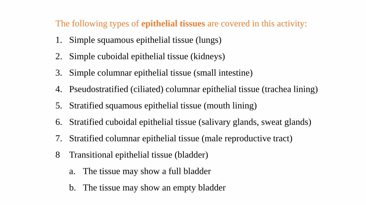
(f) Transitional epithelium
Description: Resembles both
stratified squamous and stratified
cuboidal; basal cells cuboidal or
columnar; surface cells dome
shaped or squamouslike, depending
on degree of organ stretch.
Function: Stretches readily and
permits distension of urinary organ
by contained urine.
Location: Lines the ureters, urinary
bladder, and part of the urethra.
Photomicrograph: Transitional epithelium lining the urinary
bladder, relaxed state (360X); note the bulbous, or rounded,
appearance of the cells at the surface; these cells flatten and
become elongated when the bladder is filled with urine.
BasementmembraneConnectivetissue
Transitional
epithelium

CFU

How does the structure of each type of tissue support its function?

Vocabulary
• Epithelial Tissue simple squamous epithelium
• simple cuboidal epithelium simple columnar epithelium
• pseudostratified columnar epithelium stratified squamous epithelium
• stratified cuboidal epithelium stratified columnar epithelium
• transitional epithelium glandular epithelium
• connective tissue loss connective tissue
• dense connective tissue cartilage
• bone blood
• epithelial membranes serous membranes
• mucous membranes cutaneous membrane
• synovial membrane

Connective Tissue Characteristics

CT- Major Cell Types

CT – Fiber Types

Connective Tissues

CT - Types

Connective Tissue Types

Connective Tissue Types

Connective Tissue Types

Connective Tissue Types

Skeletal Muscle Tissue
• Found in Muscle that attach to bone.
• Voluntary - controlled by conscious effort.
• Striations – alternating light and dark cross-markings.
MuscleFiber
Nuclei

Smooth Muscle Tissue
• Do not have striations.
• Cells are shorter than skeletal muscle and are spindle shaped, each with a centrally located nucleus.
• Found in hollow internal organs such as stomach, intestine, urinary bladder, uterus, and blood vessels.
• Involuntary – cannot be stimulated to contract by conscious effort.

Cardiac Muscle Tissue
• Cells are short, branched, and striated, usually with a single nucleus.
• Cells are interconnected by intercalated discs.
• Controlled involuntarily.
• Found in the heart.
• It circulates blood, maintains blood pressure.

Nervous Tissues
• Basic cell is called a neuron.
• They sense certain types of changes in their surroundings.
• They respond by transmitting nerve impulses along the axon to other neurons.
• Found in the brain, spinal cord and peripheral nerves.