Embed Size (px)
Citation preview

Cell Communicatio
n For cells to function in a biological
system, they must communicate with other cells and respond to their
external environment.
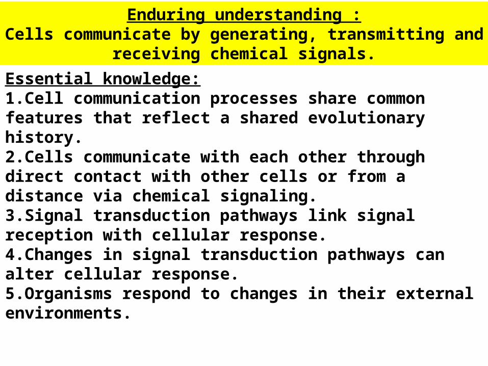
Essential knowledge:1.Cell communication processes share common features that reflect a shared evolutionary history.2.Cells communicate with each other through direct contact with other cells or from a distance via chemical signaling.3.Signal transduction pathways link signal reception with cellular response.4.Changes in signal transduction pathways can alter cellular response.5.Organisms respond to changes in their external environments.
Enduring understanding :Cells communicate by generating, transmitting and receiving
chemical signals.

Chemical Signals….
• Can direct complex processes, ranging from cell and organ differentiation to whole organism physiological responses and behaviors.
• Can allow cells to communicate without physical contact. The distance between the signal generating cell(s) and the responding cell can be small or large.

Methods of cell communicationThree general methods of cell communication:
•Diffusible chemical signals (messengers) that travel through the organism from one location to another (Close or long distances)
•Physical contact between adjacent cell plasma membranes
•Direct cytoplasmic contact via gap junctions

CELL COMMUNICATION
Part 1: An Overview of Cell Signaling
1.Cell signaling evolved earlyevolved early in
the history of life
2.Communicating cells may be
closeclose together or farfar apart
3.The three stagesthree stages of cell signaling
are reception, transduction, and
response

• Cell-to-cell communication is absolutely essential for multicellular organisms.– coordinate the activities within individual cells
that support the function of the organism as a whole.
– Use of pheromones to trigger reproduction and developmental pathways
• Important for many unicellular organisms.• finding a mate• population density (quorum sensing)• Response to external signals by bacteria that influences
cell • allowed some organisms to evolve without having a
nervous system.
Introduction

One topic of cell “conversation” is “mating”.– Ex: The yeast Saccharomyces
cerevisiae, the yeast of bread, wine, and beer, identifies its mates by chemical signaling.
– There are two sexes, a and alpha, each of which secretes a specific signaling molecule, a factor and alpha factor respectively.
– These factors each bind to receptor proteins on the other mating type.
1-Cell signaling evolved early in the history of
life

Cell signaling has remained important in the microbial world. – Myxobacteria, soil-dwelling bacteria,
use chemical signals to communicate nutrient availability.
– When food is scarce, cells secrete a signal to other cells leading them to aggregate and form thick-walled spores.

2-Communicating cells may be close together
or far apart

• In synaptic signaling, a nerve cell produces the neurotransmitter that diffuses to a single cell that is almost touching the sender.–An electrical signal passing along the nerve cell triggers secretion of the neurotransmitter into the synapse.
Example of Localized signaling

(a) Paracrine signaling. A secreting cell acts on nearby target cells by discharging molecules of a local regulator (a growth factor, for example) into the extracellular fluid.
(b) Synaptic signaling. A nerve cell releases neurotransmitter molecules into a synapse, stimulating the target cell.
Local regulator diffuses through extracellular fluid
Target cell
Secretoryvesicle
Electrical signalalong nerve celltriggers release ofneurotransmitter
Neurotransmitter diffuses across
synapse
Target cell is stimulated
Local signaling

*Gap Junctions*Gap Junctions•Narrow tunnels between animal cells that consist of proteins called connexons.•Allows the movement of ions and small molecules from cell to cell.
2-Communicating cells may be close
together or far apart.
*Plasmodesmata*Plasmodesmata•Narrow channels between plant cells•A narrow tube of Endoplasmic reticulum.•Material exchange
Touching

Cells may communicate by direct contact.– Signaling
substances dissolved in the cytosol pass freely between adjacent cells.–Cells may also communicate via direct
contact between substances on their surfaces. Clip; Start at 5:30
Touching

Long-Distance Signaling• Endocrine (hormone) signaling
– Specialized cells release hormone molecules, which travel (usually by diffusion through cells or through the circulatory system) to target cells elsewhere in the organism

• Plants also use hormones to signal at long distances.-In plants, hormones
may travel in vessels, but more often travel from cell to cell or by diffusion in air (EthyleneEthylene).-Ethylene gas in fruit ripening
Long-Distance Signaling

1. In reception, a chemical signal binds to a cellular protein, typically at the cell’s surface.
2. In transduction, binding leads to a change in the receptor that triggers a series of changes along a signal-transduction pathway.
3. In response, the transduced signal triggers a specific cellular activity.
3. The three stages of cell signaling: reception, transduction, & response
EK: Signal transduction pathways link signal reception with cellular response.
McGraw-Hill Dehydration Response
Example Clip

CELL COMMUNICATIONPart 2: Signal Reception and the
Initiation of Transduction
(1) Reception: A signal molecule
(ligand), binds to a receptor
protein, causing the protein to
change shape

• A cell targeted by a particular chemical signal has a receptor protein that recognizes the signal molecule.– Recognition: receptor on target cell is
complementary in shape.complementary in shape.
• The ligand attaches to the receptor, the receptor typically undergoes a change in shape.
1. This may activate the receptor so that it can interact with other molecules inside the cell. 2. Can lead to the aggregation of receptors.
**Signal molecule does not enter cell.***
1. A signal molecule binds to a receptor protein causing the protein
to change shape
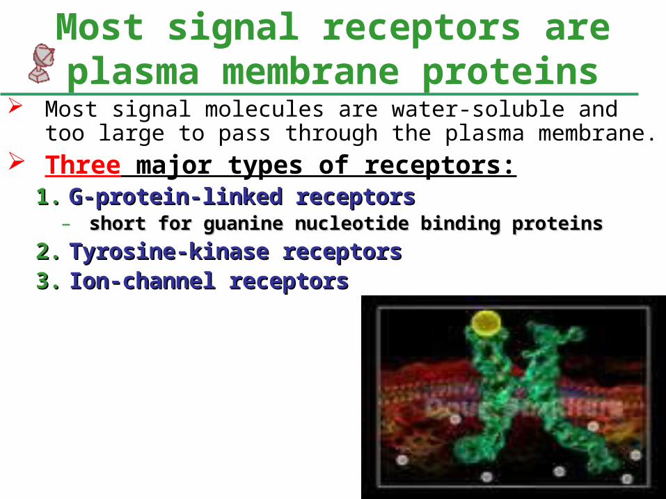
Most signal molecules are water-soluble and too large to pass through the plasma membrane.
Three major types of receptors:1.1. G-protein-linked receptorsG-protein-linked receptors
– short for guanine nucleotide binding proteinsshort for guanine nucleotide binding proteins
2.2. Tyrosine-kinase receptorsTyrosine-kinase receptors3.3. Ion-channel receptorsIon-channel receptors
Most signal receptors are plasma membrane proteins

• PROJECTS START HERE

1. G-protein-linked receptors consists of a receptor protein associated with a G-protein on the cytoplasmic side.– The receptor consists of seven alpha helices
spanning the membrane.– Effective signal
molecules include yeast mating factors, epinephrine, other hormones, and neurotransmitters.
G protein-linked receptorsG protein-linked receptors

G protein-coupled receptors
G protein-coupled receptors
An example of a G protein-linked receptor is the epinephrine receptor. Epinephrine stimulates glycogen breakdown
G-protein-linked receptors and G-proteins mediate a host of critical metabolic and developmental processes (e.g., blood vessel growth and development).
G protein-linked receptorsG protein-linked receptors

•Uses the exchange of Guanosine diphosphate (GDP) for Guanosine triphosphate (GTP) as a molecular "switch" to allow or inhibit biochemical reactions inside the cell
G protein-linked receptorsG protein-linked receptors

G protein-coupled receptors
G protein-coupled receptors Large family all Large family all
with 7 with 7 membrane-membrane-spanning regionsspanning regions
Receptor
G protein
Precursor
Ionchannel
Secondmessenger
Effectorenzyme
Receptor coupled to G Receptor coupled to G protein, and G protein protein, and G protein stimulates effector (stimulates effector (enzyme that enzyme that promotes formation of intracellular “second promotes formation of intracellular “second messenger”)messenger”)

Guanosine triphosphate

The G-protein system cycles between on & off.
1. When a G-protein-linked receptor is activated by binding with an extracellular signal molecule, the receptor binds to an inactive G protein in membrane.
2. This leads the G protein to substitute GTP for GDP.
3. The G protein then binds with another membrane protein, often
an enzyme, altering its activity and leading to
a cellular response.


The whole system can be shut down quickly when the extracellular signal molecule is no longer present.


G-Protein receptors:
• Diseases such as diabetes and certain forms of pituitary cancer, among many others, are thought to have some root in the malfunction of G proteins
• G-protein receptor systems are extremely widespread and diverse in their functions.– They play an important role during embryonic
development and sensory systems.

Ligand binding changes confirmation of the receptor so that specific ions can flow through itIon movement alters the electric potential across the plasma membrane
found in high numbers on neuron plasma membranes
ligand-gated channels for sodium and potassiumAlso found on the plasma membrane of muscle cells
binding of acetylcholineacetylcholine results in ion movement and eventual contraction of muscle
Ion-Channel receptorsIon-Channel receptors

Closed Open
Extracellular side
Cytoplasmic side
Binding

3.Ligand-gated ion channels: protein pores that open or close in response to a chemical signal.– This allows or blocks ion
flow, such as Na+ or Ca2+.– Binding by a ligand to the
extracellular side changes the protein’s shape and opens the channel.
– Ion flow changes the concentration inside the cell.
– When the ligand dissociates, the channel closes.

This colored scanning electron micrograph shows the synapses, or connections, between two nerve fibers (in purple) and a nerve cell (yellow). The picture is magnified 10,000 times.
• Ligand-gated ion channels are very important in the nervous system.– EX: binding of a
neurotransmitter to a neuron, allowing the inward flow of Na2+ that leads to the depolarization of the neuron and the propagation of a nervous impulse to adjacent cells.

Way Cool Alert!•Normally, the enzyme acetylcholinesterase converts acetylcholine into the inactive metabolites choline and acetate. •The devastating effects of nerve agents (Sarin gas for example) are due to their inhibition of this enzyme, resulting in continuous stimulation of the muscles, glands and central nervous system.•Botulinus toxin is produced by the anerobic bacillus Clostridium botulinum, which may be found in improperly canned food, and is one of the most potent toxins known. •This toxin (the agent responsible for botulism) blocks the release of vesicles. This, of course, leads to muscle paralysis and, if the diaphragm becomes affected, can be fatal.
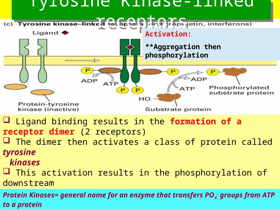
Ligand binding results in the formation of a receptor dimer (2 receptors) The dimer then activates a class of protein called tyrosine kinases This activation results in the phosphorylation of downstream targets by these tyrosine kinases (stick phosphate groups onto tyrosines within the target protein)
Tyrosine Kinase-linked receptorsTyrosine Kinase-linked receptors
Protein Kinases= general name for an enzyme that transfers PO4- groups from
ATP to a protein
Activation:
**Aggregation then phosphorylation

– an extracellular signal-binding sites
– a single alpha helix spanning the membrane, and
– an intracellular tail with several tyrosines.
An individual tyrosine-kinase receptor consists of several parts:

• When ligands bind to two receptor polypeptides, the polypeptides aggregate, forming a dimer.
• This activates the tyrosine-kinase section of both.
• These add phosphates to the tyrosine tails of the other polypeptide.

• The fully-activated receptor proteins activate a variety of specific relay proteins that bind to specific phosphorylated tyrosine molecules.– One tyrosine-kinase receptor dimer may
activate ten or more different intracellular proteins simultaneously.
• These activated relay These activated relay proteins trigger many proteins trigger many different transduction different transduction pathways and pathways and responsesresponses.

The tyrosine-kinase receptor system is especially effective when the cell needs to regulate and coordinate a variety of activities and trigger several signal pathways at once.– Extracellular growth factorsgrowth factors often bind to tyrosine-
kinase receptors.

Signalmolecule
Signal-binding sitea
CYTOPLASM
Tyrosines
Signal moleculeHelix in the
Membrane
Tyr
Tyr
Tyr
Tyr
Tyr
TyrTyr
Tyr
Tyr
Tyr
Tyr
Tyr
Tyr
Tyr
Tyr
Tyr
Tyr
Tyr Tyr
Tyr
Tyr
Tyr
Tyr
Tyr
Tyr
Tyr
Tyr
Tyr
Tyr
Tyr
Dimer
Receptor tyrosinekinase proteins(inactive monomers)
P
P
P
P
P
PTyr
Tyr
Tyr
Tyr
Tyr
TyrP
P
P
P
P
PCellularresponse 1
Inactiverelay proteins
Activatedrelay proteins
Cellularresponse 2
Activated tyrosine-kinase regions(unphosphorylateddimer)
Fully activated receptortyrosine-kinase(phosphorylateddimer)
6 ATP 6 ADP

Insulin (Click)
Polypeptide hormone that regulates carbohydrate metabolism.

• PROJECTS END HERE

Hormone(testosterone)
EXTRACELLULARFLUID
Receptorprotein
DNA
mRNA
NUCLEUS
CYTOPLASM
Plasmamembrane
Hormone-receptorcomplex
New protein
• Other signal receptors are dissolved in the cytosol or nucleus of target cells.
• The signals pass through the plasma membrane.
• Include the hydrophobic steroid and thyroid hormones of animals.
Communication through diffusion

Action of Lipid-Soluble Action of Lipid-Soluble HormonesHormones• Hormone diffuses
through phospholipid bilayer & into cell
• Binds to receptor turning on/off specific genes
• Causing the synthesis of new proteins
• New proteins alters cell’s activity

• Testosterone, like other hormones, travels through travels through the bloodthe blood and enters cells throughout the body.
• In the cytosol, they bind and activate bind and activate receptorreceptor proteins.
• These activated activated proteins enter the proteins enter the nucleusnucleus and turn turn on geneson genes that control male sex characteristics.
EXAMPLE
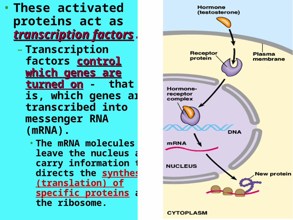
• These activated proteins act as transcription transcription factorsfactors.– Transcription factors
control which genes control which genes are turned onare turned on - that is, which genes are transcribed into messenger RNA (mRNA).• The mRNA molecules
leave the nucleus and carry information that directs the synthesis (translation) of specific proteins at the ribosome.

CELL COMMUNICATIONPart 3: Signal-Transduction
Pathways Pathways relay signalsrelay signals from
receptors to cellular responses Protein phosphorylationphosphorylation, a common
mode of regulation in cells, is a
major mechanism of signal
transduction Certain small molecules and ionssmall molecules and ions are
key components of signaling
pathways (second messengerssecond messengers)
Insulin SignalingClip

Some Features of Signal-Transduction
Pathways • Secondary messengers
• Usually a multi-step pathway
• Protein phosphorylation
• Amplification (small number of signal molecules can produce a large cellular response)
Mr. Anderson: Signal Transduction Pathways

• Pathway acts like falling dominoes.– The signal-activated receptor
activates another protein, which activates another and so on, until the protein that produces the final cellular response is activated.
• The original signal molecule is not passed along the pathway and may not even enter the cell.– Its information is passed oninformation is passed on.
Pathways relay signals from receptors to cellular
responses

• The phosphorylation of proteins by a specific enzyme (a protein kinase) is a widespread cellular mechanism for regulating protein activity.– Most protein kinases act on
other substrate proteins, unlike the tyrosine kinases that act on themselves.
Protein phosphorylation is a major mechanism of signal transduction
**Phosphorylation is the addition of a phosphate (PO4) group to a protein or a small molecule
**Many enzymes and receptors are switched "on" or "off" by phosphorylation and dephosphorylation.
phosphorylation
dephosphorylation

• Phosphorylation of a protein typically converts it from an inactive form to an active form.– The reverse (inactivation) is
possible too for some proteins.
• A single cell may have hundreds of different protein kinases, each specific for a different substrate protein.– Fully 1% of our genes may
code for protein kinases.
• Abnormal activity of protein kinases can cause abnormal cell growth and contribute to the development of cancer.
phosphorylation
dephosphorylation

• Turning off a signal-transduction pathway - protein phosphatases.– Rapidly remove
phosphate groups from proteins.
• When an extracellular signal molecule is absent, active phosphatase molecules predominate, and the signaling pathway and cellular response are shut down.
phosphorylation
dephosphorylation
ON
OFF

• Many signaling pathways involve small, nonprotein, water-soluble molecules or ions, called second messengers.– These molecules rapidly
diffuse throughout the cell.
– Two of the most important are cAMP and Ca2+.
– Inositol triphosphate and GMP are others
Certain signal molecules and ions are key components of signaling pathways
(second messengers)

– This occurs because the receptor activates adenylyl cyclase, which converts ATP to cAMP.
– cAMP is short-lived as phosphodiesterase converts it to AMP.
Epinephrine stimulates glycogen breakdown
•Binding by epinephrine leads to increases in the concentration of cyclic AMP or cAMP.
cAMP
McGraw-Hill Animation

Activate protein kinase(phosphorylate protein)
Biologicalresponse
(dephosphorylate byphosphoproteinphosphatase)
Receptor
Gs
ATPcAMP
Adenylylcyclase
cAMP:-Main purpose: activation of protein kinases.-also used to regulate the passage of Ca2+ through ion channels.
cAMP is synthesised from ATP by adenylyl cyclase. Adenylate cyclase is located at the cell membranes. It is activated by the hormones glucagon and adrenaline and by G protein
cAMP controls many biological processes, including glycogen decomposition into glucose (glycogenolysis), and lipolysis
cAMP

•Hormones and other signals can trigger the formation of cAMP.– Binding by the signal to a receptor
activates a G protein that activates adenylyl cyclase in the plasma membrane.
–The cAMP from the adenylyl cyclase diffuses through the cell and activates a serine/threonine kinase, called protein kinase A which phosphorylates
other proteins.
cAMP

• Certain microbes cause disease by disrupting the G-protein signaling pathways.– The cholera bacterium,
colonizes the small intestine and produces a toxin that modifies a G protein that regulates salt and water secretion.
– The modified G protein is stuck in its activestuck in its active form, continuously stimulating productions of cAMP.
– This causes the intestinal cells to secrete large amounts of water and salts into the intestines, leading to profuse diarrhea and death if untreated.
The toxin acts as an enzyme that changes the G protein so that it can no longer switch itself off
cAMP

• Many signal molecules in animals induce responses in their target cells via signal-transduction pathways that increase the cytosolic concentration of Ca2+.– In animal cells, increases in Ca2+ may cause contraction of muscle
cells, secretion of some substances, and cell division.– In plant cells, increases in Ca2+ trigger responses for coping with
environmental stress, including drought.
• Cells use Ca2+ as a second messenger in both G-protein pathways and tyrosine-kinase pathways.
Ca2+.

Other secondary messengers:Other secondary messengers:
inositol triphosphate (IP3)- -stimulates the release of calcium ions from the smooth endoplasmic reticulum

321
IP3 quickly diffuses throughthe cytosol and binds to an IP3–gated calcium channel in the ERmembrane, causing it to open.
4 The calcium ionsactivate the nextprotein in one or moresignaling pathways.
6 Calcium ions flow out ofthe ER (down their con-centration gradient), raisingthe Ca2+ level in the cytosol.
5
DAG functions asa second messengerin other pathways.
Phospholipase C cleaves aplasma membrane phospholipidcalled PIP2 into DAG and IP3.
A signal molecule bindsto a receptor, leading toactivation of phospholipase C.
EXTRA-CELLULARFLUID
Signal molecule(first messenger)
G protein
G-protein-linkedreceptor
Various proteinsactivated
Endoplasmicreticulum (ER)
Phospholipase CPIP2
IP3
(second messenger)
DAG
Cellular response
GTP
Ca2+
(second messenger)
Ca2+
IP3-gatedcalcium channel

CELL COMMUNICATION
Part 4: Cellular Responses to Signals
• In response to a signal, a cell
may regulate activities in the
cytoplasm or transcription in
the nucleus•Elaborate pathways amplify
and specify the cell’s response
to signals

• Ultimately, a signal-transduction pathway leads to the regulation of one or more cellular activities.– This may be a change in an ion channel or a
change in cell metabolism.– EX:, epinephrine activates enzymes that
catalyze the breakdown of glycogen.
In response to a signal, a cell may regulate activities in the
cytoplasm or transcription in the nucleus
click

• The stimulation of glycogen breakdown by epinephrine
involves a G-protein- linked receptor, a G Protein
adenylyl cyclase and cAMP, and several protein kinases before glycogen phosphorylaseis activated.
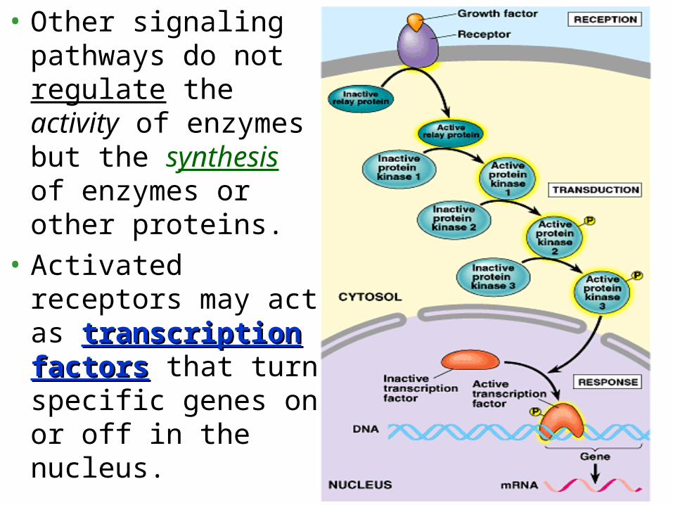
• Other signaling pathways do not regulate the activity of enzymes but the synthesis of enzymes or other proteins.
• Activated receptors may act as transcription transcription factorsfactors that turn specific genes on or off in the nucleus.

• Signaling pathways with multiple steps have two benefits.– They amplify the response to a signal.– They contribute to the specificity of the response.
• At each catalytic step in a cascade, the number of activated products is much greater than in the preceding step.– In the epinephrine-triggered pathway, binding by a
small number of epinephrine molecules can lead to the release of hundreds of millions of glucose molecules.
Pathways amplify and specify the cell’s response to signals

• Various types of cells may receive the same signal but produce very different responses.– For example, epinephrine triggers liver or striated
muscle cells to break down glycogen, but cardiac muscle cells are stimulated to contract, leading to a rapid heartbeat.
• These differences result from a basic observation:– Different kinds of cells have different collections of
proteins.• STRUCTURE AND FUNCTION.
click

• The response of a particular cell to a signal depends on its particular particular collection of receptor proteinscollection of receptor proteins, relay proteins, and proteins needed to carry out the response.

• As important as activating mechanisms are, we must say something about inactivating mechanisms.– For a cell to remain alert and capable of
responding to incoming signals, each molecular change in its signaling pathways must last only a short time.
– If signaling pathway components become locked into one state, the proper function of the cell can be disrupted.
– Binding of signal molecules to receptors must be reversible, allowing the receptors to return to their inactive state when the signal is released.
– Similarly, activated signals (cAMP and phosphorylated proteins) must be inactivated by appropriate enzymes to prepare the cell for a fresh signal.

• Conditions where signal transduction is blocked or defective can be deleterious, preventative or prophylactic.– Diabetes, heart disease, neurological disease,
autoimmune disease, cancer, cholera – Effects of neurotoxins, poisons, pesticides •
Drugs (Hypertensives, Anesthetics, Antihistamines and Birth Control Drugs)

Extracellular Signaling Review
1. Signaling molecules are released by signaling cells2. The signal is called the ligand3. The ligand binds to its specific receptor on a target
cell4. This ligand-receptor interaction induces a
conformational or shape-change in the receptor5. Produces a specific response - called the cellular
response6. Can include a vast array of compounds
e.g. small amino acid derivatives, small peptides, proteins

Cell-to-cell communication by extracellular signaling usually involves six steps
• (1) synthesis of the signaling molecule by the signaling cell• (2) release of the signaling molecule by the signaling cell• (3) transport of the signal to the target cell• (4) detection of the signal by a specific receptor protein –
receptor-ligand specificity• (5) a change in cellular metabolism, function, or development
= cellular response– triggered by the receptor-ligand complex – specific to the ligand-
receptor complex
• (6) removal of the signal, which usually terminates the cellular response – degredation of ligand