Embed Size (px)
DESCRIPTION
Cell Division and Mitosis. Chapter 9. 9.1 Dividing Cells. Eukaryotic organisms Mitosis Meiosis Prokaryotic organisms Prokaryotic fission. Roles of Mitosis. Multicelled organisms Growth Cell replacement Some protistans, fungi, plants, animals Asexual reproduction. Chromosome. - PowerPoint PPT Presentation
Citation preview
Cell Division and Mitosis
Chapter 9
9.1 Dividing Cells
Eukaryotic organisms
– Mitosis
– Meiosis
Prokaryotic organisms
– Prokaryotic fission
Roles of Mitosis
• Multicelled organisms
– Growth
– Cell replacement
• Some protistans, fungi, plants, animals
– Asexual reproduction
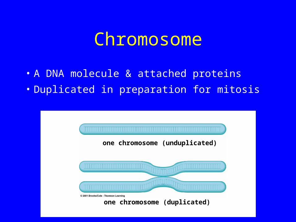
Chromosome
• A DNA molecule & attached proteins
• Duplicated in preparation for mitosis
one chromosome (unduplicated)
one chromosome (duplicated)
Chromosome Number
• Sum total of chromosomes in a cell
• Somatic cells– Chromosome number is diploid (2n)
– Two of each type of chromosome
• Gametes– Chromosome number is haploid (n)
– One of each chromosome type
Human Chromosome Number
• Diploid chromosome number (n) = 46
• Two sets of 23 chromosomes each– One set from father– One set from mother
• Mitosis produces cells with 46 chromosomes--two of each type
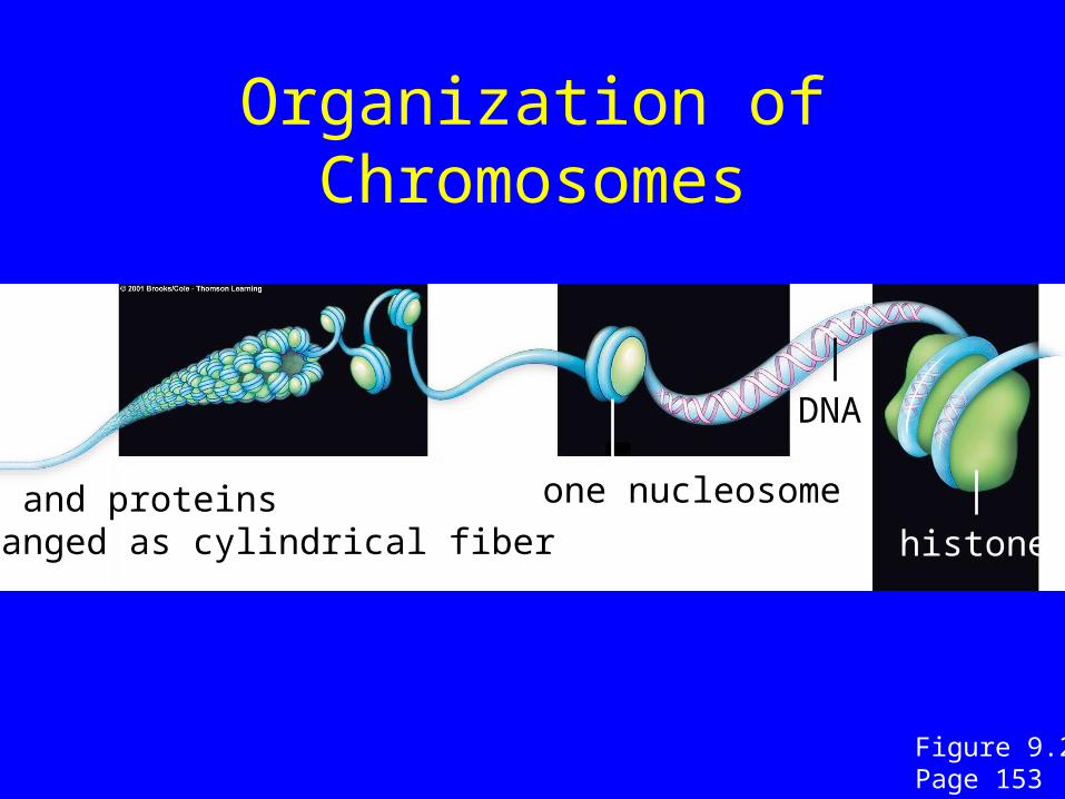
Organization of Chromosomes
DNA and proteinsarranged as cylindrical fiber
DNA
histone
one nucleosome
Figure 9.2Page 153
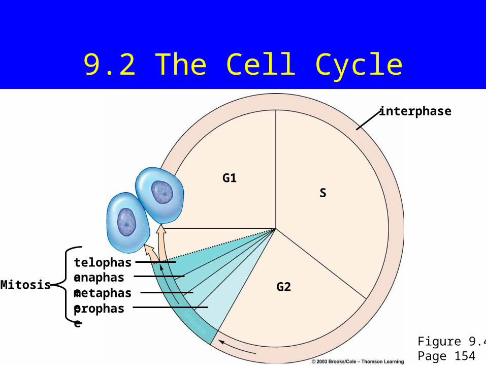
9.2 The Cell Cycle
G1S
G2Mitosis
telophaseanaphasemetaphaseprophase
interphase
Figure 9.4Page 154
Interphase
• Usually longest part of the cycle
• Cell increases in mass
• Number of cytoplasmic components
doubles
• DNA is duplicated
Mitosis
• Period of nuclear division
• Usually followed by cytoplasmic division
• Four stages:Prophase
Metaphase
Anaphase
Telophase

Control of the Cycle
• Once S begins, the cycle automatically
runs through G2 and mitosis
• The cycle has a built-in molecular brake
in G1
• Cancer involves a loss of control over
the cycle, malfunction of the “brakes”
Stopping the Cycle
• Some cells normally stop in interphase
– Neurons in human brain
– Arrested cells do not divide
• Adverse conditions can stop cycle
– Nutrient-deprived amoebas get stuck in interphase
The Spindle Apparatus
• Consists of two distinct sets of
microtubules
– Each set extends from one of the cell poles
– Two sets overlap at spindle equator
• Moves chromosomes during mitosis
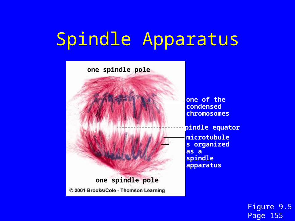
Spindle Apparatus
one spindle pole
one of the condensed chromosomes
spindle equator
microtubules organized as a spindle apparatus
one spindle pole
Figure 9.5Page 155
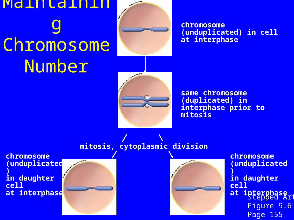
Maintaining Chromosome
Number
mitosis, cytoplasmic divisionchromosome (unduplicated) in daughter cell at interphase
chromosome (unduplicated) in daughter cell at interphase
chromosome (unduplicated) in cell at interphase
same chromosome (duplicated) in interphase prior to mitosis
Stepped ArtFigure 9.6Page 155
9.3 Stages of Mitosis
Prophase
Metaphase
Anaphase
Telophase
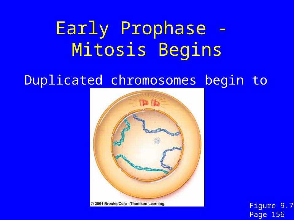
Early Prophase - Mitosis Begins
Duplicated chromosomes begin to condense
Figure 9.7 Page 156
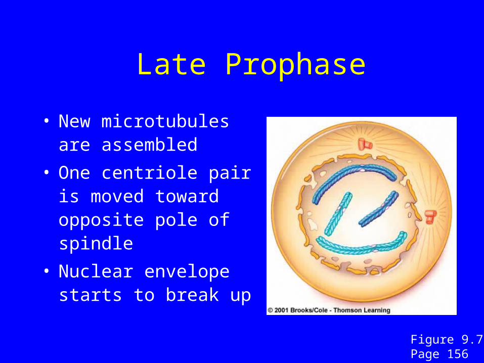
Late Prophase
• New microtubules are assembled
• One centriole pair is moved toward opposite pole of spindle
• Nuclear envelope starts to break up
Figure 9.7 Page 156
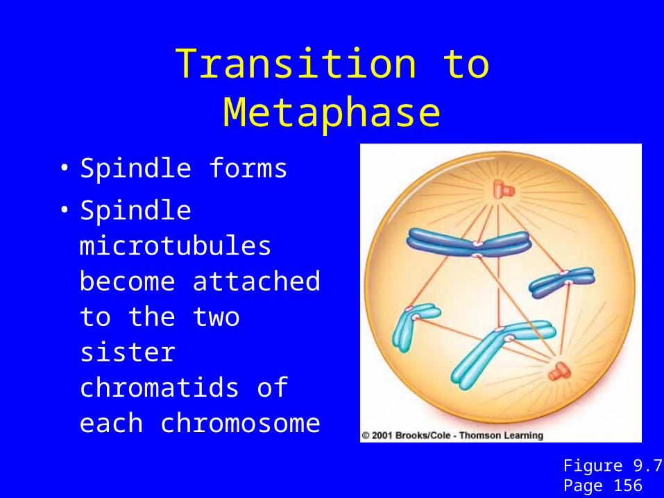
Transition to Metaphase
• Spindle forms
• Spindle microtubules become attached to the two sister chromatids of each chromosome
Figure 9.7 Page 156
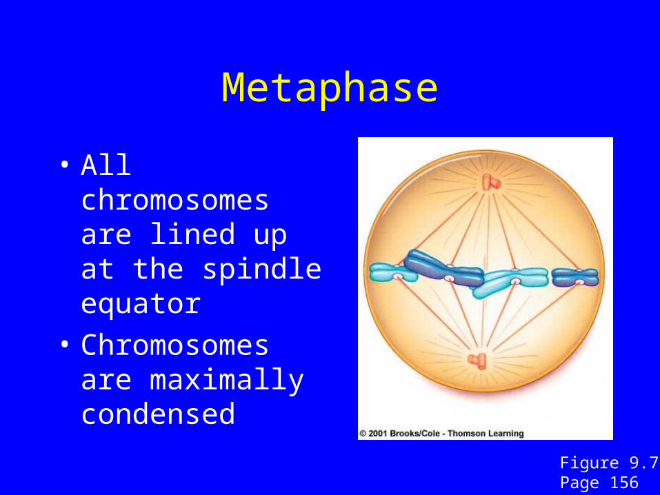
Metaphase
• All chromosomes are lined up at the spindle equator
• Chromosomes are maximally condensed
Figure 9.7 Page 156
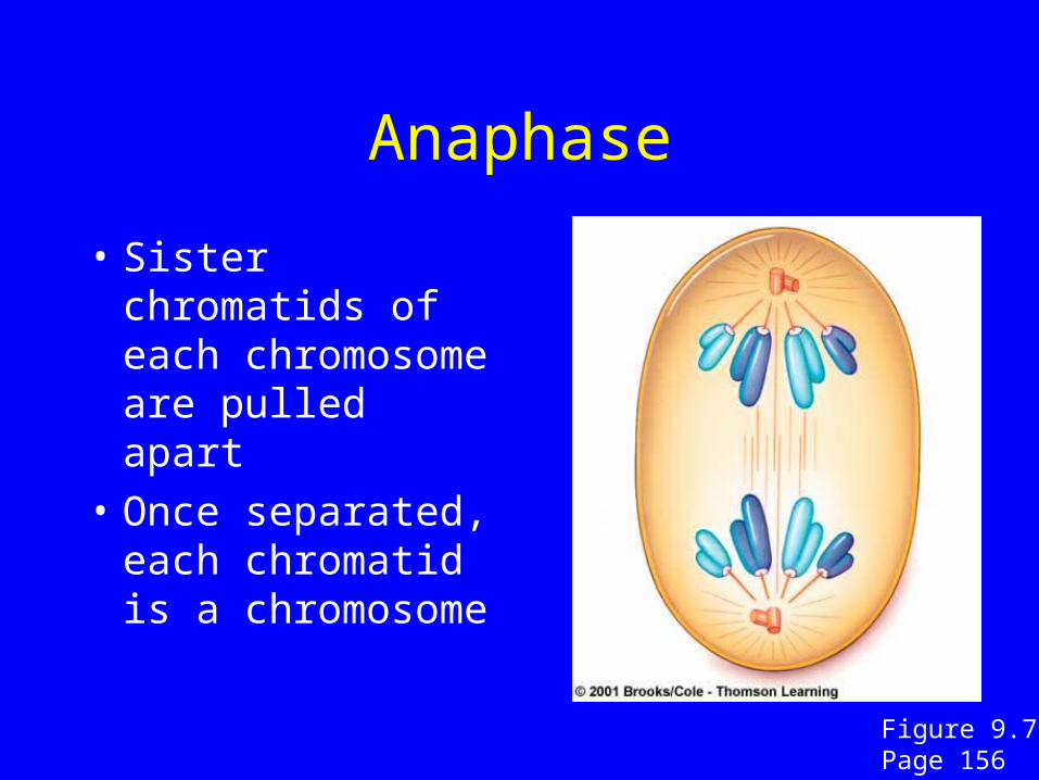
Anaphase
• Sister chromatids of each chromosome are pulled apart
• Once separated, each chromatid is a chromosome
Figure 9.7 Page 156
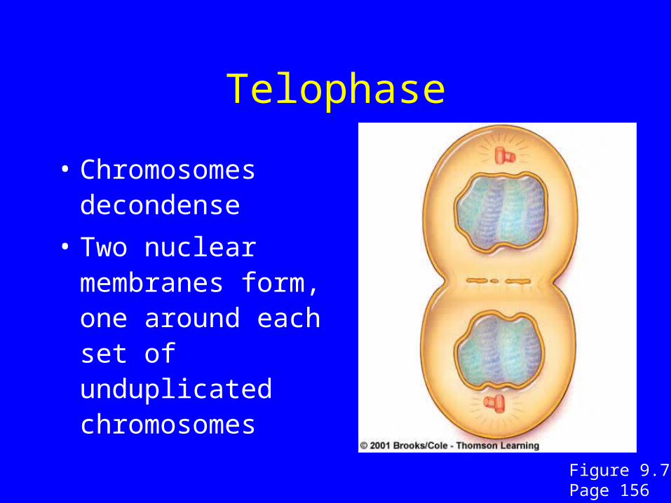
Telophase
• Chromosomes decondense
• Two nuclear membranes form, one around each set of unduplicated chromosomes
Figure 9.7 Page 156
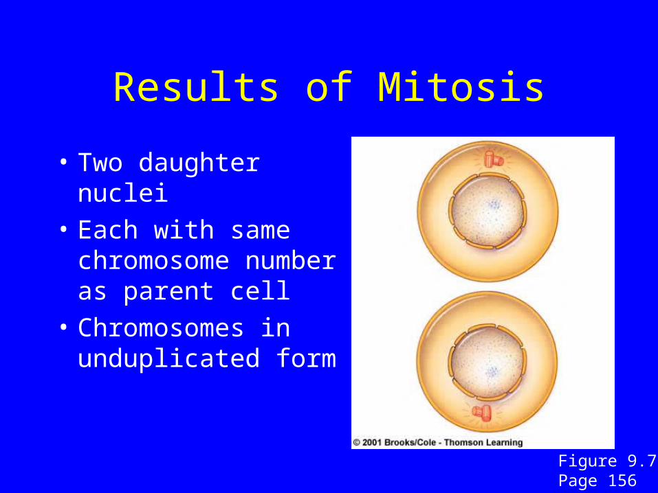
Results of Mitosis
• Two daughter nuclei
• Each with same chromosome number as parent cell
• Chromosomes in unduplicated form
Figure 9.7 Page 156
9.4 Cytoplasmic Division
• Usually occurs between late anaphase
and end of telophase
• Two mechanisms
– Cell plate formation (plants)
– Cleavage (animals)
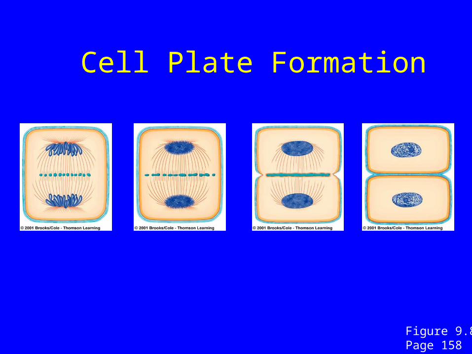
Cell Plate Formation
Figure 9.8Page 158
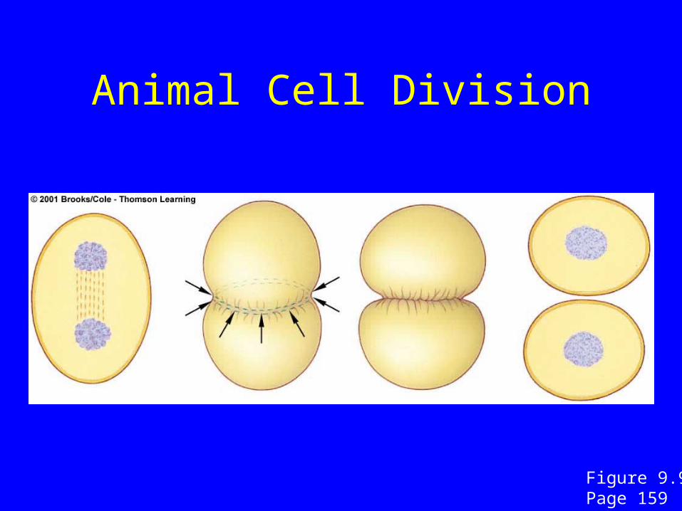
Animal Cell Division
Figure 9.9Page 159