Embed Size (px)
DESCRIPTION
Cell Growth & Division Question: Why do cells divide?. Why do we need to make more cells?. Q: Who has bigger cells?. A: Same cell size, RonRon has MORE!. From One Cell to Many. Sea Urchin Cell Division. Why do we need to make more cells?. - PowerPoint PPT Presentation
Citation preview
Cell Growth & DivisionCell Growth & Division
Question: Question: Why do cells divide?Why do cells divide?
Cell Growth & DivisionCell Growth & Division
Question: Question: Why do cells divide?Why do cells divide?
•Why do we need to Why do we need to make more cells?make more cells?
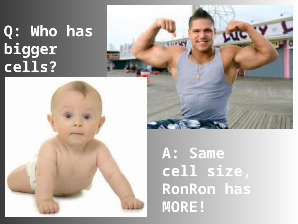
Q: Who has bigger cells?
A: Same cell size, RonRon has MORE!
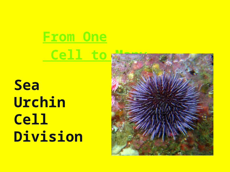
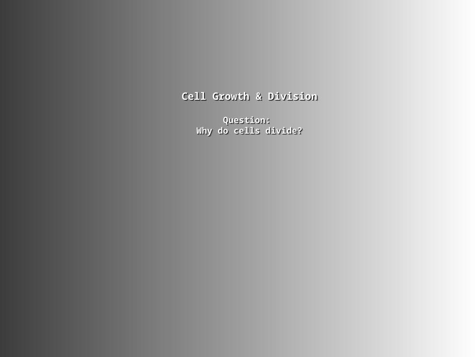
From One Cell to Many
Sea Urchin Cell Division
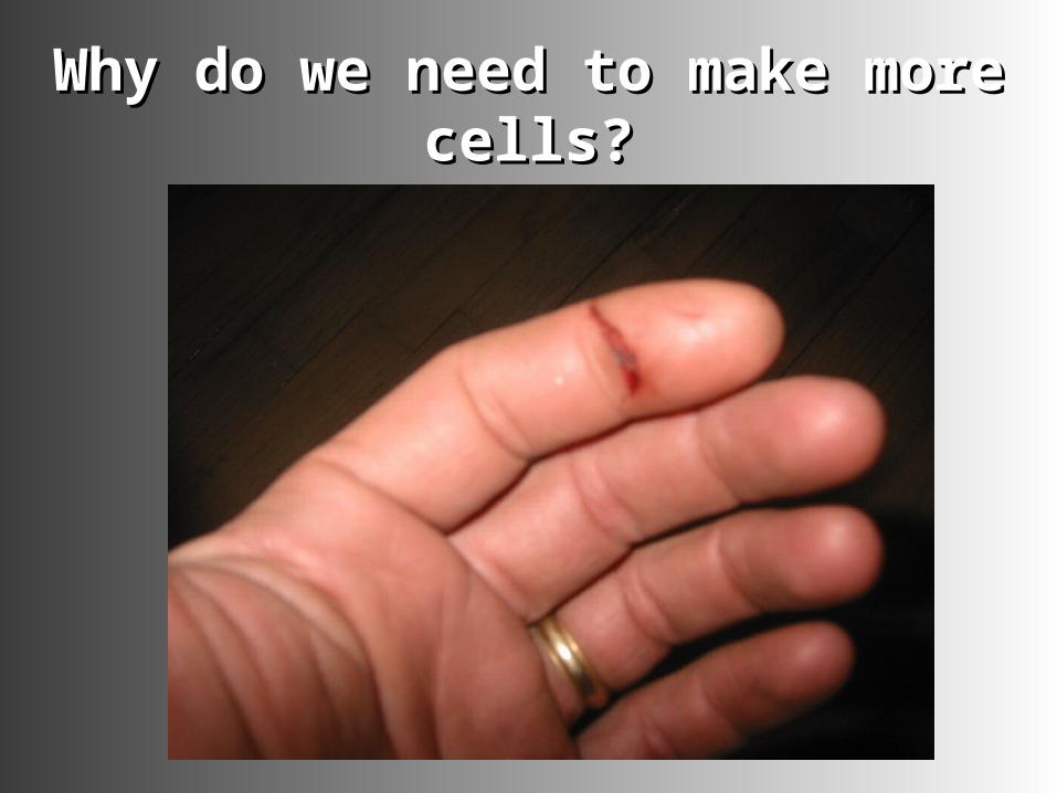
Why do we need to make more Why do we need to make more cells?cells?
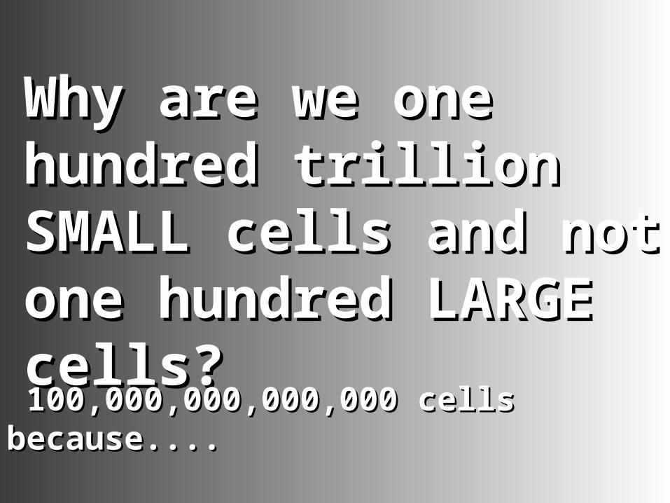
Why are we one Why are we one hundred trillion hundred trillion SMALL cells and not SMALL cells and not one hundred LARGE one hundred LARGE cells?cells?
100,000,000,000,000 cells 100,000,000,000,000 cells because....because....
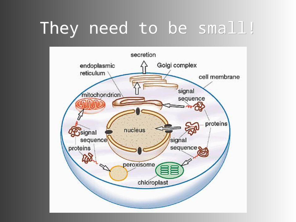
They need to be small!They need to be small!
I. Why do Cells I. Why do Cells Divide?Divide?
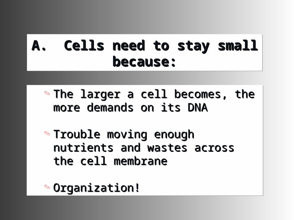
A. Cells need to stay small A. Cells need to stay small because:because:
A. Cells need to stay small A. Cells need to stay small because:because:
The larger a cell becomes, the The larger a cell becomes, the more demands on its DNAmore demands on its DNA
Trouble moving enough nutrients Trouble moving enough nutrients and wastes across the cell and wastes across the cell membranemembrane
Organization!Organization!
The larger a cell becomes, the The larger a cell becomes, the more demands on its DNAmore demands on its DNA
Trouble moving enough nutrients Trouble moving enough nutrients and wastes across the cell and wastes across the cell membranemembrane
Organization!Organization!
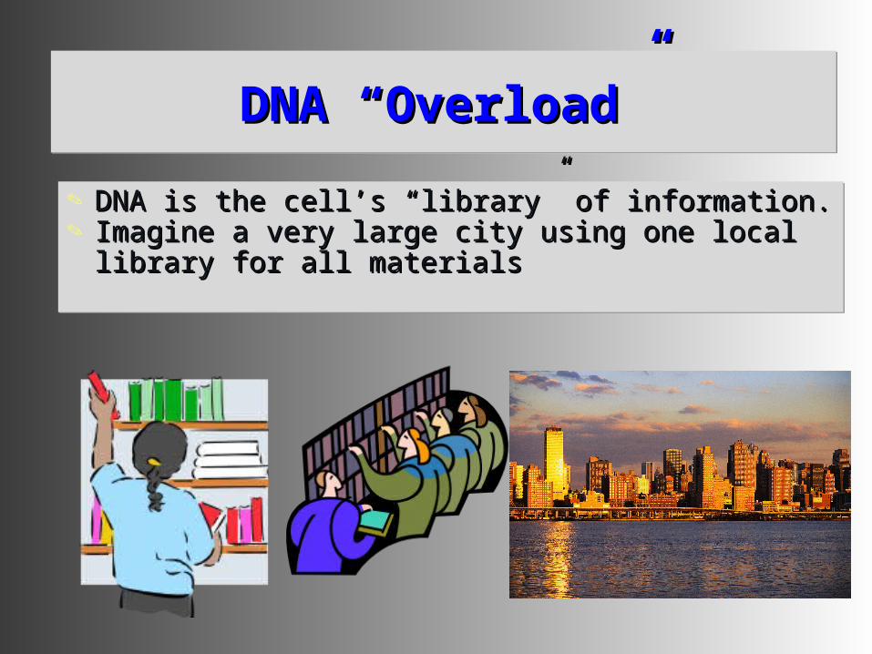
DNA DNA ““OverloadOverload””DNA DNA ““OverloadOverload””
DNA is the cellDNA is the cell’’s s ““librarylibrary”” of information. of information. Imagine a very large city using one local Imagine a very large city using one local
library for all materialslibrary for all materials
DNA is the cellDNA is the cell’’s s ““librarylibrary”” of information. of information. Imagine a very large city using one local Imagine a very large city using one local
library for all materialslibrary for all materials
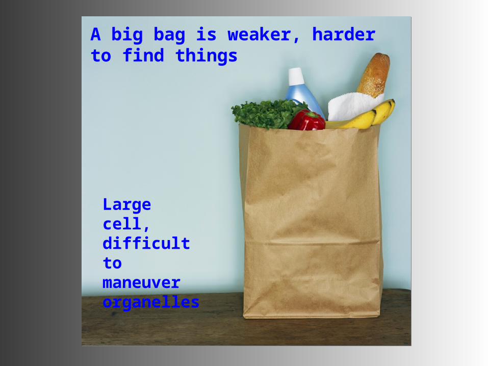
A big bag is weaker, harder to find things
Large cell, difficult to maneuver organelles
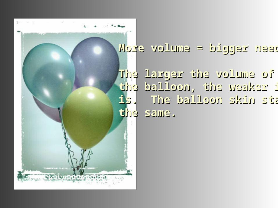
More volume = bigger needMore volume = bigger need
The larger the volume of The larger the volume of the balloon, the weaker it the balloon, the weaker it is. The balloon skin stays is. The balloon skin stays the same.the same.
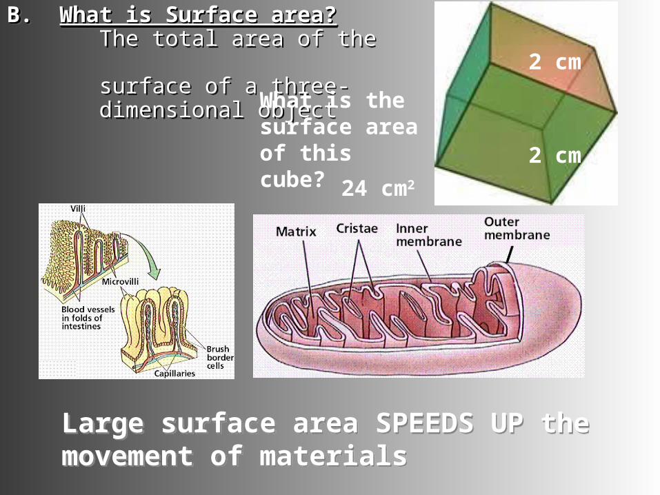
B. B. What is Surface area?What is Surface area? The total area of the surface of a three- dimensional object
B. B. What is Surface area?What is Surface area? The total area of the surface of a three- dimensional object
Large surface area SPEEDS UP the movement of materialsLarge surface area SPEEDS UP the movement of materials
What is the surface area of this cube?
24 cm2
2 cm
2 cm
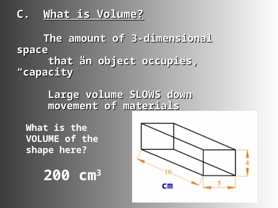
C. C. What is Volume?What is Volume?
The amount of 3-dimensional The amount of 3-dimensional space space that an object occupies, that an object occupies, “capacity”“capacity”
Large volume SLOWS down Large volume SLOWS down movement of materialsmovement of materials
C. C. What is Volume?What is Volume?
The amount of 3-dimensional The amount of 3-dimensional space space that an object occupies, that an object occupies, “capacity”“capacity”
Large volume SLOWS down Large volume SLOWS down movement of materialsmovement of materials
What is the VOLUME of the shape here?
200 cm3
cm
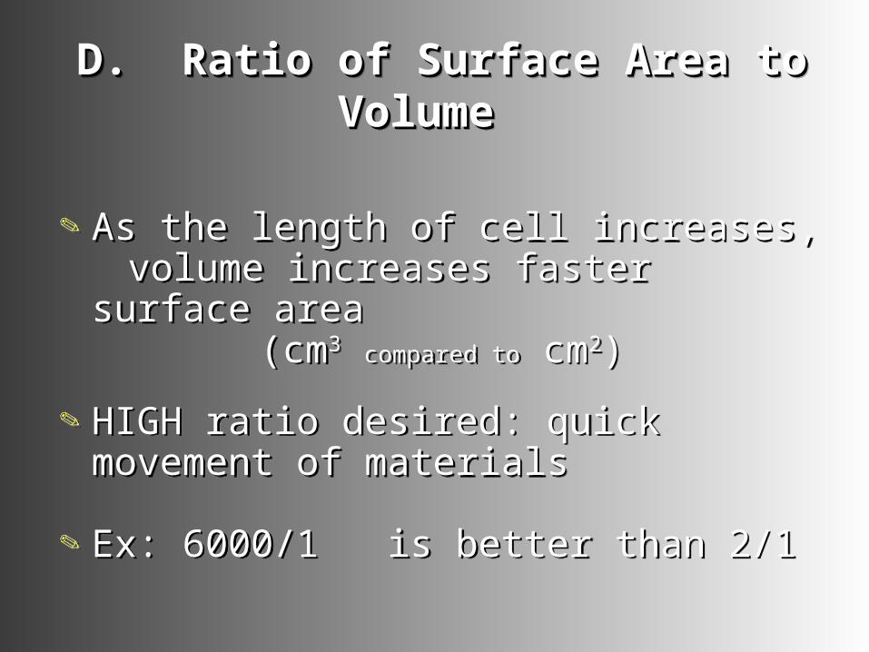
D. Ratio of Surface Area to D. Ratio of Surface Area to Volume Volume
D. Ratio of Surface Area to D. Ratio of Surface Area to Volume Volume
As the length of cell increases, volume increases faster surface area
(cm3 compared to cm2)
HIGH ratio desired: quick movement of materials
Ex: 6000/1 is better than 2/1
As the length of cell increases, volume increases faster surface area
(cm3 compared to cm2)
HIGH ratio desired: quick movement of materials
Ex: 6000/1 is better than 2/1
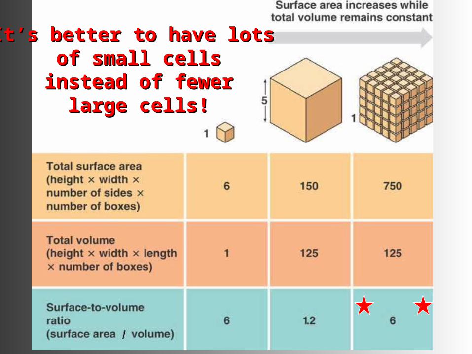
ItIt’’s better to have lots s better to have lots of small cellsof small cells
instead of fewer instead of fewer large cells!large cells!
II. ChromosomesII. Chromosomes
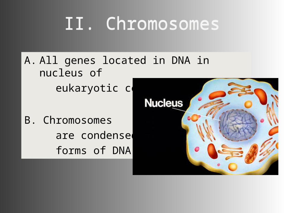
A. All genes located in DNA in nucleus of
eukaryotic cell
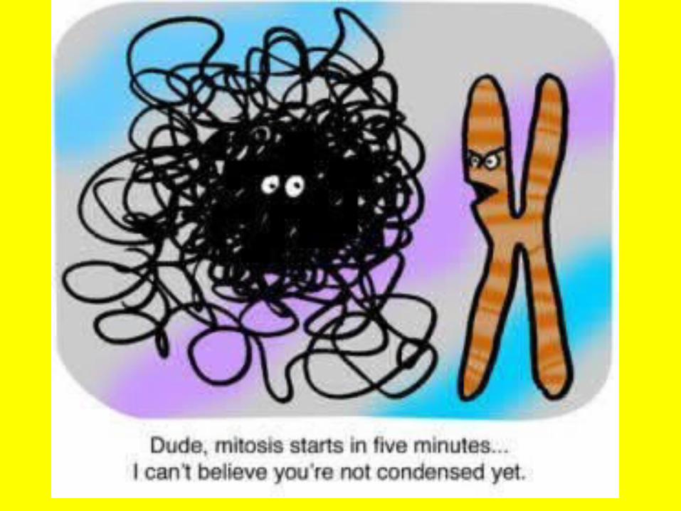
B. Chromosomes are condensed forms of DNA
A. All genes located in DNA in nucleus of
eukaryotic cell
B. Chromosomes are condensed forms of DNA
Chromosomes Chromosomes
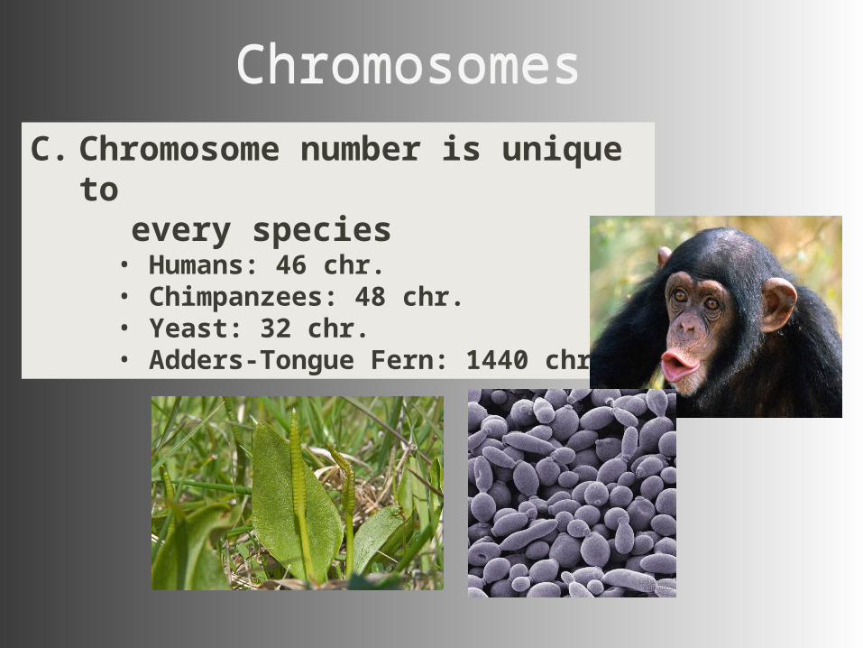
C. Chromosome number is unique to every species
• Humans: 46 chr.• Chimpanzees: 48 chr.• Yeast: 32 chr.• Adders-Tongue Fern: 1440 chr.!
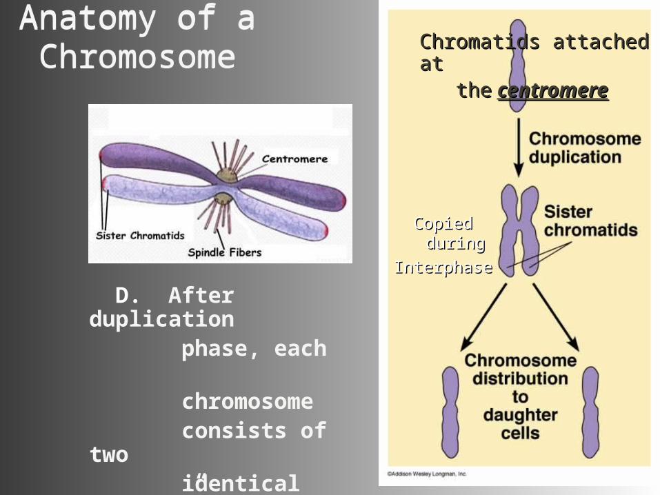
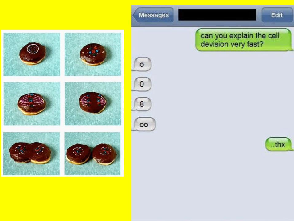
Copied duringCopied during
InterphaseInterphase
Copied duringCopied during
InterphaseInterphase
D. After duplication phase, each chromosome consists of two identical “sister” chromatids
Anatomy of a ChromosomeAnatomy of a Chromosome Chromatids attached Chromatids attached
at at thethe centromerecentromere
II. Cell DivisionII. Cell DivisionII. Cell DivisionII. Cell Division
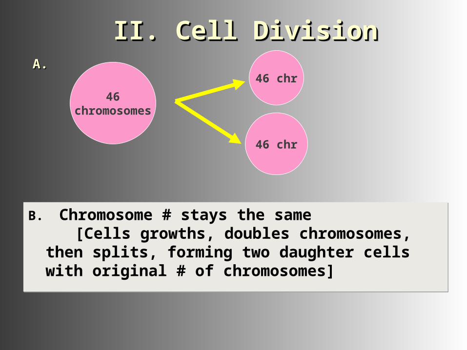
B. Chromosome # stays the same [Cells growths, doubles chromosomes, then splits,
forming two daughter cells with original # of chromosomes]
B. Chromosome # stays the same [Cells growths, doubles chromosomes, then splits,
forming two daughter cells with original # of chromosomes]
46chromosomes
46 chr
46 chr
A. A.
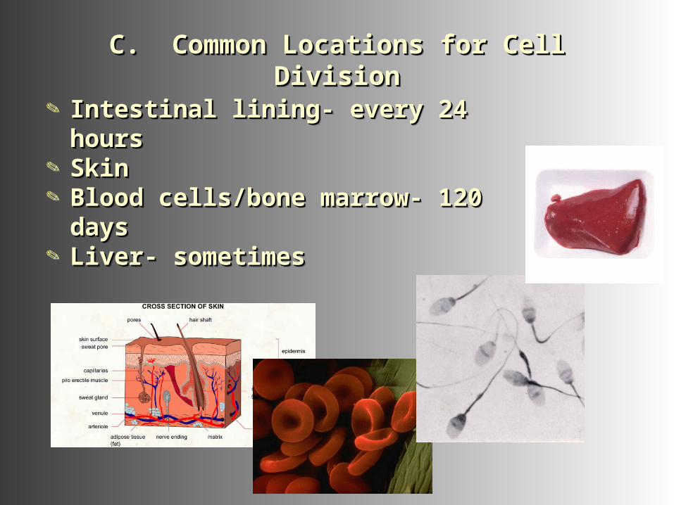
C. Common Locations for Cell DivisionC. Common Locations for Cell DivisionC. Common Locations for Cell DivisionC. Common Locations for Cell Division
Intestinal lining- every 24 hoursIntestinal lining- every 24 hours SkinSkin Blood cells/bone marrow- 120 Blood cells/bone marrow- 120
daysdays Liver- sometimesLiver- sometimes
Intestinal lining- every 24 hoursIntestinal lining- every 24 hours SkinSkin Blood cells/bone marrow- 120 Blood cells/bone marrow- 120
daysdays Liver- sometimesLiver- sometimes
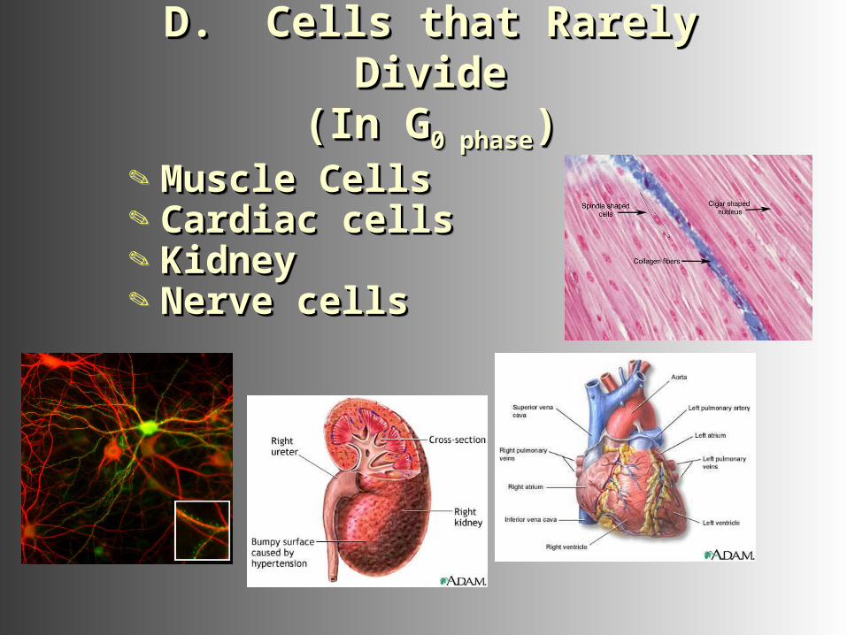
D. Cells that Rarely DivideD. Cells that Rarely Divide(In G(In G0 phase0 phase))
D. Cells that Rarely DivideD. Cells that Rarely Divide(In G(In G0 phase0 phase))
Muscle CellsMuscle Cells Cardiac cellsCardiac cells KidneyKidney Nerve cellsNerve cells
Muscle CellsMuscle Cells Cardiac cellsCardiac cells KidneyKidney Nerve cellsNerve cells
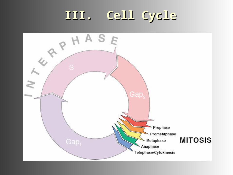
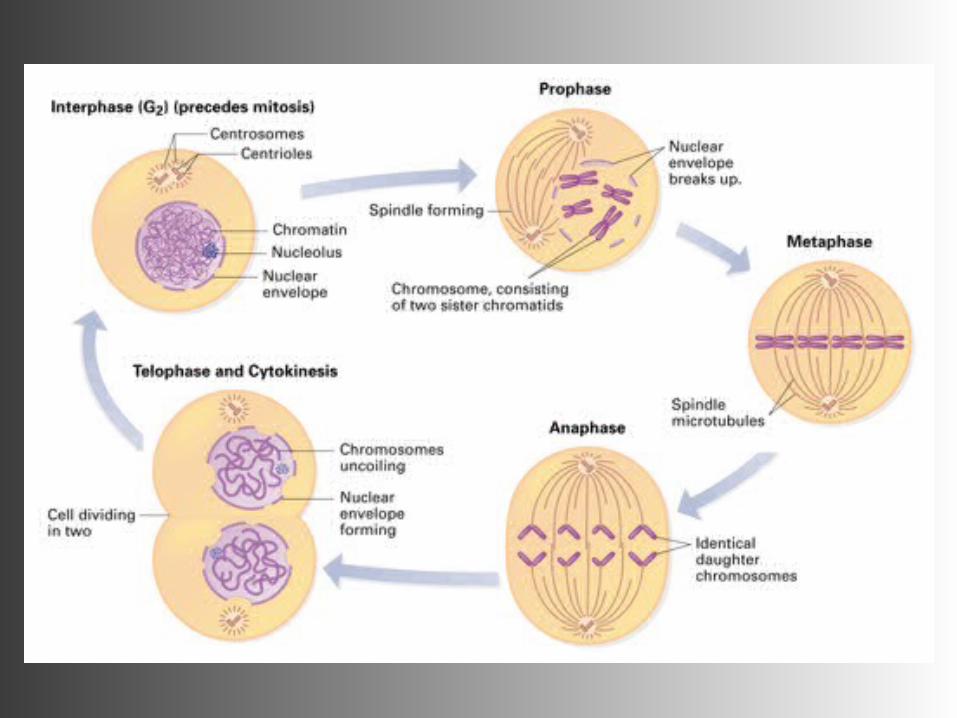
III. Cell CycleIII. Cell CycleIII. Cell CycleIII. Cell Cycle
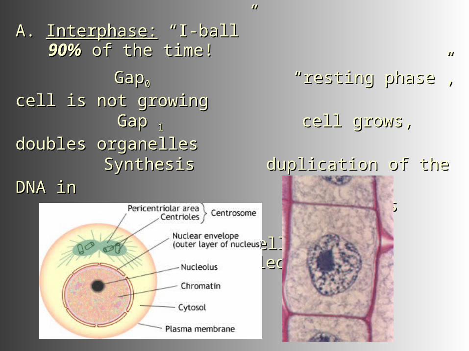
A. A. Interphase:Interphase: ““I-ballI-ball””
90%90% of the time! of the time!
GapGap00 “resting phase”, cell is not growing “resting phase”, cell is not growing
Gap Gap 1 1 cell grows, doubles organelles cell grows, doubles organelles
Synthesis duplication of the DNA in Synthesis duplication of the DNA in the cell's chromosomesthe cell's chromosomesGap Gap 2 2 cell grows, microtubules assembled cell grows, microtubules assembled
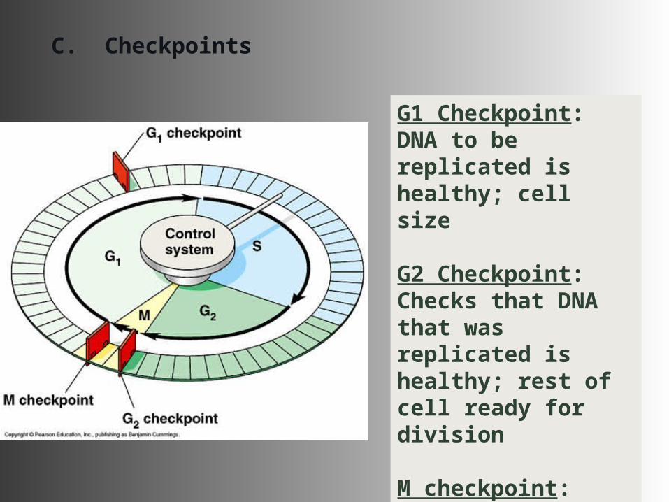
C. Checkpoints
G1 Checkpoint: DNA to be replicated is healthy; cell size
G2 Checkpoint: Checks that DNA that was replicated is healthy; rest of cell ready for division
M checkpoint:Chromosomes are properly attached to the spindle fibers.
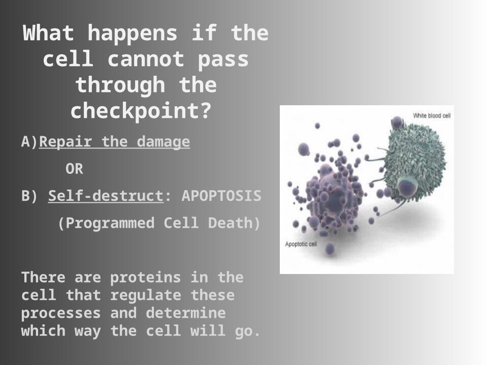
What happens if the cell cannot pass through
the checkpoint?
A)Repair the damage
OR
B) Self-destruct: APOPTOSIS
(Programmed Cell Death)
There are proteins in the cell that regulate these processes and determine which way the cell will go.
Now entering “M Phase”..
Now entering “M Phase”..
First stop, Mitosis!First stop, Mitosis!
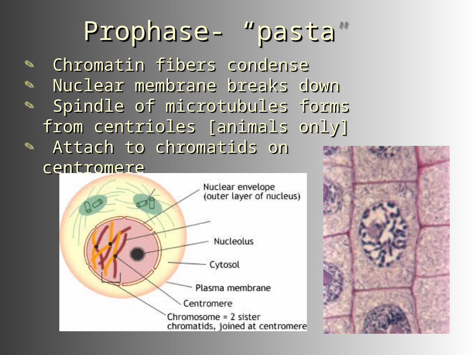
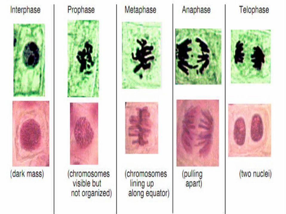
Prophase- Prophase- ““pastapasta””Prophase- Prophase- ““pastapasta”” Chromatin fibers condenseChromatin fibers condense Nuclear membrane breaks downNuclear membrane breaks down Spindle of microtubules forms from Spindle of microtubules forms from
centrioles [animals only]centrioles [animals only] Attach to chromatids on centromereAttach to chromatids on centromere
Chromatin fibers condenseChromatin fibers condense Nuclear membrane breaks downNuclear membrane breaks down Spindle of microtubules forms from Spindle of microtubules forms from
centrioles [animals only]centrioles [animals only] Attach to chromatids on centromereAttach to chromatids on centromere
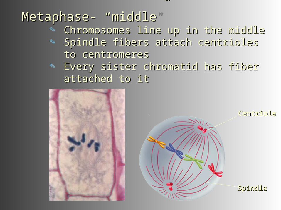
Metaphase- Metaphase- ““middlemiddle”” Chromosomes line up in the middleChromosomes line up in the middle Spindle fibers attach centrioles to Spindle fibers attach centrioles to
centromerescentromeres Every sister chromatid has fiber Every sister chromatid has fiber
attached to itattached to it
Metaphase- Metaphase- ““middlemiddle”” Chromosomes line up in the middleChromosomes line up in the middle Spindle fibers attach centrioles to Spindle fibers attach centrioles to
centromerescentromeres Every sister chromatid has fiber Every sister chromatid has fiber
attached to itattached to it
CentrioleCentriole
SpindleSpindle
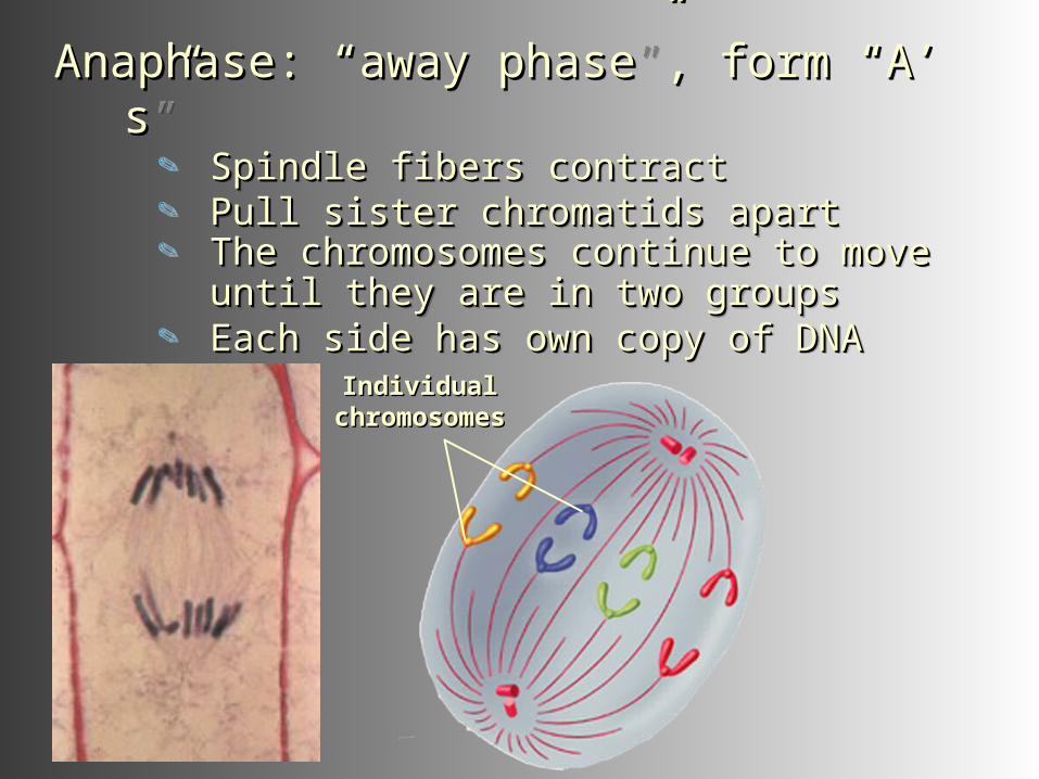
Anaphase: Anaphase: ““away phaseaway phase””, form , form ““AA ’’ss”” Spindle fibers contractSpindle fibers contract Pull sister chromatids apartPull sister chromatids apart The chromosomes continue to move The chromosomes continue to move
until they are in two groupsuntil they are in two groups Each side has own copy of DNAEach side has own copy of DNA
IndividualIndividualchromosomeschromosomes
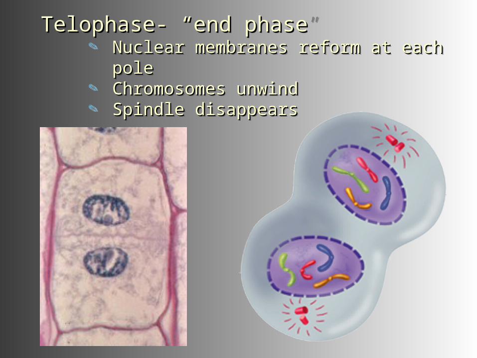
Telophase- Telophase- ““end phaseend phase”” Nuclear membranes reform at each Nuclear membranes reform at each
polepole Chromosomes unwindChromosomes unwind Spindle disappearsSpindle disappears
Telophase- Telophase- ““end phaseend phase”” Nuclear membranes reform at each Nuclear membranes reform at each
polepole Chromosomes unwindChromosomes unwind Spindle disappearsSpindle disappears
Last part of “M Phase”..Last part of “M Phase”..
Cytokinesis!Cytokinesis!
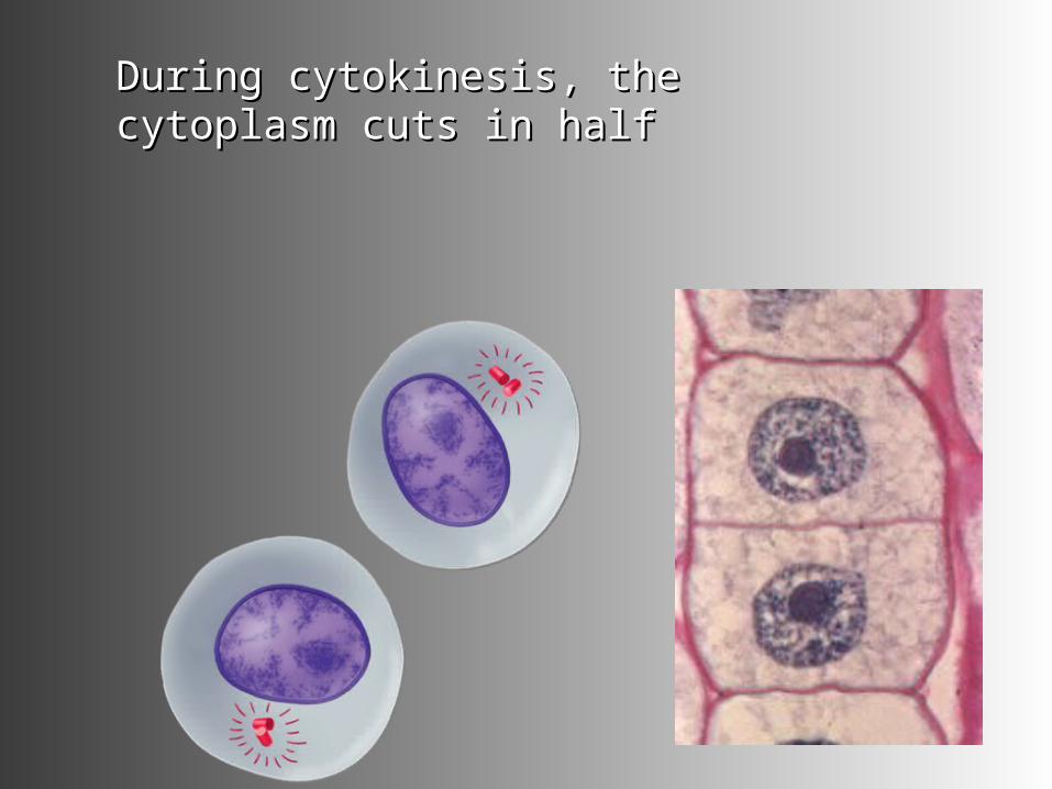
During cytokinesis, the cytoplasm During cytokinesis, the cytoplasm cuts in halfcuts in half
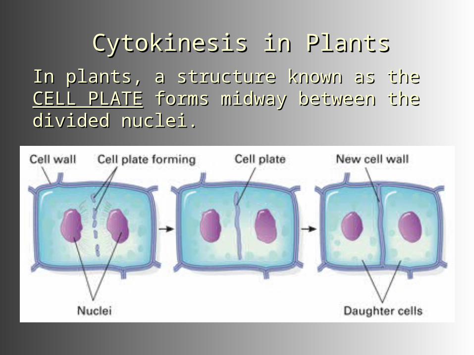
Cytokinesis in PlantsCytokinesis in PlantsCytokinesis in PlantsCytokinesis in PlantsIn plants, a structure known as the In plants, a structure known as the CELL CELL PLATEPLATE forms midway between the divided forms midway between the divided nuclei.nuclei.
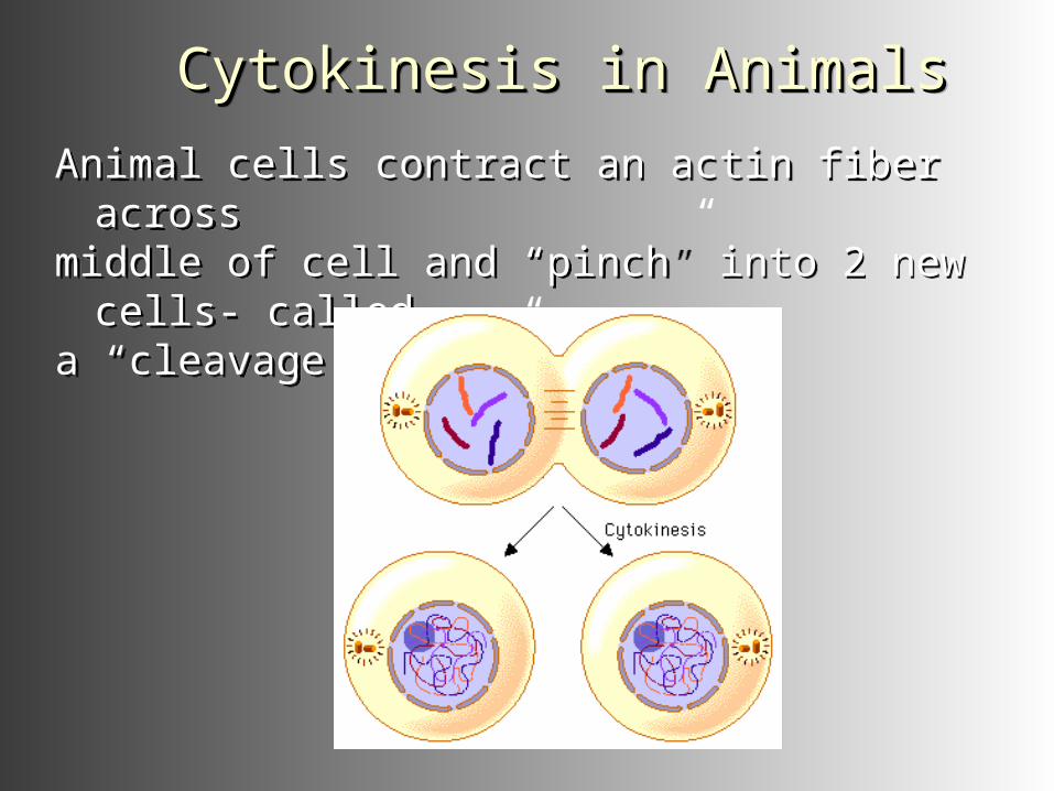
Cytokinesis in AnimalsCytokinesis in AnimalsCytokinesis in AnimalsCytokinesis in Animals
Animal cells contract an actin fiber acrossmiddle of cell and “pinch” into 2 new cells-
calleda “cleavage furrow”.
Animal cells contract an actin fiber acrossmiddle of cell and “pinch” into 2 new cells-
calleda “cleavage furrow”.
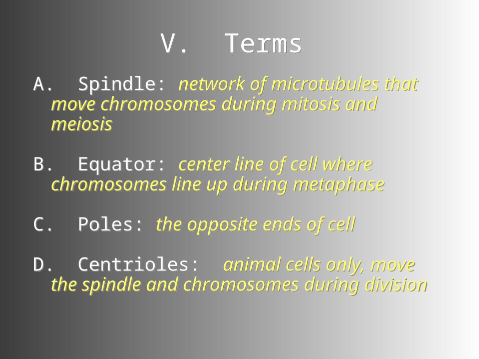
V. TermsV. TermsA. Spindle: network of microtubules that
move chromosomes during mitosis and meiosis
B. Equator: center line of cell where chromosomes line up during metaphase
C. Poles: the opposite ends of cell
D. Centrioles: animal cells only, move the spindle and chromosomes during division
A. Spindle: network of microtubules that move chromosomes during mitosis and meiosis
B. Equator: center line of cell where chromosomes line up during metaphase
C. Poles: the opposite ends of cell
D. Centrioles: animal cells only, move the spindle and chromosomes during division
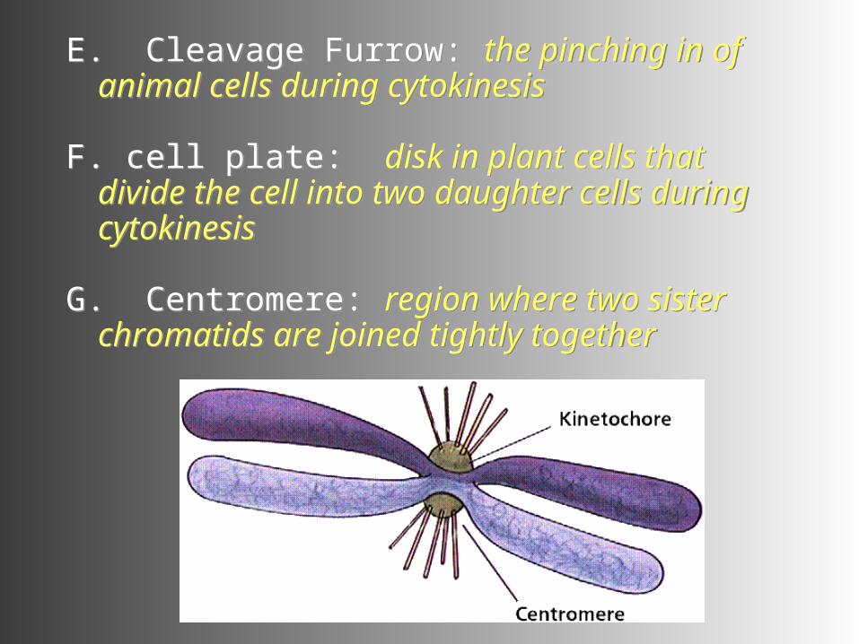
E. Cleavage Furrow: the pinching in of animal cells during cytokinesis
F. cell plate: disk in plant cells that divide the cell into two daughter cells during cytokinesis
G. Centromere: region where two sister chromatids are joined tightly together
E. Cleavage Furrow: the pinching in of animal cells during cytokinesis
F. cell plate: disk in plant cells that divide the cell into two daughter cells during cytokinesis
G. Centromere: region where two sister chromatids are joined tightly together
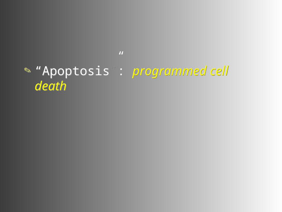
“Apoptosis”: programmed cell death
“Apoptosis”: programmed cell death
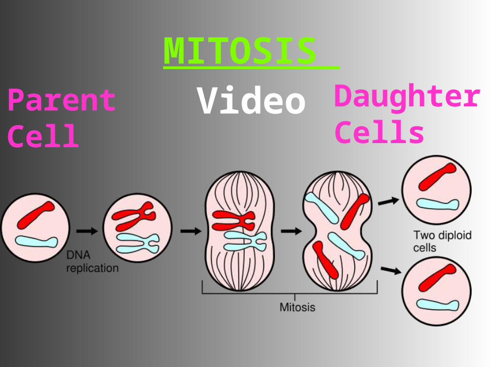
VI. Results of MitosisVI. Results of Mitosis
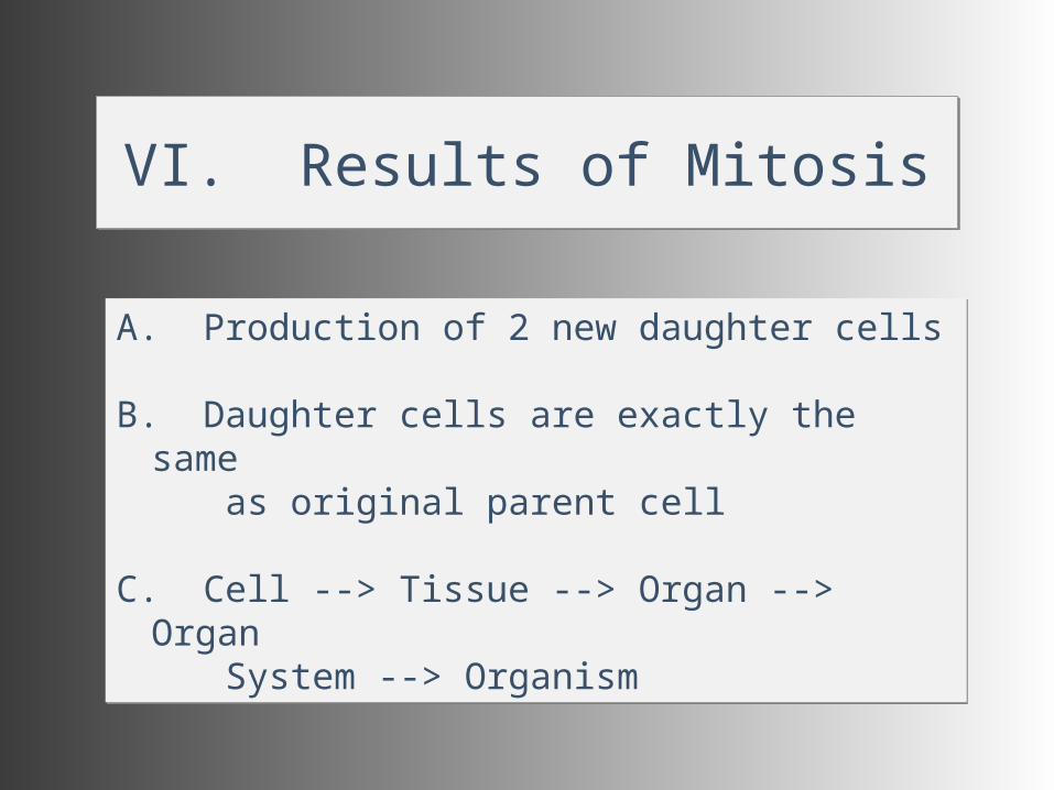
A. Production of 2 new daughter cells
B. Daughter cells are exactly the same as original parent cell
C. Cell --> Tissue --> Organ --> Organ System --> Organism
A. Production of 2 new daughter cells
B. Daughter cells are exactly the same as original parent cell
C. Cell --> Tissue --> Organ --> Organ System --> Organism