Embed Size (px)
Citation preview

CELL INJURY AND DEATH
Somphong narkpinit, M.D. Department of Pathobiology
Faculty of Science
Mahidol University
SCPA202 Basic Pathology
Objectives:
after learning, student should be able to
Describe cell injury and cell death
Describe intracellular accumulations
Describe pathologic calcification
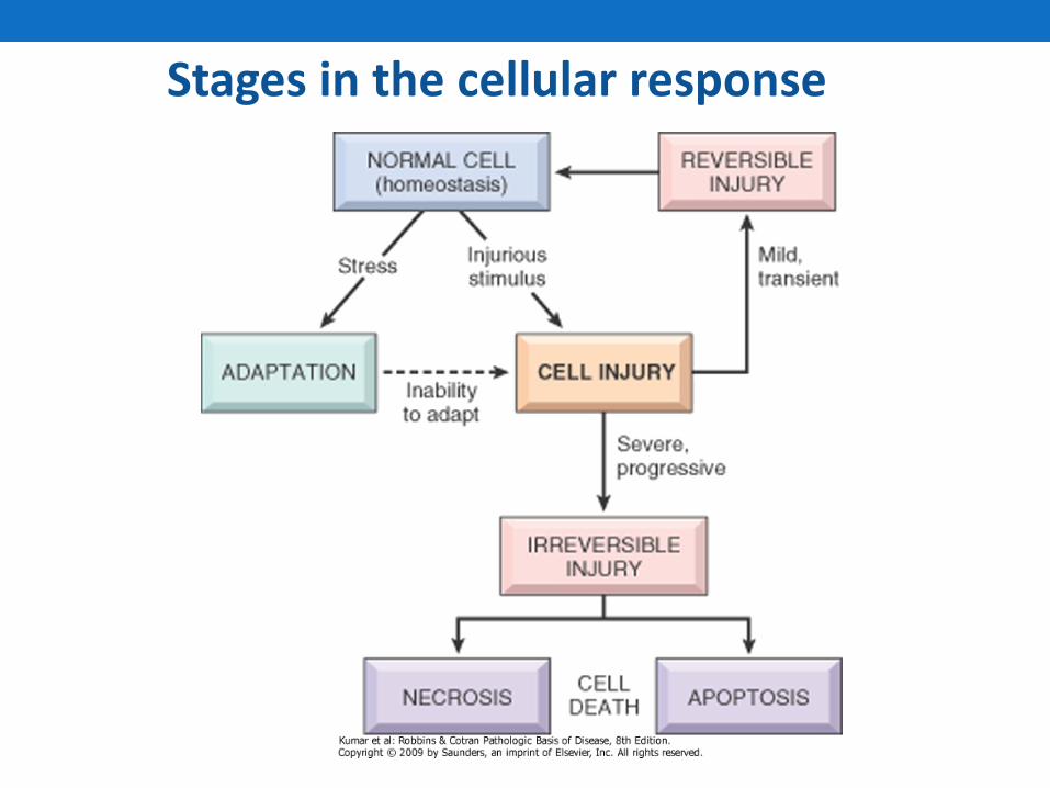
Stages in the cellular response
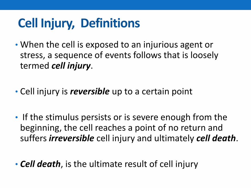
Cell Injury, Definitions
• When the cell is exposed to an injurious agent or stress, a sequence of events follows that is loosely termed cell injury.
• Cell injury is reversible up to a certain point
• If the stimulus persists or is severe enough from the beginning, the cell reaches a point of no return and suffers irreversible cell injury and ultimately cell death.
• Cell death, is the ultimate result of cell injury
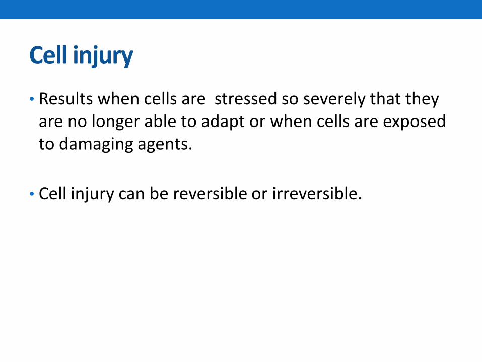
Cell injury
• Results when cells are stressed so severely that they are no longer able to adapt or when cells are exposed to damaging agents.
• Cell injury can be reversible or irreversible.

Reversible cell injury
• Functional and morphologic changes are reversible if the damaging stimulus is removed.
• The features are: decreased oxidative phosphorylation, ATP depletion and cellular swelling.
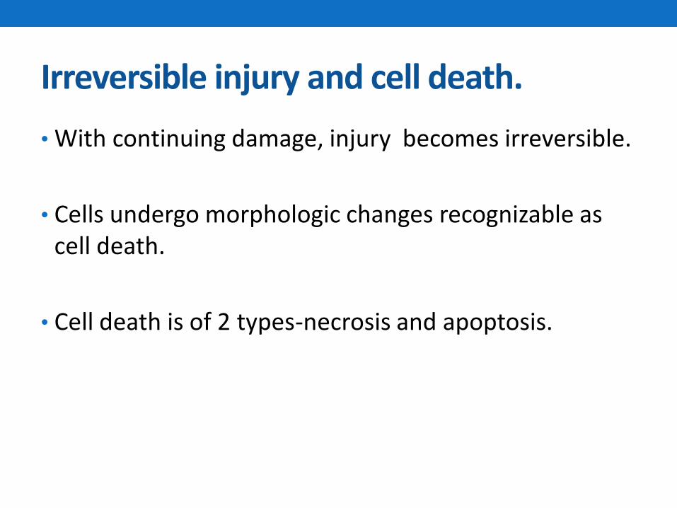
Irreversible injury and cell death.
• With continuing damage, injury becomes irreversible.
• Cells undergo morphologic changes recognizable as cell death.
• Cell death is of 2 types-necrosis and apoptosis.
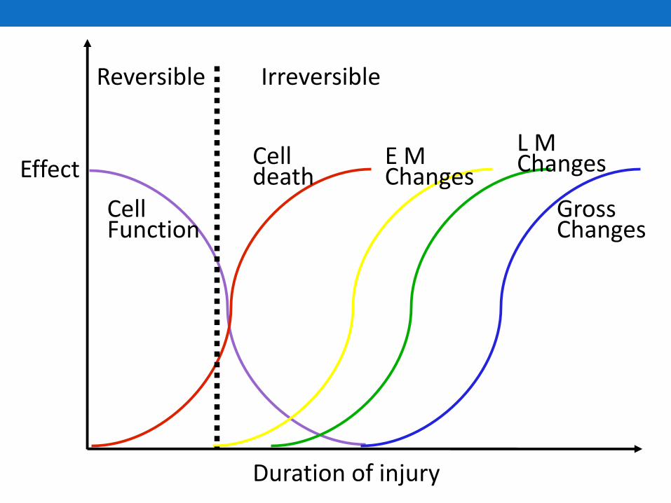
Effect
Duration of injury
Reversible Irreversible
Cell Function
Cell death
E M Changes
L M Changes
Gross Changes
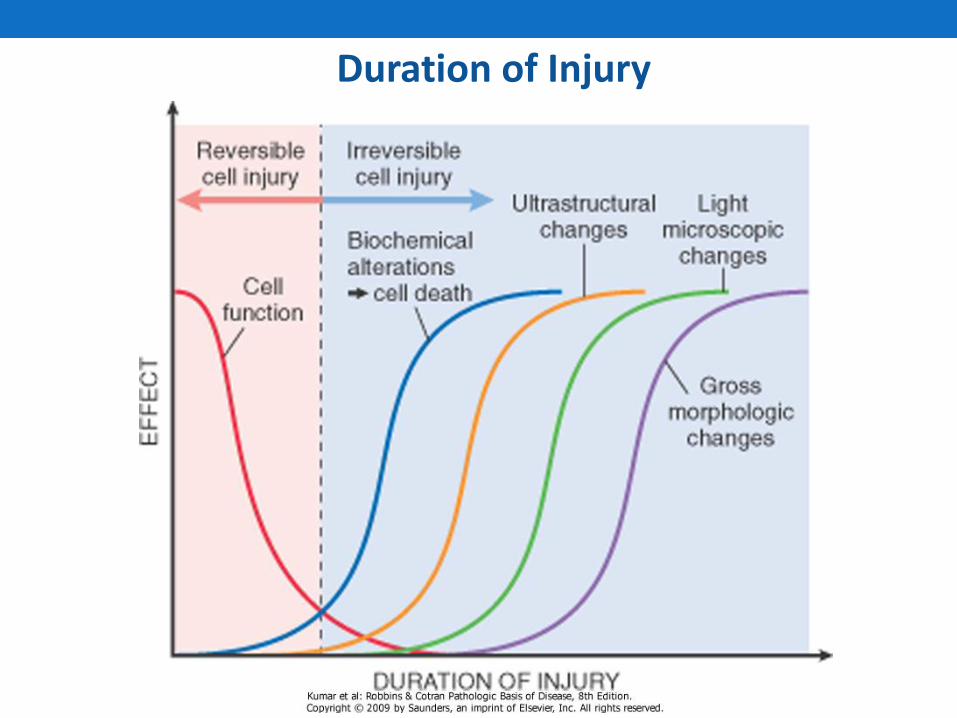
Duration of Injury
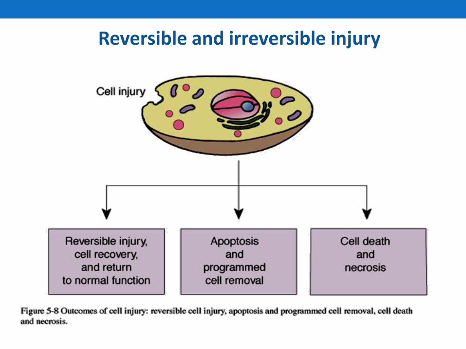
Reversible and irreversible injury
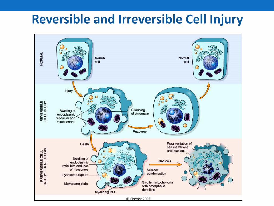
Reversible and Irreversible Cell Injury
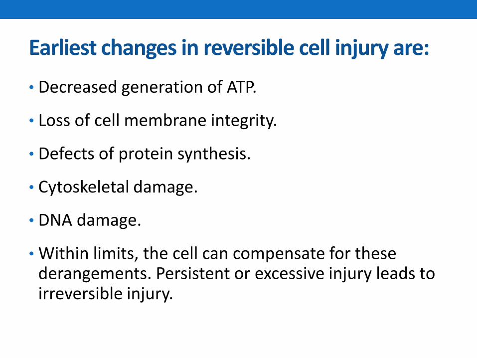
Earliest changes in reversible cell injury are:
• Decreased generation of ATP.
• Loss of cell membrane integrity.
• Defects of protein synthesis.
• Cytoskeletal damage.
• DNA damage.
• Within limits, the cell can compensate for these derangements. Persistent or excessive injury leads to irreversible injury.
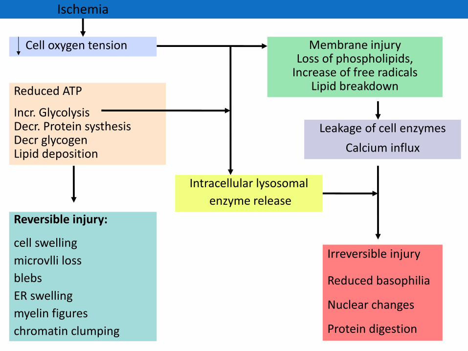
Ischemia
Cell oxygen tension Membrane injury Loss of phospholipids,
Increase of free radicals Lipid breakdown Reduced ATP
Incr. Glycolysis Decr. Protein systhesis Decr glycogen Lipid deposition
Intracellular lysosomal
enzyme release
Leakage of cell enzymes
Calcium influx
Reversible injury: cell swelling
microvlli loss
blebs
ER swelling
myelin figures
chromatin clumping
Irreversible injury
Reduced basophilia
Nuclear changes
Protein digestion
Characteristic phenomena of irreversibility:
• Inability to reverse mitochondrial dysfunction.
• Development of profound disturbances in membrane function.
• Therefore, in cardiac muscle death there is leakage of CKMB & troponin.
• In injury to bile duct epithelium & liver, serum alkaline phosphatase is raised.
• In hepatocyte injury, transaminases are raised.
Light microscopic patterns of reversible cell injury:
• Cellular swelling & fatty change.
• Morphology in cellular swelling-
Gross: Pallor, increased turgor & increase in organ weight.
Micro: small clear vacuoles seen within cytoplasm.
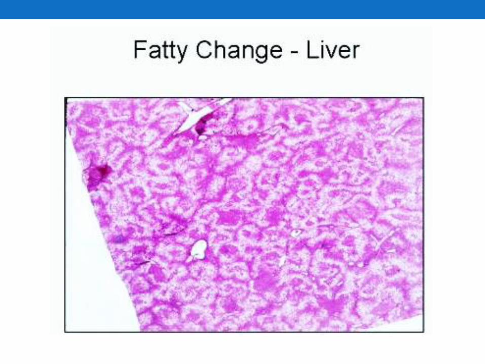
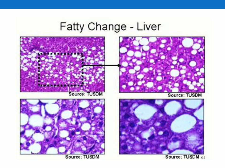
• Fatty change-seen in injured myocardial cells and hepatocytes. There is appearance of small or large lipid vacuoles in the cytoplasm.
Reversible cellular changes & accumulations
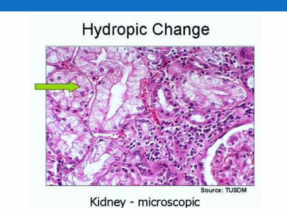
Hydropic degeneration (hydropic change)
- Only the cytoplasm is involved
- Water accumulates & the cell swells
- Large vacuoles in the cytoplasm
- Light microscopy
- Cytoplasm is pink & granular
- Electron microscopy (ultrastructural)
- Organelles are swollen
- Ribosomes displaced
- Lysosomal activity very apparent

Intracellular Accumulations
Intracellular accumulation of abnormal amounts of various substances.
(1) a normal cellular constituent accumulated in excess, such as water, lipids, proteins, and carbohydrates
(2) an abnormal substance, either exogenous, such as a mineral or products of infectious agents, or endogenous, such as a product of abnormal synthesis or metabolism
(3) a pigment.

Intracellular Accumulations
- The substance may be either the cytoplasm or the nucleus.
- In some instances, the cell may be producing the abnormal substance, and
- In others it may be merely storing products of pathologic processes occurring elsewhere in the body
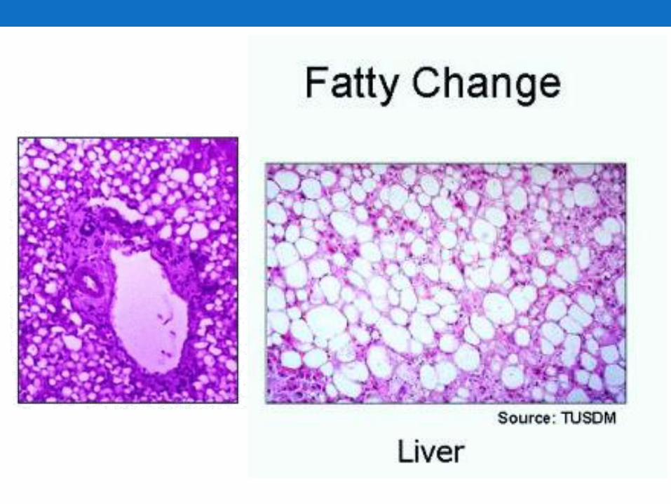
Reversible cellular changes &accumulations
fatty change ( steatosis , fatty metamorphosis)
- Characterized by accumulation of intracellular parenchymal triglycerides,
nucleus is displaced & the cells swells.
- Observed frequently in liver, heart, & kidney.
- Ex. In liver secondary to alcoholism, diabetes mellitus, malnutrition, obesity,
poisoning
- results from imbalance among the uptake, utilization & secretion of fat.
- Increased transport of triglycerides (fatty acids) to affected
cells
- Decreased mobilization of fat from cells
- Most often due to decreased production for transport
- Decreased use of fat by cells.
- Overproduction of fat in cells.
Reversible cellular changes & accumulation
Hyaline change
- Homogenous , glassy , eosinophilic appearance in H&E
stained tissue sections
- Caused most often by nonspecific accumulations of proteinaceous material
- Ex. Glomeruli tufts in diabetic glomerulosclerosis
Reversible cellular changes & accumulations
Accumulation of exogenous pigments
- Naturally colored substances not requiring tissue stain to be seen
1 - Pulmonary accumulations of carbon , silica & iron dust
2 - Plumbism ( lead poisoning)
3 - Algeria ( silver poisoning)
- May cause a permanent gray discoloration of the skin & conjunctiva
Accumulation of endogenous pigments
- Melanin :
- Most common ; brown pigment
- Formed from tyrosine via tyrosinase
- Synthesized in melanosomes of melanocytes within
the basement membrane of the epidermis &
choroid of the eye.
- Transferred by melanocytes to adjacent clusters of
keratinocytes & macrophages (melanophores) in
the subjacent dermis
- Seen also in neoplasm
- Ex. Melanocytic nevus, melanotic macule
- Ex. Melanoma


- Bilirubin
- Catabolic product of the heme moiety of hemoglobin &
myoglobin
- In pathologic conditions , accumulates & stains the blood,
sclera, mucosae , & internal organs producing a yellow
discoloration ( jaundice)
- Hemolytic jaundice
- Destruction of red blood cells.
- Obstructive jaundice
- intra or extrahepatic obstruction of the biliary tract.
- Hepatocellular jaundice Ex. Parenchyma liver damage
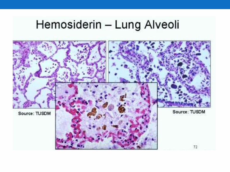
- Hemosiderin
- Iron – containing pigment , aggregates of ferritin
- In tissue appears as golden – brown amorphous
aggregates.
- Prussian blue dye – positive blue color stain
reaction.
- Exists normally in small amounts as physiologic
iron stores within tissue macrophages of the bone
marrow, liver, & spleen.

Hemosiderin Melanin
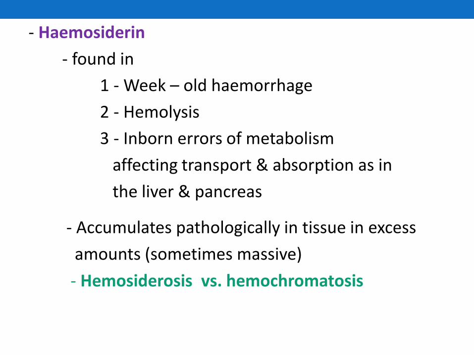
- Haemosiderin
- found in
1 - Week – old haemorrhage
2 - Hemolysis
3 - Inborn errors of metabolism
affecting transport & absorption as in
the liver & pancreas
- Accumulates pathologically in tissue in excess
amounts (sometimes massive)
- Hemosiderosis vs. hemochromatosis
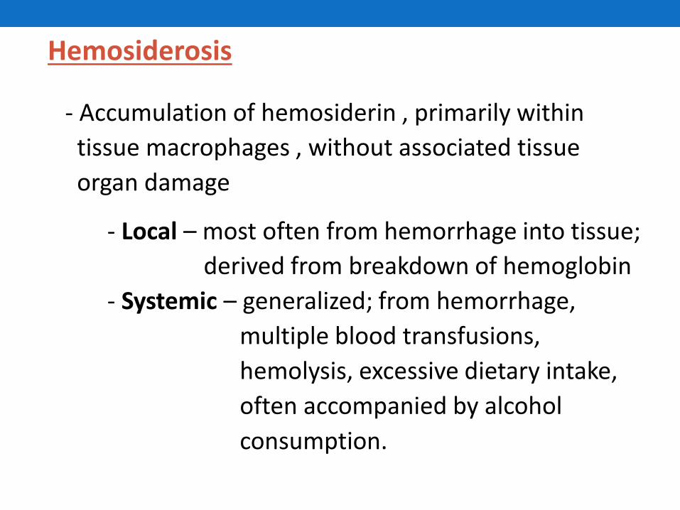
Hemosiderosis
- Accumulation of hemosiderin , primarily within
tissue macrophages , without associated tissue
organ damage
- Local – most often from hemorrhage into tissue;
derived from breakdown of hemoglobin
- Systemic – generalized; from hemorrhage,
multiple blood transfusions,
hemolysis, excessive dietary intake,
often accompanied by alcohol
consumption.

Lipofuscin
- yellowish to light brown, fat-soluble pigment; end
product of membrane lipid peroxidation
- “Wear & tear” pigment
- Commonly accumulates in elderly patients
- Found most often within hepatocytes &
at the poles of nuclei of myocardial cells.
Brown atrophy :
- accumulation of lipofuscin & atrophy of organs

Cardiac muscle Liver

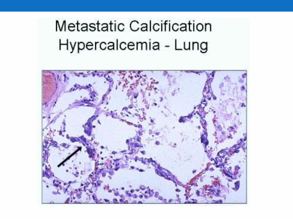
- Pathologic calcifications
- Abnormal deposition of calcium salts in soft tissue
- Deep blue-purple in nondecalcified H&E stained tissue
- May stimulate further bone deposition
- Metastatic calcification : caused by hypercalcemia
- Most often from hyperparathyroidism
- Osteolytic tumours with mobilization of Ca2+ & Po4
- Hypervitaminosis D
- Excess calcium intake
- E.g. milk – alkali syndrome – nephrocalcinosis, renal
stones caused by milk & antacid self-therapy for peptic
ulcer.
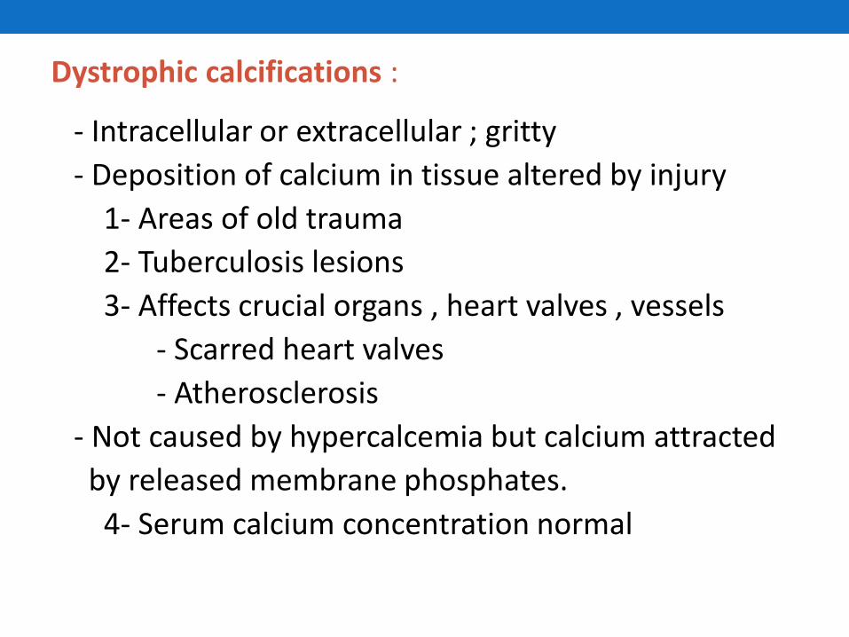
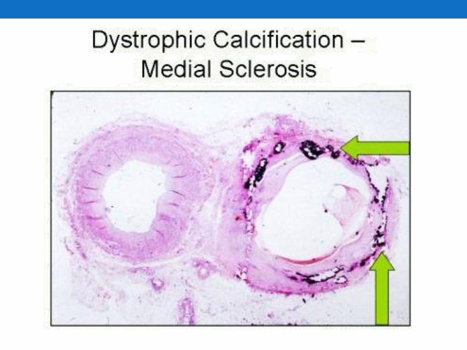
Dystrophic calcifications :
- Intracellular or extracellular ; gritty
- Deposition of calcium in tissue altered by injury
1- Areas of old trauma
2- Tuberculosis lesions
3- Affects crucial organs , heart valves , vessels
- Scarred heart valves
- Atherosclerosis
- Not caused by hypercalcemia but calcium attracted
by released membrane phosphates.
4- Serum calcium concentration normal
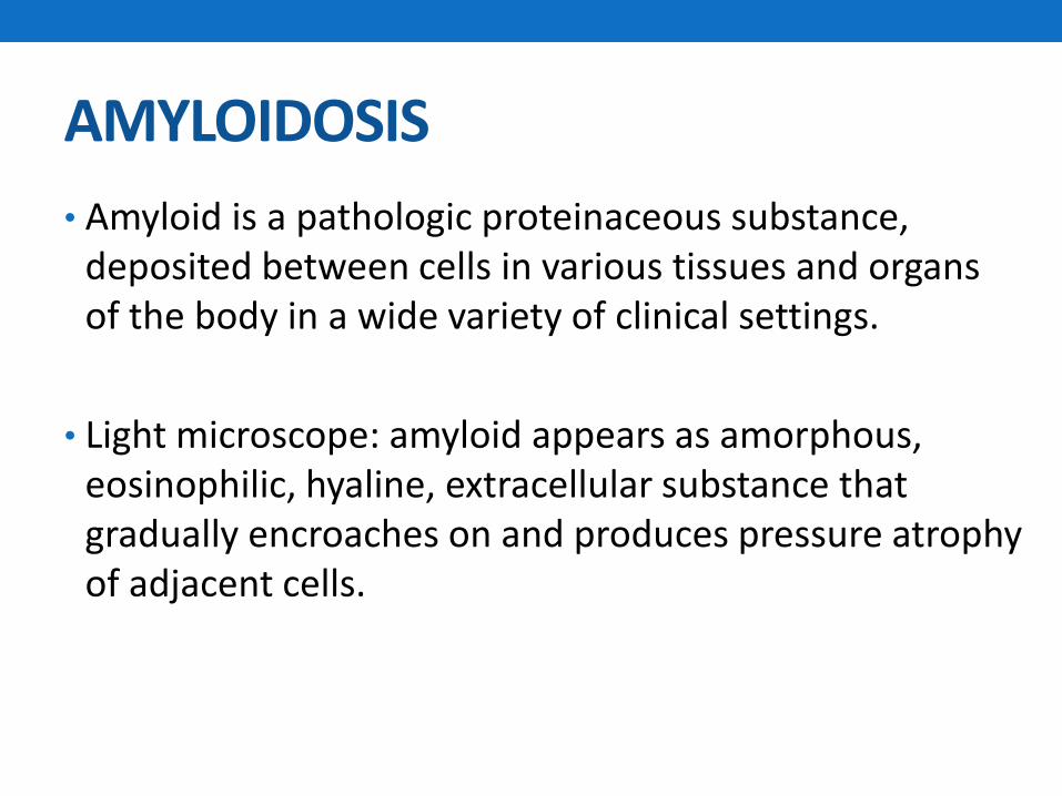
AMYLOIDOSIS
• Amyloid is a pathologic proteinaceous substance, deposited between cells in various tissues and organs of the body in a wide variety of clinical settings.
• Light microscope: amyloid appears as amorphous, eosinophilic, hyaline, extracellular substance that gradually encroaches on and produces pressure atrophy of adjacent cells.
Clinicopathologic Category Associated Diseases Major Fibril
Protein
Chemically Related
Precursor Protein
Systemic (Generalized) Amyloidosis
Immunocyte dyscrasias with amyloidosis (primary amyloidosis)
Multiple myeloma and other monoclonal B-cell proliferations
AL Immunoglobulin light chains, chiefly λ type
Reactive systemic amyloidosis (secondary amyloidosis)
Chronic inflammatory conditions
AA SAA
Hemodialysis-associated amyloidosis Hereditary amyloidosis
Chronic renal failure Aβ2 m β2-microglobulin
Familial Mediterranean fever - AA SAA
Familial amyloidotic neuropathies (several types)
- ATTR Transthyretin
Systemic senile amyloidosis - ATTR Transthyretin
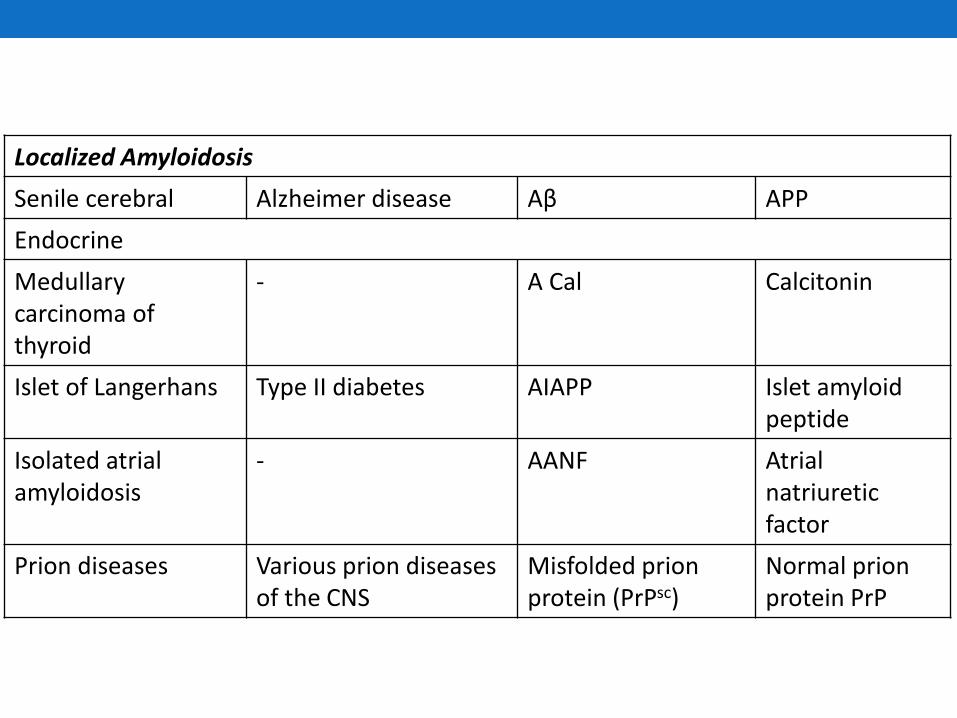
Localized Amyloidosis
Senile cerebral Alzheimer disease Aβ APP
Endocrine
Medullary carcinoma of thyroid
- A Cal Calcitonin
Islet of Langerhans Type II diabetes AIAPP Islet amyloid peptide
Isolated atrial amyloidosis
- AANF Atrial natriuretic factor
Prion diseases Various prion diseases of the CNS
Misfolded prion protein (PrPsc)
Normal prion protein PrP

NECROSIS
• There is denaturation of intracellular proteins and enzymatic digestion of the cell.
• The enzymes are derived either from the lysosomes of the dead cells themselves, in which case the enzymatic digestion is referred to as autolysis, or from the lysosomes of immigrant leukocytes, during inflammatory reactions.
Necrosis
• A spectrum of morphologic changes that follow cell death in living tissue, due to progressive degradative action of enzymes on the lethally injured cells.
• Leaked out contents of necrotic cells may elicit inflammation in the surrounding tissue.
• The morphologic appearance is due to denaturation of proteins and enzymatic digestion.
• The enzymes are derived from lysosomes of the dead cells themselves-AUTOLYSIS.
Nuclear changes in necrotic cells:
• Karyolysis-basophilia of chromatin may fade.
• Pyknosis-nuclear shrinkage and increased basophilia.
• Karyorrhexis-nuclear fragmentation.
• Disappearance of nucleus.
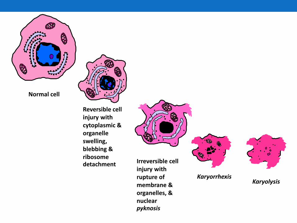
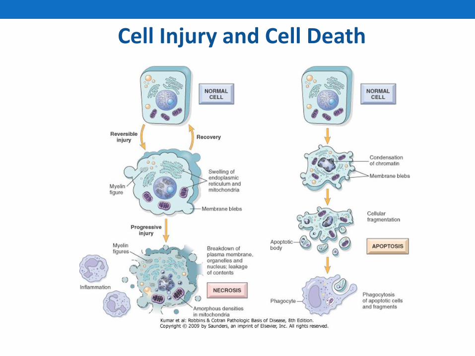
Normal cell
Reversible cell injury with cytoplasmic & organelle swelling, blebbing & ribosome detachment Irreversible cell
injury with rupture of membrane & organelles, & nuclear pyknosis
Karyorrhexis Karyolysis
Morphology of necrosis.
Necrotic cells show
• increased eosinophilia with a glassy homogeneous appearance.
• The cytoplasm becomes vacuolated and appears moth-eaten.
• Finally, calcification of the dead cells may occur.
Morphology of necrosis
By electron microscopy, necrotic cells are characterized
by :
• overt discontinuities in plasma membrane,
• marked dilation of mitochondria with the appearance
of large amorphous densities,
• intracytoplasmic myelin figures,
• amorphous osmiophilic debris, and
• aggregates of fluffy material probably representing
denatured protein
Necrosis • Definition
• Death of groups of contiguous cells in tissue or organ
• Patterns
• Coagulative
• Liquefactive
• Caseous
• Fat necrosis
• (gangrene)
• (Infarct)
• Red/haemorrhagic
• White
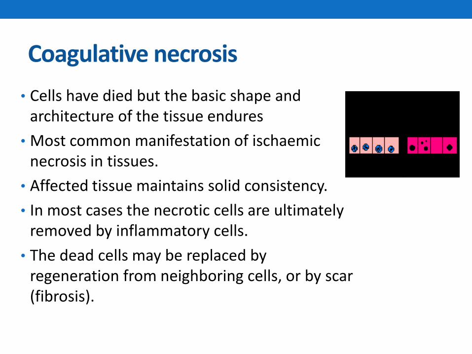
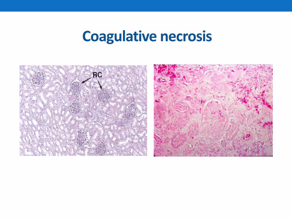
Coagulative necrosis
• Cells have died but the basic shape and architecture of the tissue endures
• Most common manifestation of ischaemic necrosis in tissues.
• Affected tissue maintains solid consistency.
• In most cases the necrotic cells are ultimately removed by inflammatory cells.
• The dead cells may be replaced by regeneration from neighboring cells, or by scar (fibrosis).
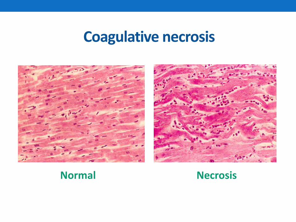
Coagulative necrosis
Necrosis Normal
Coagulative necrosis
Liquefactive necrosis
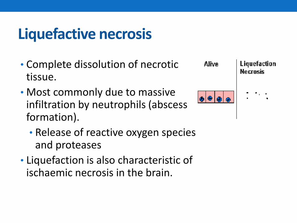
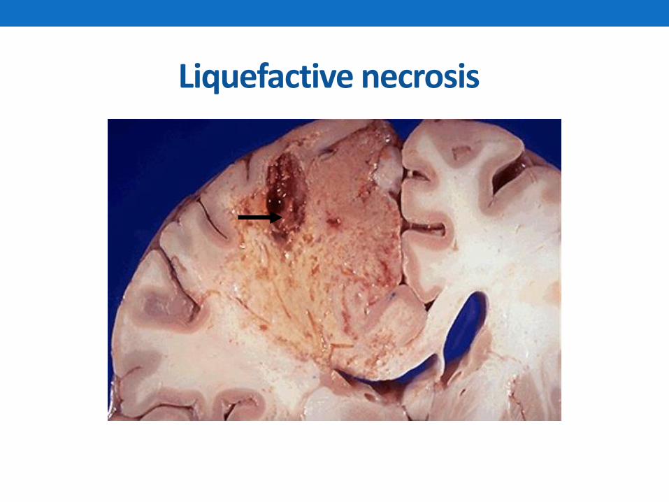
• Complete dissolution of necrotic tissue.
• Most commonly due to massive infiltration by neutrophils (abscess formation).
• Release of reactive oxygen species and proteases
• Liquefaction is also characteristic of ischaemic necrosis in the brain.
Liquefactive necrosis
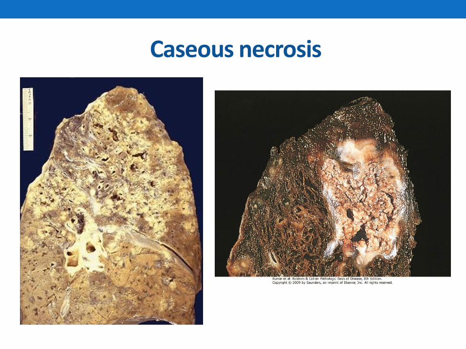
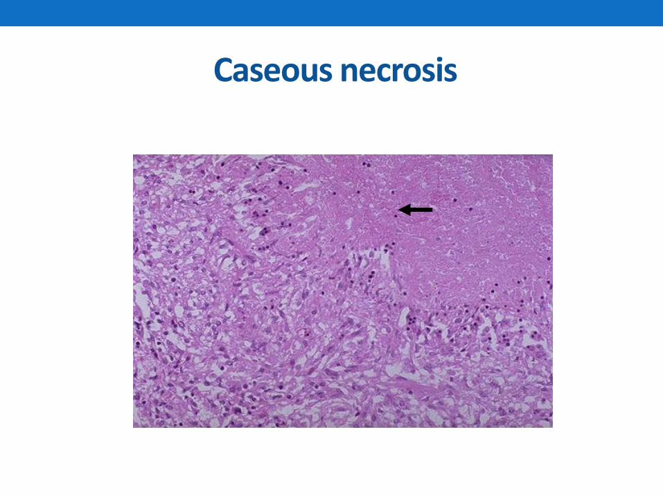
Caseous necrosis
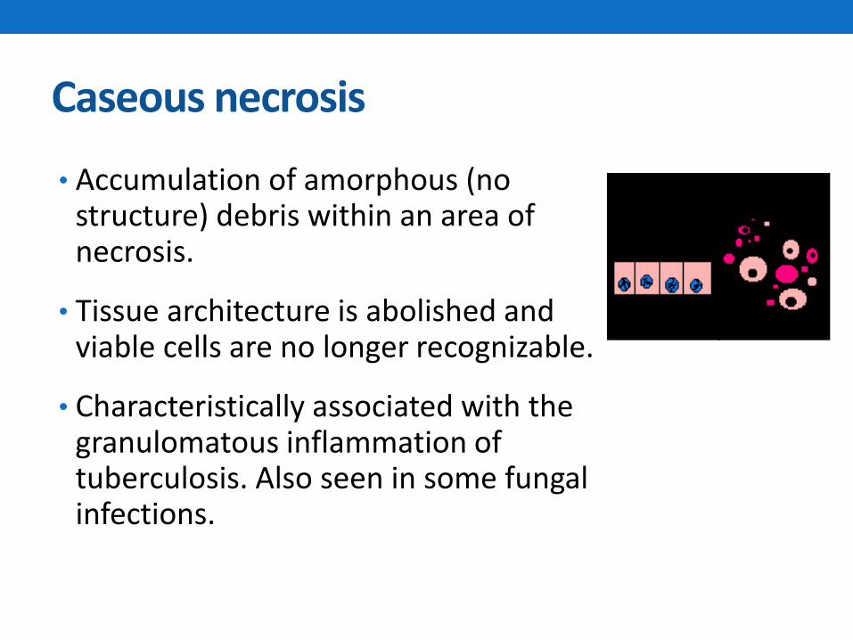
• Accumulation of amorphous (no structure) debris within an area of necrosis.
• Tissue architecture is abolished and viable cells are no longer recognizable.
• Characteristically associated with the granulomatous inflammation of tuberculosis. Also seen in some fungal infections.
Caseous necrosis
Caseous necrosis
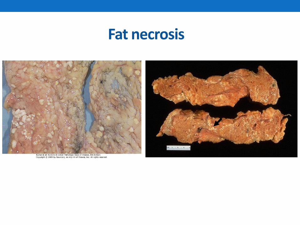
Fat necrosis
• Results from the action of lipases released into adipose tissue.
• pancreatitis, trauma.
• Free fatty acids accumulate and precipitate as calcium soaps (saponification).
• These precipitates are grossly visible as pale yellow/white nodules
• Microscopically, the digested fat loses its cellular outlines. There is often local inflammation
Fat necrosis
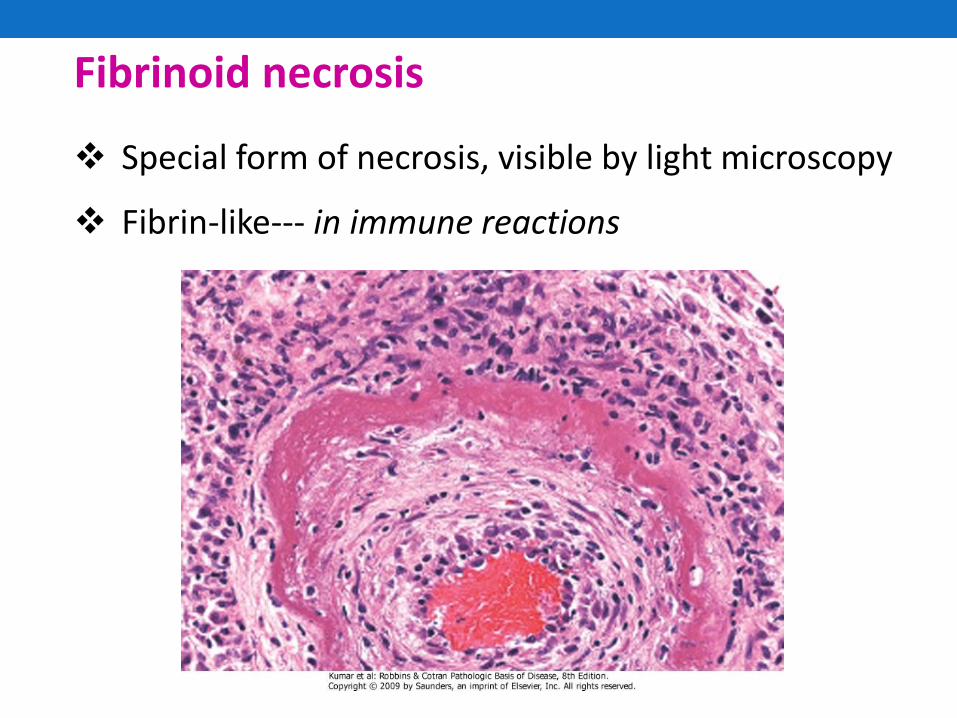
Fibrinoid necrosis
Special form of necrosis, visible by light microscopy
Fibrin-like--- in immune reactions
Gangrene ("gangrenous necrosis")
•Not a separate kind of necrosis at all, but a term for necrosis that is advanced and visible grossly.
• If there's mostly coagulation necrosis, (i.e., the typical blackening, desiccating foot which dried up before the bacteria could overgrow), we call it dry gangrene.
• If there's mostly liquefactive necrosis (i.e., the typical foul-smelling, oozing foot infected with several different kinds of bacteria), or if it's in a wet body cavity, we call it wet gangrene.
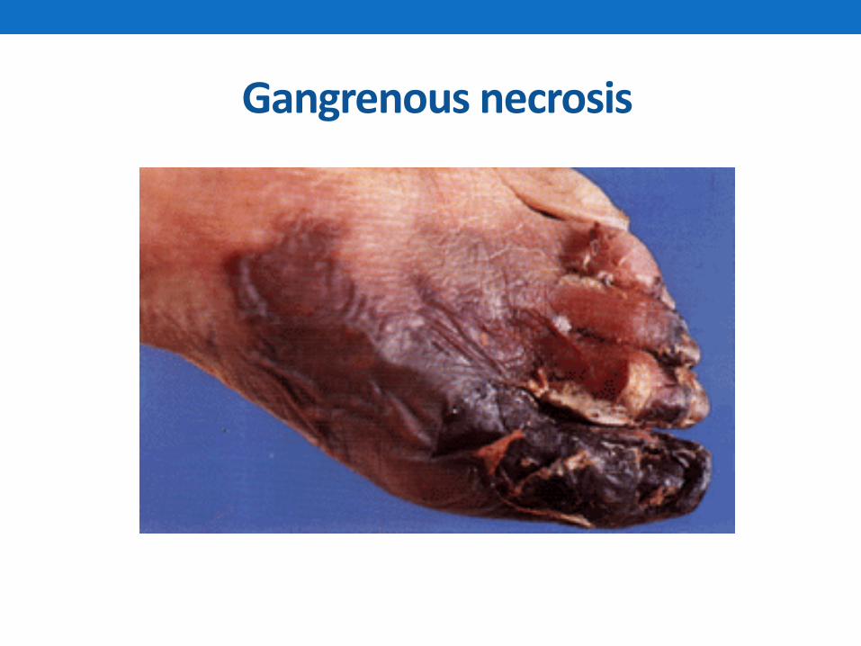
Gangrenous necrosis
Apoptosis
•Programmed cell death.
•Noxious stimuli that damage DNA result in nuclear dissolution without complete loss of cell membrane integrity.
•Can be physiologic or pathologic.
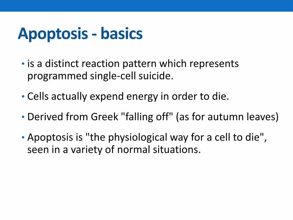
Apoptosis - basics
• is a distinct reaction pattern which represents programmed single-cell suicide.
• Cells actually expend energy in order to die.
• Derived from Greek "falling off" (as for autumn leaves)
• Apoptosis is "the physiological way for a cell to die", seen in a variety of normal situations.
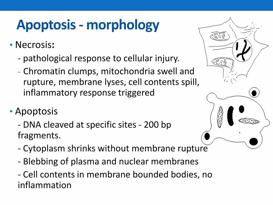
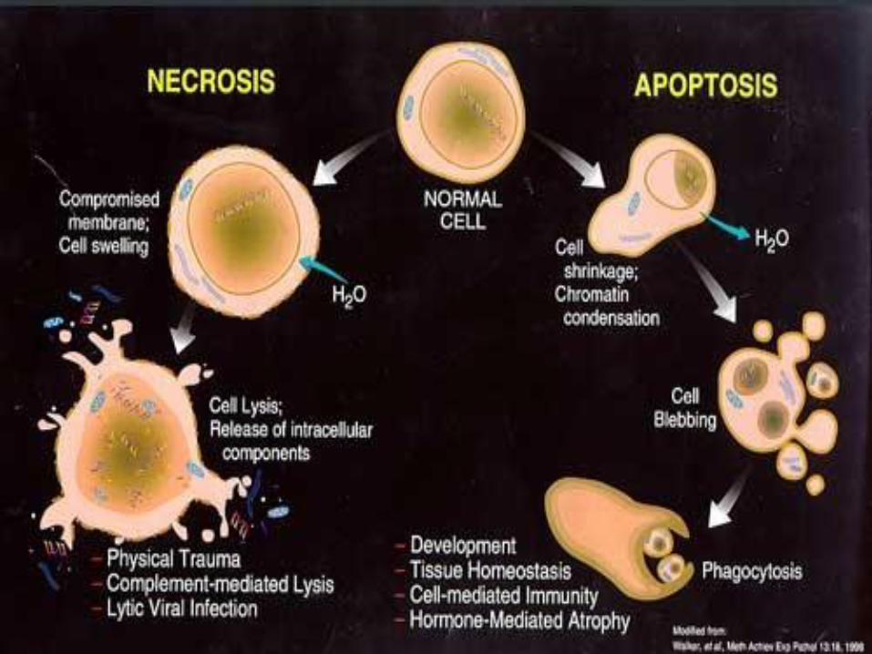
Apoptosis - morphology • Necrosis:
- pathological response to cellular injury.
- Chromatin clumps, mitochondria swell and rupture, membrane lyses, cell contents spill, inflammatory response triggered
• Apoptosis - DNA cleaved at specific sites - 200 bp fragments.
- Cytoplasm shrinks without membrane rupture
- Blebbing of plasma and nuclear membranes
- Cell contents in membrane bounded bodies, no inflammation
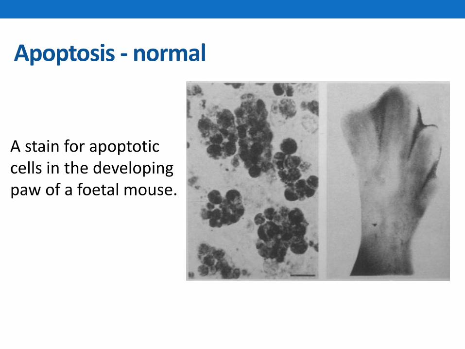
Apoptosis - normal
A stain for apoptotic cells in the developing paw of a foetal mouse.
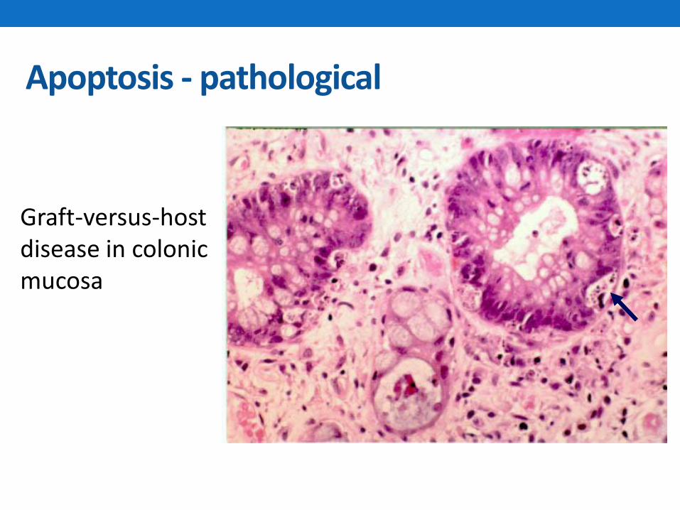
Apoptosis - pathological
Graft-versus-host disease in colonic mucosa

Apoptosis - triggers
•Withdrawal of growth stimuli
E.g. growth factors
•Death signals
E.g. TNF and Fas
•DNA damage
p53 plays an important role
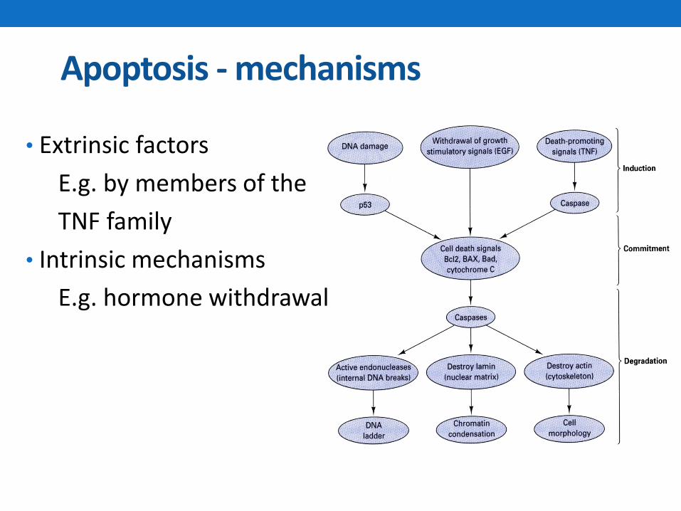
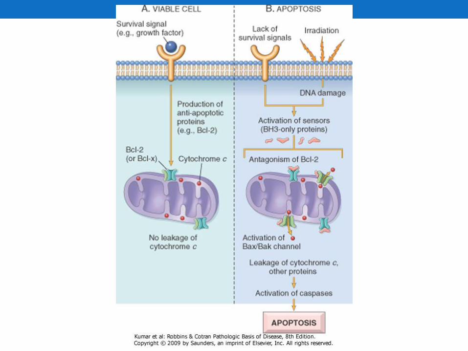
Apoptosis - mechanisms
• Extrinsic factors
E.g. by members of the
TNF family
• Intrinsic mechanisms
E.g. hormone withdrawal
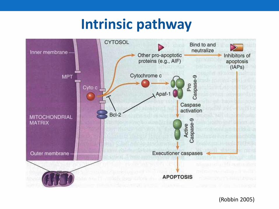
Intrinsic pathway
(Robbin 2005)
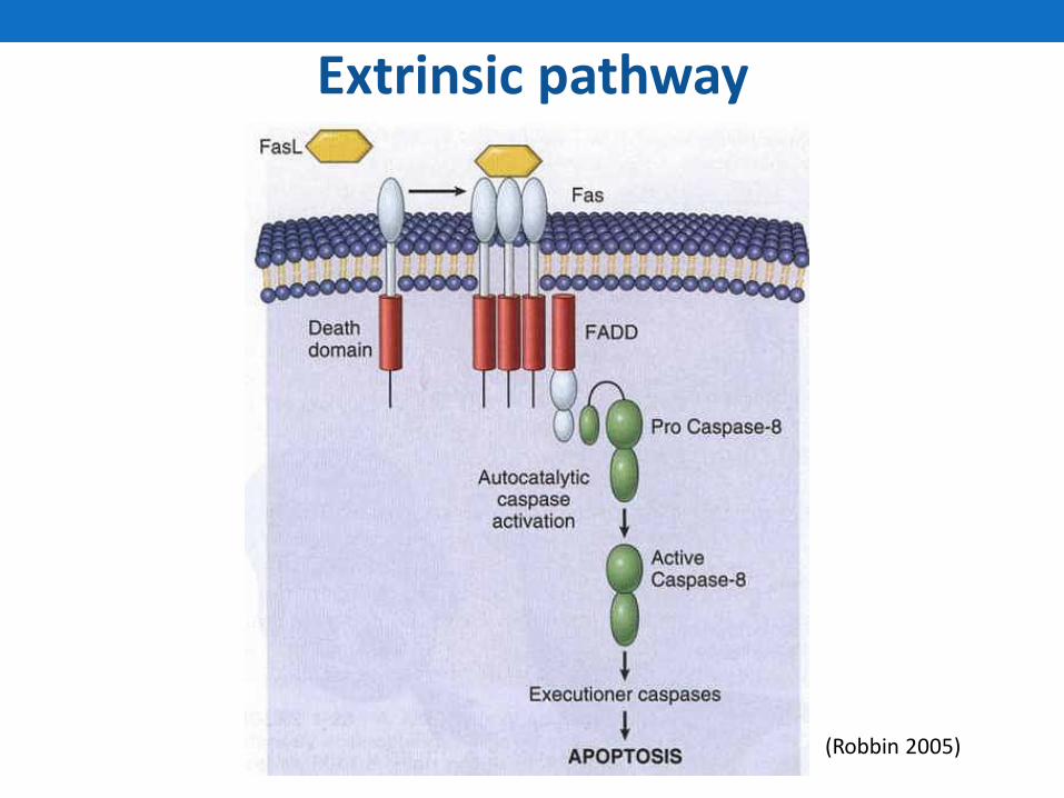
Extrinsic pathway
(Robbin 2005)
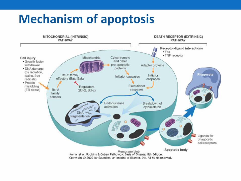
Mechanism of apoptosis
Patterns Of Cell Death
There are two principal patterns of cell death:
1- Necrosis and
2- Apoptosis.
Cell Injury and Cell Death
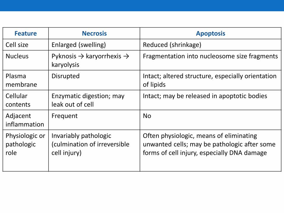
Feature Necrosis Apoptosis
Cell size Enlarged (swelling) Reduced (shrinkage)
Nucleus Pyknosis → karyorrhexis → karyolysis
Fragmentation into nucleosome size fragments
Plasma membrane
Disrupted Intact; altered structure, especially orientation of lipids
Cellular contents
Enzymatic digestion; may leak out of cell
Intact; may be released in apoptotic bodies
Adjacent inflammation
Frequent No
Physiologic or pathologic role
Invariably pathologic (culmination of irreversible cell injury)
Often physiologic, means of eliminating unwanted cells; may be pathologic after some forms of cell injury, especially DNA damage

Causes of cell injury
• Oxygen deprivation.
• Physical agents eg: mechanical trauma, burns, deep cold, barotrauma, electric shock.
• Chemical agents & drugs eg: poisons, environmental pollutants, CO, asbestos, alcohol, narcotic drugs etc.
• Infectious agents-viruses, rickettsiae, bacteria, fungi, protozoa and helminths.
• Immunologic reactions-anaphylaxis, autoimmune disorders.
• Genetic derangements.
• Nutritional imbalances-PEM, obesity, specific vitamin deficiencies etc.

Causes of Cell Injury
1) Oxygen Deprivation (Hypoxia). It is a common cause
of cell injury and cell death.
- Hypoxia can be due to :
A- inadequate oxygenation of the blood due to
Cardiorespiratory failure
B- loss of the oxygen-carrying capacity of the blood,
as in anemia or carbon monoxide poisoning.
Depending on the severity of the hypoxic state, cells may adapt, undergo injury, or die.

Causes of Cell Injury cont.
2) Physical Agents :
- Mechanical trauma,
- Burns,
- Deep cold
- Sudden changes in atmospheric pressure,
- Radiation, and electric shock
Causes of Cell Injury cont.
3) Chemical Agents and Drugs
- oxygen, in high concentrations
- poisons, such as arsenic, cyanide, or mercuric salts
- environmental and air pollutants
- insecticides, herbicides, industrial and
occupational hazards
- alcohol and narcotic drugs and therapeutic drugs
Causes of Cell Injury cont.
4) Infectious Agents e.g. bacteria, fungi, viruses and
parasites.
5) Immunologic Reactions.
6) Genetic Derangements.
7) Nutritional Imbalances

Mechanisms of cell injury
• Depletion of ATP-affects activity of Na, K-ATPase pump. This results in anaerobic glycolysis.
• Mitochondrial damage-leakage of cytochrome-C into cytosol, resulting in apoptosis.
• Influx of Ca & loss of Ca homeostasis, leading to activation of ATPases, phospholipases, proteases & endonucleases.
MECHANISM OF CELL INJURY
1. DEPLETION OF ATP:
- ATP depletion and decreased ATP synthesis are associated with both hypoxic and chemical (toxic) injury.
- ATP is required for many synthetic and degradative processes within the cell.
MECHANISM OF CELL INJURY cont.
• ATP is produced in two ways.
A- The major pathway is oxidative phosphorylation
of adenosine diphosphate.
B- The second is the glycolytic pathway, which
generate ATP in absence of oxygen using glucose
derived from body fluids or from glycogen
MECHANISM OF CELL INJURY cont.
Effects of depleted ATP
a) The activity of the plasma membrane energy-
dependent sodium pump is reduced. It causes
sodium to accumulate intracellularly and
potassium to diffuse out of the cell causing cell
swelling, and dilation of the endoplasmic
reticulum.

MECHANISM OF CELL INJURY cont.
b) If oxygen supply to cells is reduced, as in ischemia,
oxidative phosphorylation ceases and cells rely on
glycolysis for energy production (anaerobic
metabolism) resulting in depletion of glycogen
stores. Glycolysis results in the accumulation of lactic
acid which reduces the intracellular pH, resulting in
decreased activity of many cellular enzymes.
MECHANISM OF CELL INJURY cont.
c) Failure of the Ca2+ pump leads to influx of Ca2+,
with damaging effects on numerous cellular
components
d) Ribosomes detach from the RER and polysomes
breakdown into monosomes, leading to reduction
in protein synthesis. Ultimately, irreversible
damage to mitochondrial and lysosomal
membranes occurs, and cell undergoes necrosis

MECHANISM OF CELL INJURY cont.
e) In cells deprived of oxygen or glucose, proteins
may become misfolded, and trigger the unfolded
protein response leading to cell injury and even
death.

Functional and morphologic consequences of decreased intracellular ATP during cell injury.
Downloaded from: Robbins & Cotran Pathologic Basis of Disease (on 4 September 2005 02:13 PM)
© 2005 Elsevier

MECHANISM OF CELL INJURY cont.
2- Mitochondrial Damage:
Mitochondria are important targets for all types of
injury, including hypoxia and toxins.
MECHANISM OF CELL INJURY cont.
Mitochondria can be damaged by :
A- Increases of cytosolic Ca2+
B- Oxidative stress
C- Breakdown of phospholipids, and by
D- Lipid breakdown products.
MECHANISM OF CELL INJURY cont.
- Mitochondrial damage results in the formation of a high-conductance channel, called mitochondrial permeability transition, present in the inner mitochondrial membrane. In the initial phase it is reversible but once mitochondrial permeability transition is irreversble it becomes a deathblow to the cell.
- Mitochondrial damage can also be associated with leakage of cytochrome c into the cytosol.
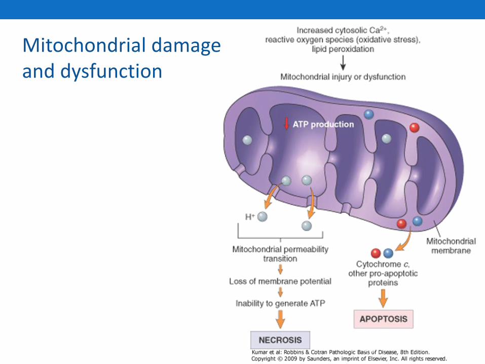
Mitochondrial damage and dysfunction
96
MECHANISM OF CELL INJURY cont.
3. INFLUX OF INTRACELLULAR CALCIUM & LOSS OF CALCIUM HOMEOSTASIS. . Ischemia causes an increase in cytosolic calcium concentration. Increased Ca2+ in turn activates a number of enzymes, e.g. - ATPases (thereby hastening ATP depletion), - Phospholipases (which cause membrane damage), - Proteases (which break down both membrane and cytoskeletal proteins), and - Endonucleases (which are responsible for DNA and chromatin fragmentation).

Influx of intracellular calcium
MECHANISM OF CELL INJURY cont.
4. ACCUMULATION OF OXYGEN-DERIVED FREE RADICALS (OXIDATIVE STRESS)
- Small amounts of partially reduced reactive oxygen forms are produced as a byproduct of mitochondrial respiration.
- Some of these free radicals can damage lipids, proteins, and nucleic acids.
- They are referred to as reactive oxygen species.
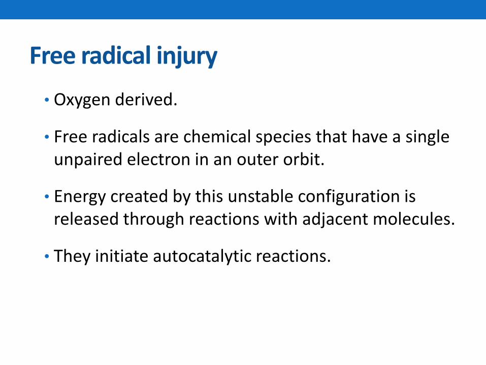
Free radical injury
• Oxygen derived.
• Free radicals are chemical species that have a single unpaired electron in an outer orbit.
• Energy created by this unstable configuration is released through reactions with adjacent molecules.
• They initiate autocatalytic reactions.

Free radicals are initiated by:
• Absorption of radiant energy(u-v or ionising):
Water is hydrolysed to(OH)&(H) free radicals.
• Enzymatic metabolism of exogenous chemicals or drugs, eg:CCl4 converted to CCl3.
• Redox reactions in the cell: (O2),(H2O2) &(OH).
• Transition metals like iron and copper.
• Nitrous oxide.

Effects of free radicals:
• Lipid peroxidation of membranes.
•Oxidative modification of proteins.
• Lesions in DNA.
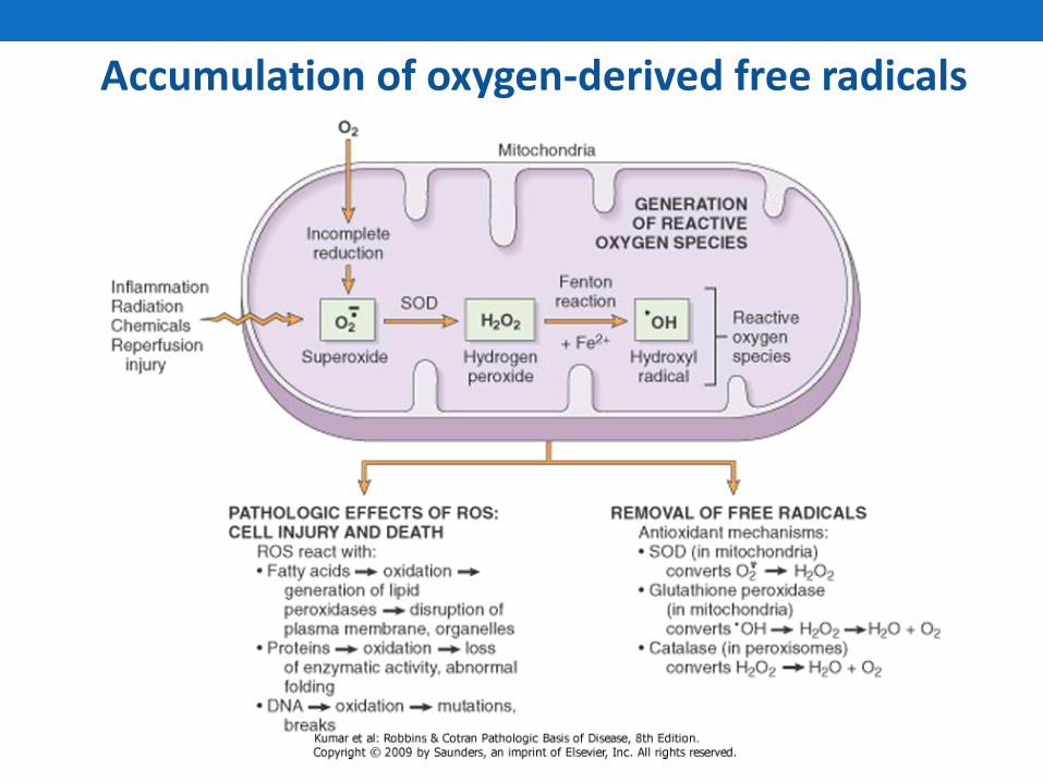
Accumulation of oxygen-derived free radicals
MECHANISM OF CELL INJURY cont.
5. Defects In Membrane Permeability: - In ischemic cells, membrane damage may be the
result of ATP depletion and calcium-modulated
activation of phospholipases.
- It can also be damaged directly by certain
bacterial toxins, viral proteins etc.
MECHANISM OF CELL INJURY cont.
The biochemical mechanisms which contribute to membrane damage are:
- Mitochondrial dysfunction
- Cytoskeletal abnormalities
- Reactive oxygen species
- Lipid breakdown products
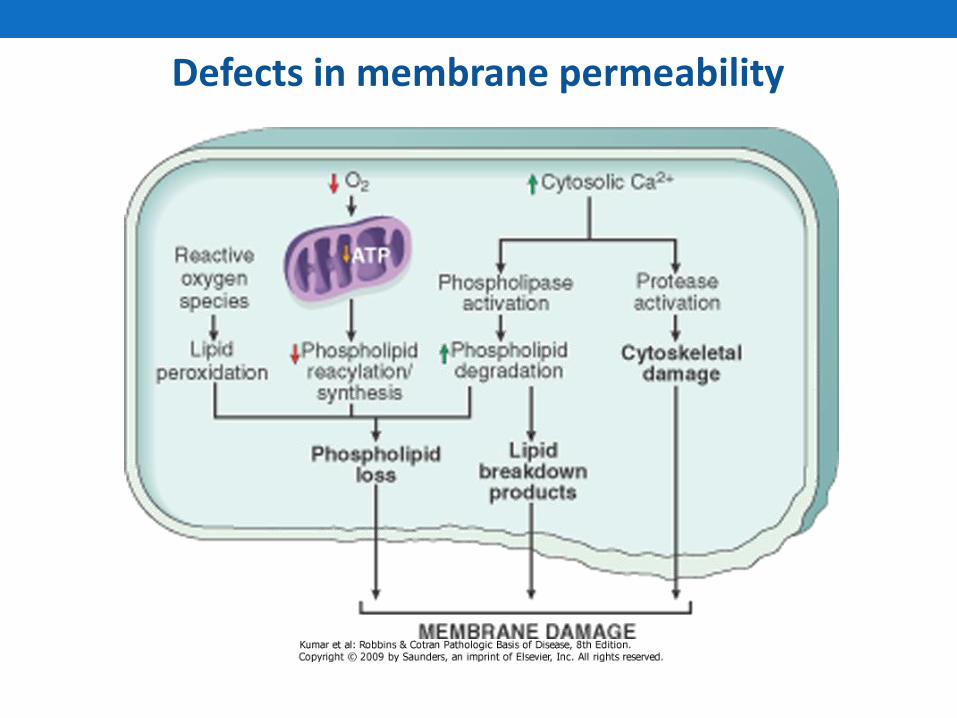
Defects in membrane permeability
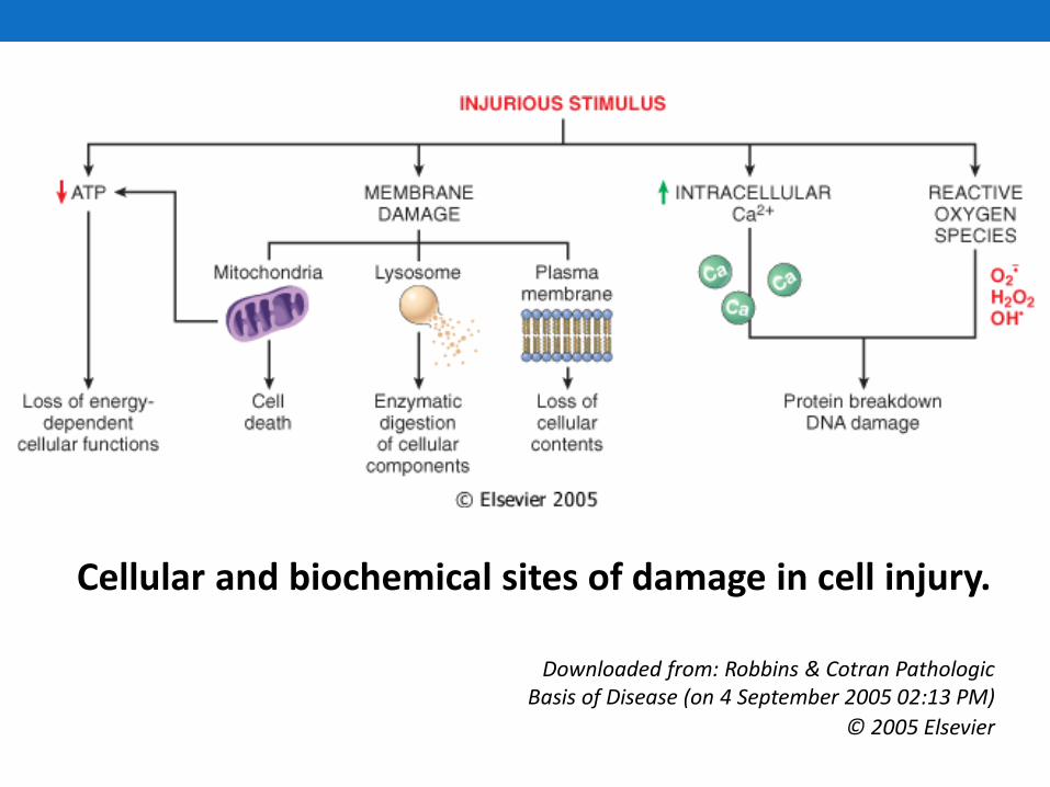
Cellular and biochemical sites of damage in cell injury.
Downloaded from: Robbins & Cotran Pathologic Basis of Disease (on 4 September 2005 02:13 PM)
© 2005 Elsevier