Embed Size (px)
Citation preview
MARINE ECOLOGY PROGRESS SERIES Mar Ecol Prog Ser
Published July 10
Cell lysis and release of particulate polysaccharides in extensive marine mucilage assessed by lipid
biomarkers and molecular probes
Franco B a l d i l v * , Andrea ~ i n a c c i ' , Alain saliot2, Laurence Mejanelle2, Patricija Mozetic 3, Valentina ~ u r k ~ , Alenka Malej
'Department of Environmental Biology, University of Siena, Via P. A. Mattioli 4, 1-53100 Siena, Italy
' ~ a b o r a t o i r e de Physique e t Chimie Marines de 1'Universite Pierre et Marie Curie, Observatoire des Sciences de I'Univers, URA CNRS 2076.4 Place Jussieu. F-75252 Paris Cedex 05, France
3National Institute of Biology, Marine Biological Station Piran. Fornace 41, SI-6330 Piran, Slovenia
ABSTRACT- During the massive mucllage event in the northern Adriatic Sea in July 1991 samples of macroaggregate were fixed in different ways: with formaldehyde, deep frozen and freeze-dried. Con- ventional microscopy (light and epifluorescence) revealed different autotrophic species embedded in gelatinous matl-ix. Cyanobacteria and heterotrophic bacteria were also identified. Scanning confocal laser microscopy (SCLMI and fluorescent molec~~ la r probes (the lectins concanavalin A and UEA-I) showed wall-free cytoplasm and particulate polysacchar~des leaklng from the envelopes of broken cells in the matrix. The extensive cell lysis was supported by the observation of cytoplasn1-free cytoskeletons, stained by the molecular probe phalloidin High concentrations of triglycerides (30Y0 of total lipids) and free fatty aclds (22'2%) along with very low concentrations of phospholipids ( 2 % ) also indicated massive cell degradation in freeze-dried material. The mucllage observations were compared with those of a natural plankton community grown under hlgh nutrlent conditions using the same tech- niques. Free polysaccharides were observed as globular flocs (marine snow) during in situ enrichment experiments and inti-acellular polysaccharides as carbon storage materials In autotrophic organisms. No strings, filaments, layers, cell lysis or lipid classes indicating strong cell biodeterioration were observed in a 1 mo controlled experiment during an algal bloom.
KEY WORDS: Marine mucilage Polysaccharides Molecular probes - SCLM . Lipid biomarker Adriatic Sea
INTRODUCTION
The exceptional event of large floating amounts of mucilage in the northern Adriatic Sea attracted public attention in 1988. The sea was covered with mucoid scum which prevented tourists from bathing. It was an economic disaster which was repeated in 1989 and 1991. A similar widespread event occurred in 1949. Before the tourist era, fishermen, local newspapers and scientific journals recorded similar events back as far as 1729 (Fonda Umani et al. 1989). The phenomenon
has been described in detail by Stachowitsch et al. (1990). The periodicity of the mucilage phenomenon was recently calculated over 120 yr, and indicated an average cycle of 5.74 yr. It was concluded that there was a 50% probability that the next event would take place in 1996, and a 90% probability that it will recur in 2005-2006 (Vollenweider et al. 1995).
Aggregation and massive sedimentation of phyto- plankton in marine and limnetic environments have been extensively studied in field and controlled exper- imental systems since these processes are of consider- able significance in the global carbon cycle (Alldredge & Jackson 1995). Empirical evidence and theoretical considerations based on coagulation theory (Kiarboe &
0 Inter-Research 1997 Resale of full article not permitted

Mar Ecol Prog Ser 153: 45-57, 1997
Hansen 1993) indicate the importance of phytoplank- ton stickiness, which varies from species to species, and gel-l~ke transparent exopolymer particles (TEP) in aggregation processes (Alldredge et al. 1993). None of these findings satisfactorily explains the mucilage phe- nomenon in the northern Adriatic, although diatoms are implicated as the main source of mucilage.
Several hypotheses to explain the origin of mucilage phenomena in the northern Adriatic have been pro- posed. (1) The first is that large floating mucilage is marine snow aggregated and consolidated to form a self-sustaining mucilage community that resists break- up (Fogg 1995). (2) Two factors leading to excessive marine snow and mucus were identified by Herndl (1992): high photosynthetic extracellular release duri.ng summer, probably stimulated by severe N and P limitation (Myklestad 1995), and development of a strong pycnocline preventing material flux to the bottom. (3) Degobbis et al. (1995) related the mucilage phenomenon to modifications in environmental condi- tions in the northern Adriatic and changes in commu- nity structure (increased diatom contribution, change ill dominant species). (4) The most recent hypothesis is that high C/P and slow-to-degrade organic matter is produced by sustained high rates of primary produc- tion and efficient bacterial phosphorus remineraliza- tion in preference to carbon, while aggregation is enhanced by mucus from bacterial capsules (Azam 1996). These hypotheses are not mutually exclusive and may even complement one another.
The aim of our paper was to shed new light on the mucilage phenomenon using novel microscope tech- niques and molecular probes for polysaccharides and other important molecular targets. The role of poly- saccharides is commonly recognized in extensive mu- cilage events (Marchetti et a1 1989, Murano et al. 1993, Faganeli et al. 1995). The lipid classes of the mucilage were also analyzed since only fatty acids had been ana- lyzed so far (Viviani et al. 1995) Lipids are useful to describe the status of mucus aggregates and they can provide an insight into biogeochemical processes (Wakeham & Lee 1989, Saliot et al. 1991). Lipid parti- tioning was also used to support microscope observa- tions. To aid interpretation of our results for large float- ing mucilage, a comparison was made with the results of an in sjtu nutrient-enrichment experiment using a natural plankton community. Various hypotheses to explain the extensive cell lysis are discussed.
MATERIALS AND METHODS
Mucilage and seawater sampling. Seawater samples were collected with a 5 1 Niskin sampler at offshore stations in the eastern part of the Gulf of Trieste (north-
ern Adriatic), during summer 1991 (June- August). In July, mucilage samples were collected at different depths by SCUBA at the same stations using a large syringe. Samples were fixed with borate-buffered for- malin (1.5% final concentration) and/or deep frozen. All analyses were carried out within 3 mo except for confocal laser microscopy and lipid analysis which were performed after 3 and 4 yr.
Enrichment experiment. Seawater for the enrich- ment experiment was sampled at an offshore station 1 m below the surface in April 1995, filtered through a 200 pm pore size plankton net to remove larger graz- ers, and dispensed into 8 l polycarbonate containers. One bottle served as a control. A mixture of inorganic nutrients was added to a second bottle to a final con- centration approximately 10 times higher than back- ground. The bottles were incubated at a depth of 2 m in the sea (exposure to daylight, water temperature between 12 and 13°C). Experimental details have already been reported (Malej et al. 1996). In these experiments all analyses including lipids and confocal microscopy were performed within 3 mo.
Light and epifluorescence microscopy. The forma- lin-preserved samples of mucilage and seawater sam- ples from the enrichment experiment were examined by light microscope. The mucilage was observed again in 1994 and 1995 for differences caused by annual storage. The phytoplankton composition was deter- mined using the sedimentation technique of Utermohl (1958), and autofluorescent cyanobacteria were counted under green excitation light (Takahashi et al. 1985). Samples for bacterial counts were stained with DAPI (4' , 6-diamidino-2-phenylindole) according to the protocol of Porter & Feig (1980) and examined by epi- fluorescence microscopy.
Molecular probe analysis. A 0.3 m1 aliquot of mucus was incubated for 1 h with 5 p1 of Con-A-FITC (con- canavalin A from Canavalia ensiformis conjugated with 1 mg ml-' fluorescein isothiocyanate, C 7642; Sigma) solution In phosphate buffer (Neu & Marshal1 1991, Gabius & Gabius 1993). This lectin specifically binds different carbohydrate residues: D(+)-glucose, N-acetyl-D-glucosamine, D(+)-mannose and methyl a-D-mannopyranoside (Haugland 1992, Gabius & Ga- bius 1993). Another lectin, UEA-l conjugated with fluorescein (L-9006; Sigma) from Ulex europeus that specifically binds sugar residues of fucose (Neu & Mar- shall 1991), was used as control for fluorescence distri- bution. To control the specificity of the lectins, com- mercial crystals of amylopectin, which consist of layers of D-glucose linked by c((1,4) and 4 % branched a(1,6) bonds, were soaked for 5 h in seawater from the Gulf of Trieste. The 2 lectins were incubated with amylopectin granules (5 mg ml-l) according to the protocol for mucilage samples.

Raldi et al.: Assess] Ing marine mucilage 4 7
To reveal stained neutral lipids in the cells, 0.5 m1 of mucilage was incubated with 10 p1 of 1 mg ml-' Nile red (N-3013, Sigma) standard solution in acetone (Greenspan & Fowler 1985). The sample was exposed for 5 min and then analyzed by scanning confocal laser microscopy (SCLM).
To localize cell-free cytoskeleton proteins of F-actin from the lysis of marine cells, a 10 p1 aliquot of 1 mg ml-' solution of phalloidin conjugated with FITC (Mol- ecular Probes Inc.) was used. The mucus sample was incubated with the molecular probe for 2 h.
SCLM analysis. A Nikon Microphot microscope was mounted on a confocal laser (MRC-500; Bio-Rad Microscience Division) to obtain images of mucilage samples. The microscope was equipped with a x60, 1.4 numerical aperture (NA) oil immersion lens (Nikon Corp.). A krypton-argon laser with maximum emission lines at 488 nm was used to measure the excitation source of the fluorescein conjugated lectins, Nile red and F-phalloidin. Images were obtained with a Bio- Rad photomultiplier pickup device and integrated and digitized with a Kalman true-running-average filter (Wolfaardt et al. 1994). The recorded video images (512 x 768 pixels) were displayed on a 7 " flat, black- and-white, high-resolution, 16 MHz video display screen (VM 1710; H. Lucius & Baer, Geretsried, Ger- many) and photographed with a Nikon F-301 camera equipped with a 105 mm lens. Image analysis of the recorded sections of the samples was carried out wlth Comos Bio-Rad software.
Iatroscan lipid analysis. Total lipid extract was ana- lyzed for lipid class composition with an Iatroscan MKV TLC/FID analyzer (Iatron Laboratories, Japan) . Samples and standards were spotted in triplicate on S111 Chroma-rods with a 1 p1 automatic syringe (SES, Germany). A stepwise method was used to separate the different lipid classes, with 3 consecutive develop- ing solvent systems of increasing polarity. After each development, partial scans of the Chroma-rods were performed to quantify the lipid classes eluted away from the initial spot (Volkman et al. 1986, Laureillard et al. 1997). The flame ionization detector (FID) was operated with a hydrogen flow of 160 m1 min-', an air flow of 1800 m1 min-', and the 'rods' were scanned in 30 S. Peak areas were integrated using 'Boreal' soft- ware (JMBS, Grenoble). Lipid classes were identified according to their retention times and quantified using external calibration. Calibration curves were con- structed for each lipid class with loads in the range 0.2 to 3.5 pg. This analytical procedure allows quantlfica- tion of 13 lipid classes: hydrocarbons, wax esters and steryl esters, methyl esters, free fatty acids, triacyl- glycerols, ketones, alcohols, sterols, diacylglycerols, chlorine pigments, monoacylglycerols and polar lipids (mainly phospholipids and glycolipids). The precision
in absolute weight is about 5 to 10% of the lipid class weight, and the standard devi.ation accounts for about 1 to 5 % of the relative abundance of any lipid class.
RESULTS
Light and epifluorescence microscopy
The analysis of fresh and formalin-preserved mu- cilage using light and epifluorescence microscopy showed that the most numerous eukaryotic organism was the diatom species Cylindrotheca closterium fol- lowed by Rhizosolenia alata f. gracillima and R. frag- ilissima. C, closterium was also rather abundant in the water samples collected before the development of vis- ible mucilage, as well as in the enrichment experiment (Table l ) , In contrast, dlatoms Pseudonitzschia deli- catissima comp. and Skeletonema costatum reached high abundance in the enrichment experiment but were rare or absent in the mucilage. Two species of dinoflagellates were abundant in the early stage of vis- ible mucilage (July 11) but were not recorded later (July 24). Synechococcus-like cyanobacteria and het- erotrophic bacteria developed well within the mucil- age (Table 1). Microscopic observations of procaryotes indicated that they were more abundant in mucus than in seawater and in the enrichment experiment. Other organisms and particles like coccolithophorids, dead and decaying copepods, fecal pellets and dinoflagel- late thecae were regularly found.
All organisms and particles were generally embed- ded in a dense mucus matrix. Primulin staining of this matrix indicated polysaccharide material in the form of fibrils and globules.
The same formalin-stored sample of mucilage was analyzed 4 yr later using the same techniques to reveal possible changes. The main difference from freshly analyzed material was faded autofluorescence. When the sample was stained with primulin, it had the same appearance, without any major c h ~ g e s in the mor- phological structure of the microorganisms embedded in the mucoid matrix.
Confocal microscope observations
The mucilage, stored in formalin at 4"C, was also analyzed 3 and 4 yr later by confocal microscopy using the lectins Con-A and UEA-I to specifically determine the carbohydrate residues. The images presented here are from many observations of different specimens of the mucilage of 1991. Con-A showed that marine eukaryotes embedded in the mucilage contained enor- mous amounts of intracellular polysaccharide (IPS)
4 8 Mar Ecol Prog Ser 153: 45-57, 1997
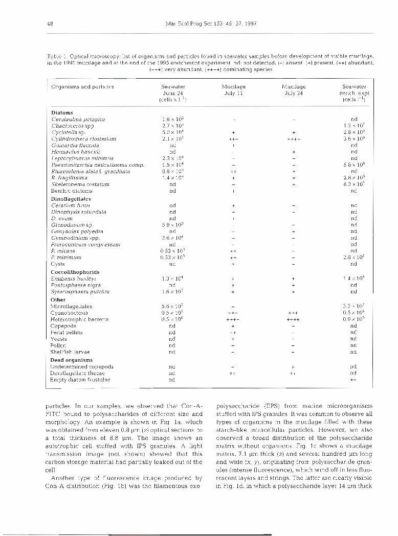
Table 1. Optical microscopy, list of organisms and particles found in seawater samples before development of visible mucilage, in the 1991 mucllage and at the end of the 1995 enr~chrnent experiment. nd: not detected; (-1 absent, (+) present, (++) abundant,
(+++) very abundant, (++++) dom~nat~ng species
Organisms and particlcs Seawater Mucilage Mucilage Seawater June 24 July 11 July 24 enrich. expt
(cells X I-') (cells I-')
Diatoms Ceratauljna pelagica 1.6 X lo3 nd Chaetoceros spp. 2.7 X lo3 - - 1.2 X 10' Cyclotella sp. 5.0 X 10" + + 2.8 X 10' Cylindrotheca closterium 2.1 103 +++ ++++ 3.6 X 10' Guinardia flaccida nd + - nd Hemiaulus hauckii nd + nd Leptocylindrus minimus 2.3 X 10' - - nd Pseudonitzschia delicatissima comp. 1.5 X lo4 - - 5.8 X 10⁸ Rh~zosolenia alata f . gracllljma 8.6 X 10³ ++ + nd R, frag~l~ssima 1.4 X 10' + + 2.8 lo5 Skeietonema costa turn nd - + 8.3 X 10' Benthic diatoms nd - nd
Dinoflagellates Ceratium fusus nd nd Dinophysis rotundata nd nd D. ovum nd nd Glenodlnium sp. 5.9 X 10³ nd Gonyaulax poiyedra nd nd Gymnodjnium spp. 3.6 X 10⁴ nd Prorocentrum compressum nd nd P. micans 0.53 X 10³ nd P. minimum 0.53 X 10³ 2.8 X lo5 Cysts nd nd
Coccolithophorids Emiliania huxleyi 1.3 X 10' 1.4 X 10⁵ Pon tospha era nigra n d nd Syracosphaera pulchra 1.6 X lo3 nd
Other Microflagellates 5.6 X 10⁵ 3.3 X 10' Cyanobacteria 0.5 X 10' 0.5 X 10⁶ Heterotrophic bacteria 0.5 X 10' 0.9 X log Copepods nd nd Fecal pellets nd nd Yeasts nd nd Pollen nd nd Shellfish larvae nd n d
Dead organisms Undetermined copepods nd - nd Dinoflagelldte thecae nd ++ nd Empty diatom frustulae nd - - ++
particles. In our samples, we observed that Con-A- FITC bound to polysaccharides of different size and morphology. An example is shown in Fig l a , which was obtained from eleven 0.8 pm (2) optical sections, to a total thickness of 8.8 pm. The image shows an autotrophic cell stuffed with IPS granules. A light transmission image [not shown) showed that this carbon storage material had partially leaked out of the cell.
Another type of fluorescence image produced by Con-A distribution (Fig. l b ) was the filamentous exo-
pol.ysacchande (EPS) from manne microorganisms stu.ffed with IPS granules. It was common to observe all types of organisms in the mucilage filled with these starch-like intracellular particles. However, we also observed a broad distribution of the polysaccharide matrix without organisms. Fig. l c shows a mucilage matrix, 7.1 pm thick [ z ) and several hundred pm long and wide (X, y), originating from polysaccharide gran- ules (intense fluorescence), which wind off in less fluo- rescent layers and strings. The latter are clearly visible in Fig. Id, in which a polysacchande layer 14 pm thick
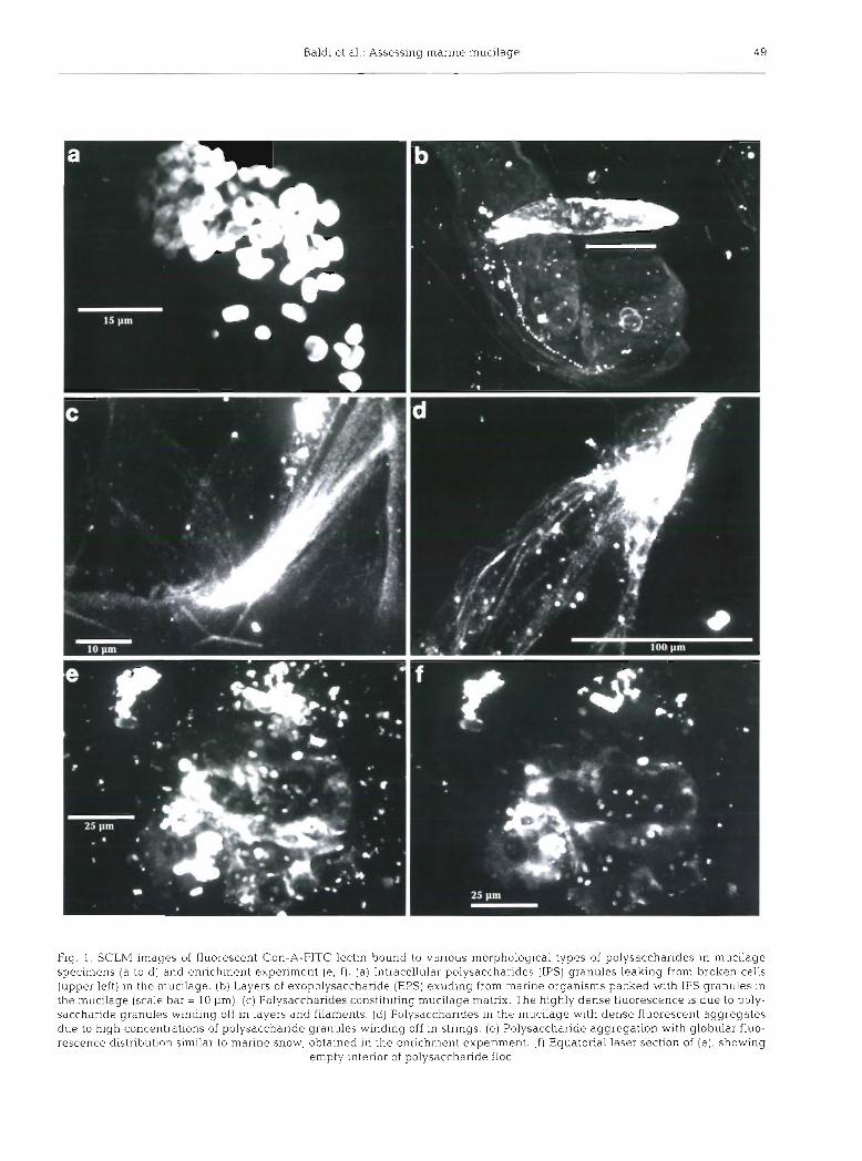
Baldi et al.: Assessing marine mucilage 49
Fig. 1. SCLM images of fluorescent Con-A-FITC lectin bound to various morphological types of polysaccharides in mucilage specin~ens (a to d) and enrichment experiment (e , f ) . (a) Intracellular polysaccharides (IPS) granules leaking from broken cells (upper left) in the mucilage. [b) Layers of exopolysacchande (EPS) exuding from marine organisms packed with IPS granules in the mucilage (scale bar = 10 urn). (c) Polysaccharides constituting mucilage matrix. The highly dense fluorescence is due to poly- saccharide granules winding off in layers and filaments. (d) Polysaccharides in the mucilage with dense fluorescent aggregates due to high concentrations of polysaccharide granules winding off in strings. (e) Polysaccharide aggregation with globular fluo- rescence distribution similar to marine snow, obtained in the enrichment experiment. ( f ) Equatorial laser section of (e), showing
empty interior of polysaccharide floe

Mar Ecol Prog Ser 153: 45-57, 1997
and more than 150 pm wide showed distinct strings originating from a dense polysaccharide aggregate.
The size and morphology of polysaccharide structure in the mucilage were very different from those ob- served in marine snow flocs formed in the enrichment experiment. These experiments were designed to fol- low the microbial populations after addition of differ- ent nutrients (Malej et al. 1996). Con-A distribution imaged very dense flocs, especially when high concen- trations of nutrients were added. In Fig. l e , the mor- phology of a selected aggregate (80 pm wide X 20 pm high) is shown. All flocs that developed in this control experiment had a globular shape with dispersed par- ticulate polysaccharides and microbial aggregates. In an equatorial section of this floc at 6 pm depth, internal hollow areas were observed (Fig. If). The type of geometry of polysaccharide aggregates in the enrich- ment experiment was completely different from that observed In the gel-like mucilage (Fig. Id , e).
To verify these observations and demonstrate the specificity of the lectins for specific sugar residues, sev- eral side experiments were performed. Fig. 2a shows thal Con-A binds partially to grazules cf hydrated amylopectin. When seawater swelled the granules, more sugar residues were available for binding Con-A. Conversely, the crystalline portion of the polysaccha- ride granule reacted feebly with the lectin. We used UEA-I which did not bind to amylopectin to demon- strate further that Con-A does not bind to all targets but only those with glucose available for binding (Fig. 2b).
Lectin UEA-I was also used to show the distribution of fucose, a minor constituent of mucilage (Murano et al. 1993, Faganeli et al. 1995). The UEA-I fluorescence distribution under the confocal microscope revealed few organisms characterized by an organized distribu- tion of small fucose-containing particles on the cell envelope (Fig. 2c). Images of lysed cells of this kind showed only feeble globular fluorescence by UEA-I (Fig. 2d).
Extensive cell lysis in the mucilage was suggested by the use of another molecular probe, phalloidin. This low molecular weight fluorochrome is widely used to study cell cytoskeleton structures. Phalloidin binds preferentially to filamentous actin (F-actin), a ubiqui- tous protein in all eukaryotic cytoskeletons. In mucil- age samples, we observed free exocellular F-actin without any cell structure in the vicinity (Fig. 2e) and associated with cell debris without envelopes (Fig. 2f).
Extensive cell lysis was evident in the mucilage especially for cylindrical diatoms, presumably belong- ing to Rhizosolenia fragilissima. The high frequency of this autotrophic organism was underestimated by opti- cal microscopy since it was mostly associated with the optically dense amorphous matrix of the mucilage
which was subsequently imaged with Con-A and con- focal microscopy. However, other types of cell debris suggested massive destruction of diatom cells: empty envelopes with extruded cytoplasm (Fig. 3a), cell envelopes, partially disintegrated or without cyto- plasm, forming thick polysaccharide strings (Fig. 3b).
Diatoms were also found in the enrichment experi- ment after 10 d and 1 mo of incubation with high con- centrations of nutrients. Cell lysis of other species of organisms was not observed in either case. The only similarity with the large mucilage of the Adriatic Sea was the high concentrations of IPS granules (Fig. 3c).
Extensive cell lysis in the mucilage was confirmed by another fluorochrome, Nile red, which stains neutral lipids. These compounds were also stored in large amounts in marine organisms in the mucilage (Fig. 3d). Neutral lipids forming layers, with a geometric distrib- ution similar to that of EPS, were also found outside cells (Fig. 3e). They were also found in layers in the matrix without adjacent eukaryotic cells. The lipids consisted of granules about the size of bacteria (Fig. 3f). In order to understand the significance of lipjds in mucilage formation, their different classes were analyzed.
Lipid biomarkers
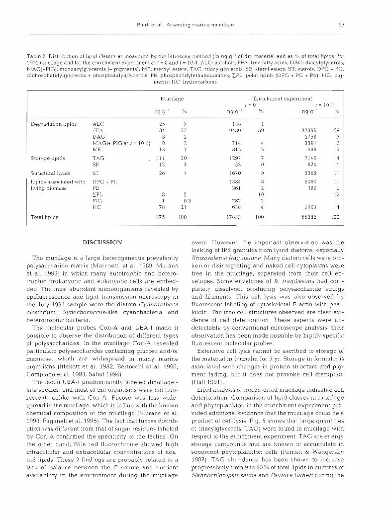
The complete lipid class distribution of mucilage is shown in Table 2 and Fig. 4. Lipids were present in detectable amounts in the Adriatic Sea mucus, and constituted 375 ng g-' of its dry weight. Tnacylglyc- erols (TAG), free fatty acids (FFA) and hydrocarbons (HC) were the most abundant lipid classes.
Enrichment experiment data over 10 d showed: a large increase in total suspended matter (from 1.88 mg I-' to 5.00 mg I-'). particulate organic carbon (from 425 pg 1-' to 893 pg I-'), and dissolved organic carbon (from 113 pM to 196 PM). These increases were accompanied by a significant decrease in total dis- solved lipids (35 pg 1-' to 5 pg I-') and an increase in total particulate lipids (34 pg I-' to 276 pg 1-') or in sus- pended matter (17.8 pg g-' to 55.3 pg g-'). Both control and nutrient-enriched experiment were characterized by large amounts of FFA, 20 and 162 pg I-' (control) and 10480 and 32396 ng g-' (enriched experiment) at t = 0 and t = 10 d respectively, amounting to 59% of total lipids, a much higher proportion than found in mucilage (22 %) (Table 2) .
The relative distributions of lipid classes (except FFA) in mucilage and in the control and nutrient- enriched culture are shown in Fig. 5. Differences are evident between the mucilage material enriched with TAG, HC and alcohols and very depleted in sterols and polar lipids.

Baldi et al.. Assessing marine mucilage 51
Fig 2. SCLh? images with different molecular probes: (a) Con-A-FITC, ( b to d) UEA-I-FITC lectins and (e, f ) phalloidin-FITC, to evaluate their specificity and different scenarios in the mucilage sample (a ) Con-A bound to glucose residues of amylopectin of outer hydrated layers (high density fluorescence), detached from the crystalline granule core (feeble fluorescence of granule penmeter). (b) UEA-I did not blnd to amylopectin. (c) UEA-I bound to fucose sugar residue of granules in this marine organism found in the mucilage. (d) UEA-I bound to fucose sugar residues of a broken organism showing faint globular shape of very different geometry to Con-A distribution in the mucilage. (e) Distribution of phalloidin bond to F-actin of a cell-free cytoskeleton belonging to a disintegrating organism in the mucilage. ( f ) Another residue of cytoskeleton without envelope imaged by phallo-
idin fluorescence
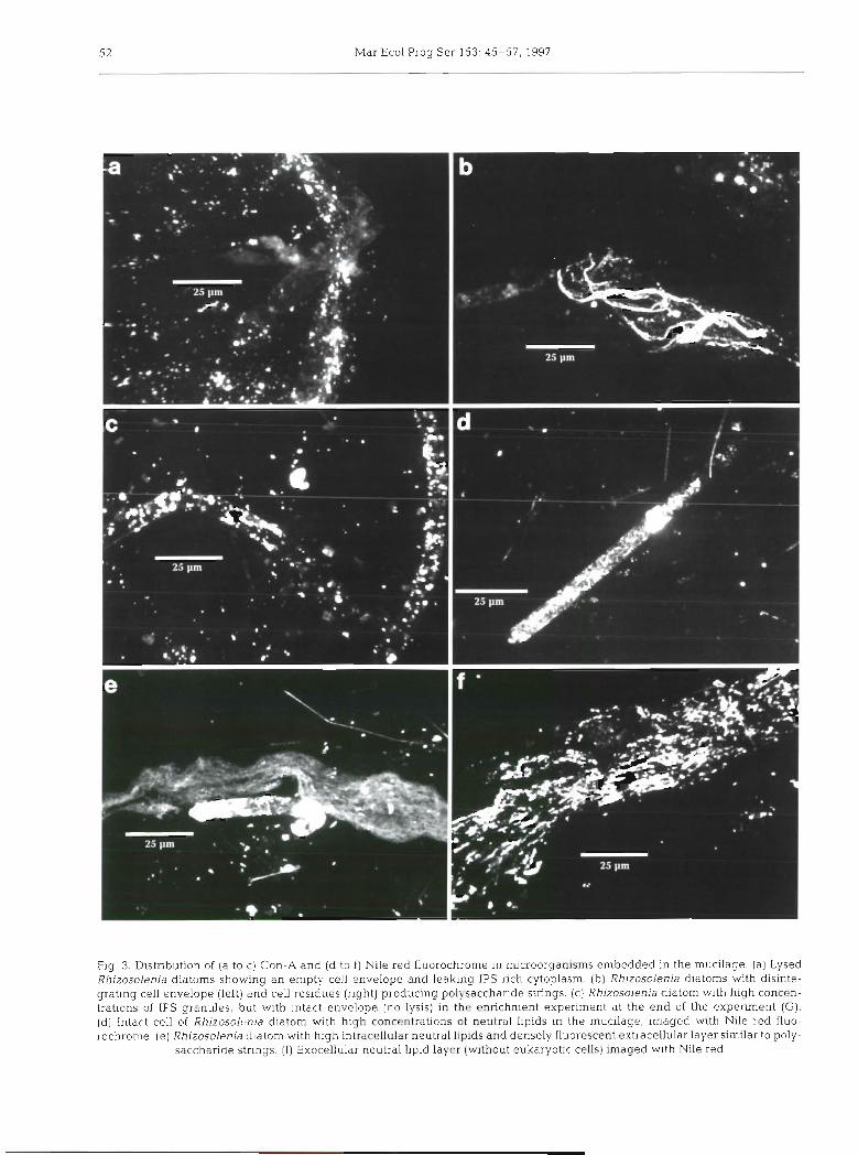
Mar Ecol Prog Ser 153: 45-57, 1997
Fig. 3. Distribution of (a to c) Con-A and (d to f ) Nile red fluorochrome in microorganisms embedded in the mucilage (a ) Lysed Rhizosolenia diatoms showing a n empty cell envelope and leaking IPS-rich cytoplasm. (b) Rhizosolenia diatoms with disinte- grating cell envelope (left) and cell residues (right) producing polysaccharide strings. (c) Rhizosolenia diatom with high concen- trations of IPS granules, but with intact envelope (no lysis) in the enrichment experiment at the end of the experiment (G). (d) Intart. cell of Rhizosolrnia diatom with high concentrations of neutral lipids in the mucilage, imaged with Nile red fluo- rochronie. (e) Rh~zosolenia diatom with high intracellular neutral lipids and densely fluorescent extracellular layer similar to poly-
saccharide strings. ( f ) Exocellular neutral lipid layer (without eukaryotic cells) imaged with Nile red
Raldi et al.: Assessing marine mucilage 53
- -
Table 2. Distribution of lipid classes as measured by the latroscan method (in ng g- ' of dry matenal and as of total liplds) for 1991 mucilage and for the enrichment experiment at t - 0 and t = 10 d . ALC: alcohols; FFA. free fatty acids, D.9G: diacylglycerols; MAG(+PIG): monoacylglycerols (+ pigments); ME- methyl esters; TAG: triac)rlglycerols; SE. stel-ol esters, ST. sterols; DPG + PG: diphosphatidylglycerols + phosphatidylglycerols, PE- phosphatidylethanolamines; EPL: polar lipids (DPG + PG + PE); PIG: pig-
ments; HC: hydrocarbons
Mucilage Enrichment experiment t = O t = l O d
"g g - ' % "g g" 0, I" ng g-' 8 8 '
-
Degradation llpids ALC 25 7 138 1 FFA 84 22 10480 59 32396 59 D AG 8 2 1758 3 MAGI+ PIG at t = 10 d ) 8 2 714 4 3391 6 ME 13 3 815 5 988 2
Storage l~p ids TAG . 111 30 1297 7 2149 4 SE 12 3 53 0 824 1
Structural lipids ST 2 6 7 1670 9 5365 10
Lipids associated with DPG + PG 1384 8 6060 11 llving biomass PE 361 2 389 1
ZPL 8 2 10 12 PIG 1 0.3 282 2 H C 78 2 1 638 4 1963 4
Total lipids 375 100 17833 100 55282 100
DISCUSSION
The mucilage is a large heterogeneous prevalently polysaccharide matrix (Marchetti et al. 1989, Murano et al. 1993) in which many autotrophic and hetero- trophic prokaryotic and eukaryotic cells are embed- ded. The most abundant microorganisms revealed by epifluorescence and light transmission microscopy in the July 1991 sample were the diatom Cylindrotheca closterium, Synechococcus-like cyanobacteria and heterotrophic bacteria.
The molecular probes Con-A and UEA-I made it possible to observe the distribution of different types of polysaccharides. In the mucilage Con-A revealed particulate polysaccharides containing glucose and/or mannose, which are widespread in many marine organisms (Ittekott et al. 1982, Bertocchi et al. 1990, Compiano et al. 1993, Saliot 1994).
The lectin UEA-I predominantly labeled dinoflagel- late species, and most of the organisms were not fluo- rescent, unlike with Con-A. Fucose was less wide- spread in the mucilage, which is in line with the known chemical composition of the mucilage (Murano et al. 1993, Faganeli et al. 1995). The fact that fucose distrib- ution was different from that of sugar residues labeled by Con-A confirmed the specificity of the lectins. On the other hand, Nile red fluorochrome showed high intracellular and extracellular concentrations of neu- tral lipids. These 2 findings are probably related to a lack of balance between the C source and nutrient availability in the environment during the mucilage
event. However, the important observation was the leaking of IPS granules from lysed diatoms, especially Rhizosolenia fragilissinla. Many diatom cells were bro- ken or disintegrating and naked cell cytoplasms were free in the mucilage, separated from their cell en- velopes. Some envelopes of R. fragilissima had com- pletely dissolved, producing polysaccharide strings and filaments. This cell lysis was also observed by fluorescent labeling of cytoskeletal F-actin with phal- loidin. The free cell structures observed are clear evi- dence of cell deterioration These aspects were un- detectable by conventional microscope analysis: their observation has been made possible by highly specific fluorescent molecular probes
Extensive cell lysis cannot be ascribed to storage of the material in formal~n fol- 3 yr. Storage in formalin is associated with changes in proteln structure and pig- ment fading, but it does not provoke cell disruption (Hall 1991).
Lipid analysis of freeze-dried mucilage indicated cell deterioration. Comparison of lipid classes in mucilage and phytoplankton in the enrichment experiment pro- vided additional evidence that the mucilage could be a product of cell lysis. Fig. 5 shows that large quantities of triacylglycerols (TAG) were found in mucilage with respect to the enrichment experiment. TAG are energy storage compounds and are known to accunlulate in senescent phytoplankton cells (Parnsh & Wangersky 1987). TAG abundance has been shown to increase progressively from 9 to 40"h of total lipids in cultures of Nannochloropsis salina and Pavlova lutheri during the
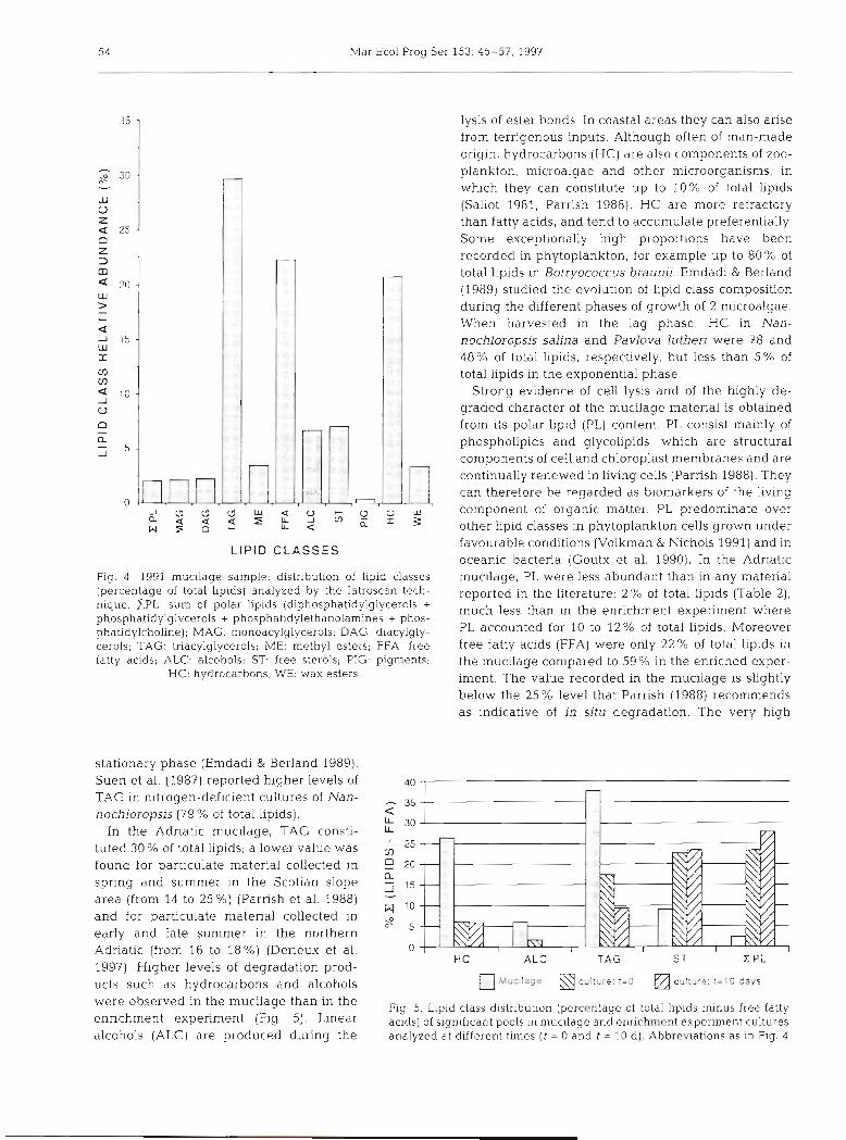
54 Mar Ecol Prog Ser 153: 45-57. 1997
L lP lD CLASSES
Fig 4 . 1991 mucllage sample: distribution of lipid classes (percentage of total hp~ds) analyzed by the Iatroscan tech- nique. ZPL: sum of polar lipids (diphosphatidylglycerols + phosphatidylglycerols + phosphatldylethanolamines + phos- phatidylcholine); MAG: monoacylglycerols; DAG: diacylgly- cerols; TAG. triacylglycerols; ME: methyl esters; FFA: free fatty acids; ALC: alcohols; ST free sterols; PIG: pigments:
HC: hydrocarbons; WE: wax esters
stationary phase (Emdadi & Berland 1989). Suen et al. (1987) reported higher levels of TAG In nitrogen-def~clent cultures of Nan- nochloropsis (79 % of total lip~ds).
In the Adriatic mucilage, TAG consti- tuted 30% of total lipids; a lower value was found for particulate material collected in spring and summer in the Scotian slope area (from 14 to 2 5 % ) (Parrish et al. 1988) and for particulate material collected in early and late summer in the northern Adriatic (from 16 to 18%) (Derieux et al. 1997). Higher levels of degradation prod- ucts such as hydrocarbons and alcohols were observed in the mucilage than in the enrichment experiment (Fig. 5). Linear alcohols (ALC) are produced during the
lysis of ester bonds. In coastal areas they can also arise from terrigenous inputs. Although often of man-made origin, hydrocarbons (HC) are also components of zoo- plankton, mlcroalgae and other microorganisms, in which they can constitute up to 10% of total lipids (Saliot 1981, Parrish 1988). HC are more refractory than fatty acids, and tend to accumulate preferentially. Some exceptionally high proportions have been recorded in phytoplankton, for example up to 80% of total lipids in Botryococcus braunii. Emdadi & Berland (1989) studied the evolution of lipid class composition during the different phases of growth of 2 microalgae. When harvested in the lag phase, HC in Nan- nochloropsis salina and Pavlova luthen were 78 and 48% of total lipids, respectively, but less than 5% of total lipids in the exponential phase.
Strong evidence of cell lysis and of the highly de- graded character of the mucilage material is obtained from its polar lipid (PL) content. PL consist mainly of phospholipids and glycolipids, which are structural components of cell and chloroplast membranes and are continually renewed in living cells (Parrish 1988). They can therefore be regarded as biomarkers of the living component of organic matter. PL predominate over other lipid classes in phytoplankton cells grown under favourable conditions (Volkman & Nichols 1991) and in oceanic bacteria (Goutx et al. 1990). In the Adr~atic mucilage, PL were less abundant than in any material reported in the literature: 2 % of total lipids (Table 2), much less than in the enrichment experiment where PL accounted for 10 to 12% of total lipids. Moreover free fatty acids (FFA) were only 22% of total lipids in the mucilage compared to 59 0/0 in the enriched exper- iment. The value recorded in the mucilage is slightly below the 25% level that Parrish (1988) recommends as indicative of in situ degradation. The very high
A L C T A G ST Z PL
m c u l t u r e : ! = l 0 days
Fig. 5. Lipid class distribution (percentage of total lipids minus free fatty acids) of s1gnifican.t pools in muc~lage and enrichment expenment cultures analyzed at different times ( t = 0 and t = 10 d). Abbrev~ations as in Fig. 4

Baldi et al.: Assessin ~g marine mucilage 55
value encountered in the enrichment experiment may be explained by large amounts of phaeophytins (Cau- wet et al. 1997). indicating the presence of senescent cells.
FFA are minor components of algae and are produced mostly during the growth phase (Emdadi & Berland 1989, Volkman & Nichols 1991). However, FFA are more abundant in diatoms than in other algae, reaching values up to 79 % of total lipids (Goutx et al. 1990). Extracellular FFA released in phytoplankton cultures accounted for 10 to 20 %, of total lipids (Parrish & Wangersky 1987). In pro- ductive natural waters, dissolved FFA often exceed the values measured In phytoplankton cultures. During the spring bloom in the North Sea, Kattner et al. (1983) re- ported that FFA abundance varied between 40 and 54 % of total lipids. Likewise, FFA were from 5 to 35 O/o of dis- solved lipids in spring and summer over the Scotian shelf (Parrish et al. 1988).
The mucilage was difficult to study by light micro- scopy because of the complex optically dense matrix of the sample. It has been suggested that polysaccharide mucilage is a product of anomalous algal exudation of EPS (Fogg 1995); however, its gel-like structure sug- gests that other mechanisms are involved. The consis- tency and mechanical resistence of this mucilage indi- cate transformation of the particulate polysaccharides into a jelly-like substance. Freeze-dried samples, thawed and resuspended in seawater, maintained their jelly structure even after 5 yr of storage (V. Turk pers. obs.).
Confocal microscope and molecular probe Con-A images of the distribution of polysaccharides in the mucilage indicated leakage of IPS granules from lysed cells and disintegration of cell envelopes. These 2 events are probably the main polysaccharide sources involved in mucilage events in the Adriatic Sea, together with the transparent exopolymer particles (TEP) formed by coalescence of EPS (Passow et al. 1994) and cell exudation (Herndl 1992, Hoagland et al. 1993). EPS exudation provoked by nutrient stress was studied in the enrichment experiment with prokary- otes and eukaryotes (Malej et al. 1997) in the northern Adriatic. Fibr~ls and layers of polysaccharides similar to those observed in the mucilage were never ob- tained.
When pure commercial polysacccharides such as amylopectin particles are suspended in seawater they do not dissolve; a partial hydration of the granule occurs after few days, but no jelly forms. The formation of gel-like mucilage in the Adriatic Sea probably involves several processes. Fibrils and layers in the mucilage matrix are probably formed by reaction of ionic polysaccharides with cations such as Ca2+ (Rees et al. 1982, Leppard 1995) which makes the mucilage dense and resistant to mechanical disruption.
Jelly could be formed by interactions between poly- saccharides and lipids and their acylglycerol derivates. Lipopolysaccharides (LPS) are sometimes exudation products of bacteria (Decho 1990, Schnaitman & Klena 1993), but the main source of LPS in mucilage is proba- bly the degradation of cell membranes and the associa- tion of polysaccharides and lipids as storage materials.
Hydration of the polysaccharides and consolidation with metals and/or lipids produces a physical-chemical res~stance of the mucilage to microbial attack. Degra- dation may then only be successful in late summer, when the mucus settles to the bottom and comes into contact with the more abundant benthic microbial community (Herndl et al. 1987).
At the moment we cannot explain cell lysis in the mucilage of the Adriatic Sea. One hypothesis is a virus attack, since virus-like particles were found by trans- mission electron microscopy (data not shown) in broken cells. This hypothesis is supported by the fact that large amounts of polysaccharides were produced in the bioreactor when the microalga Phaeocystis pouchetii was infected with PPV virus for 48 h (M. Heldal & F. Baldi pers. comm.). Another hypothesis is that lytic compounds with surfactant properties may occasionally be formed during the algal bloom. Lipids conjugated to polysaccharides are good amphipathic emulsifiers (Dasai & Banat 1997). However, these 2 hypotheses are not incompatible with others formulated previously: (1) formation of a thick false benthos at the pycnocline (Herndl 1992); (2) nutrient imbalance and carbon storage accumulation followed by anomalous EPS exu- dation (Fogg 1995); (3) formation of microbial aggre- gates due to persistent blooms caused by efficient P remineralization and carbon storage (Azam 1997). These hypotheses do not explain extensive cell lysis but raise the questlon of whether cell disruption occurs be- fore or after aggregation of marine organisms. For the answer to this question, we shall have to await the next mucilage event in the northern Adriatic Sea.
Ackno~~ledgements. We thank G. Cauwet for organic carbon analysis and J. Fillaux for Iatroscdn analysis. This research was undertaken in the framework of the PALOhlA pro- gramme. We acknowledge the support of the Cornmission of the European Community Environment R & D programme, under contract CEE-5EV-CT94-0420 and PECO under con- tract ERBCIPD-CT94-0106. The research was also supported by the Ministry of Science and Technology of the Republic of Slovenia. The authors also thank Farooq Azam and Cindy Lee for their critical reviews of the manuscript and constructive comments.
LITERATURE ClTED
Alldredge AL. Jackson GA (1995) Topical studies in oceanog- raphy. Aggregation In marine systems. Deep Sea Res 42: 273-275

Mar Ecol Prog Ser 153: 45-57, 1997
Alldredge AL, Passow U, Logan BE (1993) The abundance and significance of a class of large, transparent organic particles in the ocean. Deep Sea Res 40:1131-1140
Azam F (1997) Possible cause of massive mucilage production in the northern Adriatic sea: a novel hypothesis. In: Physl- cal and biogeochemical processes of the Adriatic Sea: eutrophic hmits of the northern Adriatic. In: Nolan C (ed) The Adnatic Sea. European Communities Ecosystems Research Reports Series, Brussels (in press)
Bertocchi CL. Navarini A, Cesaro F (1990) Polysaccharides from cvanobacteria. Carbohydr Polymers 12 127-153
Cauwet G, Terzlc S, Ahel M, Mozetic P, Turk V, Male] A (1997) Effect of nutrient addition on phytoplankton/bacte- rioplankton interactions and dissolved organic matter variability. Part IT. Biochenucal aspect. In: Physical and biogeochemical processes of the Adriatic Sea: eutrophic limits of the northern Adriatic. In: Nolan C (ed) The Adri- atlc Sea European Cornrnunitic:~ Ecosystems Research Reports Series, Brussels (in press)
Compiano AM, Romano JC, Garabetian F, Laborde P, Giraudiere 1 (1993) Monosaccharide composition of partic- ulate hydrolysable sugar fraction In surface microlayers from brackish and marine waters. Mar Chem 42:237 251
Decho AW (1990) Microbial exopolymer secretions in ocean environments: their role(s) in food webs and rnanne processes. Oceanogr Mar Biol Annu Rev 28:73-153
Degobbis D, Fonda-Umani S. Franco P, Male] A, Precali R, Smodlaka N (1995) Changes in :he northern Adriatic ecosystem and the hypertrophic appearance of gelatinous aggregates. Sci Total Environ 165:43-58
Derieux S, Moine F, Fillaux J , Pinturier L, Jan G. Laurelllard J , Saliot A (1997) Lipid chemistry of particulate and dis- solved organic matter in the North Adriatic in September 1994 and June 1995. In: Nolan C (ed) The Adriatic Sea. European Communities Ecosystems Research Reports Series, Brussels (in press)
Desai JD, Banat IM (1997) Microbial production of surfactants and their commercial potential. Microbiol Mol Biol Rev 61. 47-64
Emdadi D, Berland B (1989) Variation in lipid class composi- tion during batch growth of Nannochloropsis salina and Pavlova lutherf. Mar Chern 26:215-225
Faganeli J , Kovac K , Leskovsek H, Pezdic J (1995) Sources and flux of particulate organic matter in shallow coastal waters characterized by summer macroaggregate forma- t ~ o n . Biochemistry 29:71-88
Fogg GE (1995) Some speculations on the nature of the pelagic mucilage community of the northern Adriatic Sea. Sci Total Environ 165 59-64
Fonda U m a n ~ S. Gh~rardelli E. Specchl EM (1989) G11 episodi di mare sporco nell'Adriatico dal 1729 ai g~orni nostri. Regione Autonoma Friuli-Venezia Giulia. Direz~one reg~onale dell'Ambiente, Trieste
Gabius HJ, Gabius S (1993) Lectins and glycobiology. Springer-Verlag Inc. New York
Goutx M, Gtlnn C, Bertrand JC (1990) Lipid classes of micro- organisms as biomarkers in the marine environment. Org Geochem 16:231-237
Greenspan P, Fowler SD (1985) Spectrofluorometric studies of the lipid probe. Nile red. J Lip~d Res 26.781-789
Hall JA (1991) Long term preservation of picophytoplankton for counting by fluorescence rnicroscopy. Br Phycol J 26:169-174
Haugland RP (1992) Handbook of fluorescent probes and research chemicals. Larison KD
Herndl C J (1992) Marine snow in the northern Adriatic Sea: possible causes and consequences for a shallow ecosys- tem. Mar Microbial Food Webs 6(2):149-172
Herndl GJ, Faganeli J , Fanuko N, Peduzzi P, Turk V (1987) Role of bacteria in the carbon and nitrogen flow between water- column and sediment in a shallow marine bay (Bay of Piran, northern Adriatic Sea] PSZN l: Mar Ecol 8(3):221-236
Hoagland KD, Rosowski JR, Gretz MR, Romer SC (1993) Diatom extracellular polymeric substances: function, fine structure, chemistry and physiology. J Phycol 29-537-566
Ittekott V, Degens ET, Brockmann U (1982) Monosaccharide composition of acid-hydrolysable carbohydrates in partic- ulate matter during a phytoplankton bloom. Limnol Oceanogr 27:770-776
Kattner G, Gercken G, Hammer KD (1983) Development of lipids during a spring plankton bloom in the northern North Sea. 11. Dissolved lipids and fatty acids. Mar Chem 14:163-173
Kiarboe T, Hansen LS (1993) Phytoplankton aggregate forrna- tion: observations of patterns and mechanisms of cell sticklng and the significance of exopolymer material J Plankton Res 15:993-1018
Laureillard J , Pinturier L, Fillaux J. Saliot A (1997) Organic geochemistry of marine sediments of the Subantarctic Indian Ocean sector. lipid classes, sources and fate. Deep Sea Res (in press)
Leppard GG (1995) The characterization of algal and micro- bial mucilages and their aggregates in aquatic ecosys- tems. Sci Total Environ 165: 103-13 1
Malej A, Mozetic P, Turk V, Terzic S, Ahel M, Cauwet G (1 9971 Effect of nutrient addition on phytoplankton/bacte- rioplankton interactions and d~ssolved organic matter variability. Part I: Productivity aspect. In: Physical and biogeochemical processes of the Adriatic Sea: eutrophic 1irn.its of the northern Adriatic. In: Nolan C (ed) The Adri- atic Sea. European Communities Ecosystems Research Reports Series, Brussels (in press)
Marchetti R, Iacomini M, Torri G, Focher B (1989) Caratteriz- zazione prelimnare degli essudati di origine planctonica raccoltl in Adriatic0 nell'estate 1989. Acqua Aria 8: 883-887
Murano E, Vetere A, Toffanin R, Liuti G, Sandri G, Rizzo R (1993) Characterization of the complex mucilage pro- duced by marine microorganisms. Atti Soc Italiana Biochimica (SJB) Trieste 9
Myklestad SM (1995) Release of extracellular products by phytoplankton with special emphasis on polysaccharides. Sci Total Environ 165:155-164
Neu TR, Marshall KC (1991) Microbial 'footprints'-a new approach to adhesive polymers. B~ofouling 3:101-112
Parrish CC (1988) Dissolved and particulate marine lipid classes. a review Mar Chem 23:17-40
Parrish CC, Wangersky PJ (1987) Particulate and dlssolved lipid classes in cultures of Phaeodactylum tricornutum grown in cagc culture turbidostats with a range of nitro- gen srlpply rates Mar Ecol Prog Ser 35:119-128
Parrish CC, Wangersky PJ, Delmas RP, Ackman X (1988) Iatroscdn-measured profiles of dlssolved and partlculate marine lipid classes over the Scotian Slope and in Bedford Basin. Mar Chem 23:l-15
Passow U, Logan BE. Alldredge AL (1994) The role of partic- ulate carbohydrate exudates in the flocculation of diatoms blooms. Deep Sea Res 41 335-357
Porter KG, Feig YS (1980) The use of DAPl for identifying and counting aquatic microflora. Limnol Oceanogr 25:943-948
Rees DA, Morris ER, Thom D, Madden JK (1982) Shapes and interactions of carbohydrate chains In: Aspinall GO (ed) The polysaccharides Academic Press. New York, p 195-290

Baldi et al.: Assessing manne mucilage 5 7
Saliot A (1981) Natural hydrocarbons in seawater. In: Duursma EG, Dawson R (eds) Marine organic chemistry. Elsevier, Amsterdam, p 327-374
Saliot A (1994) Hydrates de carbone et polysaccharides In: Biogeochimie organlque marine. Oc6an1s 20: l l l -135
Saliot A, Laureillard J , Scribe P, Sicre MA (1991) Evolutionary trends in the lipid biomarker approach for ~nvestigating the biogeochemistry of organic matter in the marine envi- ronment. Mar Chem 36:233-248
Schnaitman CA. Klena JD (1993) Genetics of lipopolysaccha- ride biosynthesis in enteric bacteria. Microbiol Rev 57: 655-682
Stachowitsch M, Fanuko N, Richter M (1990) Mucus aygre- gates in the Adriatic Sea: an overview of stages and occul- rences. PSZN I - Mar Ecol 11:327-350
Suen Y, Hubbard JS, Holzer G, Tornabene TG (1987) Total lipid production of the green alga Nannochloropsis sp Q11 under different nitrogen regimes. J Phycol356:147-162
Takahashi M, Kikuchi K, Hara Y (1985) Importance of pico- cyanobacteria biomass (unicellular blue-green algae) in the phytoplankton population of the coastal waters of Japan. Mar Biol 8953-69
Utermohl H (1958) Zur Vervollkommnung der quantitativen Phytoplankton-Methodik. Mitt Int Verein Theor Angew Limnol 9: 1-38
This article was presented by E Azam, La Jolla, Calffornia, USA
Viviani R, Boni L. Cattani 0, Milandrl A, Pirini M, Poletti R, Pompei 31 (1995) Fatty acids, chlorophylls and total silicon in mucilaginous aggregates collected in a coastal area of the northern Adriatic Sea facing Emiliana-Romagna in August 1988. Sci Total Envll-on 165:193-201
Volkman JK, Everitt DA, Aallen DI (1986) Some analyses of l i p~d classes in marlne microorganisms, sediinents and seawater uslng thin-ldyer chromatography flame ionisa- tion detection. J Chromatogr 356:147-162
Volkman JK, Nichols PD (1991) Application of thin layer chro- matography-flame ionization detection to the analysis of lipids and pollutants in marine and environmental sanl- ples. J Plan Chromatogi- 4:19-26
Vollenweider RA, Montanari G , Rinaldi A (1995) Statistical inferences about the mucllage cvcnts in the Adrlat~c Sea, with special reference to recurrence patterns and claimed relationships to sun activity cycles. Sci Total Env~ron 165: 213-225
Wakeham SG, Lee C (1989) Organic geochemistry of particu- late matter in the ocean: the role of particles in oceanic sedimentary cycles. Org Geochem 14:83-96
Wolfaardt GM, Lawrence J R , Robarts RD, Caldwell SJ, Cald- well DE (1994) Mult~cellular organization in a degrada- tive biofilm community. Appl Environ Microbiol 60: 434-446
Mdn uscript first received: October 12, 1996 Rev~sed version accepted: April 16, 1997


![Heterotrophic nutrition [2015]](https://img.pdfslide.net/doc/110x75/55d39cc0bb61ebf8268b46dd/heterotrophic-nutrition-2015-55d47f014ed07.jpg)