Embed Size (px)
Citation preview

of April 5, 2018.This information is current as
Cell-Positive SelectionTurns Off RAGs and Promotes BPhosphatidylinositol 3-Kinase Signaling Basal B Cell Receptor-Directed
NemazeeAït-Azzouzene, Kamal Puri, José Luis Vela and David Laurent Verkoczy, Bao Duong, Patrick Skog, Djemel
http://www.jimmunol.org/content/178/10/6332doi: 10.4049/jimmunol.178.10.6332
2007; 178:6332-6341; ;J Immunol
Referenceshttp://www.jimmunol.org/content/178/10/6332.full#ref-list-1
, 32 of which you can access for free at: cites 64 articlesThis article
average*
4 weeks from acceptance to publicationFast Publication! •
Every submission reviewed by practicing scientistsNo Triage! •
from submission to initial decisionRapid Reviews! 30 days* •
Submit online. ?The JIWhy
Subscriptionhttp://jimmunol.org/subscription
is online at: The Journal of ImmunologyInformation about subscribing to
Permissionshttp://www.aai.org/About/Publications/JI/copyright.htmlSubmit copyright permission requests at:
Email Alertshttp://jimmunol.org/alertsReceive free email-alerts when new articles cite this article. Sign up at:
Print ISSN: 0022-1767 Online ISSN: 1550-6606. Immunologists All rights reserved.Copyright © 2007 by The American Association of1451 Rockville Pike, Suite 650, Rockville, MD 20852The American Association of Immunologists, Inc.,
is published twice each month byThe Journal of Immunology
by guest on April 5, 2018
http://ww
w.jim
munol.org/
Dow
nloaded from
by guest on April 5, 2018
http://ww
w.jim
munol.org/
Dow
nloaded from

Basal B Cell Receptor-Directed Phosphatidylinositol 3-KinaseSignaling Turns Off RAGs and Promotes B Cell-PositiveSelection1
Laurent Verkoczy,2* Bao Duong,*† Patrick Skog,* Djemel Aıt-Azzouzene,* Kamal Puri,‡
Jose Luis Vela,*† and David Nemazee3*
PI3K plays key roles in cell growth, differentiation, and survival by generating the second messenger phosphatidylinositol-(3,4,5)-trisphosphate (PIP3). PIP3 activates numerous enzymes, in part by recruiting them from the cytosol to the plasma membrane. Wefind that in immature B lymphocytes carrying a nonautoreactive Ag receptor, PI3K signaling suppresses RAG expression andpromotes developmental progression. Inhibitors of PI3K signaling abrogate this positive selection. Furthermore, immature pri-mary B cells from mice lacking the p85� regulatory subunit of PI3K suppress poorly RAG expression, undergo an exaggeratedreceptor editing response, and, as in BCR-ligated cells, fail to progress into the G1 phase of cell cycle. Moreover, immature B cellscarrying an innocuous receptor have sustained elevation of PIP3 levels and activation of the downstream effectors phospholipaseC (PLC)�2, Akt, and Bruton’s tyrosine kinase. Of these, PLC�2 appears to play the most significant role in down-regulating RAGexpression. It therefore appears that when the BCR of an immature B cell is ligated, PIP3 levels are reduced, PLC�2 activationis diminished, and receptor editing is promoted by sustained RAG expression. Taken together, our results provide evidence thatPI3K signaling is an important cue required for fostering development of B cells carrying a useful BCR. The Journal of Im-munology, 2007, 178: 6332–6341.
A ntigen recognition by the BCR may lead to distinct, andoften diametrically opposed, functional consequencescompared with the basal BCR signal. For example, basal
BCR signals are essential for continued survival of naive mature Bcells, which are uniformly nondividing (1, 2). In contrast, whenthe BCRs of these same cells are ligated either by self or foreignAgs, they undergo apoptosis or activation (3– 6). Similarly, inimmature B cells, BCR ligation promotes receptor editing anddevelopmental arrest, whereas basal surface Ig (sIg)4 signalspromote recombinase down-regulation and developmental pro-gression (7, 8).
Ligated and unligated sIg are sensed through the signaling ca-pacity of the BCR complex, which consists of sIg and the associ-ated signal transducers Ig-��, which are required for plasma mem-brane expression of sIg and for signaling through the pre-BCR and
BCR (9, 10). BCR ligation leads to phosphorylation of tyrosineswithin conserved ITAMs carried on the cytoplasmic portions ofIg-� and � (11), which in turn recruit tyrosine kinases to theircytoplasmic tails through other motifs (12). BCR signaling isinitiated by tyrosine kinases of the Src family and by Syk,which mediate ITAM phosphorylation and transphosphoryla-tion upon BCR aggregation (13). It is unclear whether the sig-nals transmitted by unligated receptors are qualitatively distinctfrom those of ligated receptors or merely represent a quantita-tive difference.
An important step in BCR signaling is the phosphorylation ofthe coreceptor CD19 (14). CD19 physically associates with theBCR through intracellular and extracellular motifs (15–17). (CD19has also been shown to facilitate pre-BCR signaling (18, 19)).BCR ligation leads to the recruitment by CD19 of PI3K via itsp85� regulatory subunit, the generation of lipid products such asphosphatidylinositol-(3,4,5)-trisphosphate (PIP3), and the atten-dant recruitment to the plasma membrane of pleckstrin homology(PH) domain-containing proteins, such as phospholipase C(PLC)�2 and cytoplasmic kinases, such as Bruton’s tyrosine ki-nase (Btk) and Akt (20). PLC�2 activation in turn promotes phos-phatidylinositol-(4,5)-bisphosphate hydrolysis to inositol trisphos-phate and diacylglycerol, mobilizing Ca2� stores and activatingprotein kinase Cs. In mature B cells, the BCR signaling complexis rapidly recruited to cholesterol-rich lipid rafts on the plasmamembrane, where several tyrosine kinases, including Lyn, areabundant (21).
Genetic experiments have shown that basal signaling through anunligated BCR is critical for peripheral B cell survival and/or im-mature B cell development (1, 22–24). This physiological signalcan be mimicked by a membrane-localized ITAM derived from anartificial chimeric Ig� protein (25), the EBV LMP2A protein (26,27), or an engineered sIg molecule devoid of Ag specificity (22).During bone marrow development, transgene-enforced expression
*Department of Immunology; †Kellogg School of Science and Technology DoctoralProgram in Chemical and Biological Sciences, The Scripps Research Institute, LaJolla, CA 92037; and ‡ICOS, Bothell, WA 98021
Received for publication January 3, 2007. Accepted for publication February27, 2007.
The costs of publication of this article were defrayed in part by the payment of pagecharges. This article must therefore be hereby marked advertisement in accordancewith 18 U.S.C. Section 1734 solely to indicate this fact.1 This work was supported by research and training grants from the National Institutesof Health (RO1AI33608 to D.N., T32HL07195 to D.A.-A., and Graduate TrainingGrant F31AI52484).2 Current address: Human Vaccine Institute, Duke University Medical Center, 106Research Drive, 4086 MSRBII, Durham, NC 27710.3 Address correspondence and reprint requests to Dr. David Nemazee, Department ofImmunology, The Scripps Research Institute, 10550 North Torrey Pines Road, MailDrop IMM-29, La Jolla, CA 92037. E-mail address: [email protected] Abbreviations used in this paper: sIg, surface Ig; Btk, Bruton’s tyrosine kinase; int,intermediate; PH, pleckstrin homology; PIP3, phosphatidylinositol-(3,4,5)-trisphos-phate; PLC, phospholipase C; Tg, transgenic.
Copyright © 2007 by The American Association of Immunologists, Inc. 0022-1767/07/$2.00
The Journal of Immunology
www.jimmunol.org
by guest on April 5, 2018
http://ww
w.jim
munol.org/
Dow
nloaded from
of prerearranged IgH or IgH/L combinations can suppress endog-enous rearrangements, demonstrating a feedback regulation pro-cess; however, autoreactive receptors that presumably generate adistinct BCR-mediated signal fail to suppress recombination andpromote instead ongoing rearrangement that often leads to receptorediting (reviewed in Ref. 28).
These results suggest that in immature B cells an unligated BCRpromotes a signal that regulates V(D)J recombination, whereas across-linked receptor promotes a distinct signal. Furthermore,studies in which the sIg is inducibly lost from immature B cellssuggest that this suppression of recombination, along with theloss of expression of maturation markers, is reversible for sometime (24).
An important aspect of the regulation of L chain recombinationinvolves the transcription rate of RAG1 and RAG2. But it is notclear how signals from an unligated BCR are distinguished fromthose of autoreactive BCRs. We found previously that one aspectof BCR-regulated RAG expression control involved the activationof NF-�B (29). Other studies have implicated effects on basal sIgsignaling through the CD19 coreceptor as important in the signalto turn off V(D)J recombination (30, 31).
In this study, we have sought to identify signaling pathways thatmay regulate B cell-positive selection, using as a readout thedown-regulation of RAG expression by an innocuous, i.e., nonli-gated, BCR, and to determine how ligated and unligated BCRssignal differentially at the immature B cell stage.
Materials and MethodsMice
The 3-83 mice (4) on the B10.D2 genetic background were bred to HYGRAG2-GFP (32) and/or p85�-deficient (33) backgrounds. The HYGRAG2-GFP reporter transgene is derived from a large bac clone that ap-pears to carry transcriptional control regions for both RAG1 and RAG2(32). Mice were maintained in The Scripps Research Institute Animal Re-sources facility; all of these studies have been reviewed and approved bythe relevant The Scripps Research Institute institutional animal care anduse review committee.
Cell culture and stimulation
Immature B cells were either isolated directly from the bone marrow of3-83 mice, or expanded in IL-7 cultures, and stimulated with BCR andcontrol mAbs at 10 �g/ml, as described (34). S23 is a mouse IgG2b anti-3-83 Id raised in a JC�-deficient mouse (D. Nemazee, unpublished obser-vation); Y3 (American Type Culture Collection designation HB-176) is anIgG2b anti-H-2Kb Ab (35). Wortmannin, LY294002, U73122, m-3M3-FBS, Akt inhibitor (1L-6-hydroxymethyl-chiro-inositol 2-(R)-2-O-methyl-3-O-octadecylcarbonate), rapamycin, SB203580, PD98059, cyclosporin A,and FK506 were obtained from Calbiochem.
Protein extracts, immunoblotting, fusion proteins withtransduction domains of HIV TAT, and EMSA
Nuclear and cytoplasmic extract fractions were prepared, as described (36).Whole cell extracts were prepared by resuspending 107 cells in 100 �l oflysis buffer (0.1% Triton X-100 in 1� PBS) supplemented with 20 mMNaF, 1 mM sodium orthovanadate, and protease inhibitors (Roche). Afterincubating for 15 min on ice, samples were cleared by 10,000 � g cen-trifugation for 10 min at 4°C, and supernatants were stored at �70°C. Afterreducing PAGE, transfer to nylon membranes was conducted using the XCell II Blot Module (Invitrogen Life Technologies). Primary Abs usedwere: p50/p105 (sc-114), p65 (sc-372), c-rel (sc-71), I�B� (sc-371),p-I�B� (sc-8404), Akt1 (sc-1618), Btk (sc-1696), and cyclin D2 (sc-593)from Santa Cruz Biotechnology; phospho-Btk (Tyr223), phospho-Akt(Thr308), phospho-PLC�2 (Tyr1217), and PLC�2 from Cell Signaling Tech-nology; GAPDH (mAb374) from Chemicon International; and fibrillarin,provided by Pollard (The Scripps Research Institute, La Jolla, CA). Sec-ondary Abs used were goat anti-rabbit IgG HRP and goat anti-mouse IgGHRP (Jackson ImmunoResearch Laboratories). Expression and purificationof TAT-superrepressor I�B� (37) and TAT-�-gal (38) were as described(29). EMSA was performed essentially as described (29).
Northern blotting and L chain rearrangement assays
Northern blots and L chain gene rearrangement assays were essentially asdescribed (29). Probes and PCR conditions/primers are described for thefollowing rearrangements: V�-J�1 (39), recombining sequence/�-deletingelement-intronic recombination sequence (40), and V�1-J�1 (7).
PIP3 assay
Assay was based on the work of Anzelon et al. (41); however, similarassays have been described by other investigators (e.g., 42). Cells werefixed and permeabilized using a kit (BD Biosciences), incubated in FACSbuffer with biotinylated anti-PI-(3,4,5)-P3 (Echelon, Z-B345), followed bystreptavidin-PerCP Cy5.5. Samples were analyzed on a FACSCalibur cy-tometer (BD Biosciences) using the FlowJo software package.
ResultsCD19 phosphorylation
Evidence for active signaling through an innocuous BCR in im-mature B cells was sought by analysis of protein tyrosine phos-phorylation in primary immature sIgM� B cells, which were gen-erated from bone marrow of 3-83 BCR transgenic (Tg) mice, asdescribed (29, 34). Positive selection in this context involves thesuppression of RAG gene expression and the up-regulation of cellsurface maturation markers. BCR ligation of the maturing cellsprevents or reverses maturation and promotes RAG gene expres-sion and receptor editing. Thus, a comparison of BCR-ligated orunligated cells provides a way to compare signals promoting ed-iting and positive selection, respectively. As shown in Fig. 1A,several prominent tyrosine-phosphorylated bands are detected in
FIGURE 1. Innocuous BCR signal promotes protein tyrosine phospho-rylations that are inhibited by prolonged BCR cross-linking. IL-7-cultured3-83 Tg bone marrow B cells were treated after IL-7 withdrawal for theindicated times with either anti-BCR or control Abs. A, Total tyrosine-phosphorylated proteins assessed in Western blot with 4G10 Ab. Lowerpanel, Shows signal of stripped blot reprobed with GAPDH Ab. B, Anal-ysis of CD19 tyrosine phosphorylation and total CD19 levels of immuno-precipitated CD19 (left panels) compared with whole cell lysates (rightpanels).
6333The Journal of Immunology
by guest on April 5, 2018
http://ww
w.jim
munol.org/
Dow
nloaded from
cells carrying innocuous receptors that are lost upon BCR ligation.One band appeared to be �95 kDa, the molecular mass of CD19.To test this, we assessed CD19 tyrosine phosphorylation at aminoacid position Y513 using a specific phosphopeptide Ab. As shownin Fig. 1B, strong CD19 phosphorylation was observed in cellscarrying an unligated receptor and the phosphorylation signal waslost in B cells treated with anti-BCR Ab. Control experiments witha pan-reactive CD19-blotting Ab showed that the level of CD19was also reduced in BCR-ligated cells compared with control cells(Fig. 1B) (the lower band in the lower left blot may be an under-glycosylated CD19 biosynthetic intermediate). To extend this anal-ysis in vivo, we assessed CD19 levels in freshly isolated bonemarrow B cells that were either innocuous (3-83 Tg on a nonde-leting H-2d background) or autoreactive (3-83 Tg central-deletingH-2k background). In B cells on the autoreactive background, lev-els of both surface CD19 and intracellular CD19 were down-mod-ulated significantly (Fig. 2). We conclude that CD19 tyrosinephosphorylation at Y513 correlates with B cell-positive selection,whereas in the context of negative selection, developing B cellscarry reduced levels of CD19 that are hypophosphorylated.
Effects of PI3K inhibitors
Because the major function of CD19 is believed to be the recruit-ment of PI3K upon phosphorylations of Y513 and Y482 (43, 44),we tested the possibility that positive selection was PI3K depen-dent. A first approach involved treating immature B cells withsignaling inhibitors (Fig. 3A). As shown in Fig. 3B (top panel), inimmature B cells carrying an innocuous BCR, PI3K inhibitorswortmannin and LY294002, but not a wide variety of other sig-naling inhibitors, were capable of eliciting a robust expression ofRAG1 and RAG2. This effect was similar to that of BCR ligationitself, only more robust (Fig. 3B, lower panel). Because wortman-nin and LY294002 may affect several PI3K, we also tested a p110�inhibitor, IC87114 (45); it too promoted RAG expression in adose-dependent manner (Fig. 3C). It therefore appears that chem-
ical inhibitors of PI3K activity suppress B cell-positive selection ina manner comparable to BCR ligation.
Effect of p85� deficiency on RAG expression in vivo
To further probe the role of PI3K in B cell positive-selection, 3-83BCR Tg mice were bred to p85�-deficient mice (33), and RAGexpression and V(D)J recombination were monitored in immatureB cells in the context of innocuous and ligated Ag receptors (Fig.4). As shown in Fig. 4A, p85-deficient cells carrying innocuousreceptors failed to strongly suppress RAG1 and RAG2 expressionas assessed by Northern blot. (In samples expressing high overallRAG transcript levels, we sometimes observed additional bandsfor RAG2, which may represent partially spliced nuclear interme-diates.) Furthermore, BCR-induced RAG expression was furtheraugmented in the absence of p85. Quantitation of these changes forRAG2 is summarized in Fig. 4B. As shown in Fig. 4C, minimaldifferences in cell recovery were observed between p85�-deficientand -sufficient cells, ruling out preferential survival of RAG-ex-pressing cells with p85� deficiency as an explanation for theseresults. Consistent with the elevated RAG expression of thep85�-deficient cells, these cells manifested elevated L chain
FIGURE 2. In vivo analysis of BCR and CD19 expression in bone mar-row B cells undergoing positive and negative selection. The 3-83 (anti-H-2Kk,b) BCR Tg mice were bred to H-2d (B10.D2) or H-2k (B10.BR) back-grounds, and their bone marrow cells were analyzed by flow cytometry forthe indicated markers. Viable (upper panels) or fixed and permeabilized(lower panels) cells were stained for expression of CD19 and Ig�. Lowerpanels, Show relative mean fluorescence intensity (MFI) for the two mark-ers within the indicated boxes. Contour plots show data from bone marrowcells using a lymphocyte gate.
FIGURE 3. Effect of PI3K inhibitors on RAG expression in immature Bcells carrying ligated or unligated BCR. A, Experimental design. B, Effectof inhibitors on RAG1 and RAG2 expression as assessed by Northern blot.The 3-83 Tg bone marrow B cells cultured for 24 h in the presence ofcontrol Ab (upper panel) or anti-BCR (lower panel) alone (untreated, lane1) or for the last 6 h with 50 nM wortmannin, 7.5 �M LY294002, 100 nMrapamycin, 10 �M SB203580, 20 �M PD98059, 1 �g/ml cyclosporin A,and 100 ng/ml FK506 (lanes 2–8, respectively). Molecular targets of eachinhibitor (— ) are indicated above each lane. For both groups (control Aband anti-BCR), relative RAG1 and RAG2 mRNA signal (calculated bynormalizing RAG signal to 18S RNA content and setting untreated samplesat 100%) is shown under each lane. Data are representative of at least threeexperiments. C, Dose/Response analysis of the effect of a specific inhibitorof the PI3K p110� catalytic component (IC87114) on RAG1 and RAG2expression.
6334 A KEY ROLE FOR PI3K IN B CELL-POSITIVE SELECTION
by guest on April 5, 2018
http://ww
w.jim
munol.org/
Dow
nloaded from
gene recombination as assessed by semiquantitative PCR assays(Fig. 4D). Analysis of ex vivo bone marrow B cells from p85�-deficient mice carrying an innocuous BCR was consistent with theabove mentioned results using IL-7-cultured B cells. As shown inFig. 4E, p85�-deficient cells clearly overexpressed RAG2 com-pared with p85�-sufficient controls, as measured using a RAG2/GFP reporter transgene (32). Hence, p85� function appears to beimportant for the innocuous BCR-mediated suppression of RAGexpression in vivo.
Effects of p85 deficiency on NF-�B activity in immature B cells
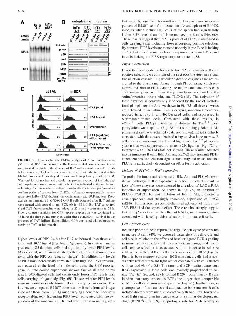
Because RAG expression was up-regulated in p85-deficient cells,and we previously observed that NF-�B/rel transcription factorsregulate RAG expression, we tested the effects of p85� deficiencyon the spontaneous and sustained BCR-induced activation of NF-�B. As shown in Fig. 5A, p85� deficiency was indeed correlatedwith elevated NF-�B nuclear activity, as detected by EMSA. BCRligation increased activity in wild-type cells relative to control(lanes 1 and 2), whereas in p85�-deficient cells NF-�B/rel activitywas high even in the absence of BCR ligation. Elevated NF-�B/relwas generally correlated with reduced levels of cytoplasmic I�B�and increases of p65 levels in the nucleus (Fig. 5B). However, inp85��/� cells, the nuclear levels of p50, c-rel, and p65 were allsubstantially increased, and the p65 and I�B� components foundin the nucleus had a distinctly slower electrophoretic mobility (Fig.5B, lanes 7 and 8).
We previously showed that the BCR ligation-induced RAG ex-pression in immature B cells was regulated by NF-�B components
and could be inhibited by a dominant-negative I�B� superrepres-sor protein (29). Consistent with the prediction that suppression ofNF-�B-induced RAG activity was downstream of BCR-inducedPI3K activity, I�B� superrepressor protein-TAT fusion proteinwas able to suppress RAG expression in cells treated with wort-mannin, BCR Ab, or both (Fig. 5C). A control TAT-fusion proteinhad no effect in this RAG2/GFP reporter assay. These results sug-gested that a BCR-regulated and p85�-dependent PI3K activitysuppresses activation of NF-�B in immature B cells. Furthermore,virtually all markers of B cell maturity were reduced in p85�-deficient B cells, including up-regulation of MHC class II, IgD,CD21, PirA/B, and CD23, and down-modulation of CD93, IgM,and CD24 (data not shown), consistent with prior studies ofsplenocytes of p85�-deficient mice (46, 47). Collectively, thesedata indicate that p85�-deficient immature B cells progress poorly,or slowly, in development in vivo, and that they have reducedability to undergo (innocuous) BCR-mediated positive selection.
PIP3 levels are increased in primary B cells carrying innocuousreceptors
Because the foregoing evidence suggested a strong correlation be-tween PI3K activity and BCR-mediated regulation of RAG ex-pression, we directly assessed p85�-deficient and sufficient pri-mary B cells, with and without BCR ligation, for PIP3 levels andRAG gene activity. As measured by intracellular staining withanti-PIP3 Ab (41), p85-sufficient B cells carrying unligated recep-tors obtained from IL-7-cultured bone marrow had significantly
FIGURE 4. Analysis of RAG expression and Lchain gene recombinations in p85-deficient 3-83bone marrow B cell cultures and primary ex vivocells. A, Northern analysis of RAG expression inBCR-stimulated and control cells that were eitherp85 deficient or sufficient. Lower panel, Showsethidium bromide-stained gel before transfer to as-sess RNA loading. B, Statistical summary ofchanges in RAG2 mRNA expression over multipleexperiments. C, Assessment of cell recovery in3-83/p85�/� and 3-83/p85�/� cultured cells at timeof harvest for mRNA analysis (24 h post-IL-7 with-drawal). D, PCR analysis of L chain and RS recom-binations in genomic DNA of B cells cultured withand without BCR ligation for 2 days post-IL-7 with-drawal. E, Elevated RAG expression in p85�/� Bcells in vivo as assessed using a GFP reporter. Bcells were from 3-83 Tg/RAG2-GFP backgroundthat either were p85�/� or p85�/�. B220-gated cellsfrom bone marrow or spleen of the indicated micewere assessed for GFP expression by flow cytom-etry. Note that cells from two independent mice/group were analyzed.
6335The Journal of Immunology
by guest on April 5, 2018
http://ww
w.jim
munol.org/
Dow
nloaded from
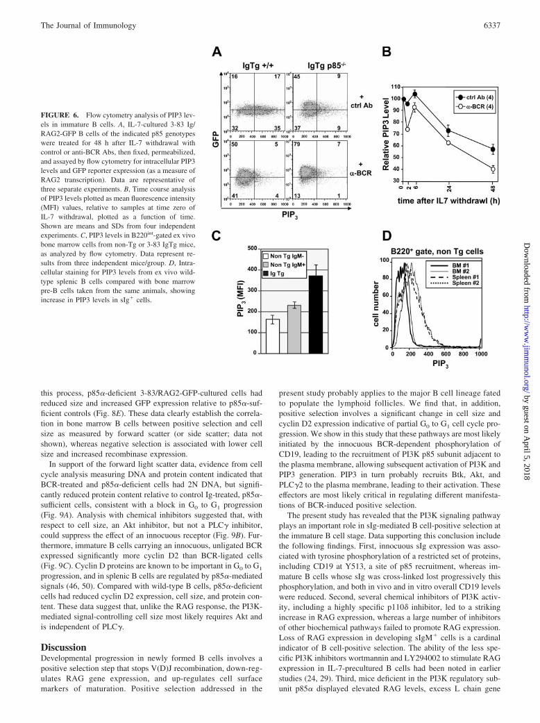
higher levels of PIP3 24 h after IL-7 withdrawal than those cul-tured with BCR ligand (Fig. 6A, cf left panels). In contrast, and aspredicted, p85-deficient cells had significantly lower PIP3 levels.(As expected, wortmannin-treated cells had reduced immunoreac-tivity with the PIP3 Ab (data not shown)). In addition, low levelsof PIP3 immunoreactivity correlated with high RAG2 expression,as measured at the level of single cells using the GFP reportergene. A time course experiment showed that at all time pointstested, BCR-ligated cells had consistently lower PIP3 levels thancells carrying unligated sIg (Fig. 6B). To see whether PIP3 levelswere increased in newly formed B cells carrying innocuous BCRin vivo, we compared B220int bone marrow B cells from wild-typemice with those from 3-83 Tg mice carrying a bone fide innocuousreceptor (Fig. 6C). Increasing PIP3 levels correlated with the ex-pression of the innocuous BCR, and were lowest in non-Tg cells
that were sIg negative. This result was further confirmed in a com-parison of B220� cells from bone marrow and spleen of B10.D2mice, in which mature sIg� cells of the spleen had significantlyhigher PIP3 levels than sIg� bone marrow pre-B cells (Fig. 6D).These results suggest that PIP3, a product of PI3K, is increased incells carrying a sIg, including those undergoing positive selection.By contrast, PIP3 levels are reduced not only in pre-B cells lackinga BCR, but also in immature B cells expressing a ligated BCR, andin cells lacking the PI3K regulatory component p85.
Enzyme activation
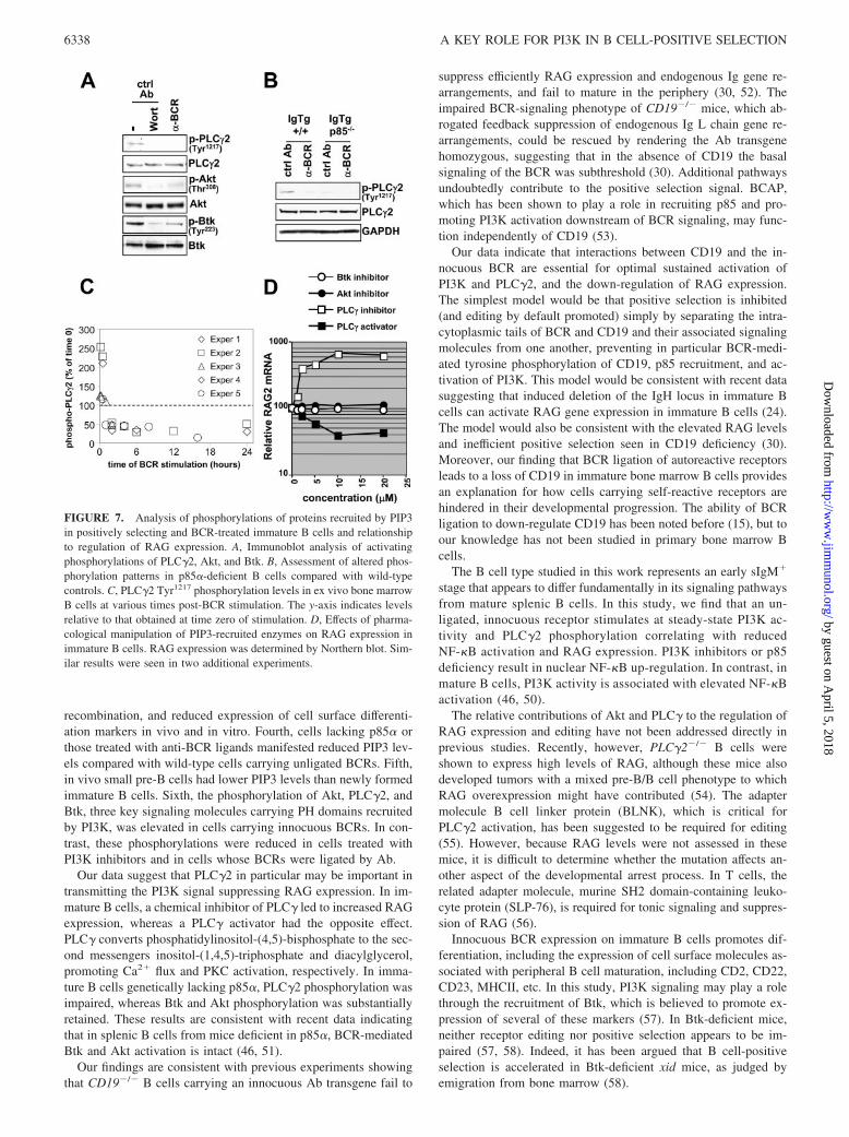
Given the clear evidence for a role for PIP3 in regulating B cell-positive selection, we considered the next possible steps in a signaltransduction cascade, in particular cytosolic enzymes that are re-cruited to the plasma membrane through PH domains, which rec-ognize and bind to PIP3. Among the major candidates in B cellsare three enzymes, as follows: the protein tyrosine kinase Btk, theserine/threonine kinase Akt, and PLC�2 (48). The activation ofthese enzymes is conveniently monitored by the use of well-de-fined phosphopeptide Abs. As shown in Fig. 7A, all three enzymesare activated in immature B cells carrying innocuous receptors,reduced in activity in anti-BCR-treated cells, and suppressed inwortmannin-treated cells. Consistent with these results, inp85��/� cells, PLC�2 activation, as detected by Tyr1217 phos-phorylation, was impaired (Fig. 7B), but surprisingly Btk and Aktphosphorylation was retained (data not shown). Results entirelyconsistent with these were obtained using ex vivo bone marrow Bcells because innocuous B cells had high-level Tyr1217 phosphor-ylation that was suppressed by either BCR ligation (Fig. 7C) ortreatment with IC87114 (data not shown). These results indicatedthat in immature B cells Btk, Akt, and PLC�2 may transmit PI3K-dependent positive selection signals from unligated BCRs, and thatPLC�2 is particularly dependent on p85� for its activation.
Linkage of PLC�2 to RAG expression
To probe the functional relevance of Btk, Akt, and PLC�2 down-stream pathways to B cell-positive selection, the effects of inhib-itors of these enzymes were assessed in a readout of RAG mRNAinduction or suppression. As shown in Fig. 7D, an inhibitor ofPLC� (U73122), but not inhibitors of Btk or Akt, promoted adose-dependent, and strikingly increased, expression of RAG2mRNA. Furthermore, a specific chemical activator of PLC� (m-3M3-FBS) had the opposite effect. These results strongly suggestthat PLC�2 is critical for the efficient RAG gene down-regulationassociated with B cell-positive selection in immature B cells.
Cell size/cell cycle
Because p85� has been reported to regulate cell cycle progressionin mature B cells (49), we assessed parameters of cell cycle andcell size in relation to the effects of basal or ligated BCR signalingin immature B cells. Several lines of evidence suggested that Bcell-positive selection is associated with an increase in cell sizerelative to unselected B cells that lack an innocuous BCR (Fig. 8).First, in bone marrow cultures, BCR-stimulated cells had a con-sistently reduced forward light scatter compared with cells treatedwith control Ab (Fig. 8A). The time- and BCR ligation-dependentRAG expression in these cells was inversely proportional to cellsize (Fig. 8B). Second, newly formed B220int bone marrow B cellsin vivo that carry innocuous BCRs are larger than comparablesIgM� pre-B cells from wild-type mice (Fig. 8C). Furthermore, ina comparison of innocuous and autoreactive bone marrow B cellsisolated directly from mice, autoreactive cells had �5% lower for-ward light scatter than innocuous ones at a similar developmentalstage (B220int) (Fig. 8D). Supporting a role for PI3K activity in
FIGURE 5. Immunoblot and EMSA analysis of NF-�B activation inp85�/� and p85�/� immature B cells. IL-7-expanded bone marrow B cellswere treated for 24 h in the absence of IL-7 with control or anti-BCR Abbefore assay. A, Nuclear extracts were incubated with the indicated radio-labeled probes and mobility shift monitored on polyacrylamide gels. B,Western blots of nuclear and cytoplasmic protein fractions of the indicatedcell populations were probed with Abs to the indicated epitopes. Immu-noblotting for the nuclear-localized protein fibrillarin was performed toconfirm purity of preparations. C, Effect of membrane-permeable, super-repressive I�B� (TAT-I�B�sr) on wortmannin- and BCR-induced RAGexpression. Immature 3-83/RAG2-GFP B cells obtained after IL-7 culturewere treated with control or anti-BCR Ab for 48 h. I�B�-TAT or control�-gal-TAT fusion proteins were added at 22 h and wortmannin at 24 h.Flow cytometry analysis for GFP reporter expression was conducted at48 h. At the time points surveyed under these conditions, survival in thepresence of TAT-I�B�sr did not differ �10% compared with cultures notreceiving TAT fusion protein.
6336 A KEY ROLE FOR PI3K IN B CELL-POSITIVE SELECTION
by guest on April 5, 2018
http://ww
w.jim
munol.org/
Dow
nloaded from
this process, p85�-deficient 3-83/RAG2-GFP-cultured cells hadreduced size and increased GFP expression relative to p85�-suf-ficient controls (Fig. 8E). These data clearly establish the correla-tion in bone marrow B cells between positive selection and cellsize as measured by forward scatter (or side scatter; data notshown), whereas negative selection is associated with lower cellsize and increased recombinase expression.
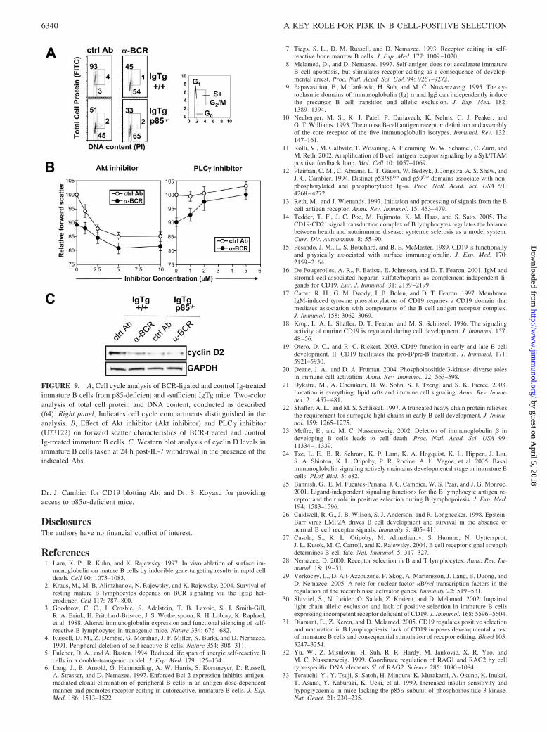
In support of the forward light scatter data, evidence from cellcycle analysis measuring DNA and protein content indicated thatBCR-treated and p85�-deficient cells had 2N DNA, but signifi-cantly reduced protein content relative to control Ig-treated, p85�-sufficient cells, consistent with a block in G0 to G1 progression(Fig. 9A). Analysis with chemical inhibitors suggested that, withrespect to cell size, an Akt inhibitor, but not a PLC� inhibitor,could suppress the effect of an innocuous receptor (Fig. 9B). Fur-thermore, immature B cells carrying an innocuous, unligated BCRexpressed significantly more cyclin D2 than BCR-ligated cells(Fig. 9C). Cyclin D proteins are known to be important in G0 to G1
progression, and in splenic B cells are regulated by p85�-mediatedsignals (46, 50). Compared with wild-type B cells, p85�-deficientcells had reduced cyclin D2 expression, cell size, and protein con-tent. These data suggest that, unlike the RAG response, the PI3K-mediated signal-controlling cell size most likely requires Akt andis independent of PLC�.
DiscussionDevelopmental progression in newly formed B cells involves apositive selection step that stops V(D)J recombination, down-reg-ulates RAG gene expression, and up-regulates cell surfacemarkers of maturation. Positive selection addressed in the
present study probably applies to the major B cell lineage fatedto populate the lymphoid follicles. We find that, in addition,positive selection involves a significant change in cell size andcyclin D2 expression indicative of partial G0 to G1 cell cycle pro-gression. We show in this study that these pathways are most likelyinitiated by the innocuous BCR-dependent phosphorylation ofCD19, leading to the recruitment of PI3K p85 subunit adjacent tothe plasma membrane, allowing subsequent activation of PI3K andPIP3 generation. PIP3 in turn probably recruits Btk, Akt, andPLC�2 to the plasma membrane, leading to their activation. Theseeffectors are most likely critical in regulating different manifesta-tions of BCR-induced positive selection.
The present study has revealed that the PI3K signaling pathwayplays an important role in sIg-mediated B cell-positive selection atthe immature B cell stage. Data supporting this conclusion includethe following findings. First, innocuous sIg expression was asso-ciated with tyrosine phosphorylation of a restricted set of proteins,including CD19 at Y513, a site of p85 recruitment, whereas im-mature B cells whose sIg was cross-linked lost progressively thisphosphorylation, and both in vivo and in vitro overall CD19 levelswere reduced. Second, several chemical inhibitors of PI3K activ-ity, including a highly specific p110� inhibitor, led to a strikingincrease in RAG expression, whereas a large number of inhibitorsof other biochemical pathways failed to promote RAG expression.Loss of RAG expression in developing sIgM� cells is a cardinalindicator of B cell-positive selection. The ability of the less spe-cific PI3K inhibitors wortmannin and LY294002 to stimulate RAGexpression in IL-7-precultured B cells had been noted in earlierstudies (24, 29). Third, mice deficient in the PI3K regulatory sub-unit p85� displayed elevated RAG levels, excess L chain gene
FIGURE 6. Flow cytometry analysis of PIP3 lev-els in immature B cells. A, IL-7-cultured 3-83 Ig/RAG2-GFP B cells of the indicated p85 genotypeswere treated for 48 h after IL-7 withdrawal withcontrol or anti-BCR Abs, then fixed, permeabilized,and assayed by flow cytometry for intracellular PIP3levels and GFP reporter expression (as a measure ofRAG2 transcription). Data are representative ofthree separate experiments. B, Time course analysisof PIP3 levels plotted as mean fluorescence intensity(MFI) values, relative to samples at time zero ofIL-7 withdrawal, plotted as a function of time.Shown are means and SDs from four independentexperiments. C, PIP3 levels in B220int-gated ex vivobone marrow cells from non-Tg or 3-83 IgTg mice,as analyzed by flow cytometry. Data represent re-sults from three independent mice/group. D, Intra-cellular staining for PIP3 levels from ex vivo wild-type splenic B cells compared with bone marrowpre-B cells taken from the same animals, showingincrease in PIP3 levels in sIg� cells.
6337The Journal of Immunology
by guest on April 5, 2018
http://ww
w.jim
munol.org/
Dow
nloaded from
recombination, and reduced expression of cell surface differenti-ation markers in vivo and in vitro. Fourth, cells lacking p85� orthose treated with anti-BCR ligands manifested reduced PIP3 lev-els compared with wild-type cells carrying unligated BCRs. Fifth,in vivo small pre-B cells had lower PIP3 levels than newly formedimmature B cells. Sixth, the phosphorylation of Akt, PLC�2, andBtk, three key signaling molecules carrying PH domains recruitedby PI3K, was elevated in cells carrying innocuous BCRs. In con-trast, these phosphorylations were reduced in cells treated withPI3K inhibitors and in cells whose BCRs were ligated by Ab.
Our data suggest that PLC�2 in particular may be important intransmitting the PI3K signal suppressing RAG expression. In im-mature B cells, a chemical inhibitor of PLC� led to increased RAGexpression, whereas a PLC� activator had the opposite effect.PLC� converts phosphatidylinositol-(4,5)-bisphosphate to the sec-ond messengers inositol-(1,4,5)-triphosphate and diacylglycerol,promoting Ca2� flux and PKC activation, respectively. In imma-ture B cells genetically lacking p85�, PLC�2 phosphorylation wasimpaired, whereas Btk and Akt phosphorylation was substantiallyretained. These results are consistent with recent data indicatingthat in splenic B cells from mice deficient in p85�, BCR-mediatedBtk and Akt activation is intact (46, 51).
Our findings are consistent with previous experiments showingthat CD19�/� B cells carrying an innocuous Ab transgene fail to
suppress efficiently RAG expression and endogenous Ig gene re-arrangements, and fail to mature in the periphery (30, 52). Theimpaired BCR-signaling phenotype of CD19�/� mice, which ab-rogated feedback suppression of endogenous Ig L chain gene re-arrangements, could be rescued by rendering the Ab transgenehomozygous, suggesting that in the absence of CD19 the basalsignaling of the BCR was subthreshold (30). Additional pathwaysundoubtedly contribute to the positive selection signal. BCAP,which has been shown to play a role in recruiting p85 and pro-moting PI3K activation downstream of BCR signaling, may func-tion independently of CD19 (53).
Our data indicate that interactions between CD19 and the in-nocuous BCR are essential for optimal sustained activation ofPI3K and PLC�2, and the down-regulation of RAG expression.The simplest model would be that positive selection is inhibited(and editing by default promoted) simply by separating the intra-cytoplasmic tails of BCR and CD19 and their associated signalingmolecules from one another, preventing in particular BCR-medi-ated tyrosine phosphorylation of CD19, p85 recruitment, and ac-tivation of PI3K. This model would be consistent with recent datasuggesting that induced deletion of the IgH locus in immature Bcells can activate RAG gene expression in immature B cells (24).The model would also be consistent with the elevated RAG levelsand inefficient positive selection seen in CD19 deficiency (30).Moreover, our finding that BCR ligation of autoreactive receptorsleads to a loss of CD19 in immature bone marrow B cells providesan explanation for how cells carrying self-reactive receptors arehindered in their developmental progression. The ability of BCRligation to down-regulate CD19 has been noted before (15), but toour knowledge has not been studied in primary bone marrow Bcells.
The B cell type studied in this work represents an early sIgM�
stage that appears to differ fundamentally in its signaling pathwaysfrom mature splenic B cells. In this study, we find that an un-ligated, innocuous receptor stimulates at steady-state PI3K ac-tivity and PLC�2 phosphorylation correlating with reducedNF-�B activation and RAG expression. PI3K inhibitors or p85deficiency result in nuclear NF-�B up-regulation. In contrast, inmature B cells, PI3K activity is associated with elevated NF-�Bactivation (46, 50).
The relative contributions of Akt and PLC� to the regulation ofRAG expression and editing have not been addressed directly inprevious studies. Recently, however, PLC�2�/� B cells wereshown to express high levels of RAG, although these mice alsodeveloped tumors with a mixed pre-B/B cell phenotype to whichRAG overexpression might have contributed (54). The adaptermolecule B cell linker protein (BLNK), which is critical forPLC�2 activation, has been suggested to be required for editing(55). However, because RAG levels were not assessed in thesemice, it is difficult to determine whether the mutation affects an-other aspect of the developmental arrest process. In T cells, therelated adapter molecule, murine SH2 domain-containing leuko-cyte protein (SLP-76), is required for tonic signaling and suppres-sion of RAG (56).
Innocuous BCR expression on immature B cells promotes dif-ferentiation, including the expression of cell surface molecules as-sociated with peripheral B cell maturation, including CD2, CD22,CD23, MHCII, etc. In this study, PI3K signaling may play a rolethrough the recruitment of Btk, which is believed to promote ex-pression of several of these markers (57). In Btk-deficient mice,neither receptor editing nor positive selection appears to be im-paired (57, 58). Indeed, it has been argued that B cell-positiveselection is accelerated in Btk-deficient xid mice, as judged byemigration from bone marrow (58).
FIGURE 7. Analysis of phosphorylations of proteins recruited by PIP3in positively selecting and BCR-treated immature B cells and relationshipto regulation of RAG expression. A, Immunoblot analysis of activatingphosphorylations of PLC�2, Akt, and Btk. B, Assessment of altered phos-phorylation patterns in p85�-deficient B cells compared with wild-typecontrols. C, PLC�2 Tyr1217 phosphorylation levels in ex vivo bone marrowB cells at various times post-BCR stimulation. The y-axis indicates levelsrelative to that obtained at time zero of stimulation. D, Effects of pharma-cological manipulation of PIP3-recruited enzymes on RAG expression inimmature B cells. RAG expression was determined by Northern blot. Sim-ilar results were seen in two additional experiments.
6338 A KEY ROLE FOR PI3K IN B CELL-POSITIVE SELECTION
by guest on April 5, 2018
http://ww
w.jim
munol.org/
Dow
nloaded from

Cell size regulation by innocuous BCR was suggested by sev-eral lines of evidence. In vivo, pre-B cells, which lack sIg, werefound to be smaller (as measured by forward light scatter) thanimmature B cells carrying a transgene-encoded innocuous BCR.Furthermore, BCR cross-linking in vitro reduced immature B cellsize relative to unligated control. Similarly, BCR-ligated or p85�-deficient cells had reduced forward scatter and total protein contentcompared with wild-type B cells with unligated receptors. How-ever, in contrast to PI3K-mediated RAG expression, in transmit-ting signals regulating cell size, PLC� is apparently not important.Rather, analysis with chemical inhibitors suggested that Akt, adifferent PI3K-recruited enzyme, most likely is important. Indeed,Akt is known to regulate cell size in many organisms and contexts,in part by regulating glycolysis and nutrient transport (59, 60).
The significance of tying cell size to BCR signaling in immatureB cells is unknown; however, it is tempting to speculate that it hasan important physiological role. BCR ligation leads to a combina-tion of elevated L chain gene recombination, a block in cell cycleprogression, and reduced Akt activation, whereas innocuous BCRsignaling promotes increased cell size and cessation of recombi-nation. We would like to propose that, at the pre-B/B cell transi-tion, absence of an Ig L chain (or expression of an autoreactiveBCR) leads to loss of Akt activity and attendant glucose uptakeand metabolism. The net effect is to progressively reduce cell sizeand ultimately survival, placing a limit on the amount of time thata B cell has to generate an appropriate L chain. This limits there-fore the number of recombination or editing attempts that can beconducted, a phenomenon that has been referred to as the crashfactor (61). According to this model, expression of an appropriate,
presumably innocuous BCR supports Akt activity, rescuing thecell from death by nutrient starvation (as has been proposed forcytokine withdrawal-induced cell death, autophagy (60)). Thismodel is supported by previous studies from our laboratory show-ing that artificially extending B cell lifespan through expression ofBcl2 promotes receptor editing (6), presumably by allowing cellslacking in Akt activity to survive longer and to undergo additionalrearrangements.
One can view the promotion of immature B cell development bythe innocuous BCR as a transition in cell cycle from G0 to G1, withattendant increase in cell size and cyclin D expression. Self Agligation of the BCR disrupts this signal, promoting continuedV(D)J recombination and receptor editing. A time limit is placedon this G0 editing state by the restriction in Akt activity, condemn-ing cells to death by neglect. Rescue occurs when successful ed-iting generates an innocuous receptor. It remains to be seenwhether BCR ligands work merely by receptor down-modulation,denuding the B cell of sIg or CD19, and terminating the positiveselection signal. If this simple model is correct, it predicts thatin cells coexpressing two receptors, one autoreactive and oneinnocuous, receptor editing could be terminated. This wouldseem to be a dangerous possibility that is to be avoided; how-ever, such a phenotype has been observed in some autoantibodyTg mice (e.g., 62, 63).
AcknowledgmentsWe thank Drs. R. Rickert, A. Anzelon, and D. Fruman for advice; Drs.Kabouridis and Dowdy for TAT-fusion protein constructs; Dr. A. Feeneyfor 3-83/H-2k mice; Dr. M. Nussenzweig for RAG2/GFP reporter mice;
FIGURE 8. Relationship between cellsize and RAG expression in immature Bcells. A, Inverse correlation between RAGexpression and cell size in immature B cellstreated with control Ab or anti-BCR. Bonemarrow B cells of 3-83 IgTg/RAG2-GFPmice were stimulated by BCR Abs, controlAbs, or a stromal cell line lacking BCR li-gand for the indicated times after IL-7 with-drawal. Cells were assayed for GFP expres-sion (upper panel) and forward scatter(lower panel). B, Data from A replotted toshow relationship between %GFP� cells andforward scatter. C, Comparison of forwardscatter in ex vivo analyzed immature B cellscarrying an innocuous receptor (B220int/IgM� bone marrow cells from IgTg mice)and pre-B cells (B220int/IgM� bone marrowcells from wild-type mice). D, Comparisonof forward light scatter in negatively select-ing (3-83/H-2k) and positively selecting (3-83/H-2d) ex vivo bone marrow B cells gatedon B220int/sIg�� cells. E, Dot plot analysisof GFP expression vs cell size in p85�/� andp85�/� IgTg B cells. Relative geometricmeans of fluorescence intensity were as fol-lows: IgTg ctrl (GFP 23; forward scatter378); IgTg anti-BCR (GFP 34; forward scat-ter 331); IgTg p85�/� ctrl (GFP 31; forwardscatter 337); IgTg p85�/� anti-BCR (GFP77; forward scatter 316).
6339The Journal of Immunology
by guest on April 5, 2018
http://ww
w.jim
munol.org/
Dow
nloaded from
Dr. J. Cambier for CD19 blotting Ab; and Dr. S. Koyasu for providingaccess to p85�-deficient mice.
DisclosuresThe authors have no financial conflict of interest.
References1. Lam, K. P., R. Kuhn, and K. Rajewsky. 1997. In vivo ablation of surface im-
munoglobulin on mature B cells by inducible gene targeting results in rapid celldeath. Cell 90: 1073–1083.
2. Kraus, M., M. B. Alimzhanov, N. Rajewsky, and K. Rajewsky. 2004. Survival ofresting mature B lymphocytes depends on BCR signaling via the Ig�� het-erodimer. Cell 117: 787–800.
3. Goodnow, C. C., J. Crosbie, S. Adelstein, T. B. Lavoie, S. J. Smith-Gill,R. A. Brink, H. Pritchard-Briscoe, J. S. Wotherspoon, R. H. Loblay, K. Raphael,et al. 1988. Altered immunoglobulin expression and functional silencing of self-reactive B lymphocytes in transgenic mice. Nature 334: 676–682.
4. Russell, D. M., Z. Dembic, G. Morahan, J. F. Miller, K. Burki, and D. Nemazee.1991. Peripheral deletion of self-reactive B cells. Nature 354: 308–311.
5. Fulcher, D. A., and A. Basten. 1994. Reduced life span of anergic self-reactive Bcells in a double-transgenic model. J. Exp. Med. 179: 125–134.
6. Lang, J., B. Arnold, G. Hammerling, A. W. Harris, S. Korsmeyer, D. Russell,A. Strasser, and D. Nemazee. 1997. Enforced Bcl-2 expression inhibits antigen-mediated clonal elimination of peripheral B cells in an antigen dose-dependentmanner and promotes receptor editing in autoreactive, immature B cells. J. Exp.Med. 186: 1513–1522.
7. Tiegs, S. L., D. M. Russell, and D. Nemazee. 1993. Receptor editing in self-reactive bone marrow B cells. J. Exp. Med. 177: 1009–1020.
8. Melamed, D., and D. Nemazee. 1997. Self-antigen does not accelerate immatureB cell apoptosis, but stimulates receptor editing as a consequence of develop-mental arrest. Proc. Natl. Acad. Sci. USA 94: 9267–9272.
9. Papavasiliou, F., M. Jankovic, H. Suh, and M. C. Nussenzweig. 1995. The cy-toplasmic domains of immunoglobulin (Ig) � and Ig� can independently inducethe precursor B cell transition and allelic exclusion. J. Exp. Med. 182:1389–1394.
10. Neuberger, M. S., K. J. Patel, P. Dariavach, K. Nelms, C. J. Peaker, andG. T. Williams. 1993. The mouse B-cell antigen receptor: definition and assemblyof the core receptor of the five immunoglobulin isotypes. Immunol. Rev. 132:147–161.
11. Rolli, V., M. Gallwitz, T. Wossning, A. Flemming, W. W. Schamel, C. Zurn, andM. Reth. 2002. Amplification of B cell antigen receptor signaling by a Syk/ITAMpositive feedback loop. Mol. Cell 10: 1057–1069.
12. Pleiman, C. M., C. Abrams, L. T. Gauen, W. Bedzyk, J. Jongstra, A. S. Shaw, andJ. C. Cambier. 1994. Distinct p53/56lyn and p59fyn domains associate with non-phosphorylated and phosphorylated Ig-�. Proc. Natl. Acad. Sci. USA 91:4268–4272.
13. Reth, M., and J. Wienands. 1997. Initiation and processing of signals from the Bcell antigen receptor. Annu. Rev. Immunol. 15: 453–479.
14. Tedder, T. F., J. C. Poe, M. Fujimoto, K. M. Haas, and S. Sato. 2005. TheCD19-CD21 signal transduction complex of B lymphocytes regulates the balancebetween health and autoimmune disease: systemic sclerosis as a model system.Curr. Dir. Autoimmun. 8: 55–90.
15. Pesando, J. M., L. S. Bouchard, and B. E. McMaster. 1989. CD19 is functionallyand physically associated with surface immunoglobulin. J. Exp. Med. 170:2159–2164.
16. De Fougerolles, A. R., F. Batista, E. Johnsson, and D. T. Fearon. 2001. IgM andstromal cell-associated heparan sulfate/heparin as complement-independent li-gands for CD19. Eur. J. Immunol. 31: 2189–2199.
17. Carter, R. H., G. M. Doody, J. B. Bolen, and D. T. Fearon. 1997. MembraneIgM-induced tyrosine phosphorylation of CD19 requires a CD19 domain thatmediates association with components of the B cell antigen receptor complex.J. Immunol. 158: 3062–3069.
18. Krop, I., A. L. Shaffer, D. T. Fearon, and M. S. Schlissel. 1996. The signalingactivity of murine CD19 is regulated during cell development. J. Immunol. 157:48–56.
19. Otero, D. C., and R. C. Rickert. 2003. CD19 function in early and late B celldevelopment. II. CD19 facilitates the pro-B/pre-B transition. J. Immunol. 171:5921–5930.
20. Deane, J. A., and D. A. Fruman. 2004. Phosphoinositide 3-kinase: diverse rolesin immune cell activation. Annu. Rev. Immunol. 22: 563–598.
21. Dykstra, M., A. Cherukuri, H. W. Sohn, S. J. Tzeng, and S. K. Pierce. 2003.Location is everything: lipid rafts and immune cell signaling. Annu. Rev. Immu-nol. 21: 457–481.
22. Shaffer, A. L., and M. S. Schlissel. 1997. A truncated heavy chain protein relievesthe requirement for surrogate light chains in early B cell development. J. Immu-nol. 159: 1265–1275.
23. Meffre, E., and M. C. Nussenzweig. 2002. Deletion of immunoglobulin � indeveloping B cells leads to cell death. Proc. Natl. Acad. Sci. USA 99:11334–11339.
24. Tze, L. E., B. R. Schram, K. P. Lam, K. A. Hogquist, K. L. Hippen, J. Liu,S. A. Shinton, K. L. Otipoby, P. R. Rodine, A. L. Vegoe, et al. 2005. Basalimmunoglobulin signaling actively maintains developmental stage in immature Bcells. PLoS Biol. 3: e82.
25. Bannish, G., E. M. Fuentes-Panana, J. C. Cambier, W. S. Pear, and J. G. Monroe.2001. Ligand-independent signaling functions for the B lymphocyte antigen re-ceptor and their role in positive selection during B lymphopoiesis. J. Exp. Med.194: 1583–1596.
26. Caldwell, R. G., J. B. Wilson, S. J. Anderson, and R. Longnecker. 1998. Epstein-Barr virus LMP2A drives B cell development and survival in the absence ofnormal B cell receptor signals. Immunity 9: 405–411.
27. Casola, S., K. L. Otipoby, M. Alimzhanov, S. Humme, N. Uyttersprot,J. L. Kutok, M. C. Carroll, and K. Rajewsky. 2004. B cell receptor signal strengthdetermines B cell fate. Nat. Immunol. 5: 317–327.
28. Nemazee, D. 2000. Receptor selection in B and T lymphocytes. Annu. Rev. Im-munol. 18: 19–51.
29. Verkoczy, L., D. Ait-Azzouzene, P. Skog, A. Martensson, J. Lang, B. Duong, andD. Nemazee. 2005. A role for nuclear factor �B/rel transcription factors in theregulation of the recombinase activator genes. Immunity 22: 519–531.
30. Shivtiel, S., N. Leider, O. Sadeh, Z. Kraiem, and D. Melamed. 2002. Impairedlight chain allelic exclusion and lack of positive selection in immature B cellsexpressing incompetent receptor deficient of CD19. J. Immunol. 168: 5596–5604.
31. Diamant, E., Z. Keren, and D. Melamed. 2005. CD19 regulates positive selectionand maturation in B lymphopoiesis: lack of CD19 imposes developmental arrestof immature B cells and consequential stimulation of receptor editing. Blood 105:3247–3254.
32. Yu, W., Z. Misulovin, H. Suh, R. R. Hardy, M. Jankovic, X. R. Yao, andM. C. Nussenzweig. 1999. Coordinate regulation of RAG1 and RAG2 by celltype-specific DNA elements 5� of RAG2. Science 285: 1080–1084.
33. Terauchi, Y., Y. Tsuji, S. Satoh, H. Minoura, K. Murakami, A. Okuno, K. Inukai,T. Asano, Y. Kaburagi, K. Ueki, et al. 1999. Increased insulin sensitivity andhypoglycaemia in mice lacking the p85� subunit of phosphoinositide 3-kinase.Nat. Genet. 21: 230–235.
FIGURE 9. A, Cell cycle analysis of BCR-ligated and control Ig-treatedimmature B cells from p85-deficient and -sufficient IgTg mice. Two-coloranalysis of total cell protein and DNA content, conducted as described(64). Right panel, Indicates cell cycle compartments distinguished in theanalysis. B, Effect of Akt inhibitor (Akt inhibitor) and PLC� inhibitor(U73122) on forward scatter characteristics of BCR-treated and controlIg-treated immature B cells. C, Western blot analysis of cyclin D levels inimmature B cells taken at 24 h post-IL-7 withdrawal in the presence of theindicated Abs.
6340 A KEY ROLE FOR PI3K IN B CELL-POSITIVE SELECTION
by guest on April 5, 2018
http://ww
w.jim
munol.org/
Dow
nloaded from
34. Melamed, D., J. A. Kench, K. Grabstein, A. Rolink, and D. Nemazee. 1997. Afunctional B cell receptor transgene allows efficient IL-7-independent maturationof B cell precursors. J. Immunol. 159: 1233–1239.
35. Hammerling, G. J., E. Rusch, N. Tada, S. Kimura, and U. Hammerling. 1982.Localization of allodeterminants on H-2Kb antigens determined with monoclonalantibodies and H-2 mutant mice. Proc. Natl. Acad. Sci. USA 79: 4737–4741.
36. Dyer, R. B., and N. K. Herzog. 1995. Isolation of intact nuclei for nuclear extractpreparation from a fragile B-lymphocyte cell line. BioTechniques 19: 192–195.
37. Kabouridis, P. S., M. Hasan, J. Newson, D. W. Gilroy, and T. Lawrence. 2002.Inhibition of NF-�B activity by a membrane-transducing mutant of I�B�. J. Im-munol. 169: 2587–2593.
38. Schwarze, S. R., A. Ho, A. Vocero-Akbani, and S. F. Dowdy. 1999. In vivoprotein transduction: delivery of a biologically active protein into the mouse.Science 285: 1569–1572.
39. Hertz, M., and D. Nemazee. 1997. BCR ligation induces receptor editing inIgM�IgD� bone marrow B cells in vitro. Immunity 6: 429–436.
40. Retter, M. W., and D. Nemazee. 1998. Receptor editing occurs frequently duringnormal B cell development. J. Exp. Med. 188: 1231–1238.
41. Anzelon, A. N., H. Wu, and R. C. Rickert. 2003. Pten inactivation alters periph-eral B lymphocyte fate and reconstitutes CD19 function. Nat. Immunol. 4:287–294.
42. Perez, O. D., S. Kinoshita, Y. Hitoshi, D. G. Payan, T. Kitamura, G. P. Nolan, andJ. B. Lorens. 2002. Activation of the PKB/AKT pathway by ICAM-2. Immunity16: 51–65.
43. Tuveson, D. A., R. H. Carter, S. P. Soltoff, and D. T. Fearon. 1993. CD19 of Bcells as a surrogate kinase insert region to bind phosphatidylinositol 3-kinase.Science 260: 986–989.
44. Wang, Y., S. R. Brooks, X. Li, A. N. Anzelon, R. C. Rickert, and R. H. Carter.2002. The physiologic role of CD19 cytoplasmic tyrosines. Immunity 17:501–514.
45. Sadhu, C., B. Masinovsky, K. Dick, C. G. Sowell, and D. E. Staunton. 2003.Essential role of phosphoinositide 3-kinase � in neutrophil directional movement.J. Immunol. 170: 2647–2654.
46. Suzuki, H., S. Matsuda, Y. Terauchi, M. Fujiwara, T. Ohteki, T. Asano,T. W. Behrens, T. Kouro, K. Takatsu, T. Kadowaki, and S. Koyasu. 2003. PI3Kand Btk differentially regulate B cell antigen receptor-mediated signal transduc-tion. Nat. Immunol. 4: 280–286.
47. Donahue, A. C., K. L. Hess, K. L. Ng, and D. A. Fruman. 2004. Altered splenicB cell subset development in mice lacking phosphoinositide 3-kinase p85�. Int.Immunol. 16: 1789–1798.
48. Fruman, D. A., A. B. Satterthwaite, and O. N. Witte. 2000. Xid-like phenotypes:a B cell signalosome takes shape. Immunity 13: 1–3.
49. Glassford, J., E. Vigorito, I. Soeiro, P. A. Madureira, G. Zoumpoulidou,J. J. Brosens, M. Turner, and E. W. Lam. 2005. Phosphatidylinositol 3-kinase isrequired for the transcriptional activation of cyclin D2 in BCR activated primarymouse B lymphocytes. Eur. J. Immunol. 35: 2748–2761.
50. Piatelli, M. J., C. Wardle, J. Blois, C. Doughty, B. R. Schram, T. L. Rothstein,and T. C. Chiles. 2004. Phosphatidylinositol 3-kinase-dependent mitogen-acti-vated protein/extracellular signal-regulated kinase kinase 1/2 and NF-�B signal-ing pathways are required for B cell antigen receptor-mediated cyclin D2 induc-tion in mature B cells. J. Immunol. 172: 2753–2762.
51. Hess, K. L., A. C. Donahue, K. L. Ng, T. I. Moore, J. Oak, and D. A. Fruman.2004. Frontline: the p85� isoform of phosphoinositide 3-kinase is essential for asubset of B cell receptor-initiated signaling responses. Eur. J. Immunol. 34:2968–2976.
52. Buhl, A. M., D. Nemazee, J. C. Cambier, R. Rickert, and M. Hertz. 2000. B-cellantigen receptor competence regulates B-lymphocyte selection and survival. Im-munol. Rev. 176: 141–153.
53. Yamazaki, T., K. Takeda, K. Gotoh, H. Takeshima, S. Akira, and T. Kurosaki.2002. Essential immunoregulatory role for BCAP in B cell development andfunction. J. Exp. Med. 195: 535–545.
54. Wen, R., Y. Chen, L. Bai, G. Fu, J. Schuman, X. Dai, H. Zeng, C. Yang,R. P. Stephan, J. L. Cleveland, and D. Wang. 2006. Essential role of phospho-lipase C�2 in early B-cell development and Myc-mediated lymphomagenesis.Mol. Cell. Biol. 26: 9364–9376.
55. Hayashi, K., T. Nojima, R. Goitsuka, and D. Kitamura. 2004. Impaired receptorediting in the primary B cell repertoire of BASH-deficient mice. J. Immunol. 173:5980–5988.
56. Roose, J. P., M. Diehn, M. G. Tomlinson, J. Lin, A. A. Alizadeh, D. Botstein,P. O. Brown, and A. Weiss. 2003. T cell receptor-independent basal signaling viaErk and Abl kinases suppresses RAG gene expression. PLoS Biol. 1: E53.
57. Middendorp, S., and R. W. Hendriks. 2004. Cellular maturation defects in Bru-ton’s tyrosine kinase-deficient immature B cells are amplified by premature B cellreceptor expression and reduced by receptor editing. J. Immunol. 172:1371–1379.
58. Cariappa, A., T. J. Kim, and S. Pillai. 1999. Accelerated emigration of B lym-phocytes in the Xid mouse. J. Immunol. 162: 4417–4423.
59. Kozma, S. C., and G. Thomas. 2002. Regulation of cell size in growth, devel-opment and human disease: PI3K, PKB and S6K. BioEssays 24: 65–71.
60. Edinger, A. L., and C. B. Thompson. 2002. Akt maintains cell size and survivalby increasing mTOR-dependent nutrient uptake. Mol. Biol. Cell 13: 2276–2288.
61. Coleclough, C. 1983. Chance, necessity and antibody gene dynamics. Nature303: 23–26.
62. Li, Y., H. Li, and M. Weigert. 2002. Autoreactive B cells in the marginal zonethat express dual receptors. J. Exp. Med. 195: 181–188.
63. Gerdes, T., and M. Wabl. 2004. Autoreactivity and allelic inclusion in a B cellnuclear transfer mouse. Nat. Immunol. 5: 1282–1287.
64. Williams, C. D., D. C. Linch, M. J. Watts, and N. S. Thomas. 1997. Character-ization of cell cycle status and E2F complexes in mobilized CD34� cells beforeand after cytokine stimulation. Blood 90: 194–203.
6341The Journal of Immunology
by guest on April 5, 2018
http://ww
w.jim
munol.org/
Dow
nloaded from