Embed Size (px)
Citation preview

CELL SENESCENCE IN VITRO IS EQUIVALENT TO CELL AGING IN VIVO
Leonard Hayflick, Ph.D.
Professor of Anatomy
University of California, San Francisco
Copyright © 2009. All rights reserved.

Presentation Outline
Presentation objectiveBrief introduction to cell senescence in vitroPhenotype of senescent cellsStem cells (an aside)Senescent human cells in aging both in vivo and in vitroSenescence in cells in human bone and vertebral discsHeat shock proteins (hsp) in vivo and in vitroSenescence in cells in human atherosclerosis in vivo and in vitro DNA and DNA repair in senescent cells Brief history of events leading to telomere, telomerase discoveriesTelomere attrition in aging human cellsSenescent cells and cancerImmunosenescenceConclusions
PRESENTATION OBJECTIVE
To provide evidence - quoted mostly from the work of others -that has persuaded them to regard the aging of cells in vitro to be equivalent to the aging of cells in vivo.
This will be an unusual presentation in that I will ask you to read more than I will talk.
Warning: The quotations I ask you to read will be numerous.

INTRODUCTION TO CELL SENESCENCE IN VITRO


WI-38 Phases I and II Phase III
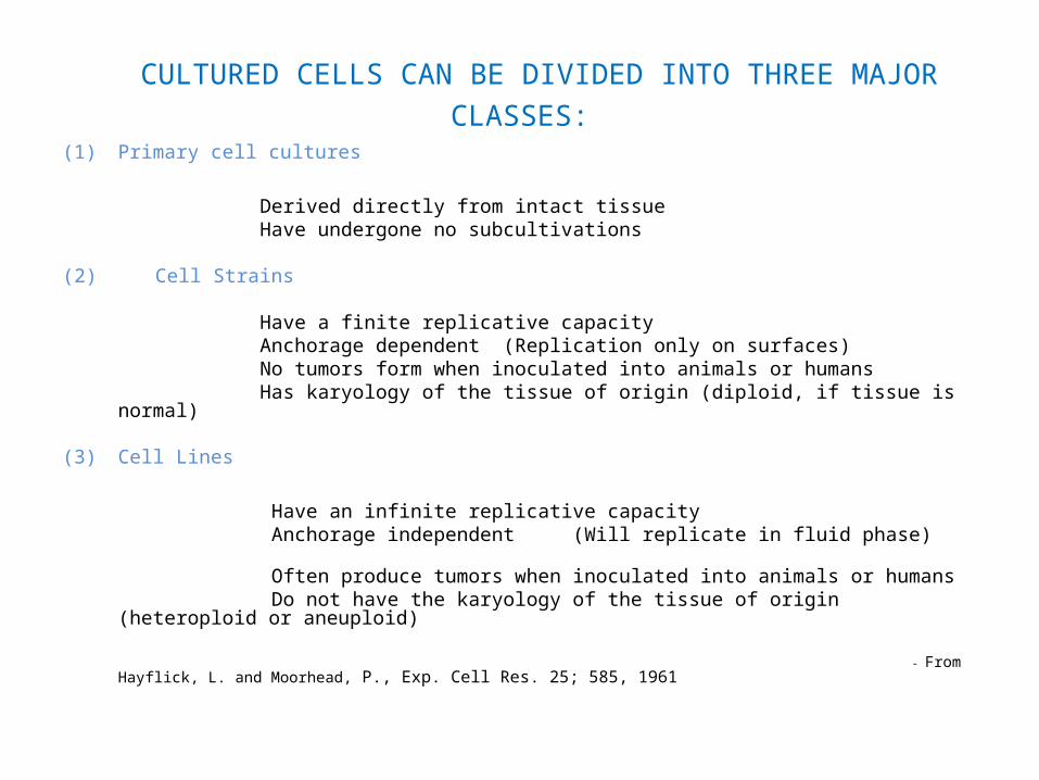
CULTURED CELLS CAN BE DIVIDED INTO THREE MAJOR CLASSES:
(1) Primary cell cultures
Derived directly from intact tissue Have undergone no subcultivations
(2) Cell Strains Have a finite replicative capacity Anchorage dependent (Replication only on surfaces) No tumors form when inoculated into animals or humans Has karyology of the tissue of origin (diploid, if tissue is normal) (3) Cell Lines
Have an infinite replicative capacity Anchorage independent (Will replicate in fluid phase) Often produce tumors when inoculated into animals or humans Do not have the karyology of the tissue of origin (heteroploid or aneuploid) - From Hayflick, L. and Moorhead, P., Exp. Cell Res. 25; 585, 1961

MAJOR PROPERTIES OF NORMAL HUMAN DIPLOID CELL STRAINS
Fibroblasts are capable of 40-60 population doublings (PDs) if derived from human embryonic tissue. Many studies find fewer doublings occurring in older donors. Some do not.
Prior to Phase III, cells have all of the properties of normal cells (karyology, biochemistry, morphology, finite lifetime)
Cells are anchorage dependent (cannot grow in suspension).
Cells cannot replicate in immune compromised animals or in terminal cancer patients.
Cells have the broadest human virus spectrum known.
During cryogenic preservation cells “remember” at what population doubling they are arrested and, when reconstituted, continue to replicate until the 50 population doubling limit is reached. (WI-38 has been frozen for 47 years which is the longest time that any viable normal human cell has ever been frozen.)
- From: Hayflick, L. and Moorhead, P.S. Exp. Cell Res. 25; 585, 1961 and Hayflick,L. Exp. Cell Res., 37; 614, 1965

FIRST SUGGESTION THAT PHASE III REPRESENTS “AGING IN VITRO”
“The third possible explanation for entry into Phase III may bear directly upon problems of ageing, or more precisely “senescence”. This concept, although vague at the level of the whole organism, may have some validity in explaining the phenomenon at the cellular level, at least as an operational concept.”
- Hayflick, L. and Moorhead, P.S. Exp. Cell Res. 25; 585, 1961

Objective
“The Holy Grail of replicative senescence studies has been to demonstrate that aging in vivo is accompanied by the accumulation of cells showing characteristics of replicative senescence identified in cell culture.”
- Effros RB, “From Hayflick to Walford: the role of T cell replicative senescence in human aging. “ Exp. Gerontol. 2004;
39: 885-90.

PHENOTYPE OF SENESCENT CELLS IN VITRO (Used by authors to provide evidence for similar properties in old cells in vivo)
Irreversible growth arrest Hayflick and Moorhead, 1961
Increased population doubling time Hayflick and Moorhead, 1961
Arrest in transition phase from G1 to S phase Chen, et al, 2000
No growth factors or mitogens can move senescent cells to S phase from G1 Cristofalo VJ, et al. J Gerontol 1989; 44: 55
Larger and flattened cell morphology Eagner et al., 2001
Increased aneuploidy Moorhead and Saksela, 1965
Senescent associated β-Gal expression Dimri et al., PNAS USA, 92, 9363,1995
(A manifestation of increased lysosomal engorgement.) Kurz DJ, et al. J Cell Sci. 2000; 113:3613
Shortened telomeres Harley, et al., 1990, Deng et al., 2003
Altered gene expression Cristofalo et al., 1998; Kim et al., 2008
Polymorphic nuclei, vacuolization Rosen et al J Cell Physiol 1981;107:123;van der Loo B, et al.,Exp Cell Res 1998; 241: 309
Upregulation of p53, p21 and p16 Q. Yang, Cytogenet Genome Res 122:211, 2008
Long survival time in the senescence phase Matsumura T.,et al., 1979, Exp Cell Res 1998; 241: 309
Resistance to apoptosis Spaulding, CS, et al., Exp. Gerontol.,1994, 29, 601

(An aside): CONTRARY TO POPULAR BELIEF CULTURED NORMAL STEM CELLS ARE NOT IMMORTAL
In keeping with our original postulate, all normal cell populations are mortal and only those that acquire cancer-like abnormalities are immortal.
(Hayflick and Moorhead, Exp. Cell Res., 1963)
Recent reports of the abnormalities that eventually occur in stem cells:
Spits, C. et al., Recurrent chromosomal abnormalities in human embryonic stem cells. Nature Biotechnology 26, 1361 - 1363 (2008) Tamra E., et al., Characterization of human embryonic stem cells with features of neoplastic progression, Nature Biotechnology 27, 91 - 97 (2009) Lefort, N. et al, Human embryonic stem cells reveal recurrent genomic instability at 20q11.21, Nature Biotechnology 26, 1364 - 1366 (2008)

SENESCENT HUMAN CELLS IN AGING BOTH IN VIVO AND IN VITRO

SENESCENT HUMAN CELLS IN AGING BOTH IN VIVO AND IN VITRO
NORMAL TISSUE:
Skin Dimri GP, et al.Proc Natl Acad Sci USA 1995; 92: 9363
Liver Paradis V, et al., Hum Pathol 2001; 32: 327; Satyanarayana, A. et al., EMBO J., 22,4003, 2003
Liver stellate cells Krizhanovsky, V. et al., Cell 134, 657–667, August 22, 2008
Abdominal aorta endothelial cells Aviv H, et al. Atherosclerosis 2001; 159: 281
Hematopoietic compartment Park IK, et al., Nature,423, 302, 2003
Satellite cells Decary S, , et al. Hum Gene Ther 1997; 8: 1429
Periodontal ligament cells Silv´erio, KG , et al Connect. Tissue Res., 1–8, 2008
Keratinocytes Soroka, Y. et al.,Experimental Gerontology 43 (2008) 947
Corneal endothelium Engelmann K, et al., Invest Ophthalmol Vis Sci 1988; 29: 1656

SENESCENT HUMAN CELLS IN AGING BOTH IN VIVO AND IN VITRO (continued)
CELLS FROM PATHOLOGICAL TISSUE Prostate (enlarged) (Choi J, et al. Urology 2000; 56: 160)
Rheumatoid arthritis (Schmid M, et al.,Z. Rheumatol 2004; 63: 483; Vallejo, et al.,J. Immunol., 162: 6572,1999)
Osteoarthritis (Price JS, et al., Aging Cell 2002; 1: 57; Dai SM, et al.Arthritis Rheum. 2006; 54: 818)
Hepatitis (Paradis V, et al., Hum Pathol 2001; 32: 327)
Liver cirrhosis (Wiemann SU, et al. FASEB J 2002; 16: 935)
Emphysema and Smoking Nobukuni S, et al Respirology 7: 217, 2002;Noordhoek JA et al,. Exp Lung Res 29: 291–302,2003; Holz O., et al., EurRespir J. 24:575, 2004.)
Chronic wound tissues (Mendez MV, et al. J Vasc Surg 1998; 28: 876; Harding KG, et al., Int. Wound J. 1998; 2:364.)
Atherosclerotic tissue (Minamino T, et al. J Cardiol 2003; 41: 39-40; Hayashi T, et al. PNAS USA 2006; 103: 17018; Repin VS, et al. Atherosclerosis 1984; 50: 35; Burrig KF. Arterioscler Thromb 1991; 11: 1678. Vasile DJ, et al. FASEB J 2001; 15: 458).
Myeloblasts in Duchenne Muscular Dystrophy (Webster C & Blau HM. Soma Cell Genet 1990; 16: 57.Wright,W., Exp. Cell Res., 157; 343, 1985.Bischoff, R., Myolgy (2nd. Ed., McGraw Hill, 1994, 1, 97)
Abdominal aortic aneuryisms Liao S, et al. ,J Surg Res 92: 85, 2000.; Bennett MR, et al., Circ Res 82: 704, 1998.
Alzheimer’s Disease “It is noteworthy that becoming senescent, fibroblasts produce increased amounts of amyloid and apolipoprotein (Petropoulou C, et al., . FEBS Lett 509:287–297, 2001. Adler MJ, et al., Proc Natl Acad Sci U S A 88: 16–20, 1991.)”
-l Muller M., ANTIOXIDANTS & REDOX SIGNALING Volume 11, Number 1, 2009

SENESCENT HUMAN CELLS IN AGING BOTH IN VIVO AND IN VITRO (continued)
“Research carried out over the last ten years has shown that cellular senescence occurs in vivo both in the vasculature and in other organ systems…and supports the concept that the number of vascular cell replications that have taken place in the vessel wall increases not only as a function of chronological age but also as a function of the hemodynamic or biochemical stress to the vascular bed.” - Erusalimsky, JD and Kurz, D., J Exp. Gerontol. 40, 634-642, 2005
“Accumulation of abnormal proteins, decreased protein degradation, and diminished response to stress are well documented characteristics in several aged organisms and cell models, including human diploid fibroblasts (HDF) during their replicative senescence in vitro (Grune T. Biogerontology 2000; 1: 31-40; Soti C & Csermely P. Biogerontology 2000; 1: 225-33; Cell Stress Chaperones 2002; 7: 186-90; Exp Gerontol 2003; 38: 1037-40; Soti C, et al.,Aging Cell 2003; 2: 39-45.) .”
- Bonelli M, et al. Exp Gerontol 2004; 39: 423-32.
“…senescent cells accumulate in vitro with increasing (donor) age.” -Lombard DB, et al. DNA repair, genome stability and aging. Cell 2005; 120: 497

SENESCENT HUMAN CELLS IN AGING BOTH IN VIVO AND IN VITRO (continued)
“Like fibroblasts. vascular endothelial and smooth muscle cells have a limited replicative life span (Rosen EM, Mueller et al., J Cell Physiol 1981; 107: 123-37.) Furthermore, human endothelial cells undergoing replicative senescence in culture show characteristic changes in morphology, such as an increase in size, nuclear polymorphism, and vacuolization, (EM, Mueller et al., J Cell Physiol 1981; 107: 123; Nichols WW, et al. J
Cell Physiol 1987; 132: 453.) reminiscent of changes that have been found in endothelial cells covering the surface of aortic, carotid, and coronary atherosclerotic lesions at various stages of disease progression (Repin VS, et al. Atherosclerosis 1984; 50: 35; Tokunaga O, et al., Am J Pathol 1989; 135: 967; Burrig KF. Arterioscler Thromb 1991; 11: 1678).”
-van der Loo B, et al., Exp Cell Res 1998; 241: 309.
“…the role of secretory phospholipase A2 ( SPLA2 ) in cellular senescence suggests that SPLA2 – mediated cellular senescence contributes to the age-related decline of tissue structure and/or the genesis and progression of various inflammatory diseases associated with age.” - Kim, HJ, J. of Gerontol. Biol. Sci., 2009, 64, 351

SENESCENT CELLS IN HUMAN BONE AND VERTEBRAL DISCS IN VIVO

SENESCENT CELLS IN HUMAN BONE AND VERTEBRAL DISCS IN VIVO
“..the number of proliferative precursor cells on trabecular bone surfaces is higher in younger subjects. There is a marked decrease in precursor numbers in the second and third decade of life to a level which is maintained into old age.”
(Shigeno Y. et al J. Bone and Joint
Surg.1995;77B No.1: 139)
“More senescent cells in herniated vs non herniated discs.”
( Roberts et al. Eur. Spine J 2006;15 (Suppl 3): S312–6)
“Quantitative analysis of immunohistochemical localization of SA-β -gal identified a sizeable population of senescent cells in the aging/degenerating disc. These cells persist and accumulate over time within the disc.” (Gruber et al. Spine 2007;32:321)
“Our findings indicate that disc cell senescence occurs in vivo and is accelerated in intravertebral disk (IVD) degeneration. Furthermore, the senescent phenotype is associated with increased catabolism, implicating cellular senescence in the pathogenesis of IVD degeneration.” (Le Maitre et al. Arthr. Res. Ther. 2007;9: R45)
“Senescent cells in degenerate intervertebral discs produce matrixdegeneration – a major problem in disc degeneration.” (Gruber et al. Spine 2007;32:1181)

BEHAVIOR OF HEAT SHOCK PROTEINS (hsp) IN SENESCENT CELLS IN VIVO AND IN VITRO

HEAT SHOCK PROTEINS (hsp) IN VIVO AND IN VITRO
“hsp70 protein and mRNA levels were lower in late passage cells and cells from old donors than in early passage cell and cells from young donors. Binding activity of the heat shock transcription factor HSF1 was significantly higher in early passage cells and cells from young donors in comparison to late passage cells and cells from old donors. Levels of HSF1 decreased significantly in late passage cells and cells from old donors in comparison to early passage cells and cells from young donors. The induction of hsp70 by hyperthermia in fibroblasts is significantly lower in late passage fibroblasts and in fibroblasts from old donors. The decline in hsp70 expression during cellular senescence in vitro and in cells derived from old human subjects is paralleled by a decrease in the levels of HSF1.
“The induction of hsp70 by heat shock in IMR-90 (Liu et al. Biol Chem 1989; 12037-45.) or WI-38 (Luce &
Cristofalo, Exp Cell Res 1992; 202: 9-16. ) human fibroblasts and human T cells (Effros et al. J Gerontol 1994, 49; 265: B65-70.) was significantly lower for late passage cells compared to early passage cells.“ “ Thus, a decline in the induction of hsp70 by heat shock is observed both in cells derived from young and old human subjects and with cellular senescence in vitro.”.
-Gutsmann-Conrad A, et al. Exp Cell Res 1998; 241: 404-13.

HEAT SHOCK PROTEINS (hsp) IN VIVO AND IN VITRO (CONTINUED)
“At the end of their replicative potential in vitro, late passage
WI-38 human diploid fibroblasts (HDF) have a low basal expression of heat shock protein 72 (HSP72) and an attenuated ability to induce it in response to heat shock.”
- Bonelli M, et al. Exp. Gerontol. 2004; 39: 423

SENESCENCE IN CELLS FROM HUMAN ATHEROSCLEROSIS IN VIVO AND IN VITRO

SENESCENCE IN CELLS FROM HUMAN ATHEROSCLEROSIS IN VIVO AND IN VITRO
Atherosclerotic arteries (De Bono DP, Heart, 80;110, 1998).
Hemodynamic stress (Okuda K, et al. Atherosclerosis 152: 391, 2000)
“Thus, accumulating evidence implicates a critical role of endothelial cell
senescence in the initiation and/or progression of atherosclerosis. Since NO evidently delays cellular senescence, eNOS may play a pivotal role in the regulation of the senescence program in endothelial cells and serve as a potential target of a novel prophylactic and/or therapeutic strategy of age associated vascular disorders.”
-Hayashi, T., et al., (Review) Pharmacology & Therapeutics 120 ; 333, 2008
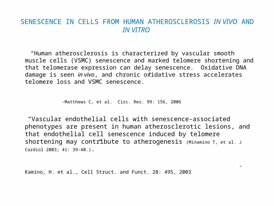
SENESCENCE IN CELLS FROM HUMAN ATHEROSCLEROSIS IN VIVO AND IN VITRO
“Human atherosclerosis is characterized by vascular smooth muscle cells (VSMC) senescence and marked telomere shortening and that telomerase expression can delay senescence. Oxidative DNA damage is seen in vivo, and chronic oxidative stress accelerates telomere loss and VSMC senescence.”
-Matthews C, et al. Circ. Res. 99: 156, 2006
“Vascular endothelial cells with senescence-associated phenotypes are present in human atherosclerotic lesions, and that endothelial cell senescence induced by telomere shortening may contribute to atherogenesis (Minamino T, et al. J Cardiol 2003; 41: 39-40.).”
-Kamino, H. et al., Cell Struct. and Funct. 28: 495, 2003

PROPERTIES OF DNA , AND DNA REPAIR IN HUMAN CELL SENESCENCE, IN VITRO AND IN VIVO

PROPERTIES OF DNA , AND DNA REPAIR IN HUMAN CELL SENESCENCE, IN VITRO AND IN VIVO
“Many genetic studies of DNA repair factors support the important role of DNA repair in the prevention of cellular senescence…” -Lou, Z. et al. Exp. Cell Res. (Review), 2006, 312, 2641
“Recent advances in cDNA array technology have made it possible to analyze global gene expressions, and a variety of genes involved in cell cycle regulation, immune and inflammation, cytoskeleton, stress response, and metabolism are known to be altered during cellular senescence.
- Shelton DN, et al, Curr Biol 1999; 9: 939; Ly DH, et al. Science 2000; 287: 2486; Zhang H, et al.; PNAS USA 2003; 100: 3251
“Zhang H, et al.;PNAS USA 2003; 100: 3251. identified transcriptional fingerprints unique to senescence and suggested that replicative senescence might be associated with alterations of chromatin structure, after investigating global changes in gene transcription occurring during replicative senescence in human fibroblasts using 31 k cDNA microarrays.”
“Although our results are generally in line with those of previous studies, we observed alterations in the expression of many novel genes in replicative-senescent fibroblasts.”
-Yoon I.K.,et al. Exp Gerontol 2004; 39: 1369-78.
“DNA damage and mutations have been shown to accumulate in human T lymphocytes with increasing age in vivo ( King, CM et al., Mutat. Res., 377,137-,1997) and an accumulation of oxidative DNA damage occurs during the replicative lifespan of T cell clones cultured in vitro( Hyland P, et al. Mech Ageing Dev 2000; 121:203; Hyland P, et al. Mech Ageing Dev 2001; 121(11) : 1151)”
- Annett K, et al. Exp Gerontol 2004; 39: 491
“..(lymphocytes from the) older population (60–70 years old) showed higher basal levels of DNA damage and was more sensitive to the effects of the DNA-damaging agents than.. (younger) adult .. (40–50 years old), who, in turn, (were) more sensitive than the younger population (5–10 years old). A decline of the repair efficiency with age to the DNA damage induced by the two agents was also observed.”
-S. M. Piperakis, et al., Cell Biol Toxicol (2009) 25:65

A BRIEF HISTORY (in 6 text slides) OF THE DISCOVERY OF MORTAL NORMAL CELLS, IMMORTAL CANCER CELLS AND WHY THE
TELOMERE REPLICOMETER AND TELOMERASE MAY EXPLAIN BOTH.
(Critical background for what follows.)

A BRIEF HISTORY… (1 of 6 slides)
1965- One of the most important observations that we made was that there are two classes of cultured cells, - mortal and immortal and that these two classes of cells have in vivo counterparts.
This was the first demonstration that cell populations could be classified into two distinct
categories mainly characterized by whether or not they possessed the property of mortality or immortality. It is from recognition of this relationship that there grew the present enormous cancer research area that studies how normal mortal cells can be immortalized to become immortal cancer cells.
Had we not shown that normal cells are mortal and cancer cells are immortal, the concept of immortalization would not have been appreciated.
I expressed this concept in the form of an equation (Hayflick,1965):

FIRST PUBLICATION OF THE DISTINCTIONS BETWEEN MORTAL AND IMMORTAL CELLS IN VITRO AND IN VIVO
Cell Lines : Transplantable Tumors = Cell Strains : Normal Somatic Tissue (In vitro) (In vivo) (In vitro) (In vivo)
1. Heteroploid 1. Diploid
2. Cancer cells 2. Normal cells
3. Indefinite replication 3. Finite replication
(From: Hayflick, L., Exp. Cell Res., 37, 614, 1965)
It is not possible to understand that cancer cell immortality is unique until it was discovered that normal cells are mortal.
A BRIEF HISTORY .. Slide 2 of 6
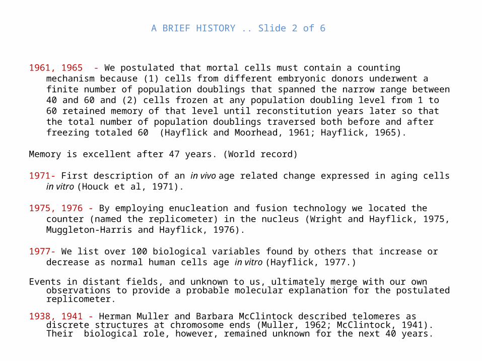
1961, 1965 - We postulated that mortal cells must contain a counting mechanism because (1) cells from different embryonic donors underwent a finite number of population doublings that spanned the narrow range between 40 and 60 and (2) cells frozen at any population doubling level from 1 to 60 retained memory of that level until reconstitution years later so that the total number of population doublings traversed both before and after freezing totaled 60 (Hayflick and Moorhead, 1961; Hayflick, 1965).
Memory is excellent after 47 years. (World record)
1971- First description of an in vivo age related change expressed in aging cells in vitro (Houck et al, 1971).
1975, 1976 - By employing enucleation and fusion technology we located the counter (named the replicometer) in the nucleus (Wright and Hayflick, 1975, Muggleton-Harris and Hayflick, 1976).
1977- We list over 100 biological variables found by others that increase or decrease as normal human cells age in vitro (Hayflick, 1977.)
Events in distant fields, and unknown to us, ultimately merge with our own observations to provide a
probable molecular explanation for the postulated replicometer. 1938, 1941 - Herman Muller and Barbara McClintock described telomeres as discrete structures at
chromosome ends (Muller, 1962; McClintock, 1941). Their biological role, however, remained unknown for the next 40 years.

A BRIEF HISTORY … slide 3 of 6
1960’s, late to early 1970's - It was found that DNA polymerase did not replicate the 3' end of linear duplex DNA. (“the end-replication problem”) (Olovnikov, 1971, 1973, 1996; Watson, 1972). An explanation was vital because the loss of genes at each round of DNA replication was not possible.
Alexey Olovnikov, who had just heard a lecture in which my work was discussed,
wondered how normal cells might have a limited capacity to replicate as he entered a Moscow subway station (Olovnikov, 1996).
When the train stopped at the station he had a remarkable flash of insight when he
visualized an analogy between the approaching train, which represented the DNA polymerase, and the track, which represented the DNA. If the train engine were to be imagined as the polymerase that replicated the DNA track, the first segment of DNA would not be replicated because it was underneath the engine at the start. This was analogous to the “end-replication problem."
Olovnikov realized that this repeated shortening of DNA at each round of replication
would shorten the molecule to some short critical length and might explain our finding that normal cells can only replicate a specific number of times (Olovnikov, 1996).

A BRIEF HISTORY … slide 4 of 6
Olovnikov further reasoned that the telomeres might consist of repeated nonsense nucleotide sequences that behaved like a buffer. At each round of DNA replication the buffer would simply loose what portion of the DNA molecule was not copied (the nonsense telomeric ends) and thus protect downstream genes. The length of the buffer would thus determine the number of rounds possible for DNA replication. Olovnikov’s imaginative solution to the “end replication problem”,(Olovnikov, 1971, 1973) languished in the literature until several discoveries made in the late 1970's and 1980’s emerged to support his armchair speculation and, subsequently proved him to be correct. 1978, - Elizabeth Blackburn, working with the ciliated protozoan, Tetrahymena, found that telomeres consisted of a simple sequence of hexameric repeats of the nucleotides TTGGGG (Blackburn and Gall, 1978). In humans the sequence is (TTAGGG)n. Like other eukaryotic organisms, the telomeres in human cells consist of thousands of repeats of this sequence. 1990 - Calvin Harley, who had worked for several years with my system of senescent human cells, and Carol Greider (a Blackburn student) decided to explore the possibility that the limited proliferative capacity of cultured normal cells might be explained by diminishing telomere length. They discovered that mean telomere length decreased by 2 to 3 kilobase pairs (kbp) during the entire in vitro lifetime of several strains of cultured normal human diploid fibroblasts (Harley et al 1990).

Telomere
Subtelomeric region
Subtelomeric region
Telomere
centromereTelomeric DNArepeats (human):(TTAGGG)n
Metaphasechromosome
Telomere Location

SCHEMATIC DIAGRAM OF TELOMERE ATTRITION

A BRIEF HISTORY … slide 5 of 6
Telomeric attrition is not a clock (chronometer) because it does not measure time, it counts DNA replications which is why I named it a replicometer.
2000, 2002 - It is now known that once a threshold number of telomeric
(TTAGGG)n repeats is reached, downstream events involving the P53, P21 and P16 genes presumably trigger signals for cells to stop dividing (Duncan, et al., 2000; Dhaene et al., 2000; Helder et al., 2002).
The essential remaining question is this: How does the class of cells that we
identified as immortal (cell lines and cancer cells) avoid telomere shortening because, if it occurs as it does in normal cells, it would lead to their mortality?
1985, 1990, 1992 - The answer to this critical question originated in studiesby Greider and Blackburn in 1985, who discovered the ribonucleoprotein
enzyme terminal transferase called telomerase. They found that telomere ends are synthesized de novo by telomerase by extending or maintaining the 3' end. Telomerase contains a reverse transcriptase and RNA template for the synthesis of the repeated sequence (Shippen-Lentz and Blackburn, 1990). It was reported simultaneously that cancer cells have shorter telomeres than do adjacent normal cells thus providing the first link for the role of telomeres in cancer biology (Hastie et al 1990; de Lange et al 1990).

Diagrammatic Representation of Telomerase (Courtesy of R. Effros)

A BRIEF HISTORY … slide 6 of 6
1989, 1992,1997 - Telomerase was later found to occur in most immortal human cell lines (Morin, 1989; Counter et al, 1992) and in about 90% of all human tumors studied (Chiu and Harley, 1997). Thus, the telomeres of immortal cell lines do not shorten with serial passage in vitro.
Telomerase is the only reverse transcriptase that is necessary for normal cell
activity. Today, the presence of telomerase in cancer cells (90%) is the single most sensitive and reliable property that distinguishes them from normal cells.
Conclusion - The observation that telomeres shorten as normal cells divide
provides the best evidence for the putative replicometer which we had postulated to have existed 30 years earlier. This, in combination with the discovery of the enzyme telomerase, has gone very far in explaining why normal somatic cells have a finite capacity to replicate in vivo and in vitro and how immortal cancer cells and cell lines circumvent this inevitability.
TELOMERE ATTRITION IN AGING HUMAN CELLS IN VIVO AND IN VITRO
TELOMERE ATTRITION IN AGING HUMAN CELLS IN VIVO AND IN VITRO
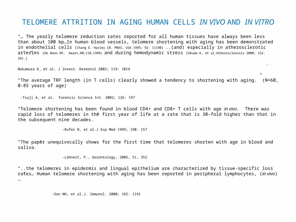
“… The yearly telomere reduction rates reported for all human tissues have always been less than about 100 bp…In human blood vessels, telomere shortening with aging has been demonstrated in endothelial cells (Chang E. Harley CB. PNAS, USA 1995; 92: 11190) ..(and) especially in atherosclerotic arteries (De
Bono DP, Heart,80;110,1998) and during hemodynamic stress (Okuda K, et al.Atherosclerosis 2000; 152: 391.)”
- Nakamura K, et al. J Invest. Dermatol 2002; 119: 1014
“The average TRF length (in T cells) clearly showed a tendency to shortening with aging.” (N=60, 0-85 years of age)
-Tsuji A, et al. Forensic Science Int. 2002; 126: 197
“Telomere shortening has been found in blood CD4+ and CD8+ T cells with age in vivo. There was rapid loss of telomeres in the first year of life at a rate that is 30-fold higher than that in the subsequent nine decades.” -Rufer N, et al.J Exp Med 1999; 190: 157
“The paper unequivocally shows for the first time that telomeres shorten with age in blood and saliva.” -Lahnert, P., Gerontology, 2005, 51, 352
“..the telomeres in epidermis and lingual epithelium are characterized by tissue-specific loss rates… Human telomere shortening with aging has been reported in peripheral lymphocytes, ( in vivo)…”
-Son NH, et al.J. Immunol. 2000; 165: 1191
TELOMERE ATTRITION IN AGING HUMAN CELLS IN VIVO AND IN VITRO (continued)
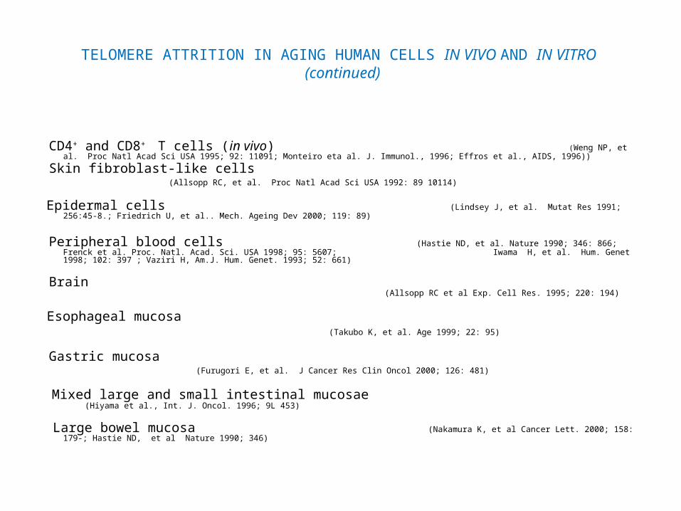
CD4+ and CD8+ T cells (in vivo) (Weng NP, et al. Proc Natl Acad Sci USA 1995; 92: 11091; Monteiro eta al. J. Immunol., 1996; Effros et al., AIDS, 1996))
Skin fibroblast-like cells (Allsopp RC, et al. Proc Natl Acad Sci USA 1992: 89 10114)
Epidermal cells (Lindsey J, et al. Mutat Res 1991; 256:45-8.; Friedrich U, et al.. Mech. Ageing Dev 2000; 119: 89)
Peripheral blood cells (Hastie ND, et al. Nature 1990; 346: 866; Frenck et al. Proc. Natl. Acad. Sci. USA 1998; 95: 5607; Iwama H, et al. Hum. Genet 1998; 102: 397 ; Vaziri H, Am.J. Hum. Genet. 1993; 52: 661)
Brain (Allsopp RC et al Exp. Cell Res. 1995; 220: 194)
Esophageal mucosa (Takubo K, et al. Age 1999; 22: 95)
Gastric mucosa (Furugori E, et al. J Cancer Res Clin Oncol 2000; 126: 481)
Mixed large and small intestinal mucosae (Hiyama et al., Int. J. Oncol. 1996; 9L 453)
Large bowel mucosa (Nakamura K, et al Cancer Lett. 2000; 158: 179-; Hastie ND, et al Nature 1990; 346)

TELOMERE ATTRITION IN AGING HUMAN CELLS IN VIVO AND IN VITRO (continued)
Vascular intima (Chang E. , PNAS, USA 1995; 92: 11190)
Kidney (Melk A, et al. J. Am. Soc. Nephrol. 2000; 11:444)
Liver (Aikata H. et al., Exp. Cell Res. 2000; 256: 578)
Thyroid and parathyroid glands (Kammori M, et al. ,Exp. Gerontol. 2002; 37: 513)
Abdominal aorta (Okuda K, et al.Atherosclerosis 2000; 152: 391)
Coronary arteries (in vivo) (Ogami, M., et al., Arterioscler. Thromb. Vasc. Biol., 24, 546, 2004)
Satellite cells (Decary S, , et al. Hum Gene Ther 1997; 8:1429)
Articular cartilage chondrocytes (in vivo) (Martin JA et al.,, J of Gerontol Bio Sci 2001; 56A: B172)
Trabicular osteoblasts (in vivo) (Kassem M, et al. Osteoporosis Int 1997; 7: 514)
TELOMERE ATTRITION IN AGING HUMAN CELLS IN VIVO AND IN VITRO
(continued)
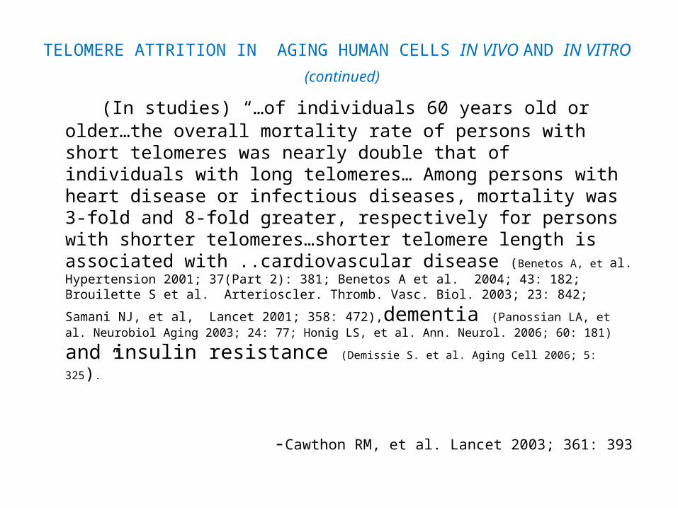
(In studies) “…of individuals 60 years old or older…the overall mortality rate of persons with short telomeres was nearly double that of individuals with long telomeres… Among persons with heart disease or infectious diseases, mortality was 3-fold and 8-fold greater, respectively for persons with shorter telomeres…shorter telomere length is associated with ..cardiovascular disease (Benetos A, et al. Hypertension 2001; 37(Part 2): 381; Benetos A et al. 2004; 43: 182; Brouilette S et al. Arterioscler.
Thromb. Vasc. Biol. 2003; 23: 842; Samani NJ, et al, Lancet 2001; 358: 472),dementia (Panossian LA,
et al. Neurobiol Aging 2003; 24: 77; Honig LS, et al. Ann. Neurol. 2006; 60: 181) and insulin resistance (Demissie S. et al. Aging Cell 2006; 5: 325).”
-Cawthon RM, et al. Lancet 2003; 361: 393

REPLICATIVE SENESCENCE, CANCER, AND CANCER PREVENTION

REPLICATIVE SENESCENCE, CANCER AND CANCER PREVENTION
“The recent reports on oncogene-induced senescence acting as a tumor suppressive principle in vivo now establish the critical mass of evidence to no longer interpret this phenomenon as a culture artifact.
The articles underscore the importance of cellular senescence in a wide spectrum of pre malignant scenarios, hereby highlighting the universal nature of this program as an anti-oncogenic fail-safe mechanism.
In summary, oncogene-induced senescence can no longer be considered a cell culture related artifact.”
- Braig M & Schmitt CA. Canc. Res. 2006; (66) 2881

REPLICATIVE SENESCENCE, CANCER AND CANCER PREVENTION (Continued)
“Premature or oncogene-induced senescence was described as a cellular response to a variety of stimuli, including activated oncogenes or excessive mitogenic signaling, DNA damage and stress signals, or in vivo in response to chemotherapy.
Collectively, results from a number of independent studies demonstrate that the growth arrest state can be bypassed by inactivation of key mediators of cellular senescence, allowing senescent cells to progress to malignancy. (Braig M, et al. Nature 2005; 436: 660; Chen Z. et al. Nature 2005; 436: 725; Michaloglou C, et al. Nature 2005; 436: 720; Collado M, et al. Nature 2005; 436: 642.)
The senescence pathway invariably invokes activation of either or both the p19ARF/p53 and p16INK4a/retinoblastoma (Rb) tumor suppressor pathways.
Taken together, it is now clear that p63 is a key mediator of epithelial cell proliferation, and that loss of this protein induces cellular senescence.”
- From Keyes WM et al. p63, A new link between senescence and aging. Cell Cycle 2006; 5, 3: 1 Feb, 260

REPLICATIVE SENESCENCE, CANCER AND CANCER PREVENTION (continued)
Cell senescence occurs in vivo after exposure to anticancer agents. -Chang BD, et al. Canc Res 1999; 59:3761
DNA damage induces cell senescence in tumor cells in vivo and in vitro. te Poele RH, et al. Canc Res 2002; 62:1876
Cell senescence is contolled by p53 and p16INK4a and contributes to the outcome of
cancer chemotherapy. Schmitt CA, et al. Cell 2002; 109: 335 “…oncogene-induced senescence (OIS) is increasingly recognized as a potentbarrier against oncogenic transformation, acting to suppress the unscheduled
proliferation of early neoplastic cells.” Thomas K and Peeper D.S. Nature Reviews | Cancer (IN PRESS)
“This is clearly the case of p53 and Rb, that have an essential role as master regulators
of senescence and, at the same time are the most frequently inactivated tumor suppressors in human cancer (Lowe, S.W.et al., (2004) Nature, 432, 307).”
Menendes et al., Current Drug Targets, 2009,10, 406

REPLICATIVE SENESCENCE, CANCER AND CANCER PREVENTION (continued)
“The recent discovery that cellular senescence is triggered by many different activated oncogenes has led to the notion that senescence, like oncogene-induced apoptosis, serves as a critical and cell-autonomous tumor preventive mechanism.” -Gerard I et al., Current Opinion in Genetics & Development 2009, 19:1
“..a senescent phenotype can be induced prematurely in early passage cells by agents that cause DNA damage (Di Leonardo,A., et al.,1994, Genes Dev., 8, 2540–2551; Chen,Q et al., 1995, Proc. Natl Acad. Sci. USA, 92, 4337–4341.; Chen,Q. and Ames,B.N. Proc. Natl Acad. Sci. USA, 91, 4130–4134. Robles,S.J. et al., 1998, Oncogene,
16, 1113–1123; Chen,J.H et al.,2004 J. Biol.Chem., 279, 49439–49446.) or disrupt heterochromatin Ogryzko,V.V., et
al.,. (1996) Mol. Cell. Biol., 16, 5210–5218), by disruption of functional telomere structures (or by overexpression of oncogenes (Serrano,M., et al (1997) Cell, 88, 593–602;Zhu,J.,et al., (1998); Takai,H.,et al. (2003, . Curr. Biol., 13, 1549–1556), Dev., 12, 2997–3007; Ferbeyre,G.,. (2002). Mol. Cell. Biol.,22, 3497–3508; Pearson,M., et al.,. (2000). Nature, 406, 207–210; Ferbeyre,G., et al., (2000). Genes Dev., 14, 2015–2027. )”
- Chen, J-H. et al., Nucleic Acids Research, 2007, Vol. 35, No. 22 7417
“These results suggest the SK (seborrhoeic keratosis) is a benign neoplasm where keratinocytes in senescent condition and G1 arrest are accumulated. … These results suggest that p16 expression is a marker of SK cells…” -Nakamura K, et al. J Invest Dermatol 2002; 119: 1014

IMMUNOSENESCENCE IN HUMAN CELLS IN VIVO AND IN VITRO
IMMUNOSENESCENCE IN HUMAN CELLS IN VIVO AND IN VITRO
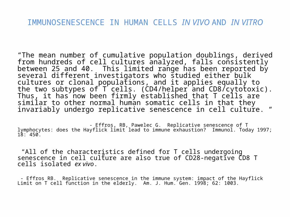
“The mean number of cumulative population doublings, derived from hundreds of cell cultures analyzed, falls consistently between 25 and 40. This limited range has been reported by several different investigators who studied either bulk cultures or clonal populations, and it applies equally to the two subtypes of T cells. (CD4/helper and CD8/cytotoxic). Thus, it has now been firmly established that T cells are similar to other normal human somatic cells in that they invariably undergo replicative senescence in cell culture. “ - Effros, RB, Pawelec G. Replicative senescence of T lymphocytes: does the Hayflick limit lead to immune exhaustion? Immunol. Today 1997; 18: 450.
“All of the characteristics defined for T cells undergoing senescence in cell culture are also true of CD28-negative CD8 T cells isolated ex vivo.” - Effros RB. Replicative senescence in the immune system: impact of the Hayflick Limit on T cell function in the elderly. Am. J. Hum. Gen. 1998; 62: 1003.

IMMUNOSENESCENCE IN HUMAN CELLS EX VIVO AND IN VITRO(Continued) (Courtesy of G. Pawalec, Tübingen, Germany)
T CELL EXPRESSION IN OLD HUMAN CELLS IN VITRO AND EX VIVO
DECREASED ACTIVITY CD28 expression Ex vivo CD8 Hoshino et al., 1993; Effros et al., 1994; Fagnoni et al., Boucher et al.,1998
CD4 Vallejo et al., 1998
In vitro CD8 Effros et al., 1994; Fiorentini et al., 1999
CD4 Adibzadeh et al., 1995; Pawelec et al., 1997
CD28 promoter binding activity Ex vivo (Binding activities of 2 factors reduced.) Vallejo et al., 1998
In vitro (Binding activity of one of these reduced.) Vallejo et al., 1999
CD154 Ex vivo Lio et al., 1998; Weyand et al., 1998; Fernandez-Gutierrez et al., 1999
In vitro Pawelec et al., 1997
CD134 Ex vivo (Unknown) In vitro Pawelec et al., 1997
IL 2R (high affinity) expression Ex vivo Froelich et al., 1988; Hara et al., 1988
In vitro Pawelec et al., 1997

IMMUNOSENESCENCE IN HUMAN CELLS EX VIVO AND IN VITRO(Continued) (Courtesy of G. Pawalec, Tübingen, Germany)
T CELL EXPRESSION IN OLD HUMAN CELLS IN VITRO AND EX VIVO
DECREASED ACTIVITY (continued)
IL 2 production Ex vivo Gillis et al., 1981; Jackola et al., 1994; Whisler et al., 1996; Liu et al., 1997; Guidi et al.,1998
(There are many more published examples; however, retention of IL 2 secretion is seen in some selected populations (e.g. The healthiest of the oldest old.) In vitro Pawelec et al., 1997
bcl-2 expression Ex vivo Aggarwal and Gupta, 1998; Gupta, 2000 In vitro (May apply only to CD4 cells) Salmon et al., 1994
(CD8 cells may have increased bcl-2) Spaulding et al., 1999Telomere lengths Ex vivo Slagboom et al., 1994; Weng et al., 1995: Frenck et al., 1998; Iwama et al.
1998; Zeichnet et al., 1999 In vitro Bodnar et al., 1996; Yamada et al., 1996; Rufer et al., 1998
Telomerase induction Ex vivo Hiyama et al., 1995
In vitro Pawelec et al., 2000
DNA repair Ex vivo Barnett and King, 1995; Frasca et al., 1999
In vitro (Not tested in T cells) Stress resistance and HSP expression Ex vivo Faasen et al., 1989 Jurivich et al., 1997
In vitro Effros et al., 1994; Jurivich et al., 1997

IMMUNOSENESCENCE IN HUMAN CELLS EX VIVO AND IN VITRO(Continued) (Courtesy of G. Pawalec, Tübingen, Germany)
T CELL EXPRESSION IN OLD HUMAN CELLS IN VITRO AND EX VIVO
INCREASED ACTIVITY
p7 (i.e. delayed down regulation )Ex vivo Arbogast et al., 1999; Tamir and Miller, 1999 In vitro Pawelec et al., 1999
p16 Ex vivo (Unknown for T cells) In vitro Pawelec, 2000 Mutation frequency Ex vivo Cole and Skopek, 1994; King et al., 1994
In vitro (Not tested in T cells) CD152 expression Ex vivo Wakikawa et alk., 1997
In vitro Engel & Pawelec unpublished IL10 production Ex vivo Cakman et al., 1996; Castle et al., 1997, 1999
In vitro Pawelec et al., 1997 DNA damage Ex vivo Turner et al., 1981; Hartwig and Korner, 1987
In vitro Hyland et al., 2000 Apoptosis AICD of CD4 cells Ex vivo Lechner et al., 1996; Aggarwal and Gupta, 1998; Aggarwal et al., 1999
In vitro Pawelec et al., 1996
CD95 and CD95L expression Ex vivo Aggarwal and Gupta, 1998 In vitro Soares et al., 2000
CD45RO+ cells Ex vivo Hannet et al., 1992; Utsuyama et al., 1992; Gabriel et al., 1993 In vitro Salmon et al., 1994

IMMUNOSENESCENCE IN HUMAN CELLS IN VIVO AND IN VITRO(CONTINUED)
NORMAL IMMUNE SYSTEM CELLS:
“Both in vivo and in vitro a significant decrease of the fraction of CD8 (+) CD28 (+) cells was observed. Comparing the proportions of other T cell populations (the CD4/cd8(+) ratio, CD56,CD57, CD27) made it possible to conclude that replicative senescence in vitro partially reflects in vivo ageing.”
-Brzezinska,A. Acta Biochemica Polonica, 52(4): 931, 2005
“As T-cells reach Phase 3 in culture they produce two proinflammatory cytokines (TNF α) and IL-6 and reduced levels of the critical antiviral cytokine interferon gamma (IF γ) (Effros RB et al., Immunol. Rev., 2005,205, 147; Dagarag MD, et al., J. Immunol., 2004,173, 6303.). Senescent T cells also no longer express a key signaling surface receptor, CD28 (Spaulding, CS., et al., Exp. Gerontol., 1999,34, 633.)
“..some aged individuals (have) more than 60% CD 28 negative cells within the CD8 T cell subset compared with the mean young adult value less than 10% (Effros RB et al., Exp. Gerontol., 1994, 29, 601. Telomere studies have confirmed that the CD28 negative cells have had undergone extensive cell division compared with other T cells from the same individual (EffroS RB, et al.,AIDS, 1996,10, F17; Monteiro J., et al., J. Immunol., 1996, 156, 3587.)
- Effros, RB, 2009, J. Gerontol., Biol. Sci., 64A, 511

IMMUNOSENESCENCE IN HUMAN CELLS IN VIVO AND IN VITRO(CONTINUED)
HUMAN DISEASES OR CONDITIONS ASSOCIATED WITH TELOMERE ATTRITION IN PERIPHERAL LEUKOCYTES
Cardiovascular mortality Cawthon RM, et al. Lancet 2003; 361: 393
Coronary atherosclerosis Samani NJ, et al. Lancet 2001; 358: 472
Premature myocardial infarction (<50 yrs.) Brouilette S, et al. Arterioscler. Thromb. Vasc. Bio.l 2003; 23: 842
Vascular dementia Von Zglinicki,T. et al. Lab. Invest. 80, 1739,2000
Hypertensives with carotid atherosclerosis Benetos A, et al. Hypertension 2004; 43: 182
Age-related calcific aortic stenosis Kurz DJ, et al., Circulation, 110,(Supple 3), 708, 2004
Increased pulse pressure Jeanclos,E., et al.,Hypertension, 36, 195, 2000
Smokers Nawrot,TS, Lancet, 363,507, 2004
Psychological stress Epel, ES, PNAS, USA, 101, 17312, 2004
Male sex Nawrot,TS, Lancet, 363,507, 2004
Wegener’s granulomatosis Vogt S, et al. Kidney Intl 2003; 63: 2144

CONCLUSIONS
“Thus it has now been firmly established that T cells are similar to other normal human somatic cells in that they invariably undergo replicative senescence in cell culture” - Effros RB. Exp Gerontol 2004; 39: 885
The recent discovery that loss of the p53-related protein p63 induces cellular senescence and causes features of accelerated aging provides further evidence that cellular senescence is intimately linked with organismal aging, and identifies p63 as a key regulator of both of these processes. - Keyes WM, et al. Cell Cycle 2006; 5, 260
“Research carried out over the last ten years has shown that cellular senescence occurs in vivo both in the vasculature and in other organ systems…and supports the concept that the number of vascular cell replications that have taken place in the vessel wall increases not only as a function of chronological age but also as a function of the hemodynamic or biochemical stress to the vascular bed.” -Erusalimsky, JD and Kurz, D., J Exp. Gerontol. 40, 634-642, 2005
“… The yearly telomere reduction rates reported for all human tissues have always been less than about 100 bp…In human blood vessels, telomere shortening with aging has been demonstrated in endothelial cells (Chang E. Harley CB. PNAS, USA 1995; 92: 11190) ..(and) especially in atherosclerotic arteries (De Bono DP, Heart,80;110,1998) and during hemodynamicstress (Okuda K, et al.Atherosclerosis 2000; 152: 391.)” - Nakamura K, et al. J Invest. Dermatol 2002; 119: 1014
“…senescent cells accumulate in vitro with increasing age.” -Lombard DB, et al. Cell 2005; 120: 497
“The average TRF length (in T cells) clearly showed a tendency to shortening with aging.” (N=60, 0-85 years of age) - Tsuji A, et al. Forensic Science Int. 2002; 126: 197
CONCLUSIONS (CONTINUED)
(In studies) “…of individuals 60 years old or older…the overall mortality rate of persons with short telomeres was nearly double that of individuals with long telomeres… Among persons with heart disease or infectious diseases, mortality was 3-fold and 8-fold greater, respectively for persons with shorter telomeres…shorter telomere length is associated with ..cardiovascular disease, dementia and insulin resistance .”
-Cawthon RM, et al. Lancet 2003; 361: 393
“The recent reports on oncogene-induced senescence acting as a tumor suppressive principle in vivo now establish the critical mass of evidence to no longer interpret this phenomenon as a culture artifact.
The articles underscore the importance of cellular senescence in a wide spectrum of pre malignant
scenarios, hereby highlighting the universal nature of this program as an anti-oncogenic fail-safe mechanism. In summary, oncogene-induced senescence can no longer be considered a cell culture related artifact.”
- Braig M & Schmitt CA. Canc. Res. 2006; (66) 2881
“Taken together, it is now clear that p63 is a key mediator of epithelial cell proliferation, and that loss of this protein induces cellular senescence.”
- Keyes WM et al. Cell Cycle 2006; 5, 260
“Once an obscure artifact of cell culture, cellular aging is emerging as a key force that shapes tissue health.”
– S. Lowe, Cold Spring Harbor Lab., Howard Hughes Medical Institute Bulletin, Feb. 2009, pp34
(Note: No data on animals was given in this presentation in which one could describe in animals the
hundreds of similar in vitro and in vivo properties to those presented here on aging human cells in vitro and in vivo.)

THANK YOU… for all of your reading!!!