Embed Size (px)
Citation preview
Current Biology 19, R823–R827, September 15, 2009 ª2009 Elsevier Ltd All rights reserved DOI 10.1016/j.cub.2009.08.012
MinireviewCell Shape and Cell Division in Fission Yeast
Matthieu Piel1 and Phong T. Tran1,2
The fission yeast Schizosaccharomyces pombe has servedas an important model organism for investigating cellularmorphogenesis. This unicellular rod-shaped fission yeastgrows by tip extension and divides by medial fission. Inparticular, microtubules appear to define sites of polarizedcell growth by delivering cell polarity factors to the celltips. Microtubules also position the cell nucleus at thecell middle, marking sites of cell division. Here, we reviewthe microtubule-dependent mechanisms that regulate cellshape and cell division in fission yeast.
IntroductionThe fission yeast Schizosaccharomyces pombe is a unicel-lular eukaryote that has a cylindrical rod shape of 4 mm diam-eter and grows by polarized tip extension from 7 to 14 mm inlength. Upon reaching 14 mm, cells stop growing and entermitosis. Cells then divide by assembling an actomyosincontractile ring at the geometrical center of the cell. Thesubsequent two daughter cells are of equal length — 7 mm.Interestingly, each daughter cell initiates growth immediatelyfrom its ‘old’ tip until the completion of S phase, at whichpoint it also initiates growth at the ‘new’ tip (i.e. the site ofthe previous cell division) in a process termed new endtake off (NETO) [1]. These seemingly simple acts of growthand division pose two important questions: how does thecell know where to divide, and how does the cell know whereto grow? The answers to these two questions appear toinvolve the dynamic microtubule cytoskeleton.
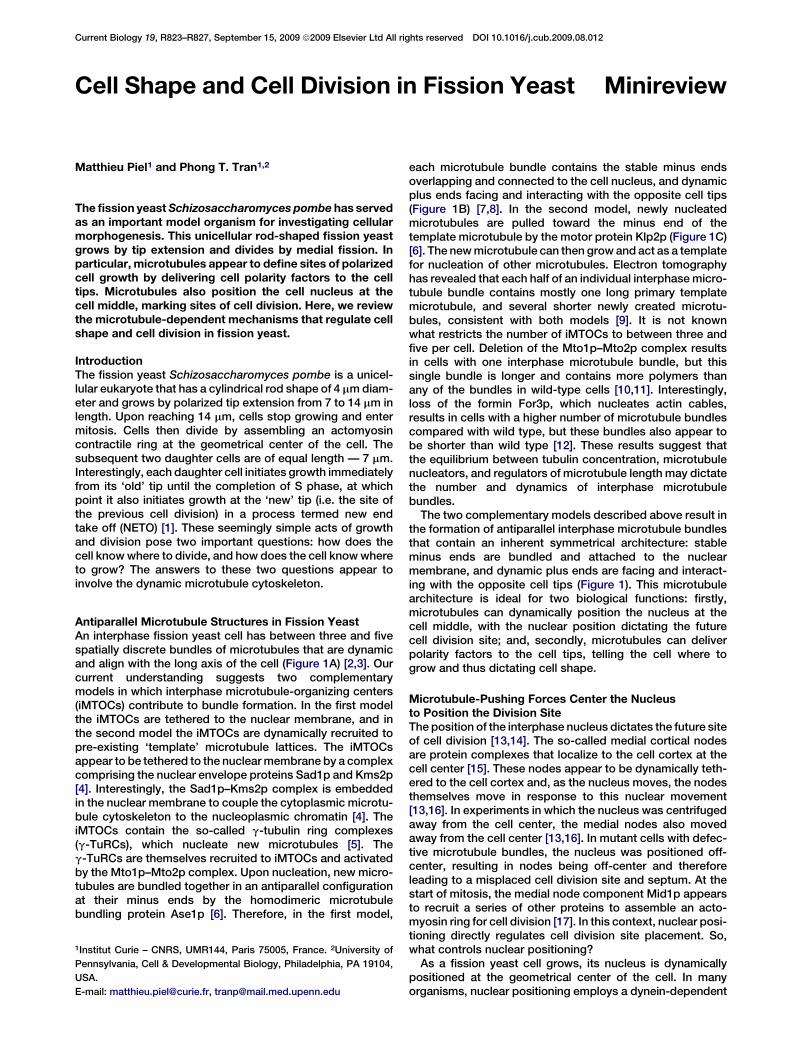
Antiparallel Microtubule Structures in Fission YeastAn interphase fission yeast cell has between three and fivespatially discrete bundles of microtubules that are dynamicand align with the long axis of the cell (Figure 1A) [2,3]. Ourcurrent understanding suggests two complementarymodels in which interphase microtubule-organizing centers(iMTOCs) contribute to bundle formation. In the first modelthe iMTOCs are tethered to the nuclear membrane, and inthe second model the iMTOCs are dynamically recruited topre-existing ‘template’ microtubule lattices. The iMTOCsappear to be tethered to the nuclear membrane by a complexcomprising the nuclear envelope proteins Sad1p and Kms2p[4]. Interestingly, the Sad1p–Kms2p complex is embeddedin the nuclear membrane to couple the cytoplasmic microtu-bule cytoskeleton to the nucleoplasmic chromatin [4]. TheiMTOCs contain the so-called g-tubulin ring complexes(g-TuRCs), which nucleate new microtubules [5]. Theg-TuRCs are themselves recruited to iMTOCs and activatedby the Mto1p–Mto2p complex. Upon nucleation, new micro-tubules are bundled together in an antiparallel configurationat their minus ends by the homodimeric microtubulebundling protein Ase1p [6]. Therefore, in the first model,
1Institut Curie – CNRS, UMR144, Paris 75005, France. 2University of
Pennsylvania, Cell & Developmental Biology, Philadelphia, PA 19104,
USA.
E-mail: [email protected], [email protected]
each microtubule bundle contains the stable minus endsoverlapping and connected to the cell nucleus, and dynamicplus ends facing and interacting with the opposite cell tips(Figure 1B) [7,8]. In the second model, newly nucleatedmicrotubules are pulled toward the minus end of thetemplate microtubule by the motor protein Klp2p (Figure 1C)[6]. The new microtubule can then grow and act as a templatefor nucleation of other microtubules. Electron tomographyhas revealed that each half of an individual interphase micro-tubule bundle contains mostly one long primary templatemicrotubule, and several shorter newly created microtu-bules, consistent with both models [9]. It is not knownwhat restricts the number of iMTOCs to between three andfive per cell. Deletion of the Mto1p–Mto2p complex resultsin cells with one interphase microtubule bundle, but thissingle bundle is longer and contains more polymers thanany of the bundles in wild-type cells [10,11]. Interestingly,loss of the formin For3p, which nucleates actin cables,results in cells with a higher number of microtubule bundlescompared with wild type, but these bundles also appear tobe shorter than wild type [12]. These results suggest thatthe equilibrium between tubulin concentration, microtubulenucleators, and regulators of microtubule length may dictatethe number and dynamics of interphase microtubulebundles.
The two complementary models described above result inthe formation of antiparallel interphase microtubule bundlesthat contain an inherent symmetrical architecture: stableminus ends are bundled and attached to the nuclearmembrane, and dynamic plus ends are facing and interact-ing with the opposite cell tips (Figure 1). This microtubulearchitecture is ideal for two biological functions: firstly,microtubules can dynamically position the nucleus at thecell middle, with the nuclear position dictating the futurecell division site; and, secondly, microtubules can deliverpolarity factors to the cell tips, telling the cell where togrow and thus dictating cell shape.
Microtubule-Pushing Forces Center the Nucleusto Position the Division SiteThe position of the interphase nucleus dictates the future siteof cell division [13,14]. The so-called medial cortical nodesare protein complexes that localize to the cell cortex at thecell center [15]. These nodes appear to be dynamically teth-ered to the cell cortex and, as the nucleus moves, the nodesthemselves move in response to this nuclear movement[13,16]. In experiments in which the nucleus was centrifugedaway from the cell center, the medial nodes also movedaway from the cell center [13,16]. In mutant cells with defec-tive microtubule bundles, the nucleus was positioned off-center, resulting in nodes being off-center and thereforeleading to a misplaced cell division site and septum. At thestart of mitosis, the medial node component Mid1p appearsto recruit a series of other proteins to assemble an acto-myosin ring for cell division [17]. In this context, nuclear posi-tioning directly regulates cell division site placement. So,what controls nuclear positioning?
As a fission yeast cell grows, its nucleus is dynamicallypositioned at the geometrical center of the cell. In manyorganisms, nuclear positioning employs a dynein-dependent
Current Biology Vol 19 No 17R824
+
++
+
–
––
–
++
++
––
A
B
CiMTOC
Nucleus
Ase1p
Microtubule
γTuRC
Ase1pKlp2p
Cls1p
Mto1p–Mto2p
Sad1p–Kms2p
+TIPs
Current Biology
Figure 1. Microtubule organization in fissionyeast.
(A) A typical fission yeast cell has betweenthree and five dynamic microtubule bundlesorganized along the long axis of the cell thatare organized by iMTOCs into antiparallelbundles with minus ends overlapping at themiddle of the cell and plus ends facing andinteracting with the cell tips. Two complemen-tary modes of microtubule organization arepresented in (B) and (C). (B) In the first model,iMTOCs are tethered to the nuclear mem-brane. The Mto1p–Mto2p complex, a compo-nent of the iMTOC, recruits g-TuRCs, whichnucleate microtubules. Microtubule polymersare then bundled into an antiparallel configu-ration by Ase1p. (C) In the second model,new microtubules nucleate on pre-existingmicrotubules. The Mto1p–Mto2p complexrecruits g-TuRCs to the lattice of a pre-exist-ing microtubule. Ase1p stabilizes the anti-parallel configuration between new and oldmicrotubules. The kinesin Klp2p slides thenew microtubule to the minus end of the oldmicrotubule (marked by the arrow), establish-ing an antiparallel bundle. Microtubule lengthis regulated by +TIP proteins and the rescuefactor Cls1p/Peg1p. A growing microtubulecan exhibit catastrophe and shrinkage (redarrow). It can then be rescued by Cls1p/Peg1pat the iMTOCand re-grow (greenarrow).
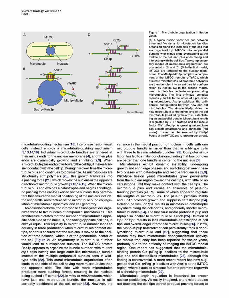
microtubule-pulling mechanism [18]. Interphase fission yeastcells instead employ a microtubule-pushing mechanism[3,13,14,19]. Individual microtubule bundles are tethered attheir minus ends to the nuclear membrane [4], and their plusends are dynamically growing and shrinking [2,3]. Whena microtubuleplus end growstoward the cell tip, it makes tran-sient contact with the cell tip. During this dwell time the micro-tubule plus end continues to polymerize. As microtubules arestructurally stiff polymers [20], this growth translates intoa pushing force [21], which moves the nucleus in the oppositedirection of microtubule growth [3,13,14,19]. When the micro-tubule plus end exhibits a catastrophe and begins shrinkage,no pushing force can be exerted on the nucleus. Key parame-ters that ensure the medial positioning of the nucleus include:the antiparallel architecture of the microtubule bundles; regu-lation of microtubule dynamics; and cell geometry.
As described above, the interphase fission yeast cell orga-nizes three to five bundles of antiparallel microtubules. Thisarchitecture dictates that the number of microtubules oppo-site each side of the nucleus, and facing opposite cell tips, isalways equal. This equality in microtubule number leads toequality in force production when microtubules contact celltips, and thus ensures that the nucleus is moved to the posi-tion of force balance, which is at the geometrical center ofthe cell (Figure 2). An asymmetry in microtubule numberwould lead to a misplaced nucleus. The iMTOC proteinRsp1p appears to organize the bundle number, with mutantrsp1 cells having one large aster-like microtubule bundleinstead of the multiple antiparallel bundles seen in wild-type cells [22]. This astral microtubule organization oftenleads to one side of the cell having more microtubules thanthe opposite side. The side with more microtubulesproduces more pushing forces, resulting in the nucleusbeing pushed off-center [22]. In mto1 or mto2 mutants, whichhave just one microtubule bundle, the nucleus is stillcorrectly positioned at the cell center [23]. However, the
variance in the medial position of nucleus in cells with onemicrotubule bundle is larger than that in wild-type cellswith three to five microtubule bundles [23]. Computer simu-lation has led to similar conclusions, finding that four bundlesare better than one bundle in centering the nucleus [3].
Microtubules exhibit dynamic instability, undergoinggrowth and shrinkage phases, and switching between thesetwo phases with catastrophe and rescue frequencies [2,3].Wild-type fission yeast microtubules grow persistentlyfrom the nuclear region toward the cell tips, with little or nocatastrophe until they make contact with the cell tips. Themicrotubule plus end carries an ensemble of plus-tip-tracking proteins (+TIPs), some of which appear to regulatethe lengths of microtubules. The plus-tip proteins Mal3pand Tip1p promote growth and suppress catastrophe [24].Deletion of mal3 or tip1 results in microtubule catastropheanywhere along the cell cortex, and generally shorter micro-tubule bundles [24]. The kinesin-8 motor proteins Klp5p andKlp6p also localize to microtubule plus ends [25]. Deletion ofklp5 or klp6 results in less microtubule catastrophe at celltips, and overall longer interphase microtubules [26]. In vitro,the Klp5p–Klp6p heterodimer can persistently track a depo-lymerizing microtubule end [27], suggesting that thesemotors may have microtubule depolymerization activity.No rescue frequency has been reported for fission yeast,probably due to the difficulty of imaging the iMTOC medialregion. One report has suggested that the microtubule-binding protein Cls1p/Peg1p localizes to the microtubuleplus end and destabilizes microtubules [28], although thisfinding is controversial. A more recent report has now sug-gested that Cls1p/Peg1p localizes with Ase1p at the iMTOCregion, where it acts as a rescue factor to promote regrowthof a shrinking microtubule [29].
Microtubule-length regulation is important for propernuclear positioning. As easily imagined, short microtubulesnot touching the cell tips cannot produce pushing forces to
Special IssueR825
position the nucleus, and long microtubules curving aroundthe cell tips would not produce efficient pushing forceseither. Indeed, mal3 and tip1 mutants have nuclear posi-tioning defects [30,31], and klp5, klp6, and cls1/peg1mutants are also expected to have nuclear positioningdefects. Interestingly, the physics of microtubule pushingsuggest that the pushing force (equivalent to the compres-sive force experienced by the microtubule as it makescontact with the cell tip) drops off quickly with the increasein microtubule length, approximated by the buckling equa-tion F = p2 � El=L2, where F is the compressive force, EI isthe flexural rigidity of the polymer, and L is the length ofthe polymer. Although there is currently no in vivo value forEI of microtubules, in vitro measurements yield an EI valueof w25 3 10224 Nm2 [20]. This value leads to the estimationthat a microtubule that is 1, 10, and 100 mm long can producepushing forces of 250, 2.5, and 0.025 pN, respectively. Forcomparison, dynein can generate 7 pN of force [32]. Giventhat a fission yeast cell is 14 mm long, each half-bundle is ex-pected to have a microtubule of 7 mm in length, producingw5 pN of force. Large forces have been shown to triggermicrotubule catastrophe in vitro [21], in vivo [25], and in silico[33]. Thus, microtubule length regulation will need to beexamined in the context of both physical forces triggeringcatastrophe and the proteins controlling the dynamic param-eters of microtubules.
Finally, cell geometry has a profound influence on mecha-nisms of nuclear positioning. As illustrated above, pushingforces from a single microtubule are productive only whenthe microtubule is in the order of 10 mm long. The interphasefission yeast has a cylindrical rod shape 14 mm long: thislength scale and shape appear to be within the productivepushing force regime. During mating, two haploid yeast cellsfuse to form a diploid. The fused diploid nucleus undergoesthe so-called horse-tail oscillation, where the nucleus ismoved back-and-forth in the cell in a dynein-dependent pull-ing mechanism [34]. These diploid cells have approximatelythe same length scale as the haploid cells, yet they use acompletely different — and opposite — mechanism ofnuclear positioning. The difference between the interphasecell and the diploid cell, which are of similar lengths, is theirshape. Diploid cells can be U-shaped, S-shaped, as well asrod-shaped: pushing would appear to be inefficient com-pared with pulling when either large length scales or complexcell shapes are involved.
It should also be noted that, while the mechanism of micro-tubule-dependent nuclear positioning described above isone pathway to position the division plane, other mecha-nisms exist. Recently, the cell tip protein kinase Pom1phas been shown to restrict the position of the kinaseCdr2p, which is a component of the medial nodes and aMid1p recruitment factor, to the cell center [35,36].
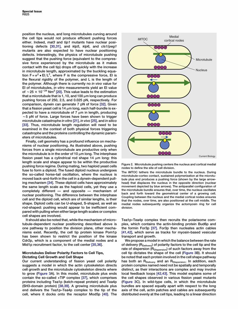
Microtubules Deliver Polarity Factors to Cell Tips,Dictating Cell Growth and Cell ShapeOur current understanding of fission yeast cell polaritysuggests a model in which the actin cytoskeleton directscell growth and the microtubule cytoskeleton directs whereto grow (Figure 3A). In this model, microtubule plus endscontain the so-called +TIP complex [37], which comprisesproteins including Tea1p (kelch-repeat protein) and Tea4p(SH3-domain protein) [38,39]. A growing microtubule plusend delivers the Tea1p–Tea4p complex to the tip of thecell, where it docks onto the receptor Mod5p [40]. The
Tea1p–Tea4p complex then recruits the polarisome com-plex, which contains the actin-binding protein Bud6p andthe formin For3p [37]. For3p then nucleates actin cables[41,42], which serve as tracks for myosin-based vesiculartransport and growth.
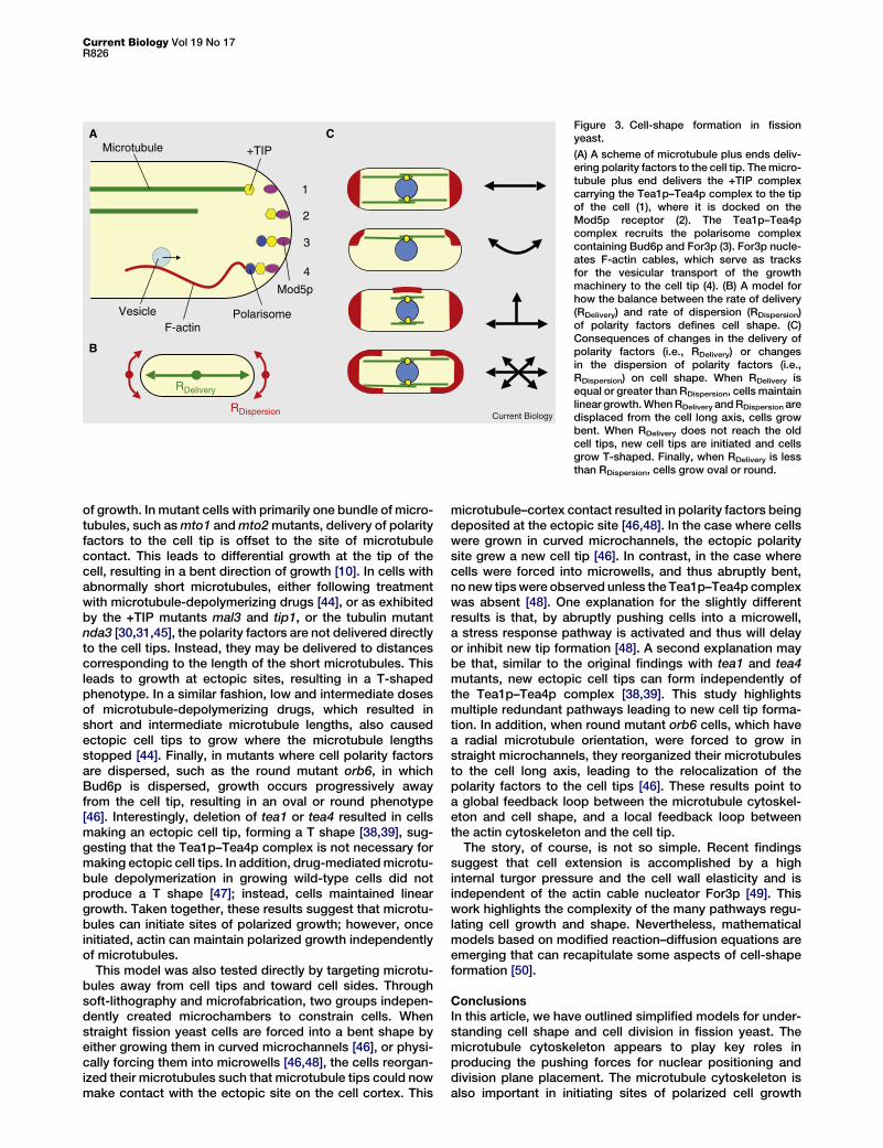
We propose a model in which the balance between the rateof delivery (RDelivery) of polarity factors to the cell tip and therate of dispersion (RDispersion) of such factors away from thecell tip dictates the shape of the cell (Figure 3B). It shouldbe noted that each protein involved in the cell shape pathwayhas both an RDelivery and an RDispersion. In addition, eachprotein complex named need not be spatially and temporallydistinct, as their interactions are complex and may involvelocal feedback loops [42,43]. This model explains some ofthe cell shapes observed in various fission yeast mutants(Figure 3C). In wild-type cells, in which the microtubulebundles are spaced equally apart with respect to the longaxis of the cell, actin patches and cables are subsequentlydistributed evenly at the cell tips, leading to a linear direction
iMTOCMedial
cortical nodes
Microtubule
Nucleus
Current Biology
Figure 2. Microtubule pushing centers the nucleus and cortical medialnodes to define the site of cell division.
The iMTOC tethers the microtubule bundle to the nucleus. Duringmicrotubule–cortex contact, sustained polymerization at the microtu-bule plus end produces a pushing force (shown by the large arrow-head) that displaces the nucleus in the opposite direction (nuclearmovement depicted by blue arrows). The antiparallel configuration ofthe microtubule bundle ensures that, over time, the nucleus oscillatesback and forth toward the geometrical center of a growing cell.Coupling between the nucleus and the medial cortical nodes ensuresthat the nodes, over time, are also positioned at the cell middle. Themedial nodes subsequently organize the actomyosin ring for celldivision.
Current Biology Vol 19 No 17R826
F-actin
Microtubule +TIP
Mod5p
Polarisome
1
2
3
4
Vesicle
A
RDelivery
RDispersion
B
C
Current Biology
Figure 3. Cell-shape formation in fissionyeast.
(A) A scheme of microtubule plus ends deliv-ering polarity factors to the cell tip. The micro-tubule plus end delivers the +TIP complexcarrying the Tea1p–Tea4p complex to the tipof the cell (1), where it is docked on theMod5p receptor (2). The Tea1p–Tea4pcomplex recruits the polarisome complexcontaining Bud6p and For3p (3). For3p nucle-ates F-actin cables, which serve as tracksfor the vesicular transport of the growthmachinery to the cell tip (4). (B) A model forhow the balance between the rate of delivery(RDelivery) and rate of dispersion (RDispersion)of polarity factors defines cell shape. (C)Consequences of changes in the delivery ofpolarity factors (i.e., RDelivery) or changesin the dispersion of polarity factors (i.e.,RDispersion) on cell shape. When RDelivery isequal or greater than RDispersion, cells maintainlinear growth. When RDelivery and RDispersion aredisplaced from the cell long axis, cells growbent. When RDelivery does not reach the oldcell tips, new cell tips are initiated and cellsgrow T-shaped. Finally, when RDelivery is lessthan RDispersion, cells grow oval or round.
of growth. In mutant cells with primarily one bundle of micro-tubules, such as mto1 and mto2 mutants, delivery of polarityfactors to the cell tip is offset to the site of microtubulecontact. This leads to differential growth at the tip of thecell, resulting in a bent direction of growth [10]. In cells withabnormally short microtubules, either following treatmentwith microtubule-depolymerizing drugs [44], or as exhibitedby the +TIP mutants mal3 and tip1, or the tubulin mutantnda3 [30,31,45], the polarity factors are not delivered directlyto the cell tips. Instead, they may be delivered to distancescorresponding to the length of the short microtubules. Thisleads to growth at ectopic sites, resulting in a T-shapedphenotype. In a similar fashion, low and intermediate dosesof microtubule-depolymerizing drugs, which resulted inshort and intermediate microtubule lengths, also causedectopic cell tips to grow where the microtubule lengthsstopped [44]. Finally, in mutants where cell polarity factorsare dispersed, such as the round mutant orb6, in whichBud6p is dispersed, growth occurs progressively awayfrom the cell tip, resulting in an oval or round phenotype[46]. Interestingly, deletion of tea1 or tea4 resulted in cellsmaking an ectopic cell tip, forming a T shape [38,39], sug-gesting that the Tea1p–Tea4p complex is not necessary formaking ectopic cell tips. In addition, drug-mediated microtu-bule depolymerization in growing wild-type cells did notproduce a T shape [47]; instead, cells maintained lineargrowth. Taken together, these results suggest that microtu-bules can initiate sites of polarized growth; however, onceinitiated, actin can maintain polarized growth independentlyof microtubules.
This model was also tested directly by targeting microtu-bules away from cell tips and toward cell sides. Throughsoft-lithography and microfabrication, two groups indepen-dently created microchambers to constrain cells. Whenstraight fission yeast cells are forced into a bent shape byeither growing them in curved microchannels [46], or physi-cally forcing them into microwells [46,48], the cells reorgan-ized their microtubules such that microtubule tips could nowmake contact with the ectopic site on the cell cortex. This
microtubule–cortex contact resulted in polarity factors beingdeposited at the ectopic site [46,48]. In the case where cellswere grown in curved microchannels, the ectopic polaritysite grew a new cell tip [46]. In contrast, in the case wherecells were forced into microwells, and thus abruptly bent,no new tips were observed unless the Tea1p–Tea4p complexwas absent [48]. One explanation for the slightly differentresults is that, by abruptly pushing cells into a microwell,a stress response pathway is activated and thus will delayor inhibit new tip formation [48]. A second explanation maybe that, similar to the original findings with tea1 and tea4mutants, new ectopic cell tips can form independently ofthe Tea1p–Tea4p complex [38,39]. This study highlightsmultiple redundant pathways leading to new cell tip forma-tion. In addition, when round mutant orb6 cells, which havea radial microtubule orientation, were forced to grow instraight microchannels, they reorganized their microtubulesto the cell long axis, leading to the relocalization of thepolarity factors to the cell tips [46]. These results point toa global feedback loop between the microtubule cytoskel-eton and cell shape, and a local feedback loop betweenthe actin cytoskeleton and the cell tip.
The story, of course, is not so simple. Recent findingssuggest that cell extension is accomplished by a highinternal turgor pressure and the cell wall elasticity and isindependent of the actin cable nucleator For3p [49]. Thiswork highlights the complexity of the many pathways regu-lating cell growth and shape. Nevertheless, mathematicalmodels based on modified reaction–diffusion equations areemerging that can recapitulate some aspects of cell-shapeformation [50].
ConclusionsIn this article, we have outlined simplified models for under-standing cell shape and cell division in fission yeast. Themicrotubule cytoskeleton appears to play key roles inproducing the pushing forces for nuclear positioning anddivision plane placement. The microtubule cytoskeleton isalso important in initiating sites of polarized cell growth
Special IssueR827
and thus determining cell shape. The actin cytoskeleton,however, directs cell growth independently of microtubules.Future work to decipher the many pathways regulating cellshape and cell division promises to be exciting and complex.
Acknowledgments
We thank the anonymous reviewers for good constructive
suggestions. We apologize that we cannot cover all aspects of fission
yeast cell shape and cell division and were obliged to leave uncited
some interesting works. The Piel Lab is supported by the ANR,
Fondation Pierre Gilles de Gennes and HFSP. The Tran Lab is
supported by NIH, ACS, ANR, FRM, LaLigue and HFSP.
References1. Mitchison, J.M., and Nurse, P. (1985). Growth in cell length in the fission
yeast Schizosaccharomyces pombe. J. Cell Sci. 75, 357–376.
2. Drummond, D.R., and Cross, R.A. (2000). Dynamics of interphase microtu-bules in Schizosaccharomyces pombe. Curr. Biol. 10, 766–775.
3. Tran, P.T., Marsh, L., Doye, V., Inoue, S., and Chang, F. (2001). A mechanismfor nuclear positioning in fission yeast based on microtubule pushing. J. CellBiol. 153, 397–411.
4. King, M.C., Drivas, T.G., and Blobel, G. (2008). A network of nuclear envelopemembrane proteins linking centromeres to microtubules. Cell 134, 427–438.
5. Sawin, K.E., and Tran, P.T. (2006). Cytoplasmic microtubule organization infission yeast. Yeast 23, 1001–1014.
6. Janson, M.E., Loughlin, R., Loiodice, I., Fu, C., Brunner, D., Nedelec, F.J., andTran, P.T. (2007). Crosslinkers and motors organize dynamic microtubules toform stable bipolar arrays in fission yeast. Cell 128, 357–368.
7. Loiodice, I., Staub, J., Setty, T.G., Nguyen, N.P., Paoletti, A., and Tran, P.T.(2005). Ase1p organizes antiparallel microtubule arrays during interphaseand mitosis in fission yeast. Mol. Biol. Cell 16, 1756–1768.
8. Yamashita, A., Sato, M., Fujita, A., Yamamoto, M., and Toda, T. (2005). Theroles of fission yeast ase1 in mitotic cell division, meiotic nuclear oscillation,and cytokinesis checkpoint signaling. Mol. Biol. Cell 16, 1378–1395.
9. Hoog, J.L., Schwartz, C., Noon, A.T., O’Toole, E.T., Mastronarde, D.N.,McIntosh, J.R., and Antony, C. (2007). Organization of interphase microtu-bules in fission yeast analyzed by electron tomography. Dev. Cell 12,349–361.
10. Janson, M.E., Setty, T.G., Paoletti, A., and Tran, P.T. (2005). Efficient forma-tion of bipolar microtubule bundles requires microtubule-bound gamma-tubulin complexes. J. Cell Biol. 169, 297–308.
11. Samejima, I., Lourenco, P.C., Snaith, H.A., and Sawin, K.E. (2005). Fissionyeast mto2p regulates microtubule nucleation by the centrosomin-relatedprotein mto1p. Mol. Biol. Cell 16, 3040–3051.
12. Feierbach, B., and Chang, F. (2001). Roles of the fission yeast formin for3pin cell polarity, actin cable formation and symmetric cell division. Curr.Biol. 11, 1656–1665.
13. Daga, R.R., and Chang, F. (2005). Dynamic positioning of the fission yeastcell division plane. Proc. Natl. Acad. Sci. USA 102, 8228–8232.
14. Tolic-Norrelykke, I.M., Sacconi, L., Stringari, C., Raabe, I., and Pavone, F.S.(2005). Nuclear and division-plane positioning revealed by optical microma-nipulation. Curr. Biol. 15, 1212–1216.
15. Wu, J.-Q., Sirotkin, V., Kovar, D.R., Lord, M., Beltzner, C.C., Kuhn, J.R., andPollard, T.D. (2006). Assembly of the cytokinetic contractile ring from a broadband of nodes in fission yeast. J. Cell Biol. 174, 391–402.
16. Almonacid, M., Moseley, J.B., Janvore, J., Mayeux, A., Fraisier, V., Nurse, P.,and Paoletti, A. (2009). Spatial control of cytokinesis by Cdr2 kinase andMid1/anillin nuclear export. Curr. Biol. 19, 961–966.
17. Wu, J.Q., Kuhn, J.R., Kovar, D.R., and Pollard, T.D. (2003). Spatial andtemporal pathway for assembly and constriction of the contractile ring infission yeast cytokinesis. Dev. Cell 5, 723–734.
18. Morris, N.R. (2003). Nuclear positioning: the means is at the ends. Curr. Opin.Cell Biol. 15, 54–59.
19. Daga, R.R., Yonetani, A., and Chang, F. (2006). Asymmetric microtubulepushing forces in nuclear centering. Curr. Biol. 16, 1544–1550.
20. Gittes, F., Mickey, B., Nettleton, J., and Howard, J. (1993). Flexural rigidity ofmicrotubules and actin filaments measured from thermal fluctuations inshape. J. Cell Biol. 120, 923–934.
21. Dogterom, M., Kerssemakers, J.W., Romet-Lemonne, G., and Janson, M.E.(2005). Force generation by dynamic microtubules. Curr. Opin. Cell Biol. 17,67–74.
22. Zimmerman, S., Tran, P.T., Daga, R.R., Niwa, O., and Chang, F. (2004).Rsp1p, a J domain protein required for disassembly and assembly of micro-tubule organizing centers during the fission yeast cell cycle. Dev. Cell 6,497–509.
23. Sawin, K.E., Lourenco, P.C., and Snaith, H.A. (2004). Microtubule nucleationat non-spindle pole body microtubule-organizing centers requires fissionyeast centrosomin-related protein mod20p. Curr. Biol. 14, 763–775.
24. Busch, K.E., and Brunner, D. (2004). The microtubule plus end-trackingproteins mal3p and tip1p cooperate for cell-end targeting of interphasemicrotubules. Curr. Biol. 14, 548–559.
25. Tischer, C., Brunner, D., and Dogterom, M. (2009). Force- and kinesin-8-dependent effects in the spatial regulation of fission yeast microtubuledynamics. Mol. Syst. Biol. 5, 250.
26. West, R.R., Malmstrom, T., Troxell, C.L., and McIntosh, J.R. (2001). Tworelated kinesins, klp5+ and klp6+, foster microtubule disassembly and arerequired for meiosis in fission yeast. Mol. Biol. Cell 12, 3919–3932.
27. Grissom, P.M., Fiedler, T., Grishchuk, E.L., Nicastro, D., West, R.R., andMcIntosh, J.R. (2009). Kinesin-8 from fission yeast: a heterodimeric, plus-end-directed motor that can couple microtubule depolymerization to cargomovement. Mol. Biol. Cell 20, 963–972.
28. Grallert, A., Beuter, C., Craven, R.A., Bagley, S., Wilks, D., Fleig, U., andHagan, I.M. (2006). S. pombe CLASP needs dynein, not EB1 or CLIP170, toinduce microtubule instability and slows polymerization rates at cell tips ina dynein-dependent manner. Genes. Dev. 20, 2421–2436.
29. Bratman, S.V., and Chang, F. (2007). Stabilization of overlapping microtu-bules by fission yeast CLASP. Dev. Cell 13, 812–827.
30. Beinhauer, J.D., Hagan, I.M., Hegemann, J.H., and Fleig, U. (1997). Mal3, thefission yeast homologue of the human APC-interacting protein EB-1 isrequired for microtubule integrity and the maintenance of cell form. J. CellBiol. 139, 717–728.
31. Brunner, D., and Nurse, P. (2000). CLIP170-like tip1p spatially organizesmicrotubular dynamics in fission yeast. Cell 102, 695–704.
32. Gennerich, A., Carter, A.P., Reck-Peterson, S.L., and Vale, R.D. (2007).Force-induced bidirectional stepping of cytoplasmic dynein. Cell 131,952–965.
33. Foethke, D., Makushok, T., Brunner, D., and Nedelec, F. (2009). Force- andlength-dependent catastrophe activities explain interphase microtubuleorganization in fission yeast. Mol. Syst. Biol. 5, 241.
34. Vogel, S.K., Pavin, N., Maghelli, N., Julicher, F., and Tolic-Norrelykke, I.M.(2009). Self-organization of dynein motors generates meiotic nuclear oscilla-tions. PLoS Biol. 7, e1000087.
35. Martin, S.G., and Berthelot-Grosjean, M. (2009). Polar gradients of theDYRK-family kinase Pom1 couple cell length with the cell cycle. Nature459, 852–856.
36. Moseley, J.B., Mayeux, A., Paoletti, A., and Nurse, P. (2009). A spatialgradient coordinates cell size and mitotic entry in fission yeast. Nature459, 857–860.
37. Martin, S.G., and Chang, F. (2005). New end take off: regulating cell polarityduring the fission yeast cell cycle. Cell Cycle 4, 1046–1049.
38. Martin, S.G., McDonald, W.H., Yates, J.R., 3rd, and Chang, F. (2005). Tea4plinks microtubule plus ends with the formin for3p in the establishment of cellpolarity. Dev. Cell 8, 479–491.
39. Tatebe, H., Shimada, K., Uzawa, S., Morigasaki, S., and Shiozaki, K. (2005).Wsh3/Tea4 is a novel cell-end factor essential for bipolar distribution ofTea1 and protects cell polarity under environmental stress in S. pombe.Curr. Biol. 15, 1006–1015.
40. Snaith, H.A., and Sawin, K.E. (2003). Fission yeast mod5p regulates polarizedgrowth through anchoring of tea1p at cell tips. Nature 423, 647–651.
41. Martin, S.G., and Chang, F. (2006). Dynamics of the formin for3p in actincable assembly. Curr. Biol. 16, 1161–1170.
42. Martin, S.G., Rincon, S.A., Basu, R., Perez, P., and Chang, F. (2007). Regula-tion of the formin for3p by cdc42p and bud6p. Mol. Biol. Cell 18, 4155–4167.
43. Snaith, H.A., Samejima, I., and Sawin, K.E. (2005). Multistep and multimodecortical anchoring of tea1p at cell tips in fission yeast. EMBO J. 24, 3690–3699.
44. Castagnetti, S., Novak, B., and Nurse, P. (2007). Microtubules offset growthsite from the cell centre in fission yeast. J. Cell Sci. 120, 2205–2213.
45. Toda, T., Umesono, K., Hirata, A., and Yanagida, M. (1983). Cold-sensitivenuclear division arrest mutants of the fission yeast Schizosaccharomycespombe. J. Mol. Biol. 168, 251–270.
46. Terenna, C.R., Makushok, T., Velve-Casquillas, G., Baigl, D., Chen, Y.,Bornens, M., Paoletti, A., Piel, M., and Tran, P.T. (2008). Physical mecha-nisms redirecting cell polarity and cell shape in fission yeast. Curr. Biol. 18,1748–1753.
47. Sawin, K.E., and Snaith, H.A. (2004). Role of microtubules and tea1p inestablishment and maintenance of fission yeast cell polarity. J. Cell Sci.117, 689–700.
48. Minc, N., Bratman, S.V., Basu, R., and Chang, F. (2009). Establishing newsites of polarization by microtubules. Curr. Biol. 19, 83–94.
49. Minc, N., Boudaoud, A., and Chang, F. (2009). Mechanical forces of fissionyeast growth. Curr. Biol. 19, 1096–1101.
50. Csikasz-Nagy, A., Gyorffy, B., Alt, W., Tyson, J.J., and Novak, B. (2008).Spatial controls for growth zone formation during the fission yeast cell cycle.Yeast 25, 59–69.