Embed Size (px)
Citation preview
CELL STRUCTURE AND FUNCTION
Lab 3
Objectives
• Be able to make a wet mount of any cellular material provided and focus on a cell.
• Be able to stain cells with either Lugol’s solution or methylene blue.
• Be able to determine the length and width of cells in micrometers and/or millimeters.
• Be able to diagram any cell observed through the microscope.
• Be able to locate the following structures in an onion cell:• cell wall• vacuole• nucleus• cytoplasm• cell membrane.
THE CELL
• The cell concept is basic to understanding the activities and characteristics of organisms.
• Cells are the units of structure and Cells are the units of structure and function of an organism.function of an organism.
• As units, they reflect the abilities of the organism as a whole. Cells are of interest because of their variety and also because of their similarities.
• In this exercise, we will examine the cell. We will try to determine the three-dimensional shape of theses cells and identify some of their structures.
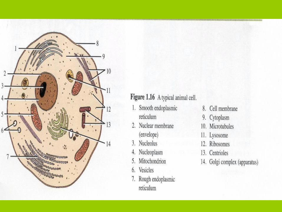
• The cell is compose by three principal compartiment:
1.- Plasmatic membrane
2.- Cytoplasm (organelles)
3.- Nucleous
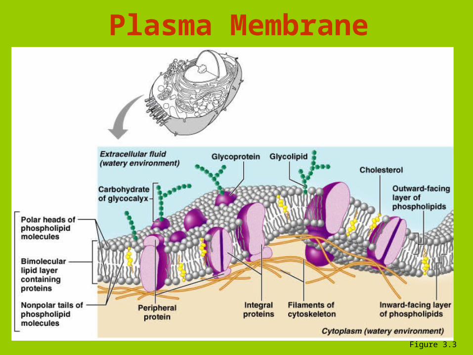
Plasma Membrane
Figure 3.3
Cytoplasma
• It is the specialized living material of cells
• It lies between the plasma membrane and the nucleus
• Numerous small structure (organelles) are part of the cytoplasma, along with the fluid that serves as the interior
environment of each cell
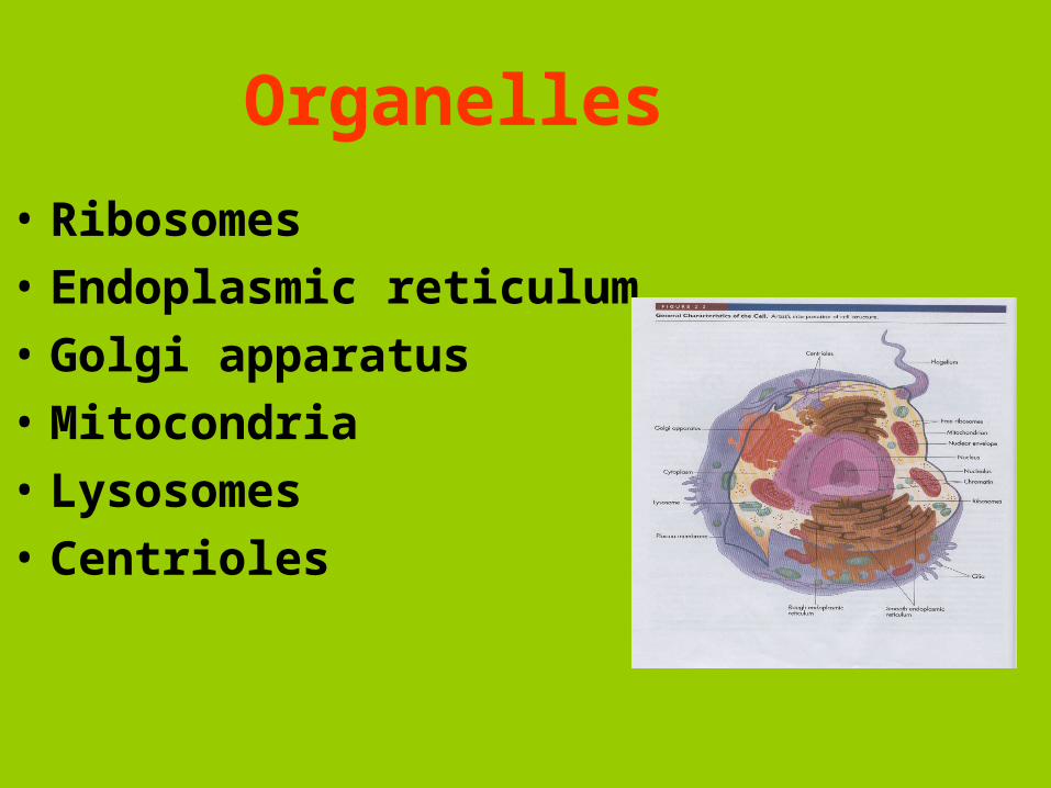
Organelles
• Ribosomes
• Endoplasmic reticulum
• Golgi apparatus
• Mitocondria
• Lysosomes
• Centrioles
Nucleus
• Contains nuclear envelope, nucleoli, chromatin, and distinct compartments rich in specific protein sets
• Gene-containing control center of the cell
• Contains the genetic library with blueprints for nearly all cellular proteins
• Dictates the kinds and amounts of proteins to be synthesized
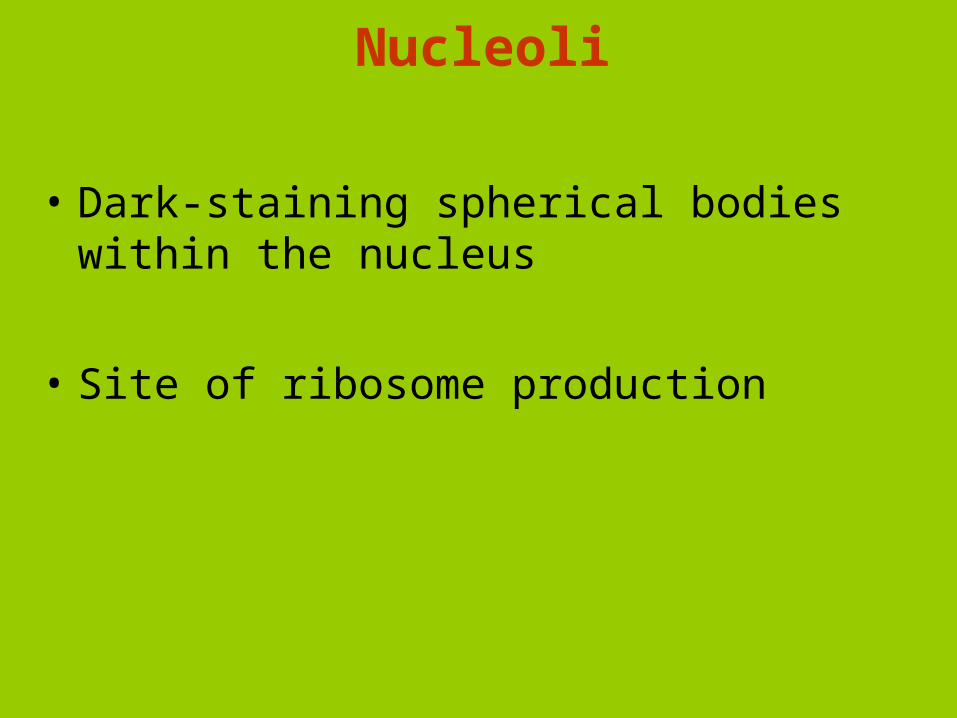
Nucleoli
• Dark-staining spherical bodies within the nucleus
• Site of ribosome production
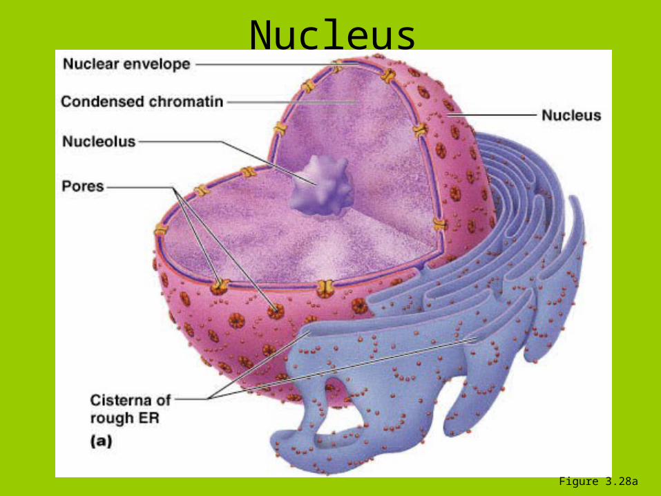
Nucleus
Figure 3.28a
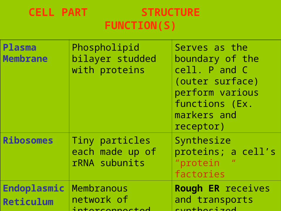
CELL PART STRUCTURE FUNCTION(S)
Plasma Membrane
Phospholipid bilayer studded with proteins
Serves as the boundary of the cell. P and C (outer surface) perform various functions (Ex. markers and receptor)
Ribosomes Tiny particles each made up of rRNA subunits
Synthesize proteins; a cell’s “protein factories”
Endoplasmic
Reticulum
(ER)
Membranous network of interconnected canals and sacs, some with ribosome (rough ER) and some without (smooth ER)
Rough ER receives and transports synthesized proteins
Smooth ER synthesizes lipids and carbohydrates
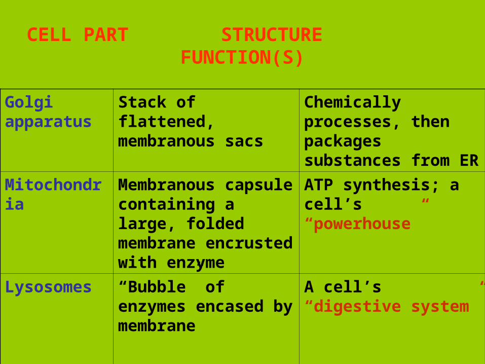
CELL PART STRUCTURE FUNCTION(S)
Golgi apparatus
Stack of flattened, membranous sacs
Chemically processes, then packages substances from ER
Mitochondria Membranous capsule containing a large, folded membrane encrusted with enzyme
ATP synthesis; a cell’s “powerhouse”
Lysosomes “Bubble” of enzymes encased by membrane
A cell’s “digestive system”
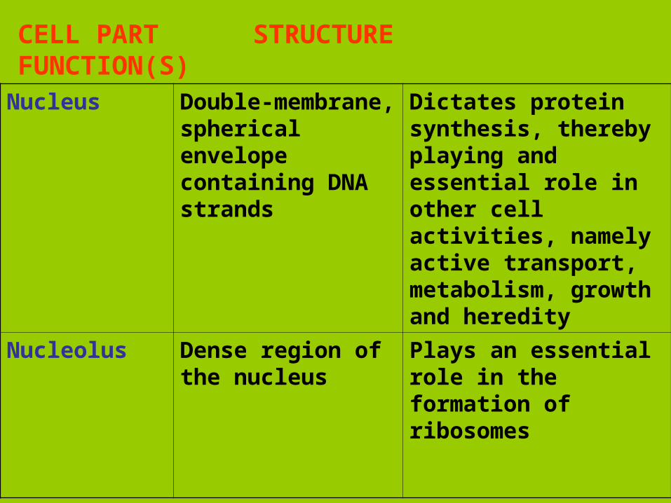
CELL PART STRUCTURE FUNCTION(S)
Nucleus Double-membrane, spherical envelope containing DNA strands
Dictates protein synthesis, thereby playing and essential role in other cell activities, namely active transport, metabolism, growth and heredity
Nucleolus Dense region of the nucleus
Plays an essential role in the formation of ribosomes
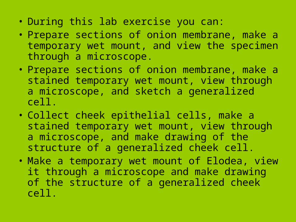
• During this lab exercise you can:• Prepare sections of onion membrane, make a
temporary wet mount, and view the specimen through a microscope.
• Prepare sections of onion membrane, make a stained temporary wet mount, view through a microscope, and sketch a generalized cell.
• Collect cheek epithelial cells, make a stained temporary wet mount, view through a microscope, and make drawing of the structure of a generalized cheek cell.
• Make a temporary wet mount of Elodea, view it through a microscope and make drawing of the structure of a generalized cheek cell.
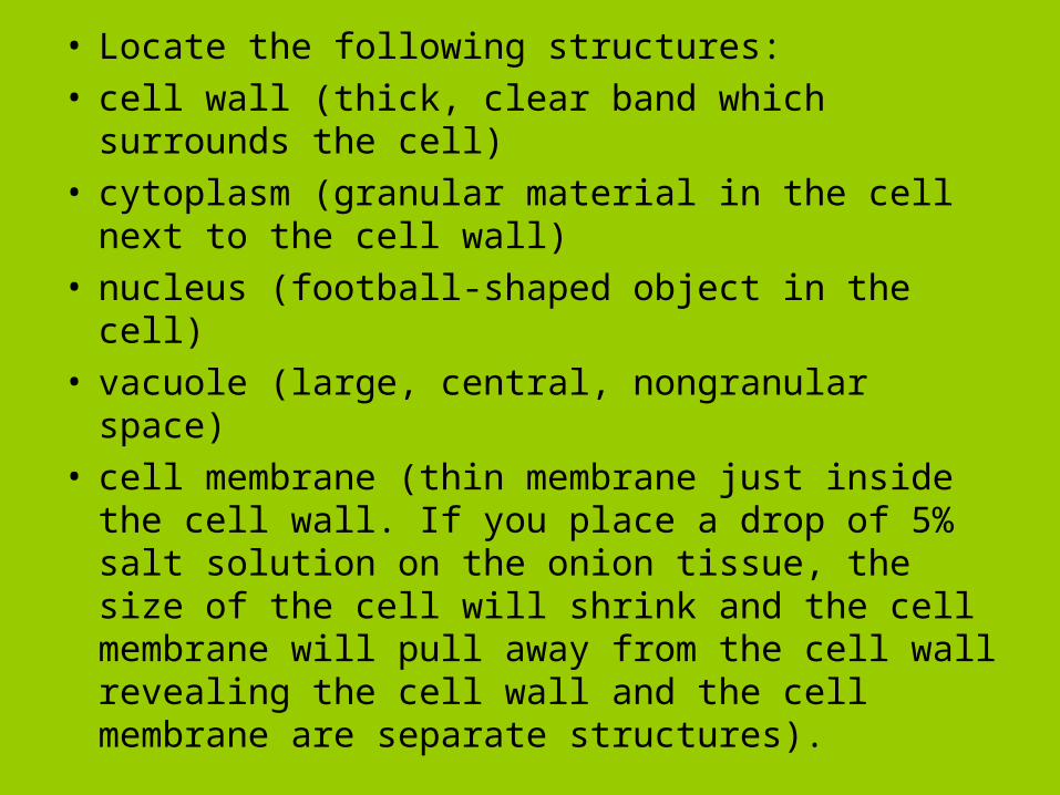
• Locate the following structures:• cell wall (thick, clear band which surrounds the
cell)• cytoplasm (granular material in the cell next to
the cell wall)• nucleus (football-shaped object in the cell)• vacuole (large, central, nongranular space)• cell membrane (thin membrane just inside the
cell wall. If you place a drop of 5% salt solution on the onion tissue, the size of the cell will shrink and the cell membrane will pull away from the cell wall revealing the cell wall and the cell membrane are separate structures).