Embed Size (px)
Citation preview
Cell Structures
Ms. KlinkhachornSeptember 12, 2011
AP Biology
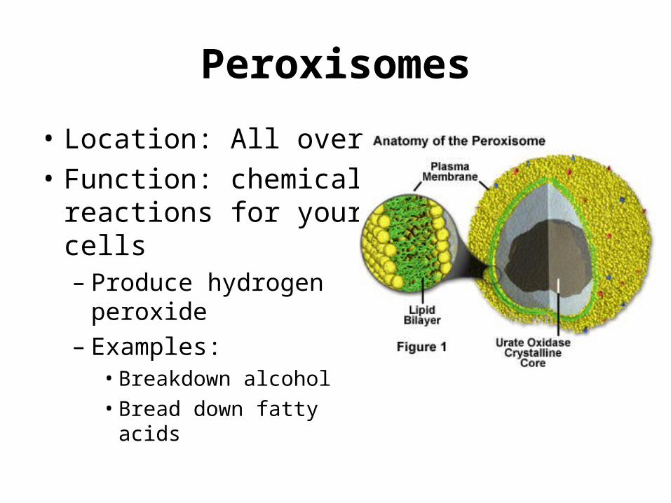
Peroxisomes
• Location: All over• Function: chemical
reactions for your cells– Produce hydrogen
peroxide – Examples: • Breakdown alcohol• Bread down fatty acids
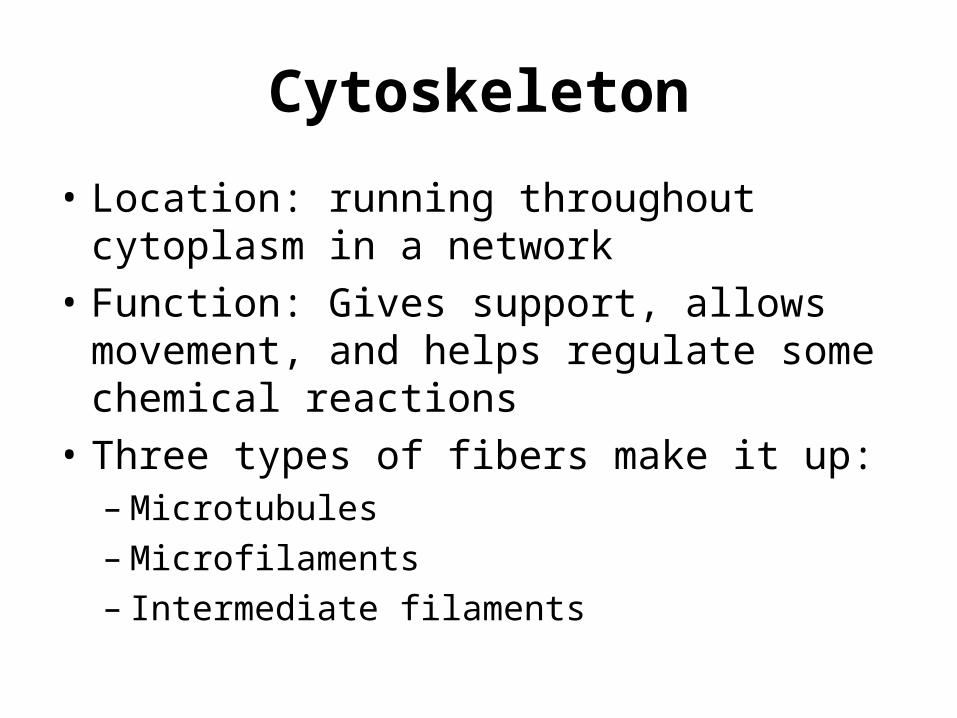
Cytoskeleton
• Location: running throughout cytoplasm in a network
• Function: Gives support, allows movement, and helps regulate some chemical reactions
• Three types of fibers make it up:– Microtubules– Microfilaments– Intermediate filaments
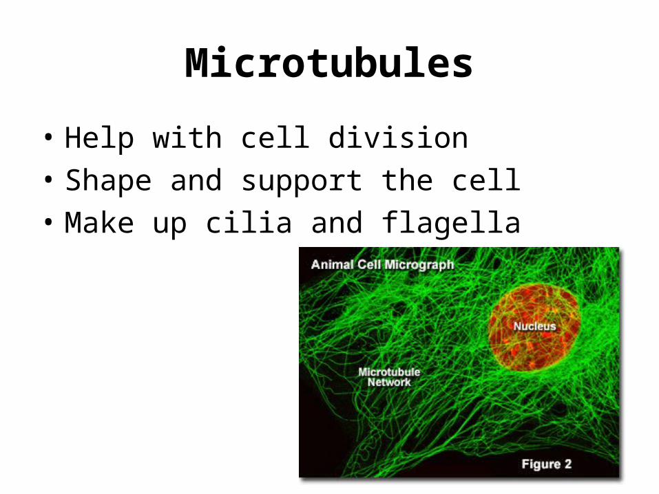
Microtubules
• Help with cell division• Shape and support the cell• Make up cilia and flagella
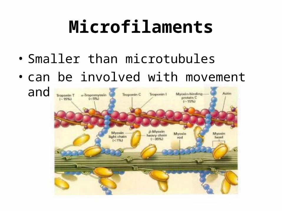
Microfilaments
• Smaller than microtubules • can be involved with movement and support
Intermediate Filaments
• microfilaments < intermediate < microtubules• Help permanently fix things in the cell– Maintain shape– Fix position of organelles
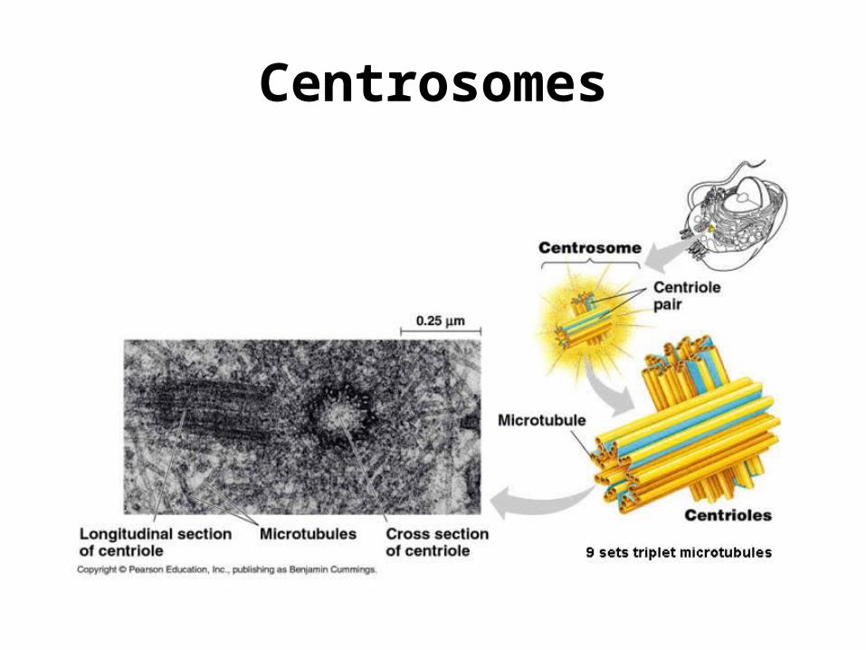
Centrosomes
• Location: near the nucleus until cell division• Function: area that microtubules grow out of– contain centrioles in animal cells
Centrosomes
Flagella and Cilia
• Made of microtubules• Function: Movement of cells – Cilia also help to move fluid over tissue
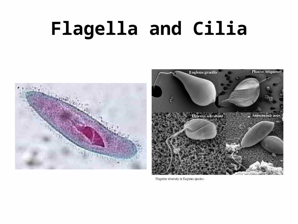
Flagella and Cilia
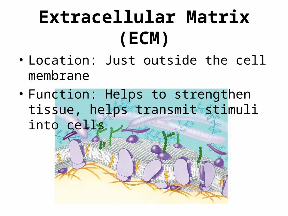
Extracellular Matrix (ECM)
• Location: Just outside the cell membrane• Function: Helps to strengthen tissue, helps
transmit stimuli into cells
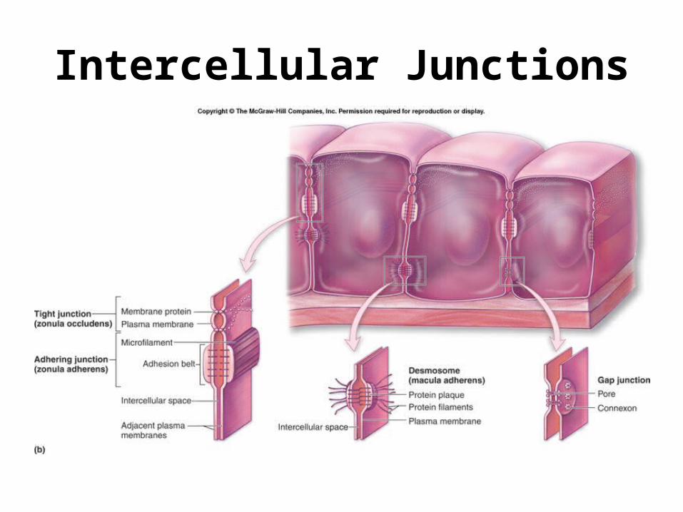
Intercellular Junctions
• Tight junctions– Places where cells fuse to make the membranes
water-tight• Desmosomes– Fasten cells together to make them strong sheets
• Gap junctions– Provide channels between cells so that molecules can
pass• Plasmodesmata (Plants only)– Similar to gap junctions in animals
Intercellular Junctions
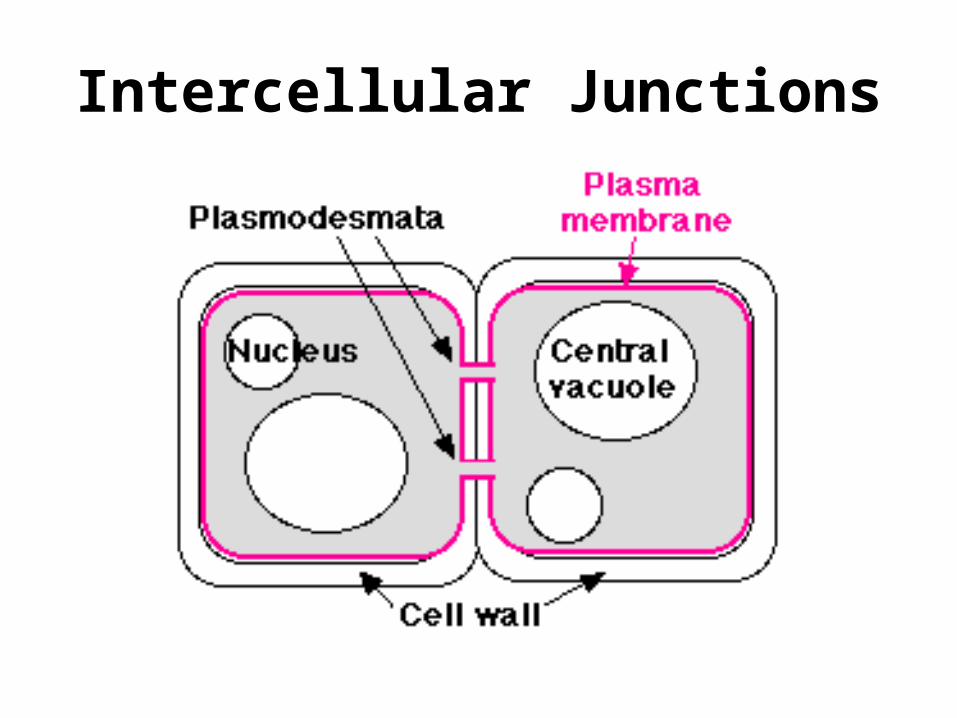
Intercellular Junctions
Membrane Structure and Function
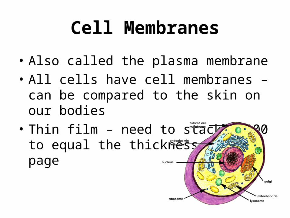
Cell Membranes
• Also called the plasma membrane• All cells have cell membranes – can be
compared to the skin on our bodies• Thin film – need to stack 8,000 to equal the
thickness of a page
Plasma Membrane
• Barrier for the cell • It is selectively permeable– Some materials go through the membrane a lot
easier than others– Some materials can’t go through at all
• What kind of materials need to be able to go in to the cell? Go out of the cell?
Other Membranes
• Membranes can also surround organelles– These organelles are membrane-bound – Create compartments within the cell itself that
have different environments
• What type of cell has membrane-bound organelles?
• What are some examples of membrane bound organelles?
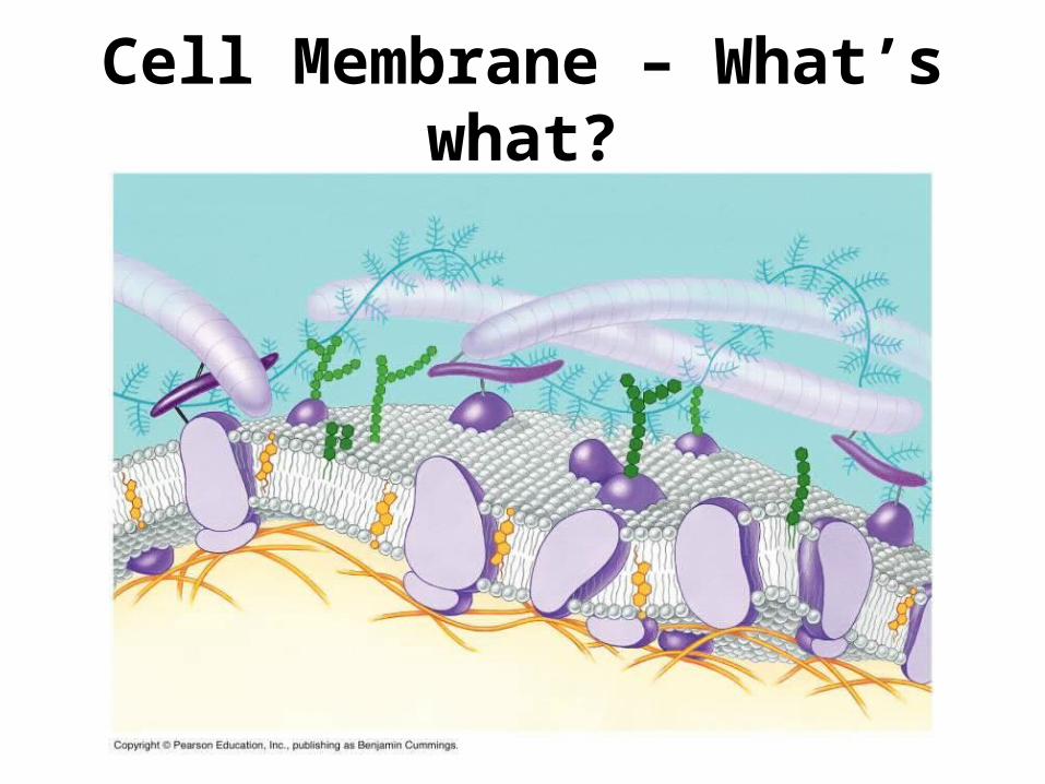
Cell Membrane – What’s what?
Membrane Composition
• Membrane composition depends on the type of cell and the kind of organelle
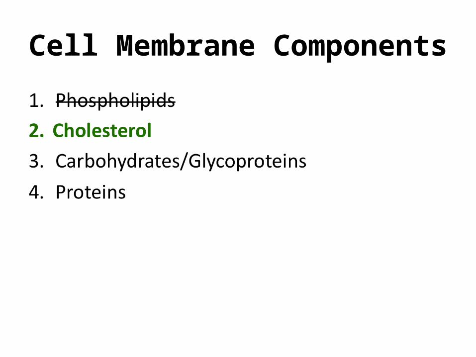
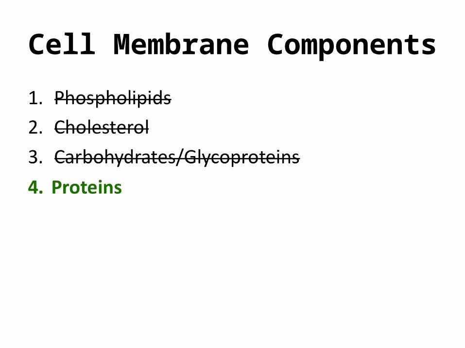
• Membranes are made up of the following:– Phospholipids– Proteins– Carbohydrates/glycoproteins– Cholesterol
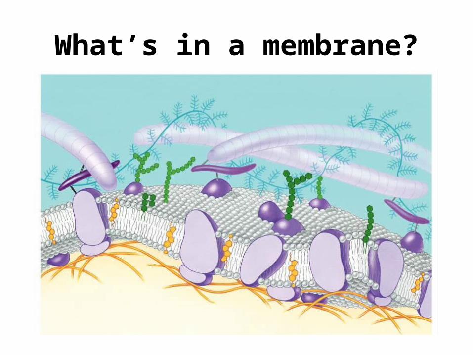
What’s in a membrane?
How do these parts behave?
• Fluid-Mosaic Model – states that components of the membrane are able to move two dimensionally without restraint– Membranes are fluid/dynamic.
• Mosaic = collage– membranes are composed of a variety of
components
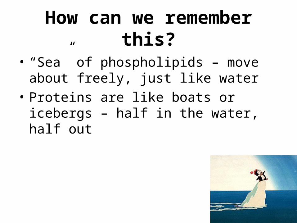
How can we remember this?
• “Sea” of phospholipids – move about freely, just like water
• Proteins are like boats or icebergs – half in the water, half out
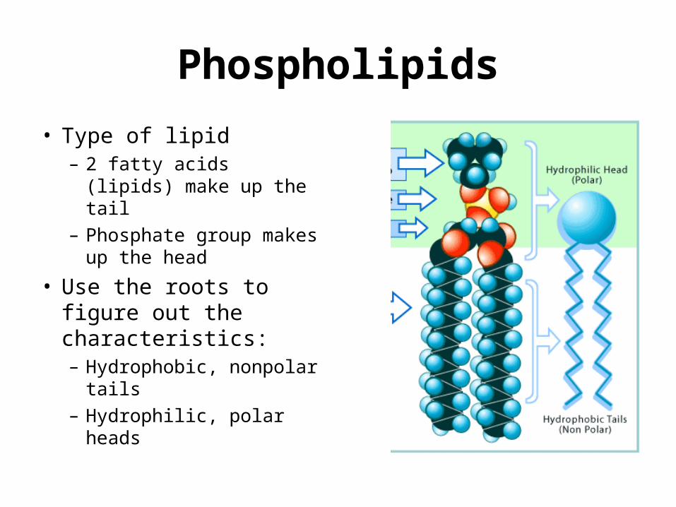
Phospholipids
• Type of lipid– 2 fatty acids (lipids)
make up the tail– Phosphate group makes
up the head
• Use the roots to figure out the characteristics:– Hydrophobic, nonpolar
tails– Hydrophilic, polar heads
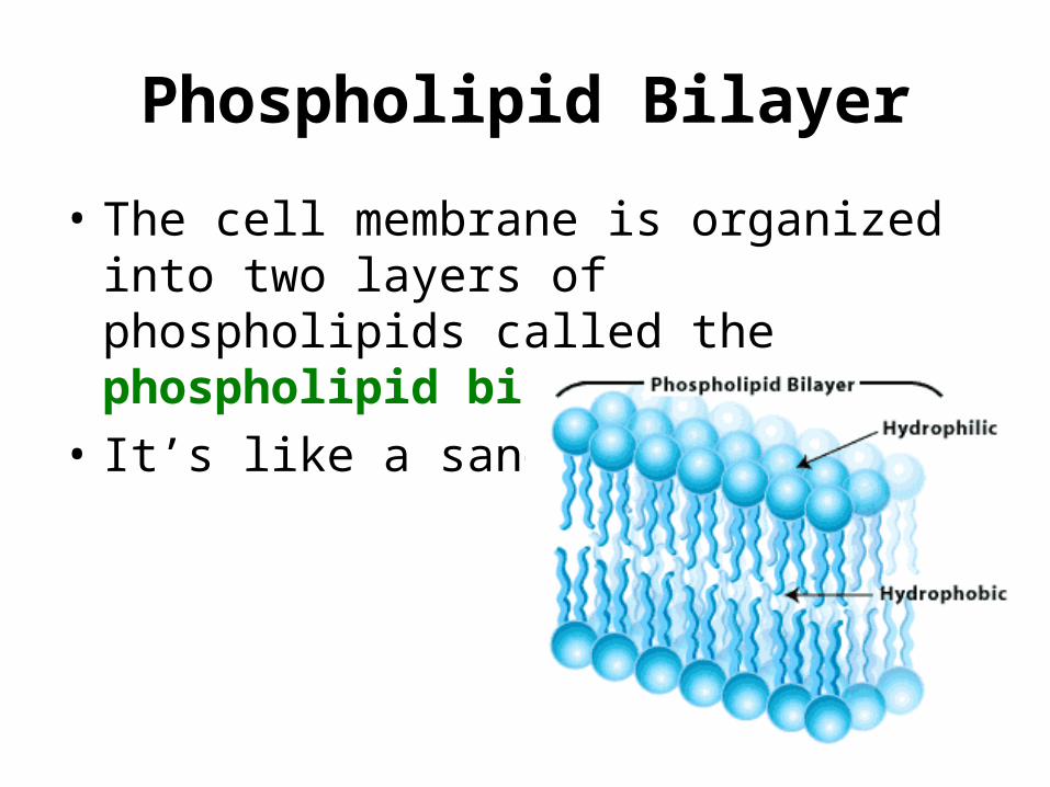
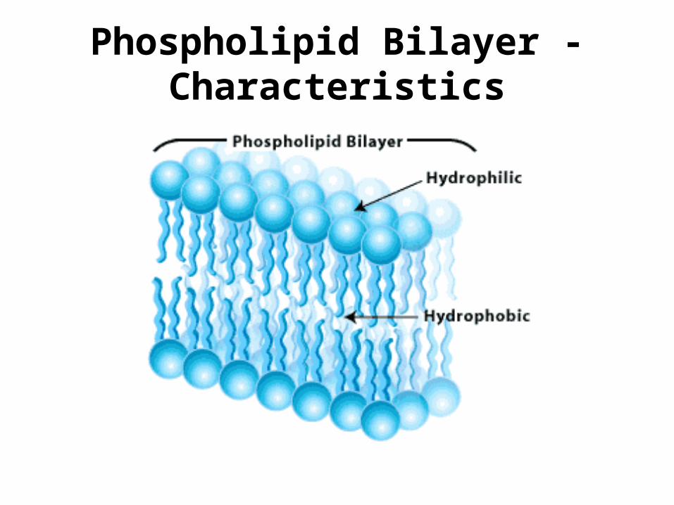
Phospholipid Bilayer
• The cell membrane is organized into two layers of phospholipids called the phospholipid bilayer
• It’s like a sandwich
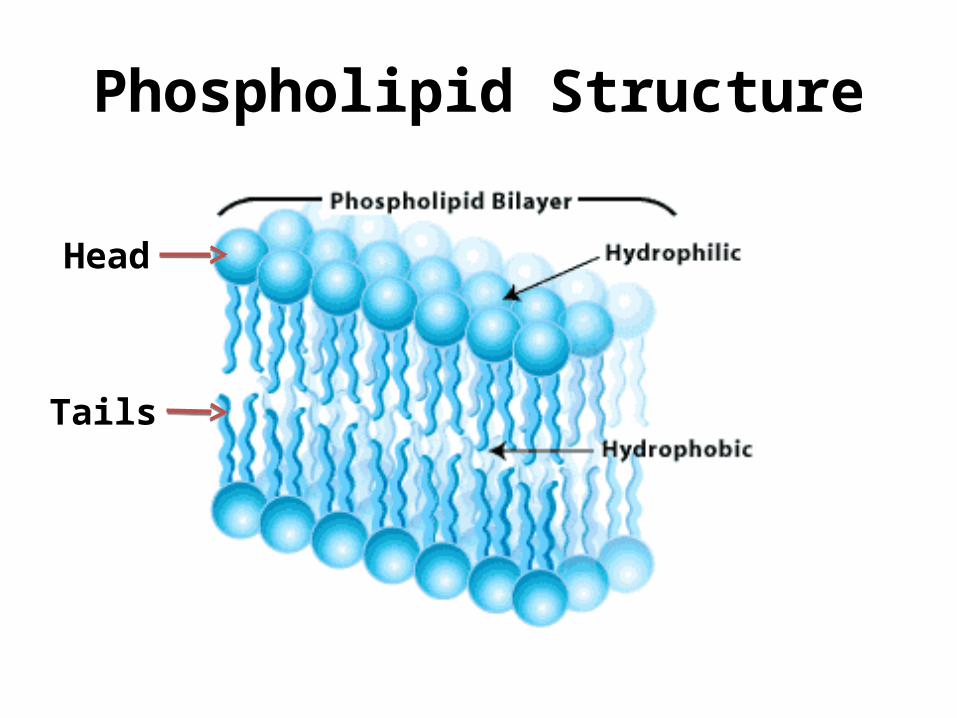
Phospholipid Structure
Head
Tails
Cell Membrane Components
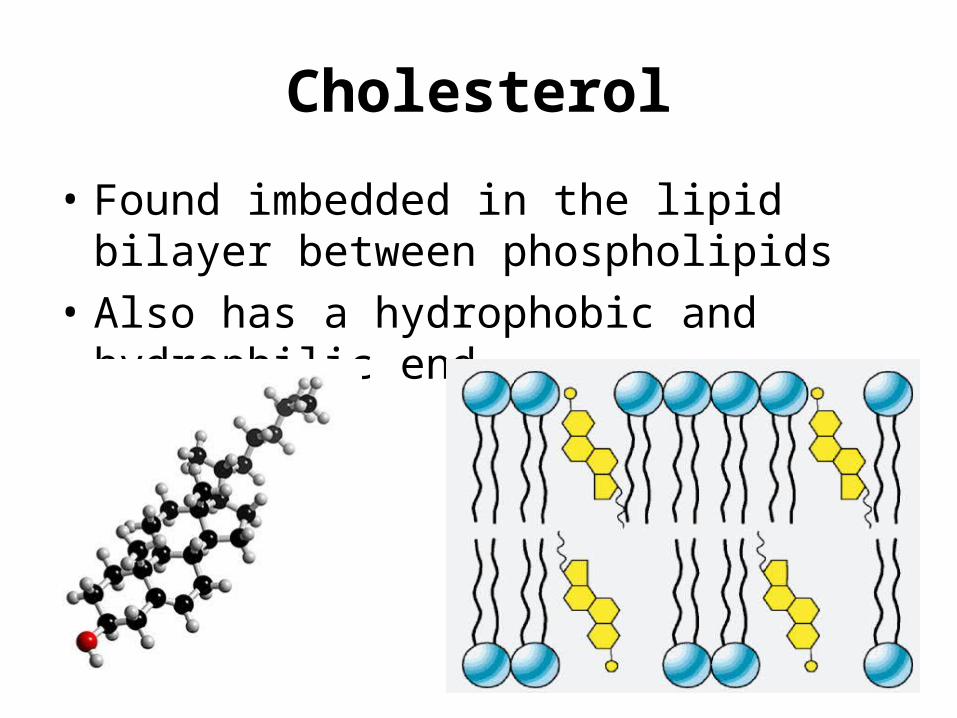
Cholesterol
• Found imbedded in the lipid bilayer between phospholipids
• Also has a hydrophobic and hydrophilic end
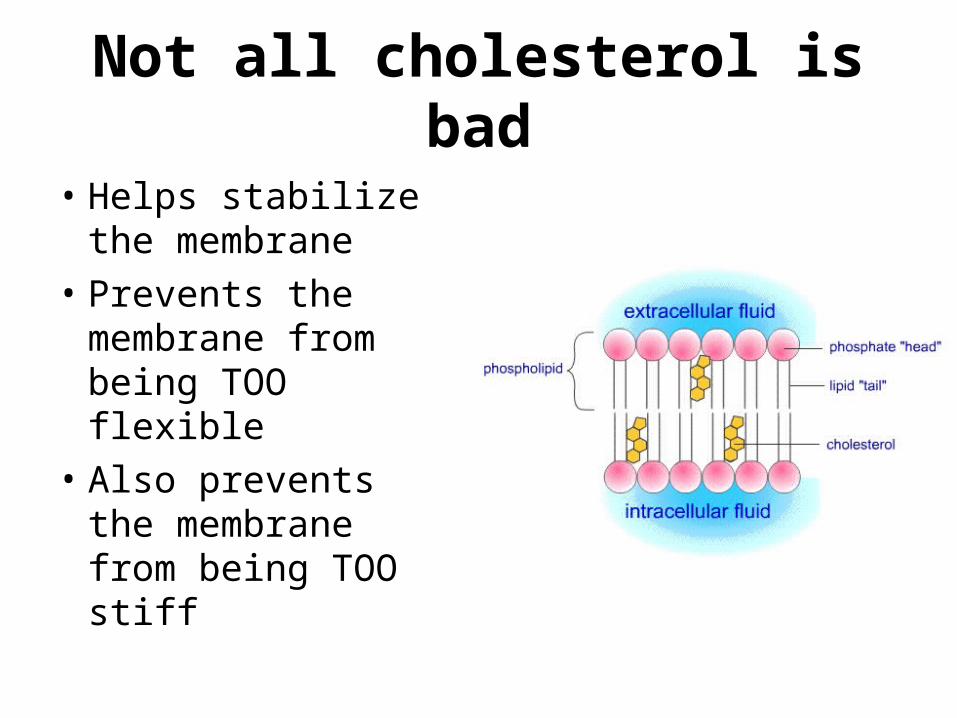
Not all cholesterol is bad
• Helps stabilize the membrane
• Prevents the membrane from being TOO flexible
• Also prevents the membrane from being TOO stiff
Cell Membrane Components
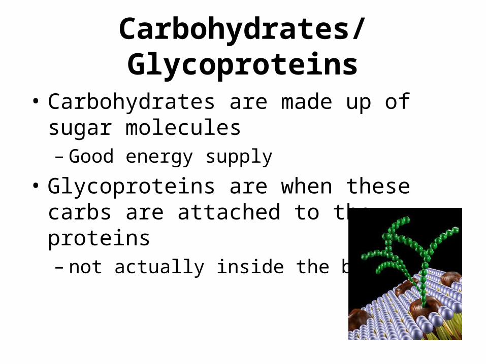
Carbohydrates/Glycoproteins
• Carbohydrates are made up of sugar molecules– Good energy supply
• Glycoproteins are when these carbs are attached to the proteins– not actually inside the bilayer
Carbohydrate Function
• Help with cell-cell recognition– Cell’s ability to distinguish neighboring cells– Helps cells figure out how to sort into tissues and
organs– Cell recognize each other based on the
carbohydrates on the surface of the cell membrane
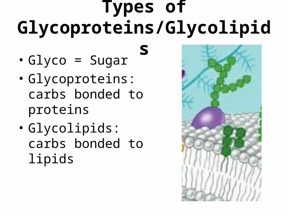
Types of Glycoproteins/Glycolipids
• Glyco = Sugar• Glycoproteins: carbs
bonded to proteins• Glycolipids: carbs bonded
to lipids
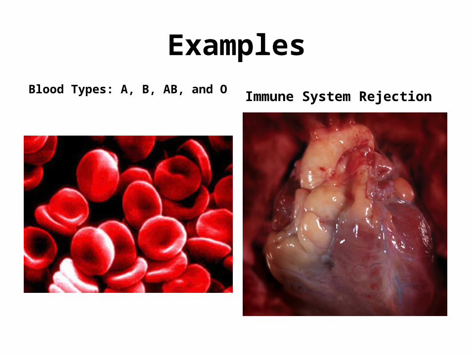
Examples
Blood Types: A, B, AB, and O Immune System Rejection
Cell Membrane Components
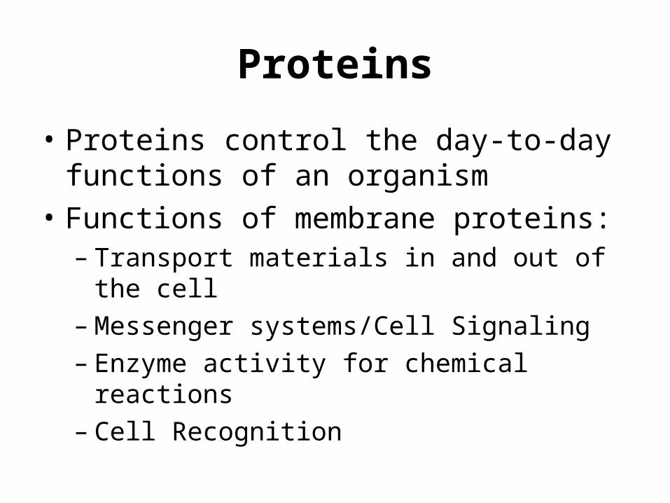
Proteins
• Proteins control the day-to-day functions of an organism
• Functions of membrane proteins:– Transport materials in and out of the cell– Messenger systems/Cell Signaling– Enzyme activity for chemical reactions– Cell Recognition
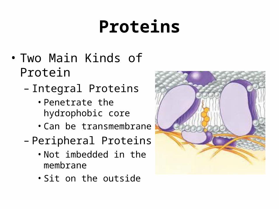
Proteins
• Two Main Kinds of Protein– Integral Proteins• Penetrate the hydrophobic
core• Can be transmembrane
– Peripheral Proteins• Not imbedded in the
membrane• Sit on the outside
Short Response
• Taken from past AP Free Response Questions
1. A major distinction between prokaryotes and eukaryotes is the presence of membrane-bound organelles in eukaryotes.a) Describe the structure and function of TWO eukaryotic
membrane-bound organelles other than the nucleus.2. Membranes are essential components of all cells.
a) Identify THREE macromolecules that are components of the plasma membrane in a eukaryotic cell and discuss the structure and function of each.
Phospholipid Bilayer - Characteristics
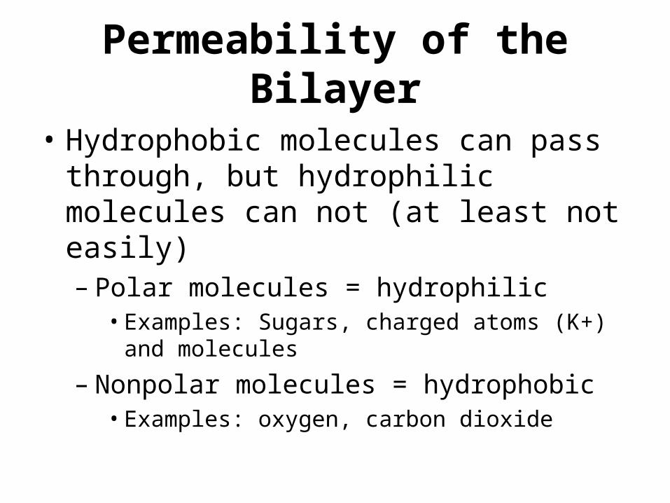
Permeability of the Bilayer
• Hydrophobic molecules can pass through, but hydrophilic molecules can not (at least not easily)– Polar molecules = hydrophilic• Examples: Sugars, charged atoms (K+) and molecules
– Nonpolar molecules = hydrophobic• Examples: oxygen, carbon dioxide
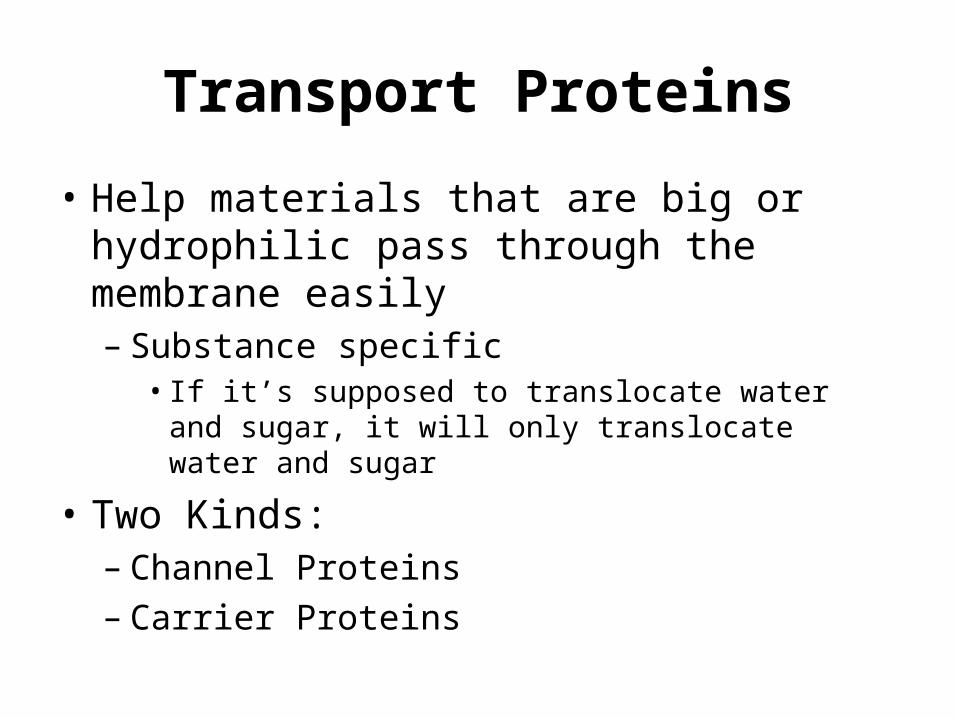
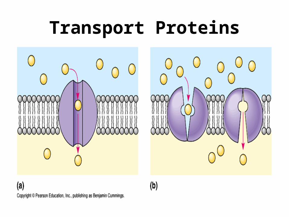
Transport Proteins
• Help materials that are big or hydrophilic pass through the membrane easily– Substance specific• If it’s supposed to translocate water and sugar, it will
only translocate water and sugar
• Two Kinds:– Channel Proteins– Carrier Proteins
Transport Proteins
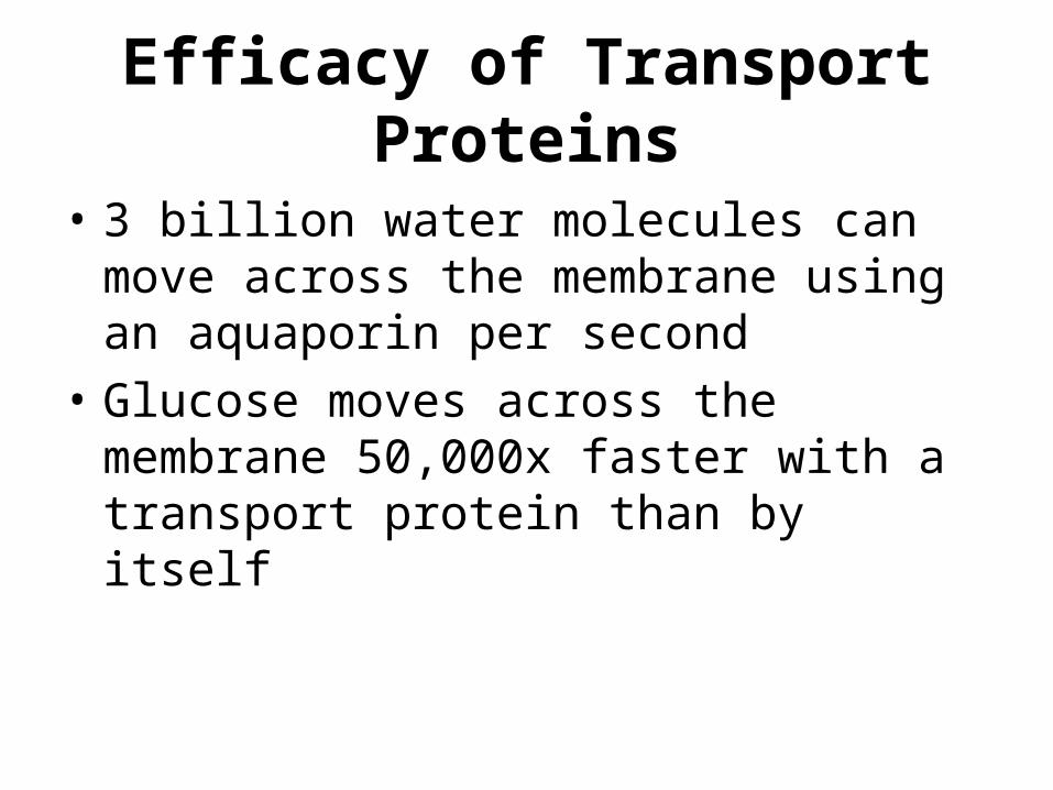
Efficacy of Transport Proteins
• 3 billion water molecules can move across the membrane using an aquaporin per second
• Glucose moves across the membrane 50,000x faster with a transport protein than by itself
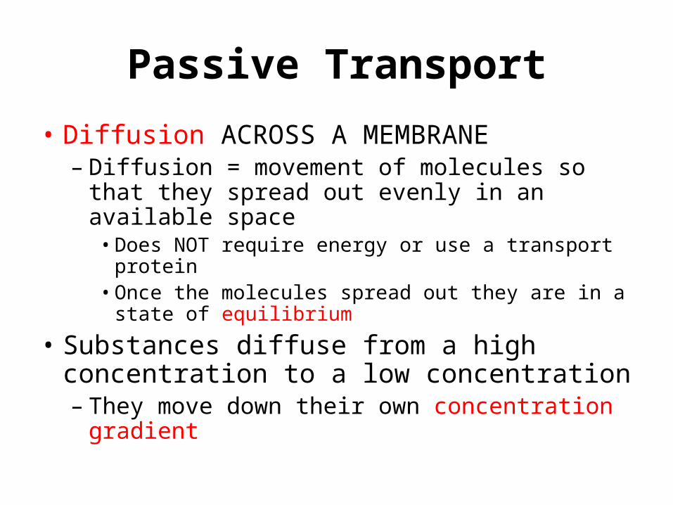
Passive Transport
• Diffusion ACROSS A MEMBRANE– Diffusion = movement of molecules so that they
spread out evenly in an available space • Does NOT require energy or use a transport protein• Once the molecules spread out they are in a state of
equilibrium
• Substances diffuse from a high concentration to a low concentration– They move down their own concentration
gradient
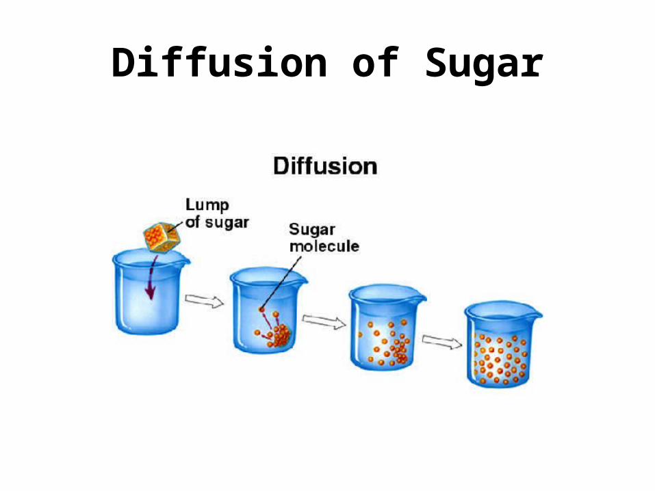
Diffusion of Sugar
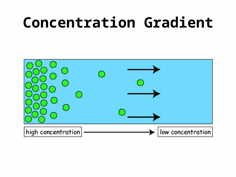
Concentration Gradient
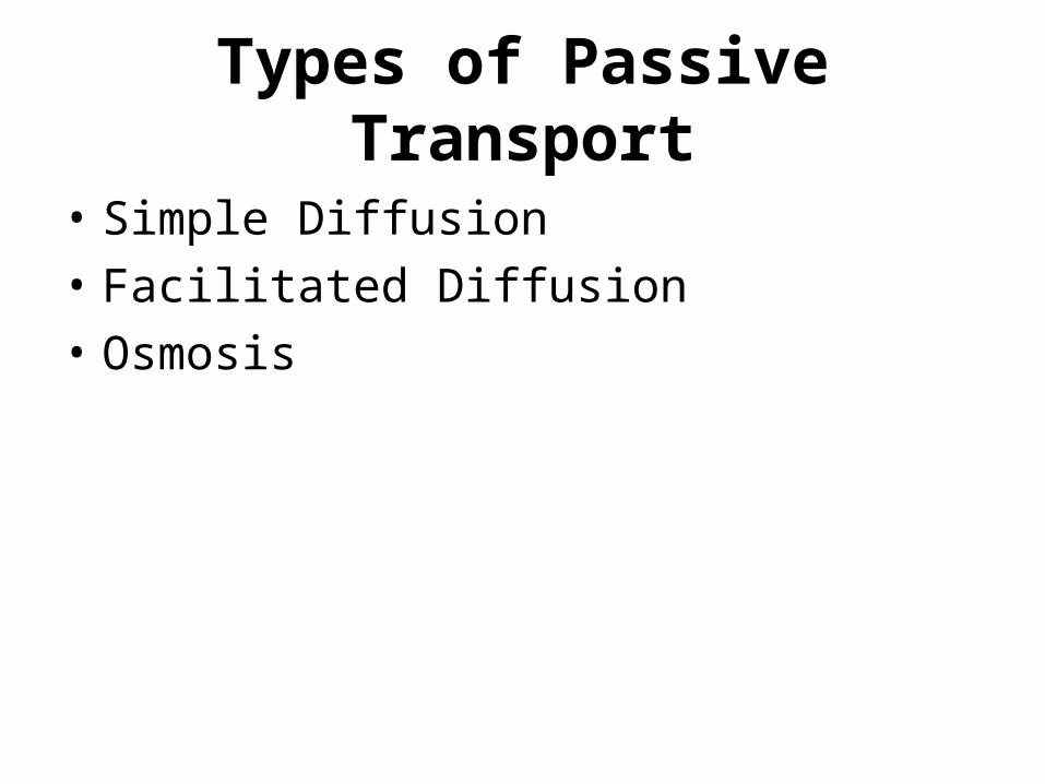
Types of Passive Transport
• Simple Diffusion• Facilitated Diffusion• Osmosis
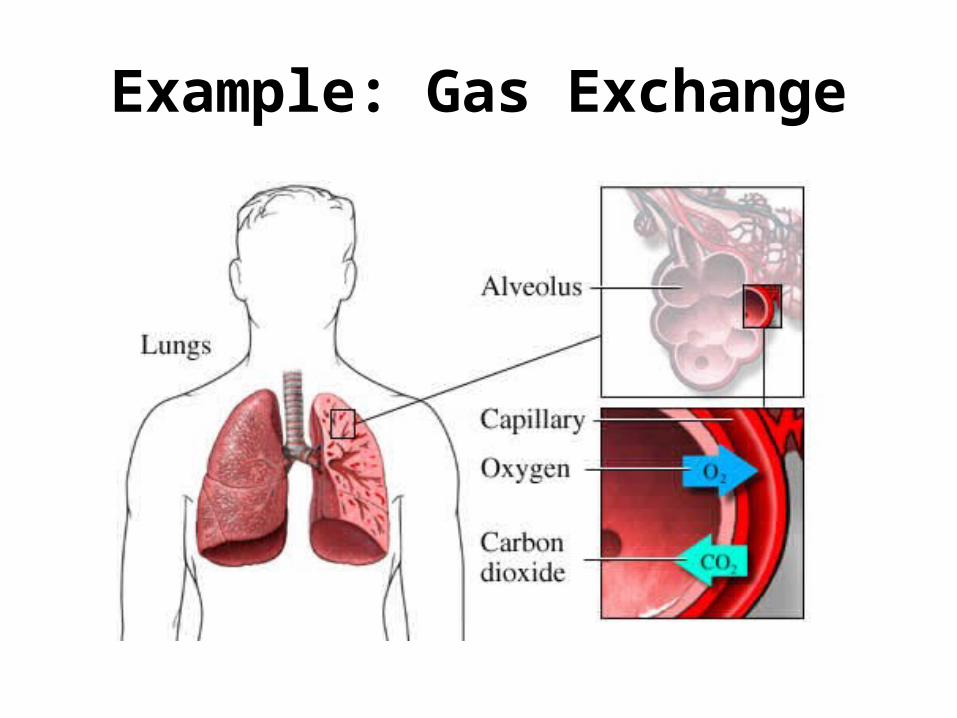
Example: Gas Exchange
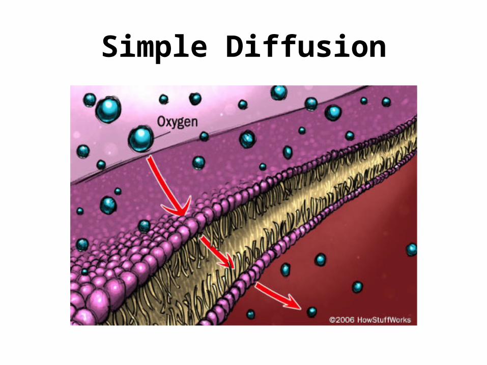
Simple Diffusion
Facilitated Diffusion
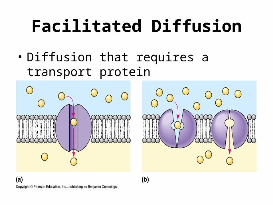
• Diffusion that requires a transport protein
Osmosis
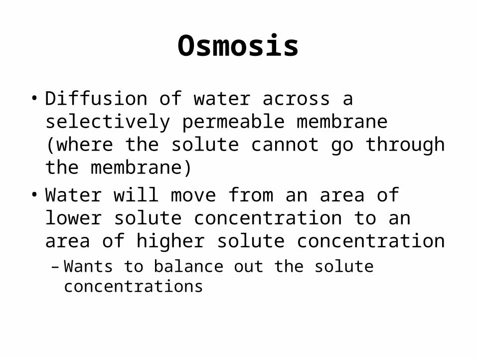
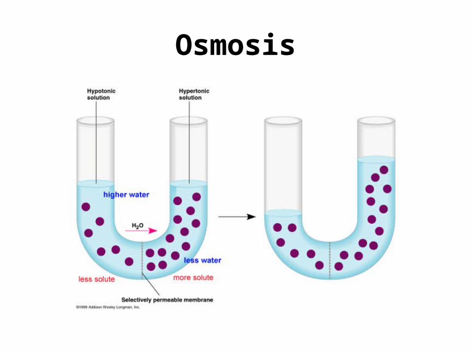
• Diffusion of water across a selectively permeable membrane (where the solute cannot go through the membrane)
• Water will move from an area of lower solute concentration to an area of higher solute concentration– Wants to balance out the solute concentrations
Osmosis
Hypertonic Solutions
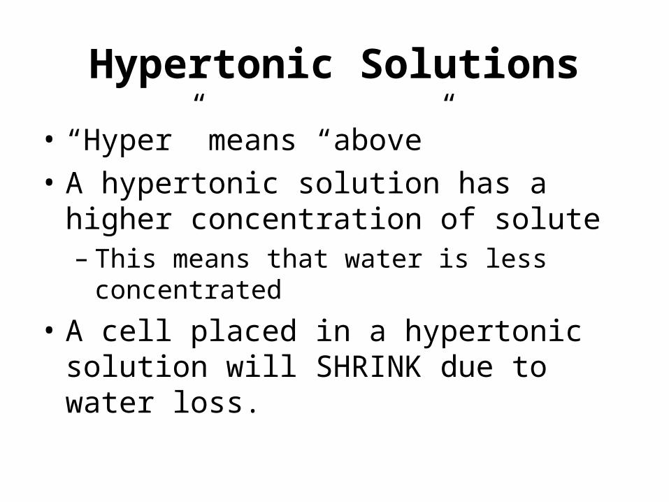
• “Hyper” means “above”• A hypertonic solution has a higher
concentration of solute– This means that water is less concentrated
• A cell placed in a hypertonic solution will SHRINK due to water loss.
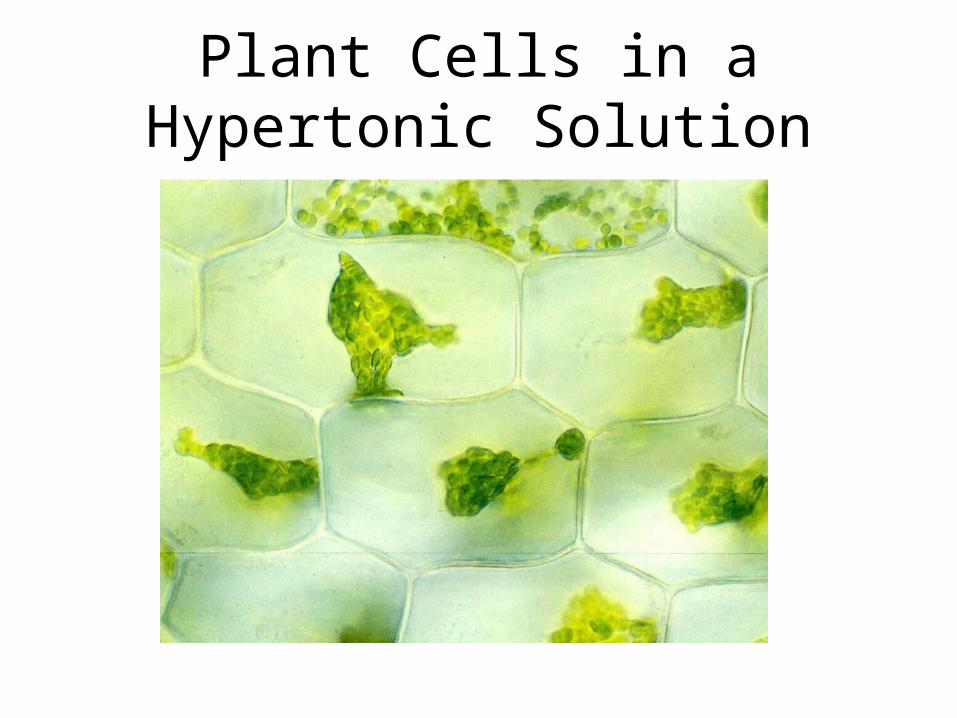
Plant Cells in a Hypertonic Solution
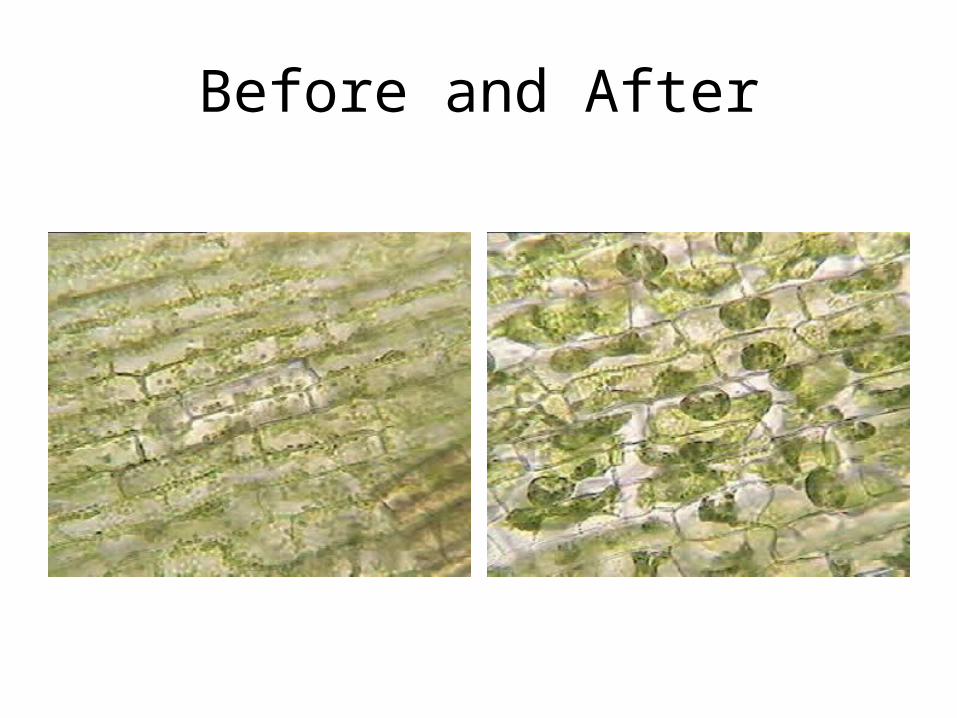
Before and After
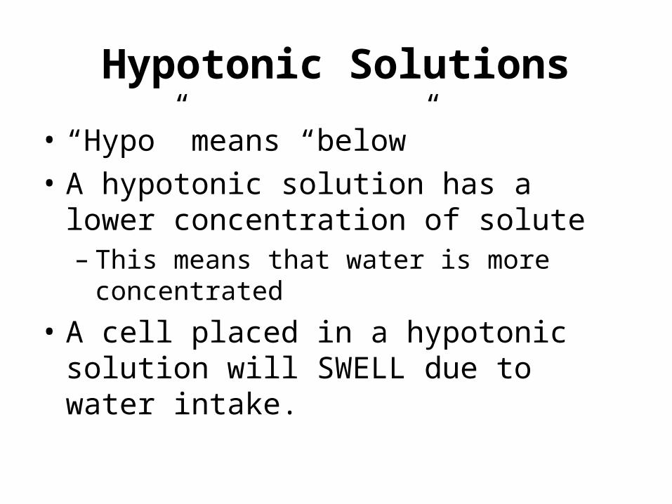
Hypotonic Solutions
• “Hypo” means “below”• A hypotonic solution has a lower
concentration of solute– This means that water is more concentrated
• A cell placed in a hypotonic solution will SWELL due to water intake.

Isotonic Solutions
• “Iso” means “equal”• An isotonic solution has an equal
concentration of solute• A cell placed in an isotonic solution will remain
unchanged• There will still be movement of water, though
there is NO NET GAIN.
DNA – November 30, 2010
Answer these on the back of your study guide:1. What is diffusion? How is it different from
passive transport?2. What do the word forms “hypo”, “hyper”,
“osmo” and “iso” mean?3. Describe what is happening in the picture.
Water Balance in Animal Cells
• If the cell swells too much, it can burst. – This is called lysis. The cell lyses.
• If the cell shrivels too much, it can die.
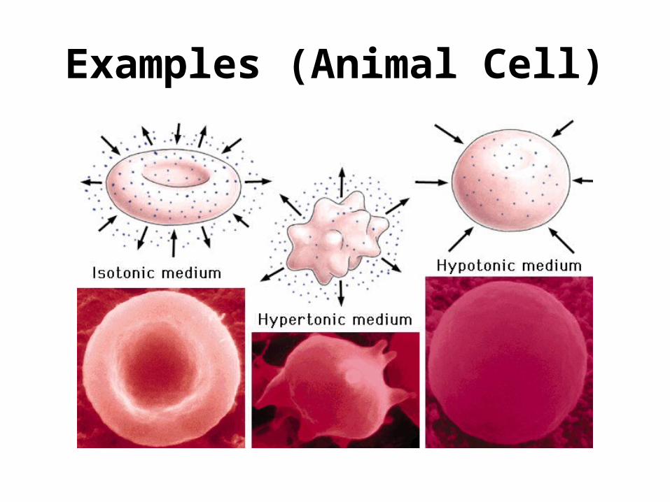
Examples (Animal Cell)
Water Balance in Plant Cells• Cells still swell in hypotonic environment, but the
wall is more rigid.– Water uptake makes the cell turgid (firm).
• This is a plant’s healthy state.• If the cell is in an isotonic environment, it is
flaccid (limp)• If the cell is in a hypertonic environment, it
plasmolyzes (a process called plasmolysis)– The cell membrane pulls away from the cell wall as
water is lost– Can cause death.
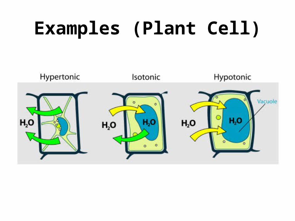
Examples (Plant Cell)
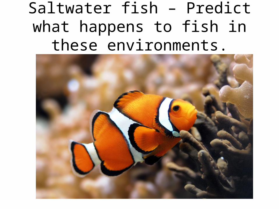
Saltwater fish – Predict what happens to fish in these environments.
Saltwater Fish
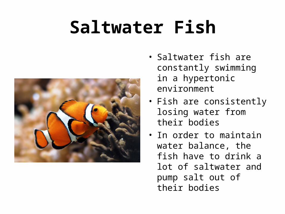
• Saltwater fish are constantly swimming in a hypertonic environment
• Fish are consistently losing water from their bodies
• In order to maintain water balance, the fish have to drink a lot of saltwater and pump salt out of their bodies
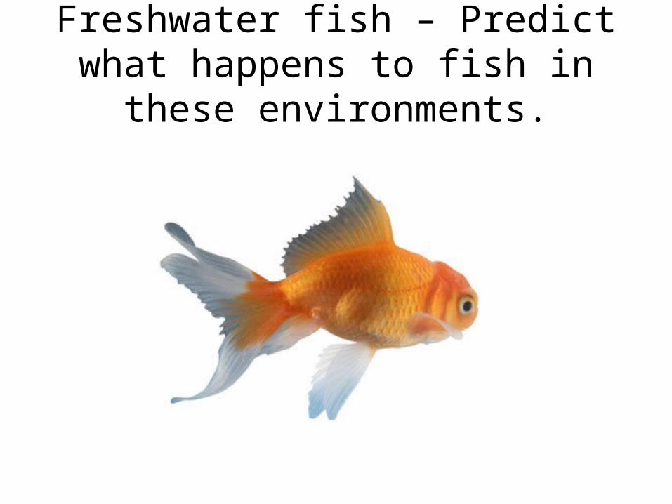
Freshwater fish – Predict what happens to fish in these environments.
Freshwater Fish
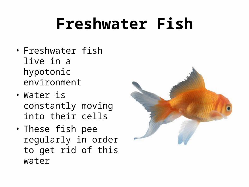
• Freshwater fish live in a hypotonic environment
• Water is constantly moving into their cells
• These fish pee regularly in order to get rid of this water
Osmoregulation
• Osmoregulation = the control of water balance
• Certain organisms are adapted to deal with this– Sea animals – Paramecium
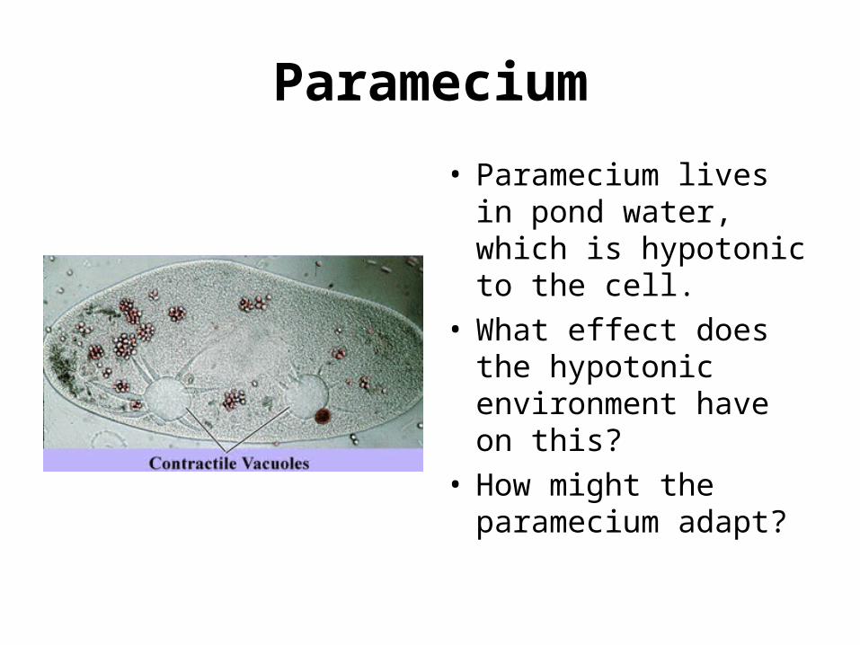
Paramecium
• Paramecium lives in pond water, which is hypotonic to the cell.
• What effect does the hypotonic environment have on this?
• How might the paramecium adapt?
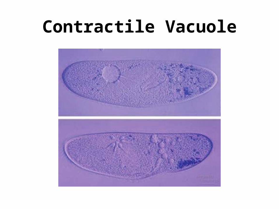
Contractile Vacuole