Embed Size (px)
Citation preview

Vol. 7, 1149-1156, September 1996 Cell Growth & DifferentIatIon 1149
Cell-Substratum Interactions Mediate Oncogene-inducedPhenotype of Lung Cancer Cells1
Linda F. Barr,2 Susan E. Campbell,Margaret B. Penno, Douglas W. Ball, andSteven B. BaylinDepartment of MedIcine, DivisIon of Pulmonary and Critical CareMedicine [L. F. B.] and Division of Endocrinology ED.W. B., S. B. B],Department of Oncology [L. F. B., S. C., S. B. B.], and Center forMedical Genetics [M. B. P.], Johns Hopkins School of MedicIne,BaltImore, Maryland 21205
Abstract
In vlvo and In vitro studies have linked small cell lungcancer (SCLC) and non-small cell lung cancer (NSCLC)cells along a differentiation continuum. The transitionof a SCLC toward a NSCLC phenotype is modeled Inculture by the simultaneous overexpresslon of myc and“as genes in cultured SCLC cells. A major phenotyplcdistinction between SCLC and NSCLC In culture Is thatSCLC cells usually grow In floating aggregates,whereas NSCLC cells and myc- plus ras-expressingSCLC cells grow as adherent spreading monolayerslike other epithellal cells. The present studies examinehow myc, ras, cell aggregation, and attachment tolaminin may interact to modulate transitions betweenthe SCLC and NSCLC phenotypes. We find that myc-expressing SCLC cells, which normally grow asanchorage-independent cells in plastic flasks, willadhere to laminin and exhibit an epithellal morphology.
In this setting, the cells express both NSCLC and SCLCmarkers, thus resembling a tumor type previouslytermed NSCLC with neuroendocrine features.
Anchorage-dependent SCLC cells simultaneouslyexpressing the myc family and an exogenous ras
oncogene move further toward the NSCLC phenotypethan the above myc-expressing cells. However, forcedsuspension of such cells restores the expression ofneuroendocrine SCLC features. These studies indicate
that cell environment, as much as gene expressionevents, profoundly affects aspects of the SCLC cellphenotype.
Received 12/28/95; revised 6/13/96; accepted 6/29/96.The costs of publication of this article were defrayed In part by thepayment of page charges. This article must therefore be hereby markedadvertisement In accordance with 18 U.S.C. Section 1734 solely to mdi-cate thIs fact.1 This work was supported by NIH Grants Ku CA01685 (L. F. B.), ROlCA48081 (S. B. B.), and P50 CA58184 (M. B. P.) and by a grant from theFrancis Family Foundation (L F. B.).2 To whom requests for reprints should be addressed, at Tumor Biology,Johns Hopkins Oncology Center, 424 North Bond Street, Baltimore, MD21231 . Phone: (410) 955-8506; Fax: (410) 614-9884.
Introduction
SCLC3 cells can evolve to display the differentiation charac-teristics of NSCLC both in vivo and in vitro (1 , 2). Also, bothin culture and in patients, cells simultaneously expressingfeatures of all the lung cancer subtypes have been wellcharacterized (3-7). Elucidating the genetic and biologicalevents that control the evolution of SCLC and NSCLC alonga differentiation continuum may help in understanding lungtumor progression and responses to therapy.
A significant difference between SCLC and NSCLC cells inculture is their aggregation and adherence profiles. SCLC
cells usually grow in floating aggregates with varying degreesof homotypic adhesion, whereas NSCLC cells, like otherepithelial cells, attach and spread on tissue culture surfaces.
The purpose of the present studies is to define the contn-bution of myc and ras gene expression events and cellularadherence to the phenotype manifested by lung cancer cellsalong the above SCLC-to-NSCLC differentiation continuum.We have determined that cell-substratum interaction has aprofound effect on the differentiation status of cells simulta-neously expressing c- or N-myc and v-Ha-ras oncogenes.The results suggest that lung cancer phenotypes may reflectinteractions between genetic alterations and cellular interac-tion with the immediate environment.
ResultsOverexpresslon of myc and ras Genes Affect Endocrineand Epithelial Features of SCLC Cells. For most of thesestudies, we have used the classic SCLC cell line 209, whichhas typical SCLC-associated neuroendocnne features (8), asexemplified in the present study by growth in tight floatingaggregates in culture (Fig. IA) and expression of transcriptsof constant markers for SCLC, such as hASH-i (Fig. 2; Ref.9), and small amounts of N-CAM (data not shown; Ref. 10).The 209 cells do not express PKC-�3 transcripts (Fig. 2; Ref.I 1), markers of NSCLC differentiation, such as TGF-a (Fig. 2;Ref. 12), or the cell surface hyaluronan-binding protein CD44(data not shown; Ref. 13), and they express only smallamounts of TGF-�3 (Fig. 2; Refs. 12 and 14).
As in previous studies (1 5), we found that the transfectionof c-myc into the 209 cell line led to looser cellular aggre-gates (Fig. 1C) and decreased the doubling time (Fig. 3) butdid not affect the expression of the characteristic SCLCmarkers dopa decarboxylase and bombesin (1 5). However,in the present study, we find the appearance of a NSCLC-
3 The abbrevIations used are; SCLC, small cell lung cancer; NSCLC,non-small cell lung cancer; 209, NCI H209 cell line; 249, NCI H249 cellline; hASH, human achaete-scute homologue; TOF, transforming growthfactor; PKC, protein kinase C; N-CAM, neural cell adhesion molecule;GAPDH, glyceraldehyde-3-phosphate dehydrogenase; hydron, poly(2-hydroxyethyl methacrylate); MiT, 3-(4,5-dimethylthiazol-2-yl)-2,5-diphe-nyltetrazolium bromide assay.

plastic lamlnin hydren
c*1
01
J�
Fig. 1 . Morphology of myc- and ras-expressing 209 cells manipulatedfor substratum interaction. Parent 209 (A), 209 ras (B), and 209 myc (C)cells grow in suspended aggregates, whereas 209 myc-ras cells (0) growin adherent monolayers in the plastic flasks. The 209 cells adhere tolaminin-coated flasks as aggregates, with epithelial-appearing cells at theperiphery growing out onto the matrix (E). The 209 ras cells disperse onthe laminin surface, but individual cells maintain rounded morphology (F).The 209 myc cells on laminin have some epithelial morphology (G). The209 myc-ras cells on laminin have extensive epithelial morphology (H). The209 myc-ras cells forced into suspension by coating the plastic flasksurface with hydron (I) appear indistinguishable from suspended 209 myccells. Culture conditions are described in “Materials and Methods.” Phasecontrast micrographs: A-E, x200; G-l, x400.
1150 Cell-Substratum Interactions in Lung Cancer Cells
.
:3 �
associated transcript, TGF-c� (Fig. 2), in these c-myc-trans-
fected cells, which still expressed low levels of the neuroen-
docrine marker hASH-i (Fig. 2). These findings support the
placement of these c-myc-expressing cells along a SCLC-
to-NSCLC differentiation continuum. Finally, as we reported
previously, c-myc expression in these cells results in high
levels of PKC-p transcript expression (Fig. 2; Ref. 11).
We have previously found that the effect of v-Ha-ras ex-
pression in SCLC cells is to increase neuroendocrine differ-
entiation, modulated in part by the simultaneous expression
of c- or N-myc (1 6). The present studies extend these ob-
servations. For the 209 cells, v-Ha-ras expression increases
the manifestations of the SCLC phenotype, as exemplified
by an increasing tightness of the cell aggregates (Fig. 1B)
and a slight increase in the transcript levels of the neuroen-
docrine marker hASH-i (Fig. 2). However, expression of
another neuroendocrine marker, N-CAM (data not shown),
and the growth rate (Fig. 3) are unchanged.
The 209 cells simultaneously expressing both exogenous
c-myc and v-Ha-ras genes manifest the anchorage-depen-
dent monolayer growth typical of most NSCLC cell lines (Fig.
1). Consistent with this increased epithelial differentiation,
these 209 myc-ras cells express very little of the neuroen-
docrine marker hASH (Fig. 2), although expression of the
N-CAM protein does not change (data not shown). Further-
more, these cells express the epithelial marker TGF-cr, al-
though in lower amounts than the 209 myc cells (Fig. 2).
Finally, these adherent 209 myc-ras cells express increased
TGF-p (Fig. 2), a molecule that may function in the cell-matrix
pathway (reviewed in Ref. 1 7). It must be noted, however,
that these 209 myc-ras cells do not manifest all of the typical
features of epithelial cells, in that individual cells do not
spread extensively on the flask surface but, rather, maintain
a somewhat rounded shape (Fig. 1D). Additionally, these
cells do not express the NSCLC-assoeiated cell surface
protein CD44 (data not shown), and there is no change in
doubling time compared with the suspended 209 myc cells
(Fig. 3).
Effects of Manipulating Cell-Substratum Interactionson the Phenotype of SCLC Cells. The epithelial differenti-
ation characteristics of cells are profoundly influenced by the
substratum environment in which they grow (for review, seeRef. 1 8). A principal molecule with which such cells interact
is laminin, a major component of the basement membrane.
Adherence of cells to laminin has previously been shown to
alter gene expression for lung cancer cells (1 9, 20). There-
fore, we investigated the effects of laminin adherence on
each of the gene insertion events under study.
The parent 209 cells attach very poorly to laminin. Most of
these cells are tightly aggregated and remain anchorage
independent. Only a few cells on the outside of the aggre-
gates are less tightly bound. These cells have a spindle
shape and appear to interact with the laminin (Fig. 1E). Ex-
posure of SCLC cells to laminin has no effect on any of the
SCLC or NSCLC markers examined for these cells (Fig. 2).
When 209 cells express an infected v-Ha-ras gene (209 ras
cells), the attachment to laminin is increased substantially.
However, the cells do not spread like typical epithelial cells,
and a rounded shape is maintained (Fig. iF). Furthermore,
these cells exhibit little marker changes from those ex-
pressed by the suspended parent cells (Fig. 2).
In contrast to the above cells, the 209 myc cells develop a
much more typical epithelial morphology when exposed to
laminin. The cells have a more flattened shape and consid-
erable spreading activity (Fig. 1 G). In addition, these cells
maintain expression of both epithelial (TGF-a) and neuroen-
docrine (hASH) markers (Fig. 2). Importantly, the expression
of the exogenous c-myc is not altered by these maneuvers
(data not shown). Thus, these cells then resemble the phe-
notype of the NSCLC cells with neuroendoerine features,
which has been well characterized in culture (3) and in pa-
tients (4-7).The most striking effect of a laminin substrate on morphol-
ogy was seen for the 209 myc-ras cells. In contrast to the
rounded appearance of these cells when adherent to plastic,
209 myc-ras cells flatten and develop cytoplasmic extension
processes on laminin (Fig. 1H). However, with the exception

Cell Growth & Differentiation 1151
Fig. 2. Phenotypic marker expressi� of myc-and ras-expressing 209 cells manipulated for sub-stratum interaction. Northern blots of polyadeny-lated RNA were sequentially stripped and re-probed, and band density was measured, asdescribed in “Materials and Methods.” A, repre-sentative experiment of two independent experi-ments. B, cumulative data of levels of expressionof the 2.8-kb transcript of hASH, the 4.8-kb tran-script of TGF-a, the 3.6-kb transcript of PKC-f3,and the 5-kb transcript of TGF-�. Graphs showthe average of two experiments. V axis units arearbitrary and represent transcript densitometriescorrected for loading by dividing by those ofGAPDH transcripts and normalized by dividingthese values by those for the densities for thetranscripts of either 209 (in the case of hASH andTGF-f3) or 209 myc (in the case of TGF-a andPKC-f3) on the same blot. Bars, SD.
A
kb
hASH � RI��42.8
t. 148. .‘
. .
TGF-Q. .�
. ..
‘� 49.5
PKC-�3
�, �43.6
TGF-�� �..iuuiP.�O�5
GAPDH i#{149}-
U U�� #{149}��EEE fEE
� �Ar�!t�i
B
Fi�hASH lOlamini’!
�
1Et�I7�TAi� �l fi
1SF-a
2.5
i 2f
J�:tI 1.5
0La �i fl
PKC.8
I
��I1H�i 0.8i 0.8� 0.4I 02 0 --
� �! fl
1SF-B
�� � U
of increases in the expression of PKC-� transcripts, laminin
growth does not elicit changes in the expression of the other
phenotypie markers examined in the 209 myc-ras cells (Fig.
2) and does not alter the expression of the exogenous e-myc
(data not shown). Furthermore, the cells do not demonstrate
changes in growth rates (data not shown). Thus, the epithelial
morphology, the expression of the epithelial cell marker
(TGF-a), and the decreased expression of the neuroendo-
crine marker (hASH-i ) suggest that the expression of oneo-
genie ras and myc, in conjunction with laminin-adherent
growth, has positioned these cells further toward a NSCLC
classification when examined in the context of a SCLC-to-
NSCLC phenotypic continuum.
To further understand the influence of cell-cell and cell-
matrix interaction on the cultured lung cancer cell pheno-
type, we next examined the effect of inducing normally ad-
herent lung cancer cells to grow in suspension using
surfaces coated with hydron [poly(2-hydroxyethyl methacry-
late) plasticizing agent]. For these studies, we used the 209
myc-ras cells, because as detailed above, these cells fully
manifest features of transition from the SCLC to the NSCLC
phenotype. For some of these studies, we also studied an-
other classic SCLC cell line, 249, which expresses the same
neuroendocrine features outlined for parent 209 cells, includ-
ing anchorage-independent growth. These cells, which have
an endogenously amplified and overexpressed N-myc gene,
also acquire epithelial characteristics, including adherence to
plastic, and expression of some NSCLC markers, such as
TGF-a, when transformed with v-Ha-ras (21).
Prevention of surface adhesion of 209 myc-ras cells first
restores many features of the SCLC morphology, including
growth as floating aggregates, which are indistinguishable
from 209 myc cells (Fig. 1 , C and I). Second, these floating
209 myc-ras cells have an increased doubling time, closer to
that of the parental 209 cells (Fig. 3). Third, abrogation of
adhesion of 209 myc-ras cells increases the expression of
transcripts for the neuroendoerine factor hASH comparedwith levels in these cells when adherent to plastic or laminin.
Fourth, levels of TGF-p and PKC-p transcripts are returned
to a pattern identical to that of the 209 myc cells (Fig. 2).
Finally, this maneuver does not alter the expression of the
exogenous c-myc (data not shown). Thus, prevention of an-
ehorage-dependent 209 myc-ras cells returns the cells to the
marker pattern more typical of the 209 parent and 209 myc
cells.
Like the 209 myc-ras cells, the 249 ras cells growing on
plastic have decreased neuroendoerine and increased epi-
thelial features when adherent to plastic (Fig. 4; Ref. 21).
Thus, ras infection of 249 cells decreases the expression of
two neuroendocrine markers, the hASH-i transcript and N-
2
1.8
1.6
1.4
1.2
1
-+-209
-�---2O9 ras
-�--2O9 myc
---209 myc-ras. . 0 . 209 myc-ras ydro
0 1 2 3 4 5 6 7 8 9
days
1152 Cell-Substratum Interactions in Lung Cancer Cells
a Fig. 3. Growth of myc- and ras-expressing209 cells manipulated for substratum interac-tion. Growth curves were performed as de-scribed in “Materials and Methods.” Shown arethe mean values of three independent experi-ments, each performed in triplicate. Bars,SE. MiT, 3-(4,5-dimethylthiazol-2-yl)-2,5-diphenyftetrazolium bromide.
CAM (Fig. 4). As was seen for the 209 myc-ras cells, forcedsuspension of these cells restores features of the endocrineSCLC phenotype. The parental 249 SCLC morphology isrestored (Fig. 4), and the levels of the hASH-i transcript andN-CAM move toward those seen in parent 249 cells (Fig. 4).No changes were seen in any of the other phenotypic mark-ers examined, and these maneuvers do not alter the expres-sion of the endogenous N-myc or exogenous v-Ha-ras genes
(data not shown). These results indicate that the cells understudy have an inherent plasticity for movement along theSCLC-to-NSCLC continuum, depending on interactions be-tween genetic alterations and cell substratum interaction.
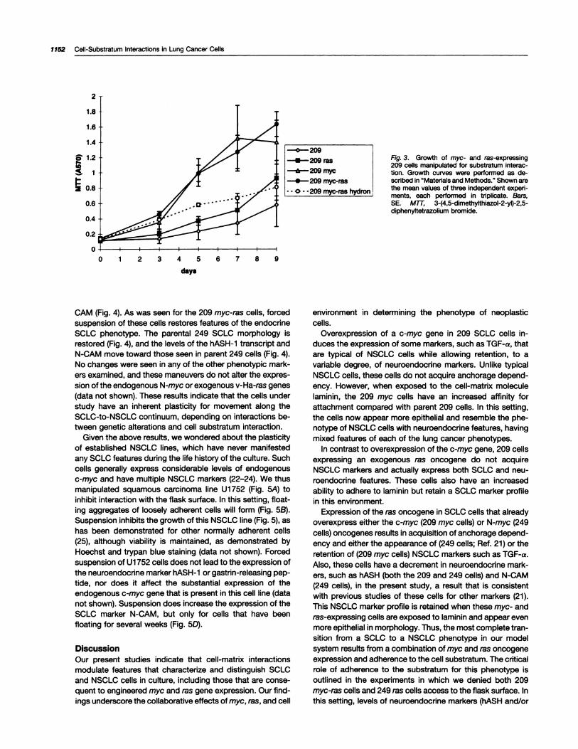
Given the above results, we wondered about the plasticityof established NSCLC lines, which have never manifestedany SCLC features during the life history of the culture. Suchcells generally express considerable levels of endogenousc-myc and have multiple NSCLC markers (22-24). We thusmanipulated squamous carcinoma line Ui 752 (Fig. 5A) toinhibit interaction with the flask surface. In this setting, float-
ing aggregates of loosely adherent cells will form (Fig. 5B).Suspension inhibits the growth of this NSCLC line (Fig. 5), as
has been demonstrated for other normally adherent cells(25), although viability is maintained, as demonstrated byHoechst and trypan blue staining (data not shown). Forcedsuspension of Ui 752 cells does not lead to the expression ofthe neuroendocrine marker hASH-i or gastrin-releasing pep-tide, nor does it affect the substantial expression of theendogenous c-myc gene that is present in this cell line (datanot shown). Suspension does increase the expression of theSCLC marker N-CAM, but only for cells that have beenfloating for several weeks (Fig. 5D).
Discussion
Our present studies indicate that cell-matrix interactionsmodulate features that characterize and distinguish SCLCand NSCLC cells in culture, including those that are conse-quent to engineered myc and ras gene expression. Our find-ings underscore the collaborative effects of myc, ras, and cell
environment in determining the phenotype of neoplasticcells.
Overexpression of a c-myc gene in 209 SCLC cells in-duces the expression of some markers, such as TGF-a, thatare typical of NSCLC cells while allowing retention, to avariable degree, of neuroendocrine markers. Unlike typicalNSCLC cells, these cells do not acquire anchorage depend-ency. However, when exposed to the cell-matrix moleculelaminin, the 209 myc cells have an increased affinity forattachment compared with parent 209 cells. In this setting,
the cells now appear more epithelial and resemble the phe-notype of NSCLC cells with neuroendocrine features, havingmixed features of each of the lung cancer phenotypes.
In contrast to overexpression of the c-myc gene, 209 cellsexpressing an exogenous ras oncogene do not acquireNSCLC markers and actually express both SCLC and neu-roendocnne features. These cells also have an increased
ability to adhere to laminin but retain a SCLC marker profilein this environment.
Expression of the ras oncogene in SCLC cells that alreadyoverexpress either the c-myc (209 myc cells) or N-myc (249cells) oncogenes results in acquisition of anchorage depend-ency and either the appearance of (249 cells; Ref. 21) or theretention of (209 myc cells) NSCLC markers such as TGF-a.Also, these cells have a decrement in neuroendocrine mark-ers, such as hASH (both the 209 and 249 cells) and N-CAM(249 cells), in the present study, a result that is consistentwith previous studies of these cells for other markers (21).
This NSCLC marker profile is retained when these myc- andms-expressing cells are exposed to laminin and appear evenmore epithelial in morphology. Thus, the most complete tran-sition from a SCLC to a NSCLC phenotype in our modelsystem results from a combination of myc and ras oncogene
expression and adherence to the cell substratum. The criticalrole of adherence to the substratum for this phenotype isoutlined in the experiments in which we denied both 209myc-ras cells and 249 ras cells access to the flask surface. Inthis setting, levels of neuroendocnne markers (hASH and/or

1’.
S.
.�. �
�% . . .‘ .
� �
D hASH � �#{149}� 2.8 kb
GAPDH � �. #{149}cf
CI O)��� �01 01
Fig. 4. Effect of substratum manipulation on pheno-typic consequences of ras expression in 249 cells. The249 parental cells (A) grow in suspended aggregates,whereas 249 ras cells (B) grow in adherent monolayersin plastic flasks. The 249 ras cells forced into suspen-sion by growth over hydron (C) look like parental 249cells. Phase-contrast micrographs; x200. D, hASHtranscript expression: representative Northem blot ofthree separate experiments. Northem blots of polya-denylated RNA were sequentially stripped and rep-robed, and band density was measured, as describedin “Materials and Methods.” E, cumulative data oflevels of expression of the 2.8-kb transcript of hASH.The graph shows the average of three separate exper-iments. Y axis units are arbitrary and represent tran-script densitometries corrected for loading by dividingby those of GAPDH transcripts. F, N-CAM expressiondetermined by fluorescence-activated cell sortinganalysis. Y axis, percentage of cells positive for fluo-rescence, measured as described in “Materials andMethods.” Shown are the mean values of three inde-pendent experiments, each performed in triplicate.Bars, SE.
E
F
Cell Growth & Differentiation 1153
hASH
0.8I
� - � �
249 249ras 249ras+h�
N-CAM
i�i�tU. 249 249 ras 249 ras +
h�
N-CAM expression) were increased, and the cells, thus re-
sumed more of their SCLC programming. In essence, this
maneuver stressed the effects on the cells of the myc gene
and lessened those of the ras gene when both oneogenes
were concordantly overexpressed. Similar to our observa-
tions, others have found altered neuroendocrine marker ex-
pression among subelones of a SCLC cell line (NCI H446)that displayed varied substratum adhesion profiles (26). Fi-nally, it is important to note that abrogation of surface ad-
hesion of a typical NSCLC cell line not basally programmed
to express SCLC features (Ui 752) did not, acutely, result in
the appearance of neuroendoerine markers. Suspension did
increase the expression of the SCLC marker N-CAM, but
only for cells that had been floating for several weeks. This is
compatible with cell selection for this trait and is similar to the
findings of others for the expression of another SCLC
marker, the growth factor gastnn-releasing peptide, in a
NSCLC line grown in serum-free media (27).There are at least three important mechanisms that may be
implicated by these results. First, the interaction of cells with
a surface affects the cell shape, and this may alter growth(25) or cell differentiation characteristics (28-30). This
change in geometry may directly affect gene expression
through altered configuration of the nuclear matrix (31). See-
ond, interaction with laminin may elicit signaling through
integrin pathways, leading to modification of gene expres-
sion events through focal adhesion kinase or other signaling
pathways (reviewed in Ref. 32). Furthermore, surface inter-
action may also alter cell polarity, which is of particular
importance for epithelial cell function. Third, increased cell-
surface interaction implies decreased cell-cell interaction.This may affect the local concentration of paracrine sub-
stances, as has been demonstrated for TGF-a (33, 34). In
addition, the cell response to growth factors may be affected
by alterations in second messengers. Indeed, one of the
most markedly affected transcripts in our study, both in
response to genetic changes (e-myc and myc-ras overex-pression) and the presence or absence of substratum adhe-
sion (209 myc-ras cells), was for the PKC-j3 gene.In addition to the biology inherent to our results, the ther-
apeutie implications of these studies merit consideration.
Cells with altered differentiation characteristics within the
same tumor may respond differently to treatment. Indeed,
earlier studies showed the correlation of neuroendoerine dif-
ferentiation of SCLC lines with ‘y-radiation sensitivity (35, 36).
Furthermore, SCLC cells attached to either plastic flasks or
laminin were found to be more resistant to a variety of agents
used to treat SCLC than the floating lines from which they
were derived (36, 37), and the adhesion of SCLC cell lines to
fibronectin led to a decreased sensitivity to an inhibitor of
polyamine biosynthesis, difluromethylornithine (38). Thus,
the phenotypie features addressed in our present studies,
which are shown to be modulated by the interaction of myc,
ras, and cellular environment, are potentially major determi-
nants of the response of lung cancer to therapeutic interven-
tion.
The ability to generalize these effects of cell environment
on activating gene expression events and lung cancer tumor
C
D
1154 Cell-Substratum Interactions in Lung Cancer Cells
-:t�: .
�:‘ �‘
�. ‘
C
�. .: . �;
- - C
#{182}�i
I� . -
0p-
U,�‘0 1 23 4567
days
---U1752
�fr”U1752hydron
N-CAM
70
�6o
‘�ir�5O�U1752 acute >Swk
hydron hydron
Fig. 5. Effect of substratum manipulation on the phenotype of Ui 752 cells. The Ui 752 parental cells (A) grow as adherent monolayers, whereas cellsforced into suspension by coating cell culture flask surfaces with hydron (B) lead to floating aggregates of viable Ui 752 cells. Phase contrast micrographs;x200. C, growth of Ui 752 in plastic and hydron-treated cell culture flasks. The graph shows the average of three separate experiments, each performedin triplicate, as described in “Materials and Methods.” Bars, SE. D, N-CAM expression determined by fluorescence-activated cell sorting analysis. The cellsanalyzed were grown either as adherent cells in plastic cell culture flasks (Ui 752) or as suspended cells for 1 week (acute hydron) or more than 6 weeks(>6 wk hydron) in flasks with hydron coating the surface. y axis, percentage of cells positive for N-CAM fluorescence, which was measured as describedin “Materials and Methods.” Shown are the mean values of three independent experiments, each performed in triplicate. Bars, SE.
biology is tempered by the number of cell lines investigated
in these initial studies. However, our present results present
a useful paradigm for future studies. In addition, threethemes in the literature support a broader application of
these current results. First, the modification of gene expres-
sion events by cell-matrix interactions is previously de-
scribed for both lung cancer cells (19, 20, 36, 37) and other
epithelial cells (reviewed in Ref. 18), including those of neu-
roendocrine gene markers (26). Second, multiple cell lines
exhibit transitions from a SCLC to a NSCLC phenotype (1 , 2)and vice versa (27). Third, the downstream effectors of on-
cogene activation and substratum receptor occupancy may
converge, suggesting an interdependence of these path-
ways (32). Because changing environmental interactions are
important components of cell population derivation during
tumorigenesis and the appearance of metastases, these
findings have implications for both understanding the biology
of, and designing the therapies for, lung cancer.
Materials and MethodsCell Culture. The NCI H209 (209) and NCI H249 (249) cells are estab-lished SCLC cell lines (8, 39). The 209 myc cell line, expressing a full-length exogenous c-myc gene behind its own intact promoters, was agenerous gift from Bruce Johnson (National Cancer Institute-Navy Med-ical Oncology Branch, Bethesda Naval Hospital, Bethesda, MD; Ref. 15)
and grows in neomycin-selective media containing 0.4 mg/mI G41 8 (Sig-ma Chemical Co.). The 209 ras, 209 myc-ras, and 249 ras cell lines were
created by infection of the 209, 209 myc, and 249 cells, respectively, with
the 1504A pseudotype of v-Ha-ras, as described previously (21 , 40). The
expression of the exogenous ras was verified by Northem hybridization(probe P2135; Oncor, Gaithersburg, MD) for all of these cell lines. U1752is a squamous cell lung cancer cell line described previously (41). All of
these cells were grown in RPMI 1640 with 9.5% fetal bovine serum(Sigma), 100 units/mI penicillin, and 100 pg/mI streptomycin, in 37”Cincubators containing 5% CO2.
Culture plates or flasks precoated with laminin were obtained from
Collaborative Research (Bedford, MA). The number of209 cells induced to
adhere to laminin was maximal by day 7 (data not shown), and this time
point was used for all studies. Poly(2-hydroxyethyl methacrylate)(hydron)
(Sigma P3932) was used as a plasticizing agent to abrogate cell-to-flaskadhesion (42). It was constituted as a 102 (w/v) stock solution in 95%
ethanol, and 0.3 or 3 ml were used to coat each well of a 24-well plate ora 75-cm2 flask surface, respectively. The coated surfaces were air dried
for 24 h and then rinsed with PBS or RPMI 1640 prior to use. The day 7
time point was used for the hydron studies as well, unless otherwise
indicated.
Growth Curves. Growth was measured by the (MTT)-assay (Sigma).Forthis assay, all cells were grown in phenol red-free RPMI 1640 (Sigma).
On day - 1 , log phase cells were triturated to one to three cell aggregatesin suspension then placed in 24-well plates in triplicate, at concentrations
of 5 x 10” cells/well. All 3-(4,5-dimethylthiazol-2-yl)-2,5-diphenyltetrazo-hum bromide-derived growth curves were compared with values gener-ated with a hemacytometer and trypan blue exclusion.
Northern Analysis. Polyadenylated RNA was isolated, and 10 �gRNA were separated by gel electrophoresis on 1 .5% agarose-formalde-hyde gels, which were alkali treated, transferred to nylon membranes
(Zeta-Probe; Bio-Rad, Hercules, CA), and hybridized as reported previ-
ously (11). cDNA probes were prepared as inserts of human TGF-f31
(ATCC 59955), human TGF-co (ATCC 59953), human PKC-p (ATCC
59289), human GAPDH (ATCC 57091 ; all obtained from American Type
Cell Growth & Differentiation 1155
Culture Collection, Rockville, MD), and hASH1 (a gift from Dr. DouglasBall, Johns Hopkins Department of Medicine, Division of Endocrinology)and were labeled by random priming with [a-�P]dCTP to a specrflc
activity of approximately 10� cpm/�g DNA(43). These blots were sequen-tially stripped and reprobed as per the manufacturer’s protocol (Bio-Rad).Images were quantitated using Phosphorlmager analysis by the Image-Quant program (Molecular Dynamics).
Flow Cytometric Analysis. Cells were prepared, and microfluorom-etry was assayed as described previously (13). The anti-N-CAM antibody(NCL-CD56) was from Novocastra Labs (Newcastle-upon-Tyne, UnitedKingdom); the anti-CD44 monoclonal antibody was derived from thehybridoma line H4C4 (44); and the fluoresceinylated goat antimouse an-thody used to detect the primary munne immunoglobulin was Obtainedfrom Boebringer Mannheim (Indianapolis, IN; Ref. 13).
AcknowledgmentsWe thank Drs. Jeremy Graff and Robert Casero for their critical review ofthe manuscript.
References1 . Abeloff, M. D., Eggleston, J. C., Mendelshohn, G., Ettinger, D. S., andBaylin, S. B. Changes in morphologic and biochemical characteristics ofsmall cell carcinoma of the lung-a clinicopathologic study. Am. J. Med.,66: 757-764, 1979.
2. Goodwin, G., Shaper, J. H., Abeloff, M. D., Mendelsohn, K. K. G., andBaylin, S. B. Analysis of cell surface protelns delineates a differentiation
pathway linking endocrine and non-endocrine human lung cancers. Proc.Nati. Acad. Si. USA, 80: 3807-381 1 , 1983.
3. Gazdar, A. F., Kadoyama, C., Venzon, D., Park, J. G., Tsai, C. M.,Unnoila, R. I., Mulshine, J. L, lhde, D. C., and Giaccone, G. Associationbetween histologic type and neuroendocnne differentiation on drug sen-
sitivity of lung cancer cell lines. NCI Monogr., 13: 191-196, 1992.
4. Gazdar, A. F., Helman, L J., Israel, M. A., Russell, E. K., Linnoila, R. I.,Mulshine, J. L, Schuller, H. M., and Park, J. G. Expression of neuroen-docrine cell markers L-dopa decarboxylase, chromogranin A, and dense
core granules in human tumors of endocrine and nonendocrine origin.Cancer Res., 48: 4078-4082, 1988.
5. Unnolla, R. I., Mulshine, J. L, Stelnberg, S. M., Funa, K., Matthews, M.J., Cotelingam, J. D., and Gazdar, A. F. Neuroendocnne differentiation inendocrine and non-endocrine lung carcinomas. Am. J. Clin. Pathol., 90:
641-652, 1988.
6. Mooi, W. J., Dewar, A., Spnngall, D., Polak, J. M., and Addis, B. J.Non-small cell lung carcinomas with neuroendocrine features. A lightmicroscopic, immunohistochemical and ultrastructural study of 11 cases.Histopathology, 13: 329-337, 1988.
7. McDowell, E. M., Wilson, T. S., and Trump, B. F. Atypical endocrinetumors of the lung. Arch. Pathol. Lab. Med., 105: 20-28, 1981.
8. Camey, D. N., Gazdar, A. F., Bepler, G., Guccion, J. G., Marangos, P.J., Moody, T. W., Zweig, M. H., and Minna, J. D. Establishment andidentification of small cell lung cancer cell lines having classic and variant
features. Cancer Res., 45: 2913-2923, 1985.
9. BaIl, D. W., Azzoli, C. G., Baylin, S. B., Chi, D., Dou, S., Donis-Keller, H.,Cumaraswamy, A., Borges, M., and Nelkin, B. D. Identification of a humanachaete-scute homolog highly expressed in neuroendocrine tumors.
Proc. NatI. Aced. Sci. USA, 90: 5648-5652, 1993.
10. Carbone, D. P., Koros, A. M., Unnoila, R. I., Jewett, P., and Gazdar,A. F. Neural cell adhesion molecule expression and messenger RNA
splicing patterns in lung cancer cell lines are correlated with neuroendo-crine phenotype and growth morphology. Cancer Res., 51: 6142-6149,1991.
11 . Barr, L F., Mabry, M., Nelkin, B. D., Tyler, G., May, W. S., and Baylmn,
S. B. c-myc gene-induced alterations in proteln kinase C expression: apossible mechanism facilitating myc-ras gene complementation. Cancer
Res., 51: 5514-5519, 1991.
12. Soderdahl, G., Betsholtz, C., Johansson, A., Nllsson, K., and Bergh,J. Differential expression of platelet-derived growth factor and transform-ing growth factor genes in small and non-small cell human lung carcinoma
lines. Int. J. Cancer, 41: 636-641 , 1988.
13. Penno, M. B., August, J. T., Baylin, S. B., Mabry, M., Linnoila, R. I.,
Lee, V. S., Croteau, D., Yang, X. L, and Rosada, C. Expression of cd44 inhuman lung tumors. Cancer Res., 54: 1381-1387, 1994.
14. Damstrup, L, Rygaard, K., Spang-Thomsen, M., and SkovgaardPoulsen, H. Expression oftransforrning growth factor � (TGF-�) receptors
and expression of TGF-�1, TGF-� and TGF-� in human small cell lungcancer cell lines. Br. J. Cancer, 67: 1015-1021 , 1993.
15. Johnson, B. E., Battey, J., Unnoila, I., Becker, K. L, Makuch, R. W.,Snider, R. H., Camey, D. N., and Minna, J. D. Changes in the phenotypeof human small celllung cancerceillines aftertransfection and expressionof the c-myc proto-oncogene. J. Clin. Invest., 78: 525-532, 1986.
16. Mabry, M., Nelkin, B. D., Falco, J. P., Barr. L F., and Baylin, S. B.Transitions between lung cancer phenotypes-implications for tumor pro-gression. Cancer Cells (Cold Spnng Harbor), 3: 53-58, 1991.
17. Roberts, A. B., and Spom, M. B. The transforming growth factor-as.In: M. B. Spom and A. B. Roberts(eds.), Peptide Growth Factors and TheirReceptors, pp. 419-472. Berlin: Springer-Verlag, 1990.
1 8. Streuli, C. H. Extracellular matrix and gene expression in mammary
epithellum (Review). Semin. Cell Biol., 4: 203-212, 1993.
19. Giaccone, G., Broers, J., Jensen, S., Fridman, R. I., Unnoila, R., andGazdar, A. F. Increased expression of differentiation markers can accom-pany laminin-induced attachment of small cell lung cancer cells. Br. J.Cancer, 66: 488-495, 1992.
20. Pavelic, K, MtoOb, M., Pavehc, L, Pavelic, J., Pavelic, 1, and Sarend,S. Human lung cancers growing on extracellular matrix: expression of once-
genes and growth factors. Anticancer Res., 12: 2191-2196, 1992.
21 . Falco, J. P., Baylmn, S. B., Lupu, R., Borges, M., Nelkin, B. D., Jasti, R.K., Davidson, N. E., and Mabry, M. v-ras’� induces non-small cell pheno-
type with associated growth factors and receptors, in a small cell lungcancer cell line. J. Clin. Invest., 85: 1740-1745, 1990.
22. Broers, J. L V., Viallet, J., Jensen, S. M., Pass, H., Travis, W. D.,Minna, J. D., and Linnoila, R. I. Expression of c-myc in progenitor cells ofthe bronchopulmonary epithelium and in a large number of non-small celllung cancers. Am. J. Respir. Cell Mol. Biol., 9: 33-43, 1993.
23. Griffin, C. A., and Baylin, S. B. Expression of the c-myb oncogene inhuman small cell lung carcinoma. Cancer Res., 45: 272-275, 1985.
24. Uttle, C. D., Nau, M. M., Carney, D. N., Gazdar, A. F., and Mince, J.D. Amplification and expression of the c-myc oncogene in human lungcancer cell lines. Nature (Lond.), 306: 194-196, 1983.
25. Folkman, J., and Moscona, A. Role of cell shape in growth control.
Nature (Lond.), 273: 345-349, 1978.
26. Doyle, L A., Borges, M., Hussein, A., Elias, A., and Tomiyasu, T. Anadherent subline of a unique small-cell lung cancer cell line downregulatesantigens of the neural cell adhesion molecule. J. Clin. Invest., 86: 1848-
1854, 1990.
27. Siegfried, J. M., Yi-Hong, H., DeMichele, M. A. A., Hunt, J. D., Gaither,A. Z., and Cuttita, F. Production of gastrin-releasing peptide by a non-
small cell lung carcinoma cell line adapted to serum-free and growthfactor-free conditions. J. Biol. Chem., 269: 8596-8603, 1994.
28. Shannon, J. M., and Pitelka, D. R. The influence of cell shape on theinduction of functional differentiation in mouse mammary cells in vitro. InVitro (Rockville), 17: 1016-1028, 1981.
29. Vernon, R. B., Lane, T. F., Angello, J. L, and Sage, H. Adhesion,
shape, proliferation, and gene expression of mouse Leydig cells areinfluenced by extracellular matrix in vitro. Biol. Reprod., 44: 157-170,1991.
30. Watt, F. M., Jordan, P. W., and O’Neill, C. H. Cell shape controlsterminal differentiation of human epidermal keratinocytes. Proc. NatI.Aced. Sci. USA, 85: 5576-5580, 1988.
31 . Getzenberg, R. H., Pienta, K J., Ward, W. S., and Coffey, D. S.Nuclear structure and the three-dimensional organization of DNA (Re-view). J. Cell. Biochem., 47: 288-299, 1991.
32. Zachary, I., and Rozengurt, E. Focal adhesion kinase (pl25�): apoint of convergence in the action of neuropeptides, integrmns and onco-genes (Review). Cell, 71: 891-894, 1992.
33. Laderoute, K. R., Murphy, B. J., Short, S. M., Grant, T. D., Knapp, A.M., and Sutherland, R. M. Enhancement of transforming growth factor-a

1156 Cell-Substratum Interactions in Lung Cancer Cells
synthesis in muiticellular tumor spheroids of A431 squamous carcinomacells. Br. J. Cancer, 65: 157-162, 1992.
34. Vlodavsky, I., Folkman, J., Sullivan, R., Fridman, R., Rivka, l-M.,
Sasse, J., and Klagsbum, M. Endothelial cell-derived basic fibroblastgrowth factor synthesis and deposition into subendothelial extracellularmatrix. Proc. NatI. Aced. Sci. USA, 84: 2292-2296, 1987.
35. Goodwin, G., and Baylmn, S. B. Relationships between neuroendo-
crime differentiation and sensitivity to ‘y-radiation in culture line OH-i ofhuman small cell lung cancer. Cancer Res., 42: 1361-1367, 1982.
36. Khan, M. Z., Freshney, R. I., Murray, A. M. B., Merry, S., Plumb, J. A.,and McNicol, A. M. Identification and characterisation in vitm of cells with
a non-SCLC cell-like phenotype derived from a continuous SCLC cell line.Anticancer Res., 1 1: 1687-1696, 1991.
37. Fridman, R., Giaccone, G., Kanemoto, T., Martin, G. R., Gazdar, A. F.,and Mulshine, J. L Reconstituted basement membrane (Matrigel) andlaminin can enhance the tumorigenicity and the drug resistance of small
cell lung cancer cell lines. Proc. NatI. Aced. Scm. USA, 87: 6698-6702,
1990.
38. Luk, G. D., and Baylin, S. B. Anchorage dependency effects ondifluoromethylomithine cytotoxicity in human lung carcinoma cells. Can-
cer Res., 46: 1844-1848, 1986.
39. Gazdar, A. F., Camey, D. N., Nau, M. M., and Minna, J. D. Charac-terization of variant subclasses of cell lines derived from small cell lung
cancer having distinctive biochemical, morphological and growth prop-erties. Cancer Res., 45: 2924-2930, 1985.
40. Nakagawa, T., Mabry, M., deBustros, A., IhIe, J. N., Nelkin, B. D., andBaylmn, S. B. Introduction of v-Ha-ras oncogene induces differentiation of
cultured medullarythyroid carcinoma cells. Proc. NatI. Aced. Sci. USA, 84:
5923-5927, 1987.
41 . Bergh, J., Nilsson, K., Zeck, L, and Biovanella, B. Establishment andcharacterization of a continuous lung squamous cell carcinoma cell line(U-1752). Anticancer Res., 1: 317-332, 1981.
42. Brouty-Boye, D., Tucker, R., and Folkman, J. Transformed and nec-plastic phenotype: reversibility during cuiture by cell density and cellshape. Int. J. Cancer, 26: 501-507, 1980.
43. Feinberg, A. P., and Vogelstein, B. A technique for radiolabeling DNArestriction endonuclease fragments to high specific activity. Anal. Bio-
chem., 132: 6-13, 1983.
44. Belitsos, P. C., Hildreth, J. E., and August, J. T. Homotypic cellaggregation induced by anti-CD44 antibodies and related to CD44 ex-pression. J. Immunol., 144: 1661-1670, 1990.