Embed Size (px)
Citation preview
Biointerfaces
Cell Surface Engineering to Control Cellular Interactions
Catarina A. Cust�dio[a, b] and Jo¼o F. Mano*[a, b]
Abstract: Cell surface composition determines all interac-tions of the cell with its environment, thus cell functionssuch as adhesion, migration and cell–cell interactions canpotentially be controlled by engineering and manipulatingthe cell membrane. Cell membranes present a rich repertoireof molecules, therefore a versatile ground for modification.However the complex and dynamic nature of the cell surfaceis also a major challenge for cell surface engineering thatshould also involve strategies compatible with cell viability.Cell surface engineering by selective chemical reactions orby the introduction of exogenous targeting ligands can be
a powerful tool for engineering novel interactions and con-trolling cell function. In addition to chemical conjugationand modification of functional groups, ligands of interest tomodify the surface of cells include recombinant proteins, lip-osomes or nanoparticles. Here, we review recent efforts toperform changes to cell surface composition. We focus onthe engineering of the cell surface with biological, chemicalor physical methods to modulate cell functions and controlcell–cell and cell–microenvironment interactions. Potentialapplications of cell surface engineering are also discussed.
Introduction
Living cells are sensitive to their environment. This means thatthey detect and respond to events in their surrounding envi-ronment.[1]
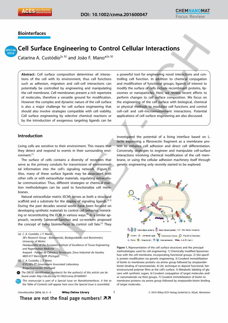
The surface of cells contains a diversity of receptors thatserve as the primary conduits for transmission of environmen-tal information into the cell’s signaling network. (Figure 1).Also, many of these surface ligands may be associated withother cells or with extracellular materials, regulating extracellu-lar communication. Thus, different strategies or chemical reac-tion methodologies can be used to functionalize cell mem-branes.
Natural extracellular matrix (ECM) serves as both a structuralscaffold and a substrate for the display of signaling ligands.[2, 3]
During the past decades several works have been focused ondeveloping synthetic materials to control cell behavior, mimick-ing or reconstituting the ECM in various ways.[4] In a similar ap-proach, recently Salmeron-Sanchez and co-workers proposedthe concept of living biointerfaces to control cell fate.[5] They
investigated the potential of a living interface based on L.lactis expressing a fibronectin fragment as a membrane pro-tein to enhance cell adhesion and direct cell differentiation.Conversely, strategies to engineer and manipulate cell-surfaceinteractions involving chemical modification of the cell mem-brane, or using the cellular adhesion machinery itself throughgenetic engineering only recently started to be explored.
[a] C. A. Cust�dio, J. F. Mano3B’s Research Group - Biomaterials, Biodegradables and BiomimeticsUniversity of MinhoHeadquarters of the European Institute of Excellence of Tissue Engineeringand Regenerative MedicineAvepark - Parque de CiÞncia e Tecnologia, Zona Industrial da Gandra,4805-017 Barco GMR (Portugal)
[b] C. A. Cust�dio, J. F. ManoICVS/3B’s PT Government Associated LaboratoryBraga/Guimar¼es (Portugal)
The ORCID identification number(s) for the author(s) of this article can befound under http ://dx.doi.org/10.1002/cnma.201600047.
This manuscript is part of a Special Issue on Nanobiointerfaces. A link tothe Table of Contents will appear here once the Special Issue is assembled.
Figure 1. Representation of the cell surface structures and the principalmethodologies used for cell engineering: 1) Chemically modified liposomesfuse with the cell membrane, incorporating functional groups. 2) Site-specif-ic protein modification via genetic engineering. 3) Covalent immobilizationof biotin to membrane proteins via amine group followed by streptavidin-biotin binding of nanomaterials. 4) LbL technique to deposit functional, het-erostructured polymer films at the cell’s surface. 5) Metabolic labeling of gly-cans with synthetic sugars. 6) Covalent conjugation of target molecules and/or nanomaterials via thiol groups. 7) Covalent immobilization of biotin tomembrane proteins via amine group followed by streptavidin-biotin bindingof target molecules.
ChemNanoMat 2016, 00, 0 – 0 � 2016 Wiley-VCH Verlag GmbH & Co. KGaA, Weinheim1
These are not the final page numbers! ��
Focus ReviewDOI: 10.1002/cnma.201600047

In this review, we will focus on the advanced techniques toengineer cell surfaces, describe their potential and challenges,highlighting the strategies that have been explored to regulatecell–cell and cell–extracellular matrix interactions.
The cell membrane is a highly complex and dynamic envi-ronment comprising lipids, proteins and carbohydrates, whichmediate extracellular communication.[6] This rich repertoire ofmolecules presents an excellent opportunity to engineer thecell membrane and a powerful tool to manipulate interactionsbetween cells and the surrounding environment. However, cellsurface engineering is particularly challenging due to the factthat the cell membrane in not a static structure.[7–9] It shouldalso be noted that any process for cell surface engineeringmust be performed using minimal alterations to the biologicalenvironment of living cells, as slight alterations of pH, tempera-ture, ionic strength and osmolality &&can have a significantinfluence &&.
Cells can be engineered by chemical modifications in thecell membrane through chemical conjugation or non-covalentinteractions. Furthermore, cells can be tailored with nanomate-rials or coated using layer-by-layer (LbL) strategies for engi-neering novel interactions and controlling cell function. Someof these engineering techniques still require optimization toimprove the efficacy and targeting effectiveness while minimiz-ing any loss of cell function. In this review we will first describewhich molecules of interest comprise the membrane and howthey are arranged, then we summarize key methodologiesused to manipulate the surface of living cells. We will then dis-cuss how these cell modifications can be applied to controlcell function or enhance the therapeutic potential of cellularproducts. Finally, we will outline future trends and perspectivesof this breakthrough field.
The cell surface
The interactions of cells with the surrounding environment aremediated by the cell membrane, thus it is worth consideringthe biomolecular composition of the membrane and howthese molecules are arranged. Cell membranes are composedof a lipid bilayer, containing proteins that span the bilayer oneither side of the two leaflets designed to perform the func-tions cell require.[6] Proteins are central molecules in cell–ECMinteractions, typically through the creation of attachmentpoints linking the cytoskeleton to extracellular binding sites.The capacity to manipulate cells interactions with the sur-rounding environment will certainly be dependent on our abil-ity to control the function of these proteins. Integrins are a su-perfamily of transmembrane cell adhesion proteins that bindto ECM ligands, cell-surface ligands, and soluble ligands. Syn-decans and lectins are a family of transmembrane core pro-teins that act synergistically with integrins as co-receptors forECM proteins that bind specific carbohydrates. Many mem-brane proteins and lipids are conjugated to polysaccharides,which comprise the glycocalyx, or cell coat, of all cells.[9] Cellsurface glycans also play crucial roles in various physiologicalevents involving cell surface recognition.[10, 11] Unfortunately,the heterogeneous nature of cell surfaces, particularly with re-
spect to glycoconjugate structures, has frustrated molecular-level studies of glycan function.
Cell surface engineering is challenging due to the fact thatthe plasma membrane is a dynamic structure: both lipid andprotein components of the membrane are continuously inter-nalized, displaced, degraded, and replaced by de novo synthe-sis.[12] Chemical conjugation and/or insertion of target mole-cules, nanomaterials or patches within the plasma membrane,are some particularly important trends in cell engineering thatwill be discussed below. These strategies to decorate cell’s sur-face would enable a close control in cell behavior, from cell ad-hesion to cell migration, proliferation or differentiation.
Strategies for Cell Surface Bioengineering
Genetic engineering
Genetic engineering is well-established as a robust and highlyversatile methodology employing the cell’s biosynthetic machi-nery to modify the genetic programming of cells.[13] The modu-lation of cell surface receptor expression through geneticmodification was recently exploited to alter cell surfaces, re-modeling extracellular communication. The base of genetic en-gineering is the inclusion of exogenous genetic material intothe cell to express or regress specific cell surface molecules toachieve the preferred outcome.[8, 14] Some recent studies havefocused on the expression of key cell surface receptors in-volved in stem cell recruitment and migration.[15] Genetic ma-nipulation of integrin expression in cells could significantly im-prove cell engraftment, increasing the efficiency of cell therapy.
Jo¼o F. Mano is a Professor at the ChemistryDepartment of University of Aveiro and princi-pal investigator at CICECO. His current re-search interests include the use of biomateri-als and cells towards the development oftransdisciplinary concepts for biomedical ap-plications, especially aimed at being used inregenerative/personalised medicine. In partic-ular, he and his group have been developingmaterials, mainly derived from biodegradablepolymers, with stimuli-responsive or bio-in-structive behaviour, or biomimetic and nano/micro-technology approaches applied to bio-materials and surfaces.
Catarina A. Cust�dio is graduated in AppliedChemistry by the Faculty of Sciences andTechnology of the New University of Lisbonand obtained her PhD on Tissue EngineeringRegenerative Medicine and Stem Cells in theUniversity of Minho and in cooperation withthe Max Planck Institute for Polymer Researchin Mainz. She is currently working as a post-doctoral research rat University of Aveiro. Herresearch interests lie at the development ofnovel bioactive hydrogels and new ap-proaches to control cell behavior through thenanoscale (bio)engineering of materials surfa-ces.
ChemNanoMat 2016, 00, 0 – 0 www.chemnanomat.org � 2016 Wiley-VCH Verlag GmbH & Co. KGaA, Weinheim2
�� These are not the final page numbers!
Focus Review

Mrksich and co-workers have used cell engineering and syn-thetic surface chemistry as complementary strategies to pro-mote unique specific ligand–receptor interactions.[16] They con-structed a chimeric receptor that contains the intracellular andtransmembrane domains of b1 integrin combined with otherspecific domains providing a new specificity for the binding ofthe receptor to ECM. Cells expressing the modified receptoradhered and spread selectively on the target substrates. Thesame group described an approach for integrating cellular ac-tivities and electrical processes in an underlying substrate bycell surface engineering.[17] Using genetic manipulation they in-troduced enzymes at the cell surface that will modify electro-active monolayers, enabling electronic transduction of biologi-cal activity. Genetic manipulation was also used to introducebio-orthogonal reactive groups into cell surface proteins, creat-ing sites for selective modification of cells. For example, Tingand co-workers developed a robust methodology to label sitespecifically cell surface proteins with biotin groups.[18] They ge-netically attach a specific peptide to either terminus of theprotein of interest and add recombinant enzyme biotin ligasethat site-specifically biotinylates a lysine side chain within thepeptide to the cell medium. These biotin groups can then betargeted with streptavidin conjugates. The same enzyme cata-lyzes ligations that also permit the derivatization of membraneproteins with ketone groups, which further extends the spec-trum of possible conjugates.[19] Membrane associated proteinscan also be site-specifically modified by using a genetically en-coded aldehyde tag.[20] Proteins bearing this aldehyde tag arethen chemically modified by selective reaction with hydrazide-or aminooxy-functionalized reagents. The precise chemicalcontrol offered by the aldehyde tag method should enable thedevelopment of new protein products for research and thera-peutic purposes. Despite the great achievements and promis-ing results, genetic manipulation of cells is technically chal-lenging dealing with regulatory and safety issues. To overcomethese issues there is growing interest in devisingnovel bioconjugation, protein engineering, chemistry,and material science approaches for cell surface en-gineering.[21]
Chemical modification
In contrast to genetic engineering that is mostlyused to manipulate proteins at the cell’s surface,chemical modification may be used to manipulatelipids, proteins or glycans. Chemical functionalgroups naturally present on this biomolecules at cellsurface are appealing sites for functionalization usingcovalent conjugation. The most commonly usedchemical groups include, amines, sulfhydryl, carboxyland carbonyl groups present in proteins and othercell surface molecules. It is worth to note that chemi-cal reactions on the cell surface should be selectivewithout any nonspecific modifications and per-formed under mild conditions.
Amine groups are widely used for chemical modifi-cation of cell membranes due to well-established
protocols, easily available conjugation linkers and mild reactionconditions. The most common reaction to modify primaryamines involves reaction with n-hydroxysuccinimide (NHS)ester. This strategy was used for the direct coupling of succini-midyl ester-functionalized polyethylene glycol (PEG) to cell sur-face amines of pancreatic islets.[22] Such modification has beenstudied toward preventing immune responses of host for suc-cessful islet transplantation
Using a similar chemistry, biotin has been covalently conju-gated on the cell membrane that can be subsequently func-tionalized through strept(avidin). Such functionalization allowsfor the immobilization of different functional molecules. Salemand co-workers reported a quick cell surface functionalizationwith biotin for the preparation of synthetic biodegradable mi-croparticle-biological-transfected-cell hybrids.[23] Microparticlesdisplaying a biotin-enriched surface can bound to the biotiny-lated cell surface with avidin as a bridging protein (Figure 2).The microparticles can easily be loaded with proteins, immu-nostimulatory molecules, or growth factors. This system hastherefore significant potential for multifunctional drug deliveryapplications. This approach has been also used to functionalizemesenchymal stem cells (MSCs) with Sialyl Lewis X (SLeX) orplatelet-derived growth factor (PDGF) sensing aptamer.[24, 25]
Thiols, present in the cysteine residues of proteins, are an-other important chemical compound used for cell surface func-tionalization. A major advantage of thiol-based functionaliza-tion strategies is the wide range of available reagents for label-ing this functional group. Cell surface thiols are present eitherin oxidized disulfide bridges or in reduced thiol groups. Themost widely used covalent reactions involve maleimide-activat-ed molecules that specifically react with thiol groups. For ex-ample, maleimide-functionalized PEG was used for chemicalmodification of red blood cells (RBCs) to camouflage the bloodgroup antigens from their antibodies.[26] The protocol involvesthe modification of a set of surface amino groups of protein as
Figure 2. A) Schematic depiction of the self-assembly of microparticle–cell hybrids.B) SEM image of self-assembled microparticle–cell hybrids (scale bar = 1.5 mm), C) fluores-cence microscopy overlay image ofHEK293 cells transfected with green fluorescent pro-tein and assembled with PLA-PEG-biotin microparticles loaded with rhodamine 123 (555/580 nm, 494/518 nm, scale bar = 1.5 mm). (Reprinted with permission from [23]. Copy-right � 2008 Wiley-VCH Verlag GmbH & Co. KGaA.)
ChemNanoMat 2016, 00, 0 – 0 www.chemnanomat.org � 2016 Wiley-VCH Verlag GmbH & Co. KGaA, Weinheim3
These are not the final page numbers! ��
Focus Review
maleimide-reactive thiols (thiolation) followed by the conjuga-tion of maleimide PEG to these sites. In another study Stephanand co-workers have conjugated nanoparticles functionalizedwith thiol-reactive maleimide head groups on cell membranesvia maleimide-based chemistry for actively targeted drug deliv-ery.[27]
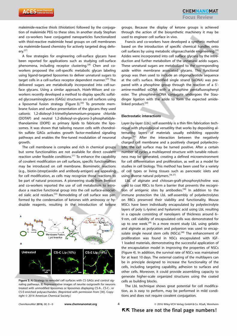
A few strategies for engineering cell-surface glycans havebeen reported for applications such as studying cell-surfacephenomena, including receptor clustering.[28] Chen and co-workers proposed the cell-specific metabolic glycan labelingusing ligand-targeted liposomes to deliver unnatural sugars totarget cells in a cell-surface receptor dependent manner.[29] Thedelivered sugars are metabolically incorporated into cell-sur-face glycans. Using a similar approach, Hsieh-Wilson and co-workers recently developed a method to display specific sulfat-ed glycosaminoglycans (GAGs) structures on cell surfaces usinga liposomal fusion strategy (Figure 3).[30] To promote mem-brane fusion and surface presentation of the glycans they usedcationic 1,2-dioleoyl-3-trimethylammonium-propane chloride(DOTAP) and neutral 1,2-dioleoyl-sn-glycero-3-phosphatidyle-thanolamine (DOPE) as primary lipids to fabricate the lipo-somes. It was shown that tailoring neuron cells with chondroi-tin sulfate GAGs activates growth factor-mediated signalingpathways and enables the fine-tuned modulation of neuronalgrowth.
The cell membrane is complex and rich in chemical groupsbut some functionalities are not available for direct covalentreaction under feasible conditions.[31] To enhance the capabilityof covalent modification on cell surfaces, specific functionalitiesmay be introduced on cell membrane. Biomimetic reactions(e.g. , biotin-(strept)avidin and antibody-antigen) are appealingfor cell modification, as cells may recognize those reactions tobe part of natural processes. A pioneering study from Bertozziand co-workers reported the use of cell metabolism to intro-duce a reactive functional group into the cell surface-associat-ed sialic acid residues.[32] Remodeling of cell surface was per-formed by the condensation of ketones with aminooxy or hy-drazide reagents, resulting in the introduction of ketone
groups. Because the display of ketone groups is achievedthrough the action of the biosynthetic machinery it may beused to engineer cell surface in vivo.
Francis and co-workers have developed a synthetic methodbased on the introduction of specific chemical handles ontocell surfaces by using metabolic oligosaccharide engineering.[33]
Azides were incorporated into cell surface glycans by the intro-duction and further metabolism of the unnatural azido sugars.These unnatural sugars are metabolized to the correspondingazide within membrane associated glycans. This functionalgroup was then used to include an oligonucleotide sequenceat the cell’s surface. Modified single strand (ssDNA) was pre-pared with a phosphine group through the reaction of a 5’-amine-modified ssDNA with a phosphine pentafluorophenylester. The phosphine-ssDNA conjugate undergoes the Stau-dinger ligation with the azide to form the expected amide-linked product.[34]
Electrostatic interactions
Layer-by-layer (LbL) self-assembly is a thin film fabrication tech-nique with physiological versatility that works by depositing al-ternating layers of materials usually exhibiting oppositecharge.[35] After the interaction between the negativelycharged cell membrane and a positively charged polyelectro-lyte, the cell surface may be turned positive. After a certainnumber of cycles a multilayered structure with tunable robust-ness may be generated, creating a defined microenvironmentfor cell differentiation and proliferation, as well as a model forstudies in cell biology. This method has been used for a varietyof cell types or living tissues such as pancreatic islets andusing diverse natural polymers.[36, 37]
LbL of alginate and chitosan-graft-phosphorylcholine wasused to coat RBCs to form a barrier that prevents the recogni-tion of antigenic sites by antibodies.[38] In addition to theimmune protection the LbL self-assembly of polyelectrolyteson RBCs preserved their viability and functionality. MouseMSCs have been individually encapsulated by polyelectrolytelayers of poly (l-lysine) and hyaluronic acid using LbL resultingin a capsule consisting of nanolayers of thickness around 6–9 nm, cell viability of encapsulated cells was demonstrated forup to one week.[39] In a more recent study LbL using gelatinand alginate as polycation and polyanion was used to encap-sulate single neural stem cells (NSCs).[40] The enhancement ofproliferation was found in NSCs encapsulated with IGF-1 loaded materials, demonstrating the successful application ofthe encapsulation model in improving the properties of NSCs(Figure 4). In addition, the survival rate of NSCs was maintainedfor at least 10 days. The external coating of the multilayers canbe in principle designed to increase the functionality of thecells, including targeting capability, adhesion to surfaces andother cells. Moreover, it could provide assembling capacity togenerate higher-scale organized structures using the coatedcells as building blocks.
The LbL technique shows great potential for cell modifica-tion, as is easy to perform, may be performed in mild condi-tions and does not require covalent conjugation.
Figure 3. A) Strategy to remodel cell surfaces with CS GAGs and control sig-naling pathways. B) Representative images of neurite outgrowth for neuronstreated with unmodified liposomes or liposomes displaying CS-A-, CS-C-, orCS-E-enriched polysaccharides. (Reprinted with permission from [30]. Copy-right � 2014 American Chemical Society.)
ChemNanoMat 2016, 00, 0 – 0 www.chemnanomat.org � 2016 Wiley-VCH Verlag GmbH & Co. KGaA, Weinheim4
�� These are not the final page numbers!
Focus Review

Nanopatches and Nanoparticles
Synthetic nanomaterials have a significant role to play in cellsurface engineering, due to their unique properties and abilityto provide functionality beyond that achievable by single mol-ecules. Alternatively to the total and uniform cell surface modi-fication achieved using LbL technique, the modification ofa portion of the cell surface using nanoparticles or nanopatch-es offers other possibilities of tailoring the cell surface. Howev-er, nanoscale objects are easily internalized by cells limiting thetailoring of the surface of the cells. The reduction of internali-zation is therefore the key to construct effective modificationof a portion of the cell membrane. For example Rubner andco-workers studied the influence of cell membrane-attachedmultilayered patches containing superparamagnetic nanoparti-cles onto lymphocytes surfaces on cell viability and migration(Figure 5 A).[41] The functional multilayered patches were suc-cessfully attached to a fraction of the surface area of living in-dividual lymphocytes. Surface-modified cells remain viable atleast 48 h following attachment of the functional patch. More-over patches carrying magnetic nanoparticles allow the cells tobe spatially manipulated using a magnetic field, extendingtheir use in regenerative medicine.[42] Using the biomolecularrecognition through the avidin-biotin interaction, Andersonand co-workers developed nanoparticulate cellular patchesthat were anchored on MSCs possessing biotinylated plasmamembrane. These nanoparticulate patches remain on themembrane of cells for days and provide a new system for cell-mediated tumoritropic drug delivery.[43] A different methodwas recently proposed by Guan and co-workers, based on theuse of microcontact printing (mCP) of polymeric biomaterialsfor functionalizing and assembling live cells (Figure 5 B).[44] Themethod started by spin-coating a thin layer of a temperature-sensitive sacrificial layer on a glass slide. A stamp coated withthe material for functionalization is then placed in contact withthe sacrificial layer. Cells are seeded on the slide at 37 8C toallow immobilization of the cells to the printed biomaterials ;subsequently a decrease to room temperature allows the re-lease of cell–biomaterial complexes. This strategy was then togenerate cell–biomaterial complexes consisting of microcon-tact-printed structures of various sizes, shapes and materials.We envisage that such methodologies could be combinedwith other surface modification of cells, as upon adhesion thedorsal portion of the cells is susceptible to be modified.
Overall the herein-described methods for cell functionaliza-tion confirm that cell–nanomaterials conjugations may act asan ideal system for cell function remodeling and drug releaseto effectively improve cell-based therapies.
Goals of Cell Engineering
In vivo cell migration
Cell based therapies have attracted increased attention in bio-medicine. One of the greatest challenges in cell-based thera-pies is to minimally invasively deliver a large quantity of viablecells to the target tissue. For example the inefficient homing ofsystemically delivered MSCs, is caused predominantly by inade-
Figure 4. Illustration of the major steps involved in the LbL encapsulation: NSCs were first suspended in the polycation solution and then centrifuged andwashed. The polycation layer was supposed to be on the cell surface. Next, the polycation-coated NSCs were put in the polyanion solution to add a secondlayer. The LbL encapsulation would be completed after several repetitions of this process. (Reprinted with permission from [40]. Copyright � 2014 AmericanChemical Society.)
Figure 5. A) Overview of a cell functionalization scheme, with confocalimages demonstrating each step. (1) A regular array of surface-bound patch-es spaced 50 mm apart. The green fluorescence is from the FITC-PAH used tofabricate the payload region. After CH27 B-cell incubation and attachment(2), a majority (85�3 %) of the surface-bound patches are occupied. The redfluorescence is from CellTracker Red CMPTX, which nonselectively tags theinterior of living cells. (3) After the temperature is reduced to 4 8C for30 min, the patches are released from the surface while remaining attachedto the cell membrane. All scale bars are 25 mm. (Reprinted with permissionfrom [41]. Copyright � 2008 American Chemical Society.) B) Procedure offunctionalizing or assembling live cells with microcontact-printed biomateri-als using spin-coated PNIPAM as the sacrificial layer. Microparticles andsingle cells are used here as a model system. (Reprinted with permissionfrom [44]. Copyright � 2014 Elsevier.)
ChemNanoMat 2016, 00, 0 – 0 www.chemnanomat.org � 2016 Wiley-VCH Verlag GmbH & Co. KGaA, Weinheim5
These are not the final page numbers! ��
Focus Review

quate expression of cell surface adhesion receptors.[45] It wasalready reported in this review that covalently conjugatedSLeX on MSC surface through a biotin-streptavidin bridge in-structs cell rolling without altering the cell phenotype and thedifferentiation potential. The conjugation of SLeX on the MSCsurface is stable, versatile, and induces a robust rolling re-sponse on P-selectin coated substrates.[24b, 46] It was also report-ed that the conjugation of stem cells with bispecific antibodiescan be directly injected and retained by injured myocardiumor targeted to injured myocardial tissues for tissue regenera-tion.[47, 48] These methods offer a simple approach to exploreengineered cell homing and potentially target any cell type tospecific tissues via the circulation.
Cell–matrix and cell–cell interactions
The precise arrangement of cells in their substrate is critical incontrolling cell function. The modification of substrates hasbeen widely explored for an accurate control of cell adhesion.More recently, many research groups have been focused theirwork in the modification of the cell surface in order to controlcell fate and biointerfaces interactions. For instance, the func-tionalization of cells with short oligonucleotides promotes spe-cific adhesive properties. Oligonucleotides offer several advan-tages, including highly specificity and ease of synthesis. Cellu-lar lipid bilayers can be modified with oligonucleotides incor-porating hydrophobic molecules at their 3’ or 5’ ends.[49–52]
Iwata and co-workers, prepared amphiphilic PEG-lipid polymersthat were attached to specific oligonucleotide sequences.[53] Byincorporating complementary DNA sequences attached to am-phiphilic PEG-lipids into the membranes of two cell popula-tions, cell–cell or cell–substrate interactions were subsequentlymediated via hybridization between the two complementaryDNA sequences A similar approach was recently described byGartner and co-workers for programming the adhesive proper-ties of cells independent of proteins, glycans, or their endoge-nous adhesion machinery.[54] They develop a strategy to chemi-cally control cell adhesion using membrane anchored single-stranded DNA oligonucleotides (Figure 6). Bertozzi and co-workers reported on non-covalent cell surface engineering asa strategy to display synthetic glycolpolymers that mimic cellsurface mucins, a class of glycoproteins with roles in modulat-ing cell–cell interactions.[11] The same group demonstrated thatthe kinetics of the assembly process is controlled by adjustingthe DNA sequence complexity, density, and cell density. Thus,cell assembly can be tuned, enabling the design of 3-dimen-sional microtissues with defined cell composition and stoichi-ometry.[55] In an alternate approach Francis and co-workersdemonstrated that cell adhesion events can be programmedthrough the attachment of synthetic ssDNA strands to the sur-faces of living cells.[33, 56] DNA strands are used to anchor cellsto specified locations on surfaces in a sequence-dependentfashion. Yousaf and co-workers developed a strategy to inducespecific and stable cell–cell contacts through chemoselectivecell-surface engineering based on liposome fusion that suc-cessfully displayed bioorthogonal functional groups on cellmembrane.[57, 58] Such strategy allows for modulation of cell ad-
hesion, the generation of stable 3D spheroid and multilayeredtissue-like structures. Since the ligation tether contains photo-active lipids, remote control of disassembly could be achievedupon UV light illumination. The use of sophisticated bioplot-ting systems could be employed in the future to assemblesuch aggregates of cells into complex 3D structures witha high geometrical precision.
Control of cellular activity
Cell surface engineering has been explored to control cellularfunctions such as proliferation and differentiation and drug de-livery. A major limitation of cell therapies is the rapid decline inviability and function of the transplanted cells. Stimulatory bio-molecules can be coupled to cells before transplantation toenhance their viability, proliferation and therapeutic poten-tial.[27] Likewise, the conjugation of drug-loaded nanoparticlesto the surfaces of therapeutic cells provides sustained cell stim-ulation, enhancing the efficacy of cell therapies while minimiz-ing the systemic side effects.[21, 59] Using such strategy, drugmolecules may be slowly released from cell-bound nanoparti-cles and primarily recaptured by particle-carrying cells.
The direct deposition approach for cell coating is quitesimple and straightforward method to modulate or control theresponse cells to their environment.[35–39, 60] Positively chargedpolymers can spontaneously bind to the outer-surface mem-brane of microorganism through electrostatic interactions. Suc-cessful application of LbL technology for encapsulation of cellswill have a major impact on a diverse set of clinical fields in-cluding tissue engineering, novel therapeutic treatments, andtargeted delivery. For instance, LbL coating of pancreatic isletswas tested to avoid immune rejection after transplanta-
Figure 6. A) Cell-surface glycans are targeted for chemical remodeling fol-lowing a 3 day incubation in azido sugar 1 and subsequent covalent modifi-cation with difluorocyclooctyne (DIFO, 2)-conjugated DNA. Protein lysineside chains are conjugated to N-hydroxysuccinimide ester-modified DNA 3.Fatty acid amides 6, 7 and dialkylphosphoglyceride-modified oligonucleo-tides 4, 5 (bold) target the lipid bilayer non-covalently. B) Incorporation ofoliognucleotides to cell surfaces. Scheme for labeling and selectively quanti-fying cell-surface oligonucleotides by flow cytometry. C) Fluorescence acti-vated cell sorting (FACS)-purified cell cluster imaged by confocal fluores-cence microscopy. (Reprinted with permission from [54]. Copyright � 2011American Chemical Society.)
ChemNanoMat 2016, 00, 0 – 0 www.chemnanomat.org � 2016 Wiley-VCH Verlag GmbH & Co. KGaA, Weinheim6
�� These are not the final page numbers!
Focus Review
tion.[36, 37] Moreover therapeutic molecules can be includedwithin the LbL protecting shell to control cell function.[60] Forexample, NSCs encapsulated with a IGF-1 loaded LbL coating,significantly enhanced the proliferation of the encapsulatedcells, demonstrating a drug-carrier function of the LbL single-cell nanocoating.[40]
Conclusions
Cells interact with the extracellular environment through themolecular receptors and ligands present on the membrane. Re-search results reviewed in this paper clearly demonstrate thatcell–cell and cell–ECM interactions can be remodeled bymeans of cell surface engineering. Cell surface engineeringrepresents a powerful tool to manipulate living cells by deco-rating the cell membrane with specific molecules of interestand specialized structures, for example nanoparticles andpatches. This provides an alternative to control the adhesionproperties of living cells without a dependence on the recep-tors that they possess. The technologies herein describedshould also have broad implications on cellular therapies thatutilize systemic administration and require targeting of cells tospecific tissues, tissue engineering or drug delivery. In addition,these results suggest therapeutic cells are promising vectorsfor actively targeted drug delivery. Many issues remain to beaddressed in cell surface engineering. The highly dynamicnature of the cell surface and the need of mild reaction condi-tions are still major challenges for living cell surface engineer-ing. Nevertheless, despite the challenges, cell membrane engi-neering has emerged as an effective method to manipulatecell function and cellular nanomodification is a promising strat-egy for improving current and future cell-based therapeuticpractices.
Acknowledgments
C.A.C. acknowledges funding support from the PortugueseFoundation for Science and Technology (FCT) (fellowshipSFRH/BPD/100594/2014). This work was also supported by Eu-ropean Research Council grant agreement ERC-2014-ADG-669858 for project ATLAS. &&Keywords ok?&&
Keywords: cell membranes · ligands · liposomes ·nanoparticles · surface engineering
[1] F. Edalat, I. Sheu, S. Manoucheri, A. Khademhosseini, Curr. Opin. Biotech-nol. 2012, 23, 820 – 825.
[2] C. A. Cust�dio, R. L. Reis, J. F. Mano, Adv. Healthcare Mater. 2014, 3,797 – 810.
[3] C. Frantz, K. M. Stewart, V. M. Weaver, J. Cell Sci. 2010, 123, 4195 – 4200.[4] W. F. Zheng, W. Zhang, X. Y. Jiang, Adv. Healthcare Mater. 2013, 2, 95 –
108.[5] A. Rodrigo-Navarro, P. Rico, A. Saadeddin, A. J. Garcia, M. Salmeron-San-
chez, Sci. Rep. 2014, 4, 5849.[6] K. Simons, J. L. Sampaio, Cold Spring Harbor Perspect. Biol. 2011, 3,
a004697.[7] L. K. Mahal, C. R. Bertozzi, Chem. Biol. 1997, 4, 415 – 422.[8] B. Kellam, P. A. De Bank, K. M. Shakesheff, Chem. Soc. Rev. 2003, 32,
327 – 337.
[9] M. D. Mager, V. LaPointe, M. M. Stevens, Nat. Chem. 2011, 3, 582 – 589.[10] X. D. Xu, H. Cheng, W. H. Chen, S. X. Cheng, R. X. Zhuo, X. Z. Zhang, Sci.
Rep. 2013, 3, &&article number?&&.[11] D. Rabuka, M. B. Forstner, J. T. Groves, C. R. Bertozzi, J. Am. Chem. Soc.
2008, 130, 5947 – 5953.[12] K. Simons, D. Lingwood, U. Coskun, M. Grzybek, FEBS J. 2009, 276, 53 –
53.[13] I. M. Verma, M. D. Weitzman, Annu. Rev. Biochem. 2005, 74, 711 – 738.[14] W. A. Zhao, G. S. L. Teo, N. Kumar, J. M. Karp, Mater. Today 2010, 13, 14 –
21.[15] M. S. Penn, A. A. Mangi, Circ. Res. 2008, 102, 1471 – 1482.[16] M. Kato, M. Mrksich, J. Am. Chem. Soc. 2004, 126, 6504 – 6505.[17] J. H. Collier, M. Mrksich, Proc. Natl. Acad. Sci. USA 2006, 103, 2021 – 2025.[18] M. Howarth, K. Takao, Y. Hayashi, A. Y. Ting, Proc. Natl. Acad. Sci. USA
2005, 102, 7583 – 7588.[19] I. Chen, M. Howarth, W. Y. Lin, A. Y. Ting, Nat. Methods 2005, 2, 99 – 104.[20] P. Wu, W. Q. Shui, B. L. Carlson, N. Hu, D. Rabuka, J. Lee, C. R. Bertozzi,
Proc. Natl. Acad. Sci. USA 2009, 106, 3000 – 3005.[21] M. T. Stephan, D. J. Irvine, Nano Today 2011, 6, 309 – 325.[22] D. Y. Lee, S. J. Park, J. H. Nam, Y. Byun, Tissue Eng. 2006, 12, 615 – 623.[23] Y. Krishnamachari, M. E. Pearce, A. K. Salem, Adv. Mater. 2008, 20, 989 –
993.[24] a) D. Sarkar, P. K. Vemula, D. P. Spelke, J. M. Karp, 2009 35th Annual
Northeast Bioengineering Conference 2009, 68 – 69; b) D. Sarkar, J. A.Spencer, J. A. Phillips, W. A. Zhao, S. Schafer, D. P. Spelke, L. J. Morten-sen, J. P. Ruiz, P. K. Vemula, R. Sridharan, S. Kumar, R. Karnik, C. P. Lin,J. M. Karp, Blood 2011, 118, e184 – e191.
[25] W. A. Zhao, S. Schafer, J. Choi, Y. J. Yamanaka, M. L. Lombardi, S. Bose,A. L. Carlson, J. A. Phillips, W. S. Teo, I. A. Droujinine, C. H. Cui, R. K. Jain,J. Lammerding, J. C. Love, C. P. Lin, D. Sarkar, R. Karnik, J. M. Karp, Nat.Nanotechnol. 2011, 6, 524 – 531.
[26] P. Nacharaju, F. N. Boctor, B. N. Manjula, S. A. Acharya, Transfusion 2005,45, 374 – 383.
[27] M. T. Stephan, J. J. Moon, S. H. Um, A. Bershteyn, D. J. Irvine, Nat. Med.2010, 16, 1035 – U1135.
[28] C. D. Rillahan, A. Antonopoulos, C. T. Lefort, R. Sonon, P. Azadi, K. Ley, A.Dell, S. M. Haslam, J. C. Paulson, Nat. Chem. Biol. 2012, 8, 661 – 668.
[29] R. Xie, S. Hong, L. Feng, J. Rong, X. Chen, J. Am. Chem. Soc. 2012, 134,9914 – 9917.
[30] A. Pulsipher, M. E. Griffin, S. E. Stone, J. M. Brown, L. C. Hsieh-Wilson, J.Am. Chem. Soc. 2014, 136, 6794 – 6797.
[31] D. S. Wade M. Fox, Micro- and Nanoengineering of the Cell Surface, Vol. 1,William Andrew, 2014.
[32] L. K. Mahal, K. J. Yarema, C. R. Bertozzi, Science 1997, 276, 1125 – 1128.[33] R. A. Chandra, E. S. Douglas, R. A. Mathies, C. R. Bertozzi, M. B. Francis,
Angew. Chem. Int. Ed. 2006, 45, 896 – 901; Angew. Chem. 2006, 118, 910 –915.
[34] E. Saxon, C. R. Bertozzi, Science 2000, 287, 2007 – 2010.[35] F. C. Picart, J.-C. Voegel, G. Decher, Layer-by-Layer Films for Biomedical
Applications, Wiley, 2014.[36] M. Germain, P. Balaguer, J. C. Nicolas, F. Lopez, J. P. Esteve, G. B. Sukhor-
ukov, M. Winterhalter, H. Richard-Foy, D. Fournier, Biosens. Bioelectron.2006, 21, 1566 – 1573.
[37] J. T. Wilson, W. Cui, V. Kozlovskaya, E. Kharlampieva, D. Pan, Z. Qu, V. R.Krishnamurthy, J. Mets, V. Kumar, J. Wen, Y. Song, V. V. Tsukruk, E. L.Chaikof, J. Am. Chem. Soc. 2011, 133, 7054 – 7064.
[38] S. Mansouri, Y. Merhi, F. M. Winnik, M. Tabrizian, Biomacromolecules2011, 12, 585 – 592.
[39] N. G. Veerabadran, P. L. Goli, S. S. Stewart-Clark, Y. M. Lvov, D. K. Mills,Macromol. Biosci. 2007, 7, 877 – 882.
[40] W. Li, T. Guan, X. Zhang, Z. Wang, M. Wang, W. Zhong, H. Feng, M. Xing,J. Kong, ACS Appl. Mater. Interfaces 2015, 7, 3018 – 3029.
[41] A. J. Swiston, C. Cheng, S. H. Um, D. J. Irvine, R. E. Cohen, M. F. Rubner,Nano Lett. 2008, 8, 4446 – 4453.
[42] E. Castro, J. F. Mano, J. Biomed. Nanotechnol. 2013, 9, 1129 – 1136.[43] H. Cheng, C. J. Kastrup, R. Ramanathan, D. J. Siegwart, M. L. Ma, S. R. Bo-
gatyrev, Q. B. Xu, K. A. Whitehead, R. Langer, D. G. Anderson, ACS Nano2010, 4, 625 – 631.
[44] Z. Wang, J. Xia, Y. Yan, A. C. Tsai, Y. Li, T. Ma, J. Guan, Acta Biomater.2015, 11, 80 – 87.
ChemNanoMat 2016, 00, 0 – 0 www.chemnanomat.org � 2016 Wiley-VCH Verlag GmbH & Co. KGaA, Weinheim7
These are not the final page numbers! ��
Focus Review
[45] R. Sackstein, J. S. Merzaban, D. W. Cain, N. M. Dagia, J. A. Spencer, C. P.Lin, R. Wohlgemuth, Nat. Med. 2008, 14, 181 – 187.
[46] D. Sarkar, P. K. Vemula, G. S. L. Teo, D. Spelke, R. Karnik, L. Y. Wee, J. M.Karp, Bioconjugate Chem. 2008, 19, 2105 – 2109.
[47] a) L. G. Lum, H. Fok, R. Sievers, M. Abedi, P. J. Quesenberry, R. J. Lee,Blood Cells Mol. Dis. 2004, 32, 82 – 87; b) R. J. Lee, Q. Z. Fang, P. A. Davol,Y. P. Gu, R. E. Sievers, R. C. Grabert, J. M. Gall, E. Tsang, M. S. Yee, H. Fok,N. F. Huang, J. F. Padbury, J. W. Larrick, L. G. Lum, Stem Cells 2007, 25,712 – 717.
[48] J. S. Yu, Y. K. Wu, Y. P. Gu, Q. Z. Fang, R. Sievers, C. H. Ding, J. E. Olgin,R. J. Lee, J. Cell. Mol. Med. 2015, 19, 1483 – 1491.
[49] G. G. Borisenko, M. A. Zaitseva, A. N. Chuvilin, G. E. Pozmogova, NucleicAcids Res. 2009, 37, &&article number?&&.
[50] K. Borjesson, J. Tumpane, T. Ljungdahl, L. M. Wilhelmsson, B. Norden, T.Brown, J. Martensson, B. Albinsson, J. Am. Chem. Soc. 2009, 131, 2831 –2839.
[51] T. Itagaki, Y. Arima, R. Kuwabara, N. Kitamura, H. Iwata, Colloids Surf. B2015, 135, 765 – 773.
[52] T. Matsui, Y. Arima, N. Takemoto, H. Lwata, Acta Biomater. 2015, 13, 32 –41.
[53] Y. Teramura, H. Chen, T. Kawamoto, H. Iwata, Biomaterials 2010, 31,2229 – 2235.
[54] N. S. Selden, M. E. Todhunter, N. Y. Jee, J. S. Liu, K. E. Broaders, Z. J. Gart-ner, J. Am. Chem. Soc. 2012, 134, 765 – 768.
[55] Z. J. Gartner, C. R. Bertozzi, Proc. Natl. Acad. Sci. USA 2009, 106, 4606 –4610.
[56] S. C. Hsiao, B. J. Shum, H. Onoe, E. S. Douglas, Z. J. Gartner, R. A. Math-ies, C. R. Bertozzi, M. B. Francis, Langmuir 2009, 25, 6985 – 6991.
[57] D. Dutta, A. Pulsipher, W. Luo, M. N. Yousaf, J. Am. Chem. Soc. 2011, 133,8704 – 8713.
[58] W. Luo, A. Pulsipher, D. Dutta, B. M. Lamb, M. N. Yousaf, Sci. Rep. 2014,4, &&article number?&&.
[59] W. Chen, L. W. Fu, X. Y. Chen, J. Controlled Release 2015, 219, 560 – 575.[60] V. Gribova, R. Auzely-Velty, C. Picart, Chem. Mater. 2012, 24, 854 – 869.
Manuscript received: February 2, 2016
Accepted Article published: && &&, 0000
Final Article published: && &&, 0000
ChemNanoMat 2016, 00, 0 – 0 www.chemnanomat.org � 2016 Wiley-VCH Verlag GmbH & Co. KGaA, Weinheim8
�� These are not the final page numbers!
Focus Review

REVIEW
Biointerfaces
Catarina A. Cust�dio, Jo¼o F. Mano*
&& –&&
Cell Surface Engineering to ControlCellular Interactions
More than skin deep : This FocusReview highlights the recent progresson engineering cell surfaces with bio-logical, chemical or physical methods tomodulate cell functions and controlcell–cell and cell–microenvironment in-teractions. &&Please provide an18x18cm frontispiece image with theproofs, and academic titles for all au-thors&&
Focus Review from J. Mano @udominho on cell #surface engineering SPACE RESERVED FOR IMAGE AND LINK
Share your work on social media! ChemNanoMat has added Twitter as a means to promote your article. Twitter is an onlinemicroblogging service that enables its users to send and read text-based messages of up to 140 characters, known as “tweets”.Please check the pre-written tweet in the galley proofs for accuracy. Should you or your institute have a Twitter account,please let us know the appropriate username (i.e., @accountname), and we will do our best to include this information in thetweet. This tweet will be posted to the journal�s Twitter account @ChemNanoMat (follow us!) upon online publication of yourarticle, and we recommended you to repost (“retweet”) it to alert other researchers about your publication.
Please check that the ORCID identifiers listed below are correct. We encourage all authors to provide an ORCIDidentifier for each coauthor. ORCID is a registry that provides researchers with a unique digital identifier. Somefunding agencies recommend or even require the inclusion of ORCID IDs in all published articles, and authors shouldconsult their funding agency guidelines for details. Registration is easy and free; for further information, see http://orcid.org/.
Catarina A. Cust�dio http://orcid.org/0000-0003-2967-700XJo¼o F. Mano
ChemNanoMat 2016, 00, 0 – 0 www.chemnanomat.org � 2016 Wiley-VCH Verlag GmbH & Co. KGaA, Weinheim9
These are not the final page numbers! ��
Focus Review