Embed Size (px)
Citation preview
Resource
CellNet: Network Biology Appliedto Stem Cell EngineeringPatrick Cahan,1,2,3,8 Hu Li,4,8 Samantha A. Morris,1,2,3,8 Edroaldo Lummertz da Rocha,5,6,7 George Q. Daley,1,2,3,9,*and James J. Collins5,9,*1Stem Cell Transplantation Program, Division of Pediatric Hematology and Oncology, Manton Center for Orphan Disease Research, Howard
Hughes Medical Institute, Boston Children’s Hospital and Dana Farber Cancer Institute, Boston, MA 02115, USA2Department of Biological Chemistry and Molecular Pharmacology, Harvard Medical School, Boston, MA 02115, USA3Harvard Stem Cell Institute, Cambridge, MA 02138, USA4Center for Individualized Medicine, Department of Molecular Pharmacology and Experimental Therapeutics, Mayo Clinic College of
Medicine, Rochester, MN 55905, USA5Howard Hughes Medical Institute, Department of Biomedical Engineering and Center of Synthetic Biology, Boston University, Boston, MA02215, USA6Wyss Institute for Biologically Inspired Engineering, Harvard University, Boston, MA 02115, USA7Graduate Program in Materials Science and Engineering, Federal University of Santa Catarina, 88040-900 Florianopolis, Brazil8Co-first author9Co-senior author
*Correspondence: [email protected] (G.Q.D.), [email protected] (J.J.C.)
http://dx.doi.org/10.1016/j.cell.2014.07.020
SUMMARY
Somatic cell reprogramming, directed differentiationof pluripotent stem cells, and direct conversions be-tween differentiated cell lineages represent power-ful approaches to engineer cells for research andregenerative medicine. We have developed CellNet,a network biology platform that more accuratelyassesses the fidelity of cellular engineering than ex-isting methodologies and generates hypotheses forimproving cell derivations. Analyzing expressiondata from 56 published reports, we found that cellsderived via directed differentiation more closelyresemble their in vivo counterparts than productsof direct conversion, as reflected by the establish-ment of target cell-type gene regulatory networks(GRNs). Furthermore, we discovered that directlyconverted cells fail to adequately silence expressionprograms of the starting population and that theestablishment of unintended GRNs is common tovirtually every cellular engineering paradigm. CellNetprovides a platform for quantifying how closelyengineered cell populations resemble their targetcell type and a rational strategy to guide enhancedcellular engineering.
INTRODUCTION
Transitions between cellular states are fundamental to develop-
ment, physiology, and pathology. Directing state transitions
in vitro is a current preoccupation of stem cell biology, as the
derived cells can be used to investigate otherwise inaccessible
cell types in development and disease, for drug screening, and
for regenerative cell therapies. Dramatic cell-state transitions
have been achieved in vitro and in vivo through the enforced
expression of transcription factors. For example, differentiated
somatic cells—fibroblasts (Takahashi and Yamanaka, 2006),
keratinocytes (Aasen et al., 2008), peripheral blood (Loh et al.,
2010; Staerk et al., 2010), and neural progenitors (Kim et al.,
2009)—have been reprogrammed to pluripotent stem cells;
fibroblasts have been converted to cells resembling myoblasts
(Davis et al., 1987), motor neurons (Vierbuchen et al., 2010), car-
diomyocytes (Ieda et al., 2010), hepatocytes (Huang et al., 2011;
Sekiya and Suzuki, 2011), and blood progenitors (Szabo et al.,
2010); B cells have been converted to macrophage-like cells
(Xie et al., 2004); and exocrine pancreas cells have been con-
verted to insulin-producing beta cells (Zhou et al., 2008). Further-
more, pluripotent stem cells can be coaxed to specific lineages
through a combination of defined growth conditions and ectopic
gene expression (Murry and Keller, 2008).
The widespread practice of cellular engineering has raised
critical questions about the relationship of the derived cells to
their native counterparts. To what extent does a cell population
engineered in vitro resemble the corresponding target cell or
tissue in both molecular and functional terms? While functional
complementation via transplantation in live animals has been
used to assess the ability of engineered cells to mimic the phys-
iology of their native counterparts, such experiments are techni-
cally challenging, lack quantitative rigor, and provide limited
insights when judging human tissue function in animal hosts.
The molecular similarity of engineered populations is typically
assessed by semiquantitative PCR, array-based expression
profiling, or RNA sequencing followed by simple clustering
analysis. However, such global analyses do not provide an intu-
itive or a quantitative means for diagnosing the deficiencies of
engineered cells, nor do they provide a systematic approach to
prioritize interventions to improve derivations of the desired
populations.
Cell 158, 903–915, August 14, 2014 ª2014 Elsevier Inc. 903
A
B
C D
E F
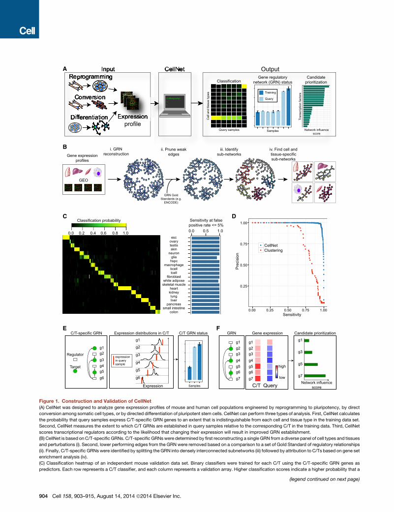
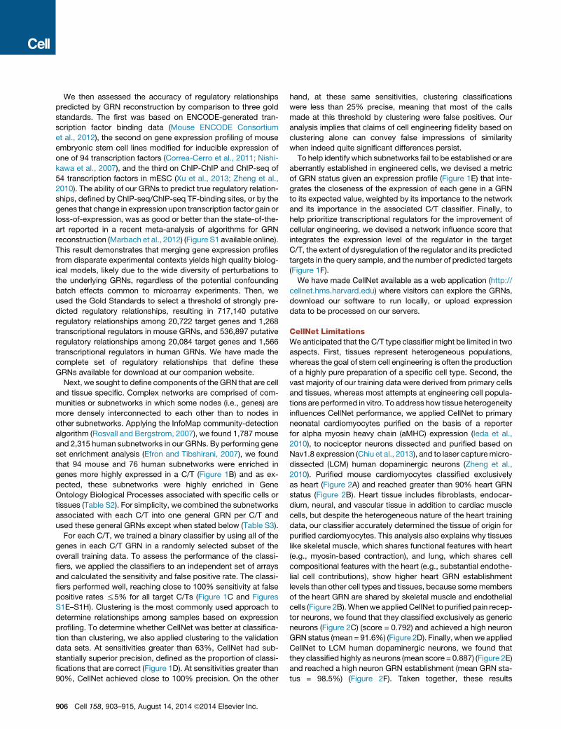
Figure 1. Construction and Validation of CellNet
(A) CellNet was designed to analyze gene expression profiles of mouse and human cell populations engineered by reprogramming to pluripotency, by direct
conversion among somatic cell types, or by directed differentiation of pluripotent stem cells. CellNet can perform three types of analysis. First, CellNet calculates
the probability that query samples express C/T-specific GRN genes to an extent that is indistinguishable from each cell and tissue type in the training data set.
Second, CellNet measures the extent to which C/T GRNs are established in query samples relative to the corresponding C/T in the training data. Third, CellNet
scores transcriptional regulators according to the likelihood that changing their expression will result in improved GRN establishment.
(B) CellNet is based on C/T-specific GRNs. C/T-specific GRNs were determined by first reconstructing a single GRN from a diverse panel of cell types and tissues
and perturbations (i). Second, lower performing edges from the GRN were removed based on a comparison to a set of Gold Standard of regulatory relationships
(ii). Finally, C/T-specific GRNs were identified by splitting the GRN into densely interconnected subnetworks (iii) followed by attribution to C/Ts based on gene set
enrichment analysis (iv).
(C) Classification heatmap of an independent mouse validation data set. Binary classifiers were trained for each C/T using the C/T-specific GRN genes as
predictors. Each row represents a C/T classifier, and each column represents a validation array. Higher classification scores indicate a higher probability that a
(legend continued on next page)
904 Cell 158, 903–915, August 14, 2014 ª2014 Elsevier Inc.
Here, we provide a network biology platform, CellNet, which
assesses the fidelity of cell fate conversions and generates spe-
cific hypotheses aimed at improving derived cell populations.
Our platform includes both novel and previously described com-
ponents, which we outline below. We describe the construction
of this platform for human and mouse cell and tissue types and
use it to assess the results of 56 published attempts at reprog-
ramming to pluripotency (most of which use the canonical
reprogramming factors Oct4, Sox2, Klf4, and Myc), directed
differentiation, and direct conversion of somatic cells. On the
basis of these analyses, we have documented quantitatively
that reprogramming is the most complete and successful of
the various cell fate conversions; indeed, CellNet confirms that
iPSC are virtually indistinguishable from ES cells in their faithful
establishment of gene regulatory networks (GRNs). Further, we
show that neurons and cardiomyocytes derived by directed dif-
ferentiation of pluripotent stem cells more completely establish
the target tissue- and cell-type GRNs than do neurons and car-
diomyocytes directly converted from fibroblasts. Moreover,
analysis of cardiomyocytes converted from cardiac fibroblasts
in situ demonstrates that the in vivo environment provides selec-
tive and/or inductive signals that more completely establish
heart GRNs. We also demonstrate that GRNs of the starting
cell type are detectable in purified populations of both directed
differentiation and in direct conversion experiments, and we
show that the establishment of unintended GRNs is common
to virtually every cellular engineering paradigm. Thus CellNet
provides a platform for assessing and improving efforts at
cellular engineering.
RESULTS
CellNet Construction and ValidationCellNet is predicated on the discovery of GRNs, which govern
the steady-state expression program of a particular cell type
as well as its transcriptional responses to environment, disease,
and age. GRNs thus act as major molecular determinants of cell-
type identity (Davidson and Erwin, 2006). We reasoned that
measuring the establishment of cell and tissue (C/T)-specific
GRNs in engineered populations would serve as both a robust
metric of cellular identity and a tool to identify aberrant regulatory
nodes. We designed CellNet to query gene expression profiles
for the extent to which C/T GRNs are established, to classify
input samples by their similarity to target cells and tissues, and
to score transcriptional regulators according to their likelihood
of improving the engineered population (Figure 1A).
query sample expresses the C/T GRN genes at a level indistinguishable from the s
source of the validation samples at a false positive rate % 5%.
(D) Precision and sensitivity curves of CellNet (blue) and hierarchical clustering (red
positives divided by the number of positive calls. Sensitivity is the number of true p
the average of all mouse C/T classifiers computed across a range of classificatio
(E) Using a gene expression profile to quantify GRN establishment or status. Ce
distributions of gene expression in the C/T based on the training data and integra
connectivity of each gene to arrive at GRN status, which is normalized to the GR
(F) Combining gene expression with GRNs to prioritize transcriptional regulators
selected target C/T, CellNet computes a network influence score, which scores e
dysregulation of target genes and the regulator, weighted by the expression of t
See Figure S1.
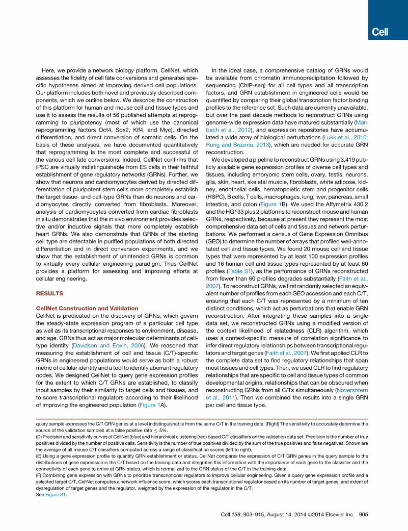
In the ideal case, a comprehensive catalog of GRNs would
be available from chromatin immunoprecipitation followed by
sequencing (ChIP-seq) for all cell types and all transcription
factors, and GRN establishment in engineered cells would be
quantified by comparing their global transcription factor binding
profiles to the reference set. Such data are currently unavailable,
but over the past decade methods to reconstruct GRNs using
genome-wide expression data have matured substantially (Mar-
bach et al., 2012), and expression repositories have accumu-
lated a wide array of biological perturbations (Lukk et al., 2010;
Rung and Brazma, 2013), which are needed for accurate GRN
reconstruction.
Wedevelopedapipeline to reconstructGRNsusing3,419pub-
licly available gene expression profiles of diverse cell types and
tissues, including embryonic stem cells, ovary, testis, neurons,
glia, skin, heart, skeletal muscle, fibroblasts, white adipose, kid-
ney, endothelial cells, hematopoietic stem and progenitor cells
(HSPC),Bcells, T cells,macrophages, lung, liver, pancreas, small
intestine, and colon (Figure 1B). We used the Affymetrix 430.2
and theHG133plus 2platforms to reconstructmouseand human
GRNs, respectively, because at present they represent the most
comprehensive data set of cells and tissues and network pertur-
bations. We performed a census of Gene Expression Omnibus
(GEO) to determine the number of arrays that profiled well-anno-
tated cell and tissue types. We found 20 mouse cell and tissue
types that were represented by at least 100 expression profiles
and 16 human cell and tissue types represented by at least 60
profiles (Table S1), as the performance of GRNs reconstructed
from fewer than 60 profiles degrades substantially (Faith et al.,
2007). To reconstructGRNs,we first randomly selected an equiv-
alent number of profiles from eachGEO accession and eachC/T,
ensuring that each C/T was represented by a minimum of ten
distinct conditions, which act as perturbations that enable GRN
reconstruction. After integrating these samples into a single
data set, we reconstructed GRNs using a modified version of
the context likelihood of relatedness (CLR) algorithm, which
uses a context-specific measure of correlation significance to
infer direct regulatory relationships between transcriptional regu-
lators and target genes (Faith et al., 2007).We first appliedCLR to
the complete data set to find regulatory relationships that span
most tissues and cell types. Then, we usedCLR to find regulatory
relationships that are specific to cell and tissue types of common
developmental origins, relationships that can be obscured when
reconstructing GRNs from all C/Ts simultaneously (Novershtern
et al., 2011). Then we combined the results into a single GRN
per cell and tissue type.
ame C/T in the training data. (Right) The sensitivity to accurately determine the
) basedC/T classifiers on the validation data set. Precision is the number of true
ositives divided by the sum of the true positives and false negatives. Shown are
n scores (left to right).
llNet compares the expression of C/T GRN genes in the query sample to the
tes this information with the importance of each gene to the classifier and the
N status of the C/T in the training data.
to improve cellular engineering. Given a query gene expression profile and a
ach transcriptional regulator based on its number of target genes, and extent of
he regulator in the C/T.
Cell 158, 903–915, August 14, 2014 ª2014 Elsevier Inc. 905
We then assessed the accuracy of regulatory relationships
predicted by GRN reconstruction by comparison to three gold
standards. The first was based on ENCODE-generated tran-
scription factor binding data (Mouse ENCODE Consortium
et al., 2012), the second on gene expression profiling of mouse
embryonic stem cell lines modified for inducible expression of
one of 94 transcription factors (Correa-Cerro et al., 2011; Nishi-
kawa et al., 2007), and the third on ChIP-ChIP and ChIP-seq of
54 transcription factors in mESC (Xu et al., 2013; Zheng et al.,
2010). The ability of our GRNs to predict true regulatory relation-
ships, defined by ChIP-seq/ChIP-seq TF-binding sites, or by the
genes that change in expression upon transcription factor gain or
loss-of-expression, was as good or better than the state-of-the-
art reported in a recent meta-analysis of algorithms for GRN
reconstruction (Marbach et al., 2012) (Figure S1 available online).
This result demonstrates that merging gene expression profiles
from disparate experimental contexts yields high quality biolog-
ical models, likely due to the wide diversity of perturbations to
the underlying GRNs, regardless of the potential confounding
batch effects common to microarray experiments. Then, we
used the Gold Standards to select a threshold of strongly pre-
dicted regulatory relationships, resulting in 717,140 putative
regulatory relationships among 20,722 target genes and 1,268
transcriptional regulators in mouse GRNs, and 536,897 putative
regulatory relationships among 20,084 target genes and 1,566
transcriptional regulators in human GRNs. We have made the
complete set of regulatory relationships that define these
GRNs available for download at our companion website.
Next, we sought to define components of the GRN that are cell
and tissue specific. Complex networks are comprised of com-
munities or subnetworks in which some nodes (i.e., genes) are
more densely interconnected to each other than to nodes in
other subnetworks. Applying the InfoMap community-detection
algorithm (Rosvall and Bergstrom, 2007), we found 1,787 mouse
and 2,315 human subnetworks in our GRNs. By performing gene
set enrichment analysis (Efron and Tibshirani, 2007), we found
that 94 mouse and 76 human subnetworks were enriched in
genes more highly expressed in a C/T (Figure 1B) and as ex-
pected, these subnetworks were highly enriched in Gene
Ontology Biological Processes associated with specific cells or
tissues (Table S2). For simplicity, we combined the subnetworks
associated with each C/T into one general GRN per C/T and
used these general GRNs except when stated below (Table S3).
For each C/T, we trained a binary classifier by using all of the
genes in each C/T GRN in a randomly selected subset of the
overall training data. To assess the performance of the classi-
fiers, we applied the classifiers to an independent set of arrays
and calculated the sensitivity and false positive rate. The classi-
fiers performed well, reaching close to 100% sensitivity at false
positive rates %5% for all target C/Ts (Figure 1C and Figures
S1E–S1H). Clustering is the most commonly used approach to
determine relationships among samples based on expression
profiling. To determine whether CellNet was better at classifica-
tion than clustering, we also applied clustering to the validation
data sets. At sensitivities greater than 63%, CellNet had sub-
stantially superior precision, defined as the proportion of classi-
fications that are correct (Figure 1D). At sensitivities greater than
90%, CellNet achieved close to 100% precision. On the other
906 Cell 158, 903–915, August 14, 2014 ª2014 Elsevier Inc.
hand, at these same sensitivities, clustering classifications
were less than 25% precise, meaning that most of the calls
made at this threshold by clustering were false positives. Our
analysis implies that claims of cell engineering fidelity based on
clustering alone can convey false impressions of similarity
when indeed quite significant differences persist.
To help identify which subnetworks fail to be established or are
aberrantly established in engineered cells, we devised a metric
of GRN status given an expression profile (Figure 1E) that inte-
grates the closeness of the expression of each gene in a GRN
to its expected value, weighted by its importance to the network
and its importance in the associated C/T classifier. Finally, to
help prioritize transcriptional regulators for the improvement of
cellular engineering, we devised a network influence score that
integrates the expression level of the regulator in the target
C/T, the extent of dysregulation of the regulator and its predicted
targets in the query sample, and the number of predicted targets
(Figure 1F).
We have made CellNet available as a web application (http://
cellnet.hms.harvard.edu) where visitors can explore the GRNs,
download our software to run locally, or upload expression
data to be processed on our servers.
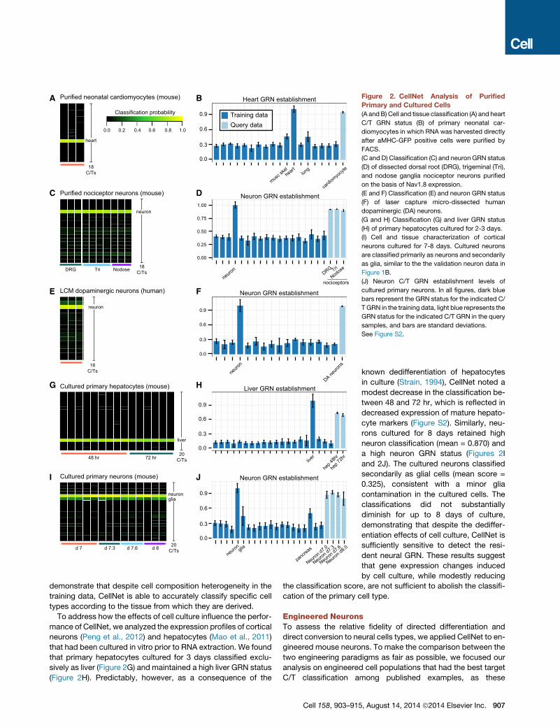
CellNet LimitationsWe anticipated that the C/T type classifier might be limited in two
aspects. First, tissues represent heterogeneous populations,
whereas the goal of stem cell engineering is often the production
of a highly pure preparation of a specific cell type. Second, the
vast majority of our training data were derived from primary cells
and tissues, whereas most attempts at engineering cell popula-
tions are performed in vitro. To address how tissue heterogeneity
influences CellNet performance, we applied CellNet to primary
neonatal cardiomyocytes purified on the basis of a reporter
for alpha myosin heavy chain (aMHC) expression (Ieda et al.,
2010), to nociceptor neurons dissected and purified based on
Nav1.8 expression (Chiu et al., 2013), and to laser capturemicro-
dissected (LCM) human dopaminergic neurons (Zheng et al.,
2010). Purified mouse cardiomyocytes classified exclusively
as heart (Figure 2A) and reached greater than 90% heart GRN
status (Figure 2B). Heart tissue includes fibroblasts, endocar-
dium, neural, and vascular tissue in addition to cardiac muscle
cells, but despite the heterogeneous nature of the heart training
data, our classifier accurately determined the tissue of origin for
purified cardiomyocytes. This analysis also explains why tissues
like skeletal muscle, which shares functional features with heart
(e.g., myosin-based contraction), and lung, which shares cell
compositional features with the heart (e.g., substantial endothe-
lial cell contributions), show higher heart GRN establishment
levels than other cell types and tissues, because somemembers
of the heart GRN are shared by skeletal muscle and endothelial
cells (Figure 2B).Whenwe appliedCellNet to purified pain recep-
tor neurons, we found that they classified exclusively as generic
neurons (Figure 2C) (score = 0.792) and achieved a high neuron
GRN status (mean = 91.6%) (Figure 2D). Finally, whenwe applied
CellNet to LCM human dopaminergic neurons, we found that
they classified highly as neurons (mean score = 0.887) (Figure 2E)
and reached a high neuron GRN establishment (mean GRN sta-
tus = 98.5%) (Figure 2F). Taken together, these results
H Liver GRN establishment
J Neuron GRN establishment
0.0
0.3
0.6
0.9
neuronglia
pancreas
Neuron d7.0
Neuron d7.3
Neuron d7.6
Neuron d8.0
0.0
0.3
0.6
0.9
liver
hep 4
8hr
hep 7
2hr
Training dataQuery data
Cultured primary hepatocytes (mouse)G
I Cultured primary neurons (mouse)
20C/Tsd 7 d 7.3 d 7.6 d 8
neuronglia
B Heart GRN establishment
heart
cardiomyo
cyte
0.0
0.3
0.6
0.9Classification probability
1.00.0 0.80.60.40.2
A Purified neonatal cardiomyocytes (mouse)
C Purified nociceptor neurons (mouse)
E
0.00
0.25
0.50
0.75
1.00
DRG Tri
Nodose
Neuron GRN establishmentD
neuron
nociceptors
F
0.0
0.3
0.6
0.9
neur
on
DA neu
rons
LCM dopaminergic neurons (human)
heart
18C/Ts
neuron
18C/Ts
neuron
16C/Ts
Neuron GRN establishment
musc ske
llung
48 hr 72 hr
liver
20C/Ts
DRG Tri Nodose
Figure 2. CellNet Analysis of Purified
Primary and Cultured Cells
(A andB) Cell and tissue classification (A) and heart
C/T GRN status (B) of primary neonatal car-
diomyocytes in which RNA was harvested directly
after aMHC-GFP positive cells were purified by
FACS.
(C and D) Classification (C) and neuron GRN status
(D) of dissected dorsal root (DRG), trigeminal (Tri),
and nodose ganglia nociceptor neurons purified
on the basis of Nav1.8 expression.
(E and F) Classification (E) and neuron GRN status
(F) of laser capture micro-dissected human
dopaminergic (DA) neurons.
(G and H) Classification (G) and liver GRN status
(H) of primary hepatocytes cultured for 2-3 days.
(I) Cell and tissue characterization of cortical
neurons cultured for 7-8 days. Cultured neurons
are classified primarily as neurons and secondarily
as glia, similar to the the validation neuron data in
Figure 1B.
(J) Neuron C/T GRN establishment levels of
cultured primary neurons. In all figures, dark blue
bars represent the GRN status for the indicated C/
TGRN in the training data, light blue represents the
GRN status for the indicated C/T GRN in the query
samples, and bars are standard deviations.
See Figure S2.
demonstrate that despite cell composition heterogeneity in the
training data, CellNet is able to accurately classify specific cell
types according to the tissue from which they are derived.
To address how the effects of cell culture influence the perfor-
mance of CellNet, we analyzed the expression profiles of cortical
neurons (Peng et al., 2012) and hepatocytes (Mao et al., 2011)
that had been cultured in vitro prior to RNA extraction. We found
that primary hepatocytes cultured for 3 days classified exclu-
sively as liver (Figure 2G) and maintained a high liver GRN status
(Figure 2H). Predictably, however, as a consequence of the
Cell 158, 903–915
known dedifferentiation of hepatocytes
in culture (Strain, 1994), CellNet noted a
modest decrease in the classification be-
tween 48 and 72 hr, which is reflected in
decreased expression of mature hepato-
cyte markers (Figure S2). Similarly, neu-
rons cultured for 8 days retained high
neuron classification (mean = 0.870) and
a high neuron GRN status (Figures 2I
and 2J). The cultured neurons classified
secondarily as glial cells (mean score =
0.325), consistent with a minor glia
contamination in the cultured cells. The
classifications did not substantially
diminish for up to 8 days of culture,
demonstrating that despite the dediffer-
entiation effects of cell culture, CellNet is
sufficiently sensitive to detect the resi-
dent neural GRN. These results suggest
that gene expression changes induced
by cell culture, while modestly reducing
the classification score, are not sufficient to abolish the classifi-
cation of the primary cell type.
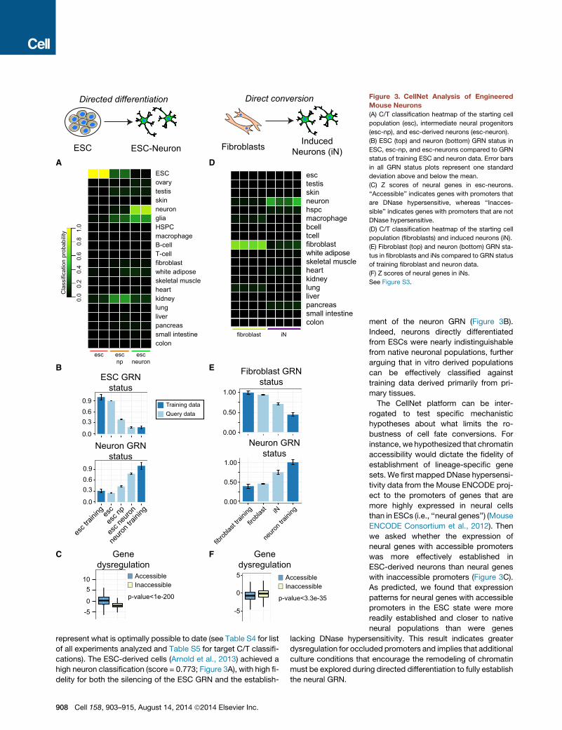
Engineered NeuronsTo assess the relative fidelity of directed differentiation and
direct conversion to neural cells types, we applied CellNet to en-
gineered mouse neurons. To make the comparison between the
two engineering paradigms as fair as possible, we focused our
analysis on engineered cell populations that had the best target
C/T classification among published examples, as these
, August 14, 2014 ª2014 Elsevier Inc. 907
fibroblast iN
colonsmall intestinepancreasliverlungkidneyheartskeletal musclewhite adiposefibroblasttcellbcellmacrophagehspcneuronskintestisesc
ESC ESC-NeuronA
Directed differentiation
Fibroblasts InducedNeurons (iN)
F
Direct conversion
B
esc escnp
escneuron
colonsmall intestinepancreasliverlungkidneyheartskeletal musclewhite adiposefibroblastT-cellB-cellmacrophageHSPCglianeuronskintestisovaryESC
C
E
0.0
0.30.60.9
esc t
rainin
ges
c
esc n
p
esc n
euron
neuro
n trai
ning
ESC GRNstatus
Neuron GRNstatus
0.0
0.30.60.9
D
0-5
510
Genedysregulation
AccessibleInaccessible
p-value<1e-200
Cla
ssifi
catio
n pr
obab
ility 1.
00.
00.
80.
60.
40.
2
Neuron GRNstatus
Fibroblast GRNstatus
0.00
0.50
1.00
0.00
0.50
1.00
fibrob
last tr
aining
firobla
st iN
neuro
n trai
ning
Genedysregulation
0
5
-5
AccessibleInaccessible
p-value<3.3e-35
Training dataQuery data
Figure 3. CellNet Analysis of Engineered
Mouse Neurons
(A) C/T classification heatmap of the starting cell
population (esc), intermediate neural progenitors
(esc-np), and esc-derived neurons (esc-neuron).
(B) ESC (top) and neuron (bottom) GRN status in
ESC, esc-np, and esc-neurons compared to GRN
status of training ESC and neuron data. Error bars
in all GRN status plots represent one standard
deviation above and below the mean.
(C) Z scores of neural genes in esc-neurons.
‘‘Accessible’’ indicates genes with promoters that
are DNase hypersensitive, whereas ‘‘Inacces-
sible’’ indicates genes with promoters that are not
DNase hypersensitive.
(D) C/T classification heatmap of the starting cell
population (fibroblasts) and induced neurons (iN).
(E) Fibroblast (top) and neuron (bottom) GRN sta-
tus in fibroblasts and iNs compared to GRN status
of training fibroblast and neuron data.
(F) Z scores of neural genes in iNs.
See Figure S3.
represent what is optimally possible to date (see Table S4 for list
of all experiments analyzed and Table S5 for target C/T classifi-
cations). The ESC-derived cells (Arnold et al., 2013) achieved a
high neuron classification (score = 0.773; Figure 3A), with high fi-
delity for both the silencing of the ESC GRN and the establish-
908 Cell 158, 903–915, August 14, 2014 ª2014 Elsevier Inc.
ment of the neuron GRN (Figure 3B).
Indeed, neurons directly differentiated
from ESCs were nearly indistinguishable
from native neuronal populations, further
arguing that in vitro derived populations
can be effectively classified against
training data derived primarily from pri-
mary tissues.
The CellNet platform can be inter-
rogated to test specific mechanistic
hypotheses about what limits the ro-
bustness of cell fate conversions. For
instance, we hypothesized that chromatin
accessibility would dictate the fidelity of
establishment of lineage-specific gene
sets. We first mapped DNase hypersensi-
tivity data from the Mouse ENCODE proj-
ect to the promoters of genes that are
more highly expressed in neural cells
than in ESCs (i.e., ‘‘neural genes’’) (Mouse
ENCODE Consortium et al., 2012). Then
we asked whether the expression of
neural genes with accessible promoters
was more effectively established in
ESC-derived neurons than neural genes
with inaccessible promoters (Figure 3C).
As predicted, we found that expression
patterns for neural genes with accessible
promoters in the ESC state were more
readily established and closer to native
neural populations than were genes
lacking DNase hypersensitivity. This result indicates greater
dysregulation for occluded promoters and implies that additional
culture conditions that encourage the remodeling of chromatin
must be explored during directed differentiation to fully establish
the neural GRN.
We tested whether GRNs associated with lineages other than
ESCs and neurons were detected in ESC-derived neurons. As
expected, we detected modestly high glia classification scores
in populations of ESC-derived neurons, which typically include
glial contaminants, and consistent with the known glia constitu-
ents that contaminate the neuron training data (Figure 1C).
Next, we applied CellNet to the direct conversion of mouse
fibroblasts to dopaminergic neurons via the ectopic expression
of Ascl1, Nr4a2, and Lmx1a (Caiazzo et al., 2011). Induced neu-
rons (iN) were classified as generic neurons (mean score = 0.340;
Figure 3D), consistent with the functional characteristics of these
cells (e.g., spontaneous action potentials resembling in vivo neu-
rons and K+ induced monoamine release), but not as robustly as
did ESC-neurons (mean score = 0.773). Despite the fact that iN
were purified on the basis of TH-GFP positivity prior to RNA
collection, they exhibited high fibroblast GRN status (Figure 3E)
and a correspondingly high dysregulation of the fibroblast
GRN regulators (Figure S3A), suggesting that the converted
cells retain fibroblast identity. To explore possible mechanisms
whereby neural genes remained silenced, we asked whether
there was an association between promoter accessibility and
dysregulation of neural genes. In contrast to the case of ESC-
neurons, we found that neural genes with inaccessible pro-
moters in fibroblasts tended to be less dysregulated than
accessible genes, suggesting that nucleosome occlusion is not
a major factor limiting establishment of endogenous GRNs in
the directly converted population, consistent with Ascl1 acting
as a pioneer factor that opens occluded chromatin (Wapinski
et al., 2013). Taken together, this analysis suggests that directed
conversion establishes a neuron GRN but fails to silence the
fibroblast GRN, resulting in a hybrid cell type.
We tested whether GRNs and constituent subnetworks
associated with lineages other than fibroblasts and neurons
were established in iN (relative to fibroblasts or neurons). We
found that the heart subnetwork 1 and pancreas subnetwork 1
were partially established (Figures S3B and S3C), and specu-
lated that the aberrant GRNs arose because one or more of
the conversion factors specifically targeted the aberrant subnet-
works. To explore this possibility, we determined the enrichment
of CellNet predicted targets of each conversion factor in each
subnetwork, finding that indeed the pancreas subnetwork 1
was significantly enriched (p value = 8.56 3 10�6 by Chi-square
test). Heart subnetwork 1 genes were not enriched as targets of
any of the conversion factors, suggesting that they are indirect
targets or that their regulatory relationships were not detected
in the GRN reconstruction process.
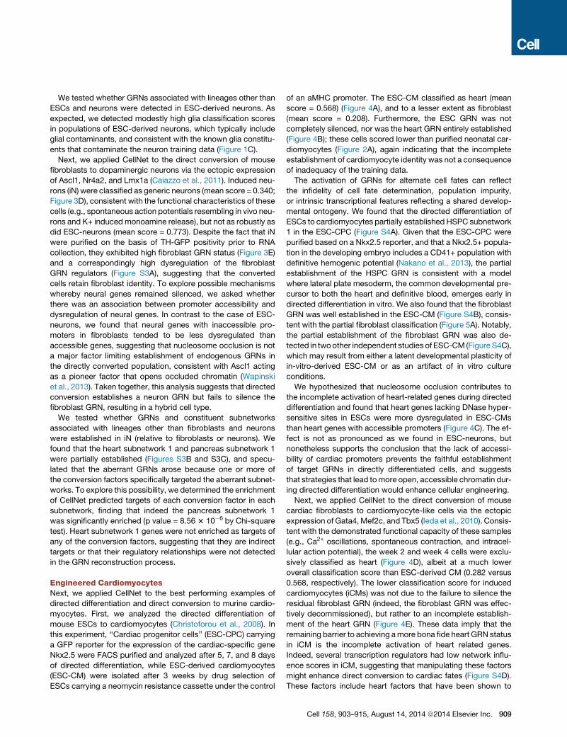
Engineered CardiomyocytesNext, we applied CellNet to the best performing examples of
directed differentiation and direct conversion to murine cardio-
myocytes. First, we analyzed the directed differentiation of
mouse ESCs to cardiomyocytes (Christoforou et al., 2008). In
this experiment, ‘‘Cardiac progenitor cells’’ (ESC-CPC) carrying
a GFP reporter for the expression of the cardiac-specific gene
Nkx2.5 were FACS purified and analyzed after 5, 7, and 8 days
of directed differentiation, while ESC-derived cardiomyocytes
(ESC-CM) were isolated after 3 weeks by drug selection of
ESCs carrying a neomycin resistance cassette under the control
of an aMHC promoter. The ESC-CM classified as heart (mean
score = 0.568) (Figure 4A), and to a lesser extent as fibroblast
(mean score = 0.208). Furthermore, the ESC GRN was not
completely silenced, nor was the heart GRN entirely established
(Figure 4B); these cells scored lower than purified neonatal car-
diomyocytes (Figure 2A), again indicating that the incomplete
establishment of cardiomyocyte identity was not a consequence
of inadequacy of the training data.
The activation of GRNs for alternate cell fates can reflect
the infidelity of cell fate determination, population impurity,
or intrinsic transcriptional features reflecting a shared develop-
mental ontogeny. We found that the directed differentiation of
ESCs to cardiomyocytes partially established HSPC subnetwork
1 in the ESC-CPC (Figure S4A). Given that the ESC-CPC were
purified based on a Nkx2.5 reporter, and that a Nkx2.5+ popula-
tion in the developing embryo includes a CD41+ population with
definitive hemogenic potential (Nakano et al., 2013), the partial
establishment of the HSPC GRN is consistent with a model
where lateral plate mesoderm, the common developmental pre-
cursor to both the heart and definitive blood, emerges early in
directed differentiation in vitro. We also found that the fibroblast
GRN was well established in the ESC-CM (Figure S4B), consis-
tent with the partial fibroblast classification (Figure 5A). Notably,
the partial establishment of the fibroblast GRN was also de-
tected in twoother independent studies of ESC-CM (Figure S4C),
which may result from either a latent developmental plasticity of
in-vitro-derived ESC-CM or as an artifact of in vitro culture
conditions.
We hypothesized that nucleosome occlusion contributes to
the incomplete activation of heart-related genes during directed
differentiation and found that heart genes lacking DNase hyper-
sensitive sites in ESCs were more dysregulated in ESC-CMs
than heart genes with accessible promoters (Figure 4C). The ef-
fect is not as pronounced as we found in ESC-neurons, but
nonetheless supports the conclusion that the lack of accessi-
bility of cardiac promoters prevents the faithful establishment
of target GRNs in directly differentiated cells, and suggests
that strategies that lead tomore open, accessible chromatin dur-
ing directed differentiation would enhance cellular engineering.
Next, we applied CellNet to the direct conversion of mouse
cardiac fibroblasts to cardiomyocyte-like cells via the ectopic
expression of Gata4, Mef2c, and Tbx5 (Ieda et al., 2010). Consis-
tent with the demonstrated functional capacity of these samples
(e.g., Ca2+ oscillations, spontaneous contraction, and intracel-
lular action potential), the week 2 and week 4 cells were exclu-
sively classified as heart (Figure 4D), albeit at a much lower
overall classification score than ESC-derived CM (0.282 versus
0.568, respectively). The lower classification score for induced
cardiomyocytes (iCMs) was not due to the failure to silence the
residual fibroblast GRN (indeed, the fibroblast GRN was effec-
tively decommissioned), but rather to an incomplete establish-
ment of the heart GRN (Figure 4E). These data imply that the
remaining barrier to achieving amore bona fide heart GRN status
in iCM is the incomplete activation of heart related genes.
Indeed, several transcription regulators had low network influ-
ence scores in iCM, suggesting that manipulating these factors
might enhance direct conversion to cardiac fates (Figure S4D).
These factors include heart factors that have been shown to
Cell 158, 903–915, August 14, 2014 ª2014 Elsevier Inc. 909
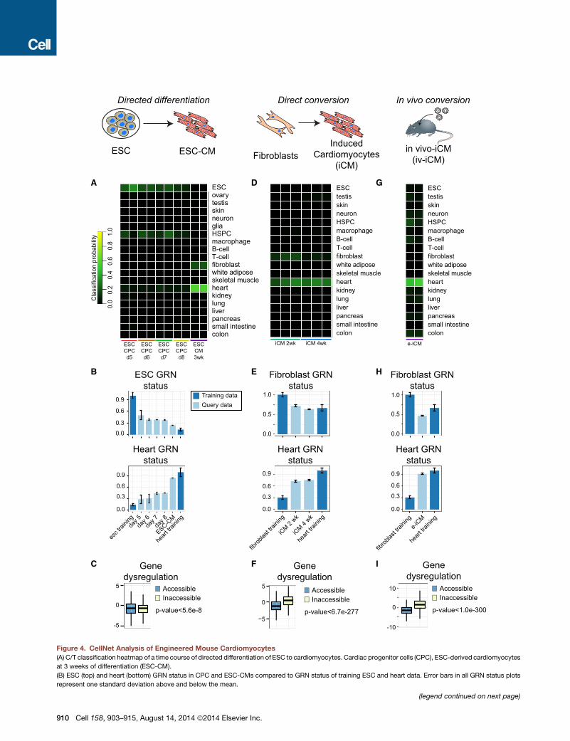
F
B
Cla
ssifi
catio
n pr
obab
ility 1.
00.
00.
80.
60.
40.
2
FibroblastsInduced
Cardiomyocytes(iCM)
A
E
Direct conversion
ESC ESC-CM
Directed differentiation
colonsmall intestinepancreasliverlungkidneyheartskeletal musclewhite adiposefibroblastT-cellB-cellmacrophageHSPCglianeuronskintestisovaryESC
ESCCPCd5
colonsmall intestinepancreasliverlungkidneyheartskeletal musclewhite adiposefibroblastT-cellB-cellmacrophageHSPCneuronskintestisESC
iCM 2wk iCM 4wk
In vivo conversion
in vivo-iCM(iv-iCM)
e-iCM
colonsmall intestinepancreasliverlungkidneyheartskeletal musclewhite adiposefibroblastT-cellB-cellmacrophageHSPCneuronskintestisESC
IC
G
H
D
ESC GRNstatus
Heart GRNstatus
0.0
0.3
0.6
0.9
esc t
rainin
gda
y 5
heart
traini
ng0.0
0.3
0.6
0.9
day 6
day 7
day 8
ESC-CM
Genedysregulation
AccessibleInaccessible
p-value<5.6e-80
-5
5
Heart GRNstatus
Fibroblast GRNstatus
0.0
0.5
1.0
fibrob
last tr
aining
iCM 2
wk
heart
traini
ng0.0
0.6
0.9
0.3
iCM 4
wk
Genedysregulation
AccessibleInaccessible
p-value<6.7e-277−5
0
5
Fibroblast GRNstatus
0.0
0.5
1.0
Heart GRNstatus
0.0
0.3
0.6
0.9
fibrob
last tr
aining
e-iCM
heart
traini
ng
AccessibleInaccessible
p-value<1.0e-300
Genedysregulation
0
10
-10
Training dataQuery data
ESCCPCd6
ESCCPCd7
ESCCPCd8
ESCCM3wk
Figure 4. CellNet Analysis of Engineered Mouse Cardiomyocytes
(A) C/T classification heatmap of a time course of directed differentiation of ESC to cardiomyocytes. Cardiac progenitor cells (CPC), ESC-derived cardiomyocytes
at 3 weeks of differentiation (ESC-CM).
(B) ESC (top) and heart (bottom) GRN status in CPC and ESC-CMs compared to GRN status of training ESC and heart data. Error bars in all GRN status plots
represent one standard deviation above and below the mean.
(legend continued on next page)
910 Cell 158, 903–915, August 14, 2014 ª2014 Elsevier Inc.
Mouse Human
Cla
ssifi
catio
n sc
ore
Cla
ssifi
catio
n sc
ore
A B
Direct
conv
ersion
Primed
conv
ersion
Directe
d diffe
rentia
tion
Reprog
ramming
0.00
0.25
0.50
0.75
1.00
Reprog
ramming
Directe
d diffe
rentia
tion
Direct
conv
ersion
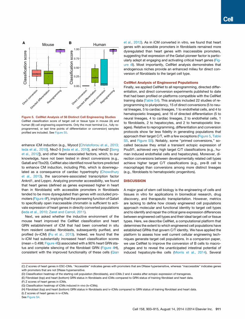
Figure 5. CellNet Analysis of 56 Distinct Cell Engineering Studies
CellNet classification score of target cell or tissue type in mouse (A) and
human (B) cell engineering experiments. Only the most terminal (i.e., fully re-
programmed, or last time points of differentiation or conversion) samples
profiled are included. See Figure S5.
enhance iCM induction (e.g., Myocd [Christoforou et al., 2013;
Ieda et al., 2010], Nkx2-5 [Ieda et al., 2010], and Hand2 [Song
et al., 2012]), and other heart-associated factors, which, to our
knowledge, have not been tested in direct conversions (e.g.,
Gata6 and Tbx20). CellNet also identified novel factors predicted
to enhance CM induction, including Phb, which is downregu-
lated as a consequence of cardiac hypertrophy (Chowdhury
et al., 2013), the sarcomere-associated transcription factor
Ankrd1, and Lrpprc. Analyzing promoter accessibility, we found
that heart genes (defined as genes expressed higher in heart
than in fibroblasts) with accessible promoters in fibroblasts
tended to be more dysregulated than genes with occluded pro-
moters (Figure 4F), implying that the pioneering function of Gata4
to specifically open inaccessible chromatin is sufficient to acti-
vate expression of heart genes in directly converted populations
(Ieda et al., 2010; Zaret and Carroll, 2011).
Next, we asked whether the inductive environment of the
mouse heart improved the CellNet classification and heart
GRN establishment of iCM that had been converted in situ
from resident cardiac fibroblasts, subsequently purified, and
profiled (iv-iCM) (Fu et al., 2013). Indeed, we found that the
iv-iCM had substantially increased heart classification scores
(mean = 0.498; Figure 4G) associated with a 90% heart GRN sta-
tus and complete silencing of the fibroblast GRN (Figure 4H),
consistent with the improved functionality of these cells (Qian
(C) Z scores of heart genes in ESC-CMs. ‘‘Accessible’’ indicates genes with prom
with promoters that are not DNase hypersensitive.
(D) Classification heatmap of the starting cell population (fibroblasts), and iCMs
(E) Fibroblast (top) and heart (bottom) GRN status in fibroblasts and iCMs comp
(F) Z scores of heart genes in iCMs.
(G) Classification heatmap of iCMs induced in vivo (iv-iCMs).
(H) Fibroblast (top) and heart (bottom) GRN status in fibroblasts and iv-iCMs com
(I) Z scores of heart genes in iv-iCMs.
See Figure S4.
et al., 2012). As in iCM converted in vitro, we found that heart
genes with accessible promoters in fibroblasts remained more
dysregulated than heart genes with inaccessible promoters,
suggesting that expression of the Gata4 pioneer factor is partic-
ularly adept at engaging and activating critical heart genes (Fig-
ure 4I). Most importantly, CellNet analysis demonstrates that
endogenous niches provide an enhanced milieu for direct con-
version of fibroblasts to the target cell type.
CellNet Analysis of Engineered PopulationsFinally, we applied CellNet to all reprogramming, directed differ-
entiation, and direct conversion experiments published to date
that had been profiled on platforms compatible with the CellNet
training data (Table S4). This analysis included 22 studies of re-
programming to pluripotency, 15 of direct conversions (5 to neu-
ral lineages, 5 to cardiac lineages, 1 to endothelial cells, and 4 to
hematopoietic lineages), and 16 of directed differentiation (5 to
neural lineages, 4 to cardiac lineages, 2 to endothelial cells, 1
to fibroblasts, 2 to hepatocytes, and 2 to hematopoietic line-
ages). Relative to reprogramming, differentiation and conversion
protocols show far less fidelity in generating populations that
approach their target C/T, with a few exceptions (Figure 5, Table
S5, and Figure S5). Notably, some ‘‘primed conversions,’’ so-
called because they entail a transient ectopic expression of
Pou5f1, achieved very high target C/T classifications (e.g., hu-
man induced endothelial cells and hepatocytes). Moreover, di-
rection conversions between developmentally related cell types
achieve higher target C/T classifications (e.g., pre-B cell to
macrophage) than conversions among more distinct lineages
(e.g., fibroblasts to hematopoietic progenitors).
DISCUSSION
A major goal of stem cell biology is the engineering of cells and
tissues in vitro for applications in biomedical research, drug
discovery, and therapeutic transplantation. However, metrics
are lacking to define how closely engineered cell populations
approach molecular and functional identity to target cell types
and to identify and repair the critical gene expression differences
between engineered cell types and their ideal target cell or tissue
types. Here, we describe CellNet, a computational platform that
determines the extent to which engineered cell populations have
established GRNs that govern C/T identity. We have applied the
platform to assess how well current cellular engineering tech-
niques generate target cell populations. In a companion paper,
we use CellNet to improve the conversion of B cells to macro-
phages and to reveal the unanticipated intestinal potential of
induced hepatocyte-like cells (Morris et al., 2014). Several
oters that are DNase hypersensitive, whereas ‘‘inaccessible’’ indicates genes
2 and 4 weeks after ectopic expression of transgenes.
ared to GRN status of training fibroblast and heart data.
pared to GRN status of training fibroblast and heart data.
Cell 158, 903–915, August 14, 2014 ª2014 Elsevier Inc. 911
insights from our analytical studies have implications for stem
cell biology and cellular engineering.
First, CellNet indicates that reprogramming of somatic cells
to pluripotency is the most faithful and complete cell-fate con-
version among the many reported studies, providing a distinct
and independent corroboration of other meta-analyses that con-
firms that iPSC and ESC are indistinguishable at the transcrip-
tome level (Cahan and Daley, 2013; Newman and Cooper,
2010). Second, we found that neurons and cardiomyocytes
derived by directed differentiation of pluripotent stem cells
more completely establish the target tissue- and cell-type
GRNs than do neurons and cardiomyocytes directly converted
from fibroblasts. While ESC-derived cells appear superior to
directly converted cells, even these cell populations lack full
establishment of target C/T GRNs. The target C/T GRNs appear
to be incompletely established because of promoter inaccessi-
bility, as indicated by a lack of DNase hypersensitive sites, sug-
gesting that developmental programs of in vitro differentiation
fail to properly induce critical pioneer factors that activate essen-
tial gene expression programs. Surprisingly, according to Cell-
Net analysis, the major barrier to direct conversion does not
appear to be restricted access to the promoters or enhancers
of target C/T genes, as these seem to be effectively activated
by virtue of the powerful action of pioneer transcription factors
included in the reprogramming cocktails. Instead, it appears
that a failure to adequately silence donor cell GRNs and the
expression of aberrant GRNs resulting in hybrid cell types repre-
sent the main molecular deficiencies of directly converted cell
populations. These observations suggest that conversions be-
tween closely related cell types, which consequently minimize
the ‘‘epigenetic distance’’ between donor and target cell, allows
the most high fidelity fate conversions, as observed for lineage
and stage conversions within the hematopoietic system (Di Tullio
et al., 2011; Doulatov et al., 2013; Riddell et al., 2014).
Third, analysis of cardiomyocyte populations induced in situ
from resident cardiac fibroblasts demonstrates that the native
tissue environment provides additional selective and/or induc-
tive signals to more completely establish the heart GRN. We
anticipate that this finding is likely to hold for most engineered
cell types.
Fourth, we have found that GRNs of the starting cell type are
detectable in purified populations of both directed differentiation
and in direct conversion experiments. In ESC-CM, the residually
expressed members of the ESC-GRN are associated with ESC
cell-cycle regulation and their impact on the tumor forming
potential and developmentally immature phenotype of ESC-
derived cells may be considerable. Directly induced neurons
retain fibroblast GRNs, which may impede the normal range of
neuron behavior. Our work has identified specific regulators of
these residual GRNs that are candidates to be targeted in future
studies in an attempt to extinguish features of the ESC cell cycle
that persist in ESC-CMs, or to silence the residual fibroblast GRN
in induced neurons.
Finally, we have discovered that the establishment of unin-
tended GRNs is common to virtually every cellular engineering
paradigm, either during the conversion or in the final products.
For example, an unexpected GRN establishment occurs during
the directed differentiation of ESC to cardiomyocytes, during
912 Cell 158, 903–915, August 14, 2014 ª2014 Elsevier Inc.
which the HSPC GRN is partially established. This may reflect
in vitro development of an aberrant cell population or may
reflect development of a common mesodermal intermediate,
which may in fact represent a signature of shared develop-
mental ontogeny that is reflected in transient GRN expression
detected in time-course experiments by CellNet. An example
of unintended GRN establishment in the final cell product is
the partial establishment of pancreas and heart subnetworks
in induced neurons. It is possible that the iN transgenes target
sufficient numbers of pancreas genes to modestly increase
the expression of their cognate targets, and we speculate that
these transcriptional regulators may prove useful in engineering
pancreas cell types. By suppressing the transcriptional regula-
tors of the pancreas GRNs, it may be possible to prevent their
establishment and thereby improve the efficiency and fidelity
of the iN conversions. Taken together, these results confirm
that off-target transcriptional events of cellular engineering are
frequent. More investigations are needed to determine how un-
intended targeting can either facilitate the conversion, lead to
less efficient protocols or result in populations with hybrid
GRNs.
We acknowledge that our current CellNet platform is limited in
several ways, which suggest important avenues for future im-
provements. First, whereas mice and humans consist of hun-
dreds of individual cell types, the limitations of the data available
for training CellNet, which requires at least 60 gene arrays per
cell or tissue type to achieve optimal performance of the GRN
reconstruction, provides adequate resolution for only 20 target
cell and tissue types in mouse and 16 in human. Moreover, the
GRNs were reconstructed based largely on tissues rather than
purified cell types. For example, the training data for the
‘‘neuron’’ C/T encompasses dissected brain and multiple types
of neurons. Consequently, CellNet compares engineered popu-
lations to a generic ‘‘neuron,’’ and at present cannot distinguish
between distinct neuronal subtypes. These limitations notwith-
standing, CellNet performs admirably as a metric for assessing
engineered cell similarities to a wide number of experimentally
and medically important target cell and tissue types, and as
demonstrated in the accompanying paper (Morris et al., 2014),
can effectively suggest hypotheses for dysregulated transcrip-
tion factors that can be further manipulated to enhance the iden-
tity and function of engineered populations. Our long-term goal is
to employ single-cell RNA sequencing data to resolve the issue
of tissue heterogeneity and to expand the platform to train on
numerous distinct cell types.
Second, the training data are largely comprised of in vivo pro-
files, whereas most cellular engineering samples to date are
derived in vitro. This limitation appears to be of minimal conse-
quence because of the strong predictive power of C/T GRNs,
which are not so confounded by signatures of cell culture as to
compromise their utility. When we compared primary cultured
cells and lines for which expression profiles were available (car-
diomyocytes, neurons, and hepatocytes), we found that CellNet
matched the cultured cells with their in vivo counterparts with
high fidelity. Ideally, however, we will need to profile cultured pri-
mary cells for many more medically relevant cell types to be able
to formally distinguish generic cell culture artifacts from defects
of cellular engineering.
Finally, as sequencing costs fall, the increased use of RNA-seq
is likely to soon replace microarrays as the transcriptomic plat-
form of choice. To that end, we are working to extend CellNet
so that it can be applied directly to RNA-seq data. CellNet is
also amenable to incorporation of additional types of global epi-
genomic data, such as analysis of DNA methylation and histone
marks, and accumulating experience with whole-genome loca-
tion analysis of transcription factor binding, such that future ver-
sions of CellNet will have improved resolution and predictive
value.
EXPERIMENTAL PROCEDURES
Training CellNet
To generate the complete training data sets, GEO was queried for expression
profiles on the Affymetrix Mouse 430.2, Illumina8v2, HG133 plus 2, MoGene
1.0, and HuGene 1.0 platforms. Affymetrix microarrays not passing the GNUse
quality control metric were not used to train CellNet. Probe intensities from raw
.CEL files were background corrected and summarized into probeset values.
Then, values of probesets mapping to the same gene were averaged. Each
array was normalized by dividing each gene expression value by the total
gene expression per array. To reconstruct cell type and tissue-specific
GRNs, gene expression profiles from GEO representing 16–20 cell and tissue
types with at least 10 perturbations per C/T were used as input to CLR (Faith
et al., 2007). The resulting expression matrix was quantile normalized to create
a data set for input to the CLR GRN reconstruction algorithm. Samples were
then limited to those as defined in Extended Experimental Procedures, prior
to re-executing CLR. An optimal regulatory relationship score was selected
based on the Gold Standard assessment and used to remove lower perform-
ing edges from the GRN. C/T-specific GRNs were identified by first splitting up
the large single GRN into subnetworks using InfoMap (Rosvall and Bergstrom,
2007). Subnetworks were attributed to specific C/Ts by determining which
subnetworks were more highly represented in gene set enrichment analysis
of each C/T versus all other C/Ts. Gene set enrichment analysis was
performed on each subnetwork to identify C/T-enriched subnetworks, and
subnetworks enriched in the same C/Ts were merged into single C/T GRNs.
For each platform, a single Random Forest classifier was trained for each C/
T (Breiman, 2001). Initially, approximately 50% of arrays were randomly
selected and used to train each Random Forest classifier. The performance
of these classifiers was evaluated by classifying the remaining, independent
arrays, which were not used to train the classifier and were from different
studies. The performance of these classifiers is presented in Figure 1B and Fig-
ures S1E–S1H. To improve the overall power of the classifiers, we then used all
arrays to train the classifiers used in the final version of CellNet.
GRN Status
We reasoned that the extent to which a GRN is established in a sample is
reflected in the expression of the genes in the GRN such that the GRN genes
should fall within a range of expression observed in the corresponding C/T in
the training data. We formalized this notion as a GRN status metric, defined in
comparison to the complete training data set. The status of C/T GRN in a query
sample is defined as the weighted mean of the Z scores of the genes in the
GRN, where the Z score is defined in reference to the expression distribution
of each gene in a C/T. The GRN status score can be weighted by the absolute
expression level of each gene in a C/T so that genes more highly expressed
have more influence on the GRN status (default) and/or by the importance to
the Random Forest classifier (default):
RGSðxÞ=Xn
i = 1
�ZscoreðgeneÞC=T � gene weightGRN
�
where n = number of genes in the GRN and gene weight is proportional to the
expression of the gene in C/T and, optionally, derived from the importance of
the gene to the Random Forest classifier. Zscore(gene) is determined in refer-
ence to the expression distribution of the gene in C/T samples in the complete
training data set.We transform and normalize the rawGRN status score so that
higher GRN status indicates a GRN status more similar to the endogenous C/T
as:
GSðxÞ= 1; 000� RGSðxÞ�Pkj RGSðyÞ
�.k
where ðPkj RGSðyÞÞ=k is the average RGS of C/T samples in the complete
training data.
Network Influence Score
To estimate the importance of transcriptional regulators to dysregulated
GRNs, we devised the following network influence score (NIS):
NISðTRÞ=Xn
i = 1
�ZscoreðtargetÞC=T �weighttarget
�
+ n � ZscoreðTRÞC=T �weightTR
where TR = transcriptional regulator and n = number of genes in the GRN. By
default, CellNet weights the target and TR by their mean expression in the C/T
samples of the complete training data set when searching for inappropriately
silenced factors.
SUPPLEMENTAL INFORMATION
Supplemental Information includes Extended Experimental Procedures, five
figures, and five tables and can be found with this article online at http://dx.
doi.org/10.1016/j.cell.2014.07.020.
AUTHOR CONTRIBUTIONS
P.C., S.A.M., and H.L. designed, performed, and interpreted analyses and
wrote the paper. E.L.d.R. performed analysis and helped to write the paper.
J.J.C. and G.Q.D. oversaw the project and helped to design the study and
write the paper.
ACKNOWLEDGMENTS
J.J.C. is supported by NIH grant R24DK092760 and the HHMI. G.Q.D. is
supported by grants from the NIH (Progenitor Cell Biology Consortium UO1-
HL100001, R24DK092760, and P50HG005550), the Ellison Medical Founda-
tion, Doris Duke Medical Foundation, and Boston Children’s Hospital Stem
Cell Program, and is an affiliate member of the Broad Institute. P.C. is sup-
ported by NIDDK (K01DK096013) and received support from NHLBI
(T32HL066987 and T32HL007623). S.A.M. is supported by a Young Investi-
gator Award from Alex’s Lemonade Stand Foundation. H.L. is supported by
Mayo Clinic Center for Individualized Medicine and Mayo Clinic Center for
RegenerativeMedicine. E.L.d.R. is supported by National Council for Scientific
and Technological Development and the program Science Without Borders
(CNPq, Brazil).
Received: January 14, 2014
Revised: May 28, 2014
Accepted: July 17, 2014
Published: August 14, 2014
REFERENCES
Aasen, T., Raya, A., Barrero, M.J., Garreta, E., Consiglio, A., Gonzalez, F., Vas-
sena, R., Bili�c, J., Pekarik, V., Tiscornia, G., et al. (2008). Efficient and rapid
generation of induced pluripotent stem cells from human keratinocytes. Nat.
Biotechnol. 26, 1276–1284.
Arnold, P., Scholer, A., Pachkov, M., Balwierz, P.J., Jørgensen, H., Stadler,
M.B., van Nimwegen, E., and Schubeler, D. (2013). Modeling of epigenome dy-
namics identifies transcription factors that mediate Polycomb targeting.
Genome Res. 23, 60–73.
Cell 158, 903–915, August 14, 2014 ª2014 Elsevier Inc. 913
Breiman, L. (2001). Random Forests. Mach. Learn. 45, 5–32.
Cahan, P., and Daley, G.Q. (2013). Origins and implications of pluripotent stem
cell variability and heterogeneity. Nat. Rev. Mol. Cell Biol. 14, 357–368.
Caiazzo, M., Dell’Anno, M.T., Dvoretskova, E., Lazarevic, D., Taverna, S., Leo,
D., Sotnikova, T.D., Menegon, A., Roncaglia, P., Colciago, G., et al. (2011).
Direct generation of functional dopaminergic neurons from mouse and human
fibroblasts. Nature 476, 224–227.
Chiu, I.M., Heesters, B.A., Ghasemlou, N., Von Hehn, C.A., Zhao, F., Tran, J.,
Wainger, B., Strominger, A., Muralidharan, S., Horswill, A.R., et al. (2013).
Bacteria activate sensory neurons that modulate pain and inflammation. Na-
ture 501, 52–57.
Chowdhury, D., Tangutur, A.D., Khatua, T.N., Saxena, P., Banerjee, S.K., and
Bhadra,M.P. (2013). A proteomic view of isoproterenol induced cardiac hyper-
trophy: prohibitin identified as a potential biomarker in rats. J. Transl. Med. 11,
130.
Christoforou, N., Miller, R.A., Hill, C.M., Jie, C.C., McCallion, A.S., and Gear-
hart, J.D. (2008). Mouse ES cell-derived cardiac precursor cells are multipo-
tent and facilitate identification of novel cardiac genes. J. Clin. Invest. 118,
894–903.
Christoforou, N., Chellappan, M., Adler, A.F., Kirkton, R.D.,Wu, T., Addis, R.C.,
Bursac, N., and Leong, K.W. (2013). Transcription factors MYOCD, SRF,
Mesp1 and SMARCD3 enhance the cardio-inducing effect of GATA4, TBX5,
and MEF2C during direct cellular reprogramming. PLoS ONE 8, e63577.
Correa-Cerro, L.S., Piao, Y., Sharov, A.A., Nishiyama, A., Cadet, J.S., Yu, H.,
Sharova, L.V., Xin, L., Hoang, H.G., Thomas, M., et al. (2011). Generation of
mouse ES cell lines engineered for the forced induction of transcription factors.
Sci Rep 1, 167.
Davidson, E.H., and Erwin, D.H. (2006). Gene regulatory networks and the
evolution of animal body plans. Science 311, 796–800.
Davis, R.L., Weintraub, H., and Lassar, A.B. (1987). Expression of a single
transfected cDNA converts fibroblasts to myoblasts. Cell 51, 987–1000.
Di Tullio, A.A., VuManh, T.P., Schubert, A.A., Castellano, G.G., Mansson, R.R.,
and Graf, T.T. (2011). CCAAT/enhancer binding protein alpha (C/EBP(alpha))-
induced transdifferentiation of pre-B cells into macrophages involves no overt
retrodifferentiation. Proc. Natl. Acad. Sci. USA 108, 17016–17021.
Doulatov, S., Vo, L.T., Chou, S.S., Kim, P.G., Arora, N., Li, H., Hadland, B.K.,
Bernstein, I.D., Collins, J.J., Zon, L.I., and Daley, G.Q. (2013). Induction of
multipotential hematopoietic progenitors from human pluripotent stem cells
via respecification of lineage-restricted precursors. Cell Stem Cell 13,
459–470.
Efron, B., and Tibshirani, R. (2007). On testing the significance of sets of genes.
Ann. Appl. Stat. 1, 107–129.
Faith, J.J., Hayete, B., Thaden, J.T., Mogno, I., Wierzbowski, J., Cottarel, G.,
Kasif, S., Collins, J.J., and Gardner, T.S. (2007). Large-scale mapping and vali-
dation of Escherichia coli transcriptional regulation from a compendium of
expression profiles. PLoS Biol. 5, e8.
Fu, J.-D., Stone, N.R., Liu, L., Spencer, C.I., Qian, L., Hayashi, Y., Delgado-Ol-
guin, P., Ding, S., Bruneau, B.G., and Srivastava, D. (2013). Direct Reprogram-
ming of Human Fibroblasts toward a Cardiomyocyte-like State. StemCell Rep.
1, 235–247.
Huang, P., He, Z., Ji, S., Sun, H., Xiang, D., Liu, C., Hu, Y., Wang, X., and Hui, L.
(2011). Induction of functional hepatocyte-like cells from mouse fibroblasts by
defined factors. Nature 475, 386–389.
Ieda, M., Fu, J.-D., Delgado-Olguin, P., Vedantham, V., Hayashi, Y., Bruneau,
B.G., and Srivastava, D. (2010). Direct reprogramming of fibroblasts into func-
tional cardiomyocytes by defined factors. Cell 142, 375–386.
Kim, J.B., Sebastiano, V., Wu, G., Arauzo-Bravo, M.J., Sasse, P., Gentile, L.,
Ko, K., Ruau, D., Ehrich, M., van den Boom, D., et al. (2009). Oct4-induced
pluripotency in adult neural stem cells. Cell 136, 411–419.
Loh, Y.-H., Hartung, O., Li, H., Guo, C., Sahalie, J.M., Manos, P.D., Urbach, A.,
Heffner, G.C., Grskovic, M., Vigneault, F., et al. (2010). Reprogramming of
T cells from human peripheral blood. Cell Stem Cell 7, 15–19.
914 Cell 158, 903–915, August 14, 2014 ª2014 Elsevier Inc.
Lukk, M., Kapushesky, M., Nikkila, J., Parkinson, H., Goncalves, A., Huber, W.,
Ukkonen, E., and Brazma, A. (2010). A global map of human gene expression.
Nat. Biotechnol. 28, 322–324.
Mao, T., Shao, M., Qiu, Y., Huang, J., Zhang, Y., Song, B., Wang, Q., Jiang, L.,
Liu, Y., Han, J.-D.J., et al. (2011). PKA phosphorylation couples hepatic
inositol-requiring enzyme 1alpha to glucagon signaling in glucosemetabolism.
Proc. Natl. Acad. Sci. USA 108, 15852–15857.
Marbach, D., Costello, J.C., Kuffner, R., Vega, N.M., Prill, R.J., Camacho,
D.M., Allison, K.R., Kellis, M., Collins, J.J., and Stolovitzky, G.; DREAM5 Con-
sortium (2012). Wisdom of crowds for robust gene network inference. Nat.
Methods 9, 796–804.
Morris, S.A., Cahan, P., Li, H., Zhao, A.M., San Roman, A.K., Shivdasani, R.A.,
Collins, J.J., George, Q., and Daley, G.Q. (2014). Dissecting Engineered Cell
Types and Enhancing Cell Fate Conversion via CellNet. Cell 158, this issue,
889–902.
Mouse ENCODE Consortium, Stamatoyannopoulos, J.A., Snyder, M., Hardi-
son, R., Ren, B., Gingeras, T., Gilbert, D.M., Groudine, M., Bender, M., Kaul,
R., Canfield, T., et al. (2012). An encyclopedia of mouse DNA elements (Mouse
ENCODE). Genome Biol. 13, 418.
Murry, C.E., and Keller, G. (2008). Differentiation of embryonic stem cells to
clinically relevant populations: lessons from embryonic development. Cell
132, 661–680.
Nakano, H., Liu, X., Arshi, A., Nakashima, Y., van Handel, B., Sasidharan, R.,
Harmon, A.W., Shin, J.-H., Schwartz, R.J., Conway, S.J., et al. (2013). Haemo-
genic endocardium contributes to transient definitive haematopoiesis. Nat
Commun 4, 1564.
Newman, A.M., and Cooper, J.B. (2010). Lab-specific gene expression signa-
tures in pluripotent stem cells. Cell Stem Cell 7, 258–262.
Nishikawa, S.-I., Jakt, L.M., and Era, T. (2007). Embryonic stem-cell culture as
a tool for developmental cell biology. Nat. Rev. Mol. Cell Biol. 8, 502–507.
Novershtern, N., Subramanian, A., Lawton, L.N., Mak, R.H., Haining, W.N.,
McConkey, M.E., Habib, N., Yosef, N., Chang, C.Y., Shay, T., et al. (2011).
Densely interconnected transcriptional circuits control cell states in human
hematopoiesis. Cell 144, 296–309.
Peng, Z.F., Chen, M.J., Manikandan, J., Melendez, A.J., Shui, G., Russo-
Marie, F., Whiteman, M., Beart, P.M., Moore, P.K., and Cheung, N.S. (2012).
Multifaceted role of nitric oxide in an in vitro mouse neuronal injury model:
transcriptomic profiling defines the temporal recruitment of death signalling
cascades. J. Cell. Mol. Med. 16, 41–58.
Qian, L., Huang, Y., Spencer, C.I., Foley, A., Vedantham, V., Liu, L., Conway,
S.J., Fu, J.-D., and Srivastava, D. (2012). In vivo reprogramming of murine
cardiac fibroblasts into induced cardiomyocytes. Nature 485, 593–598.
Riddell, J., Gazit, R., Garrison, B.S., Guo, G., Saadatpour, A., Mandal, P.K.,
Ebina,W., Volchkov, P., Yuan, G.-C., Orkin, S.H., et al. (2014). Reprogramming
committed murine blood cells to induced hematopoietic stem cells with
defined factors. Cell 157, 549–564.
Rosvall, M., and Bergstrom, C.T. (2007). An information-theoretic framework
for resolving community structure in complex networks. Proc. Natl. Acad.
Sci. USA 104, 7327–7331.
Rung, J., and Brazma, A. (2013). Reuse of public genome-wide gene expres-
sion data. Nat. Rev. Genet. 14, 89–99.
Sekiya, S., and Suzuki, A. (2011). Direct conversion of mouse fibroblasts to
hepatocyte-like cells by defined factors. Nature 475, 390–393.
Song, K., Nam, Y.-J., Luo, X., Qi, X., Tan, W., Huang, G.N., Acharya, A., Smith,
C.L., Tallquist, M.D., Neilson, E.G., et al. (2012). Heart repair by reprogram-
ming non-myocytes with cardiac transcription factors. Nature 485, 599–604.
Staerk, J., Dawlaty, M.M., Gao, Q., Maetzel, D., Hanna, J., Sommer, C.A.,
Mostoslavsky, G., and Jaenisch, R. (2010). Reprogramming of human periph-
eral blood cells to induced pluripotent stem cells. Cell Stem Cell 7, 20–24.
Strain, A.J. (1994). Isolated hepatocytes: use in experimental and clinical
hepatology. Gut 35, 433–436.
Szabo, E., Rampalli, S., Risueno, R.M., Schnerch, A., Mitchell, R., Fiebig-Co-
myn, A., Levadoux-Martin, M., and Bhatia, M. (2010). Direct conversion of
human fibroblasts to multilineage blood progenitors. Nature 468, 521–526.
Takahashi, K., and Yamanaka, S. (2006). Induction of pluripotent stem cells
from mouse embryonic and adult fibroblast cultures by defined factors. Cell
126, 663–676.
Vierbuchen, T., Ostermeier, A., Pang, Z.P., Kokubu, Y., Sudhof, T.C., and
Wernig, M. (2010). Direct conversion of fibroblasts to functional neurons by
defined factors. Nature 463, 1035–1041.
Wapinski, O.L., Vierbuchen, T., Qu, K., Lee, Q.Y., Chanda, S., Fuentes, D.R.,
Giresi, P.G., Ng, Y.H., Marro, S., Neff, N.F., et al. (2013). Hierarchical mecha-
nisms for direct reprogramming of fibroblasts to neurons. Cell 155, 621–635.
Xie, H., Ye, M., Feng, R., and Graf, T. (2004). Stepwise reprogramming of B
cells into macrophages. Cell 117, 663–676.
Xu, H., Baroukh, C., Dannenfelser, R., Chen, E.Y., Tan, C.M., Kou, Y., Kim,
Y.E., Lemischka, I.R., and Ma’ayan, A. (2013). ESCAPE: database for inte-
grating high-content published data collected from human and mouse embry-
onic stem cells. Database (Oxford) 2013, bat045.
Zaret, K.S., and Carroll, J.S. (2011). Pioneer transcription factors: establishing
competence for gene expression. Genes Dev. 25, 2227–2241.
Zheng, B., Liao, Z., Locascio, J.J., Lesniak, K.A., Roderick, S.S., Watt, M.L.,
Eklund, A.C., Zhang-James, Y., Kim, P.D., Hauser, M.A., et al.; Global PD
Gene Expression (GPEX) Consortium (2010). PGC-1a, a potential therapeutic
target for early intervention in Parkinson’s disease. Sci. Transl. Med. 2, 52ra73.
Zhou, Q., Brown, J., Kanarek, A., Rajagopal, J., andMelton, D.A. (2008). In vivo
reprogramming of adult pancreatic exocrine cells to b-cells. Nature 455,
627–632.
Cell 158, 903–915, August 14, 2014 ª2014 Elsevier Inc. 915