Embed Size (px)
Citation preview

Slide 1 / 192
Cells: The Basis of Life
Slide 2 / 192
Cells Unit Topics
· Prokaryotes
· Eukaryotes
Click on the topic to go to that section
· Viruses· Cellular Defenses
Slide 3 / 192

Prokaryotes
Return toTable ofContents
Slide 4 / 192
All cells have 4 things in common.
· They are surrounded by a plasma membrane (or cell membrane).
· They contain a semifluid substance called the cytosol/cytoplasm .
· They contain structures called chromosomes, which carry the cell's genes.
· They have ribosomes, which assemble amino acids into proteins.
All Cells
Slide 5 / 192
There are 3 key differences between prokaryotic and eukaryotic cells.
· Eukaryotic cells are usually larger than prokaryotic cells.
· Eukaryotic cells have small compartments inside them call organelles.
· Most eukaryotes (but not all) are multi-cellular organisms.
Eukaryotes vs. Prokaryotes
Slide 6 / 192

1 Which is NOT a basic feature of all cells?
A All cells are surrounded by a plasma membrane.
B All cells contain a semifluid substance called the cytoplasm.
C All cells contain structures called chromosomes, which are contained in the nucleus.
D All cells have ribosomes.
Slide 7 / 192
2 A scientist is trying to determine if a newly discovered organic object is alive. It has a plasma membrane, organelles, and DNA, but no ribsomes. Is this organic material alive?
Yes
No
Slide 8 / 192
Prokaryotes: 2 Types
Bacteria Archaea
Slide 9 / 192

We Rely on Bacteria!
Often we think of bacteria as being primarily harmful organisms. While there are harmful bacteria, most are beneficial; we depend on them.
Bacteria cover all the external surfaces of our bodies. This includes our digestive tracts since that is also considered to be external.
Bacteria live in cooperation with you; they protect you against harmful bacteria and help you digest food. Without these bacteria, which have evolved with us, as we evolved, we could not live healthy lives.
Slide 10 / 192
In fact, the number of bacterial cells living on us is greater than the number of our own cells.
Those bacteria have more unique genetic material than do our own genes. That bacterial genetic material allows them to create enzymes or products that are essential to us.
We Rely on Bacteria!
bacteria on skin - National Geographic Magazine
Slide 11 / 192
We Rely on Bacteria!
Newborn babies get innoculations of these bacteria from their mothers, so that their digestive systems can function.
There are also bacteria in many food sources, like yogurt and cheese.
Slide 12 / 192

Antibiotics
When we take antibiotics to fight a harmful bacteria, it's usually recommended to consume probiotics, such as yogurt, to replace any of our helpful bacteria that might be accidentally harmed.
In fact, most antibiotics themselves are derived from bacteria. They are created in nature by bacteria to fight other bacteria.
When then use them in the form of antibiotic pills or injections to fight harmful bacteria.
Slide 13 / 192
Archaea
Archaea
Archaea were classified as bacteria until very recently. In 1977, they were separated from bacteria into their own domain, or grouping.
Many archaea are extremophiles, organisms that live in environments where life had been considered impossible. They have be found living in areas of extreme temperature (such as hydrothermal vents), pH solutions of lower than 3 and higher than 9, and solutions with high salt, methane, or heavy metal concentrations.
Slide 14 / 192
LUCA (3.5 - 3.8 BYA)
ArchaeaWhile archaea have many cell structures and metabolic pathways in common with bacteria, research has shown that their genes and factors involved in their gene expression are more like those of eukaryotes (the class of organisms that include animals, plants, and fungi).
This has led scientists to believe that archaea developed after bacteria.
Slide 15 / 192

3 The earliest living organisms were probably:
A animalsB archaeaC bacteriaD plants
Slide 16 / 192
4 Prokaryotes can live in which of the following environments?
A the oceanB acidic lakesC hydrothermal ventsD under the Arctic iceE all of the above
Slide 17 / 192
Order/Organization
All prokaryotes are unicellular, meaning a single cell is considered an entire organism.
They can live on their own, but most form colonies, large groups (millions, billions or more) live in a tightly packed area.
They have a variety of shapes and functions.
Slide 18 / 192

Prokaryotic Shapes
Slide 19 / 192
Structures
http://www.s ingle ton-associates .org/bacteri2.htm
Prokaryotes have many different structures, each having a specific job or function. These structures within the cell operate like small molecular machines.
They are used for various functions that help maintain the life of the overall organism.
Slide 20 / 192
Cell Surface
Most prokaryotes have a cell wall.
http://www.s ingle ton-associates .org/bacteri2.htm
The cell wall is outside the cell's plasma membrane and maintains the cell's shape, provides physical protection, and prevents the cell from bursting in a hypotonic environment.
In bacteria, this cell wall is made of a strong carbohydrate fiber called peptidoglycan. In Archaea, various cell wall types exist.
Slide 21 / 192
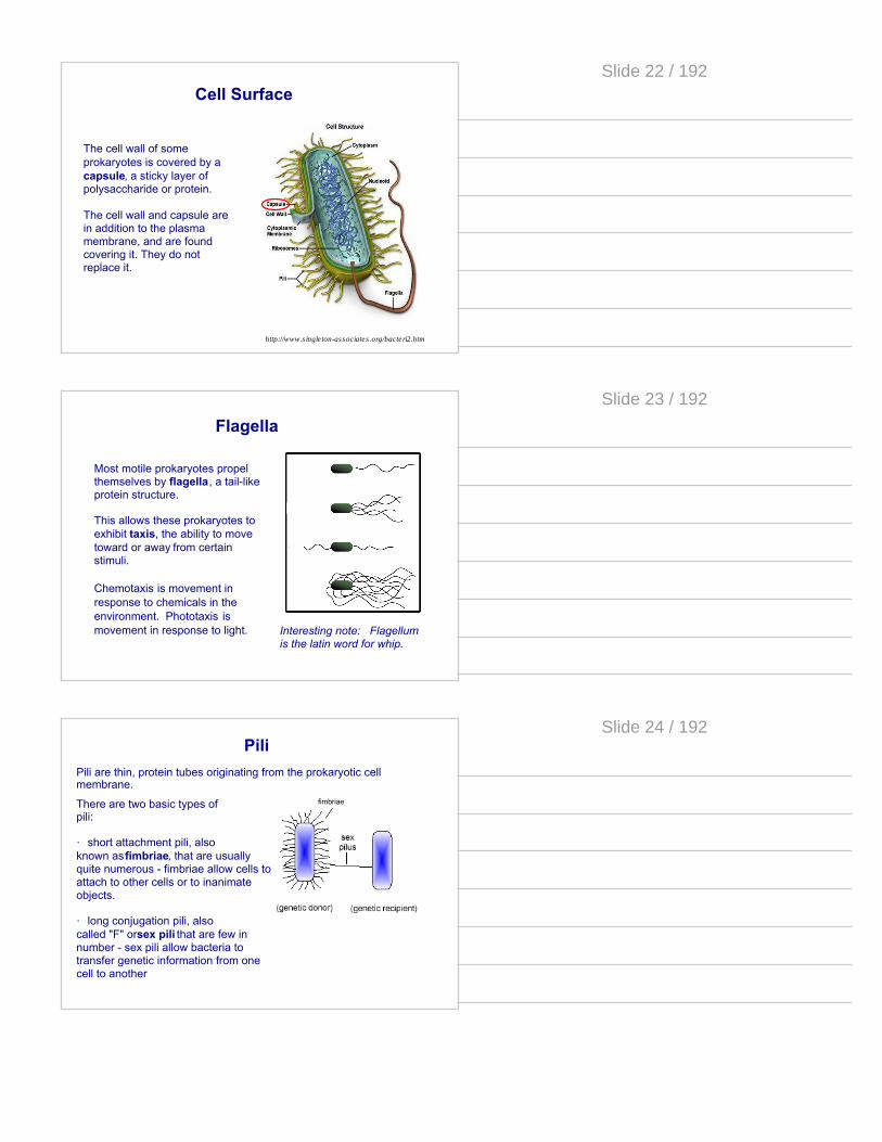
Cell Surface
The cell wall of some prokaryotes is covered by a capsule, a sticky layer of polysaccharide or protein.
The cell wall and capsule are in addition to the plasma membrane, and are found covering it. They do not replace it.
http://www.s ingle ton-associates .org/bacteri2.htm
Slide 22 / 192
Flagella
Most motile prokaryotes propel themselves by flagella, a tail-like protein structure.
This allows these prokaryotes to exhibit taxis, the ability to move toward or away from certain stimuli.
Chemotaxis is movement in response to chemicals in the environment. Phototaxis is movement in response to light. Interesting note: Flagellum
is the latin word for whip.
Slide 23 / 192
fimbriaeThere are two basic types of pili:
· short attachment pili, also known as fimbriae, that are usually quite numerous - fimbriae allow cells to attach to other cells or to inanimate objects.
· long conjugation pili, also called "F" or sex pili that are few in number - sex pili allow bacteria to transfer genetic information from one cell to another
PiliPili are thin, protein tubes originating from the prokaryotic cell membrane.
Slide 24 / 192

5 What structure allows a prokaryote to adhere to environmental surfaces?
A cell wallB sex piliC flagellumD fimbriae
Slide 25 / 192
6 What structure allows a prokaryote to exhibit taxis?
A cell wallB sex piliC flagellumD fimbriae
Slide 26 / 192
7 In bacteria, the _________ is made of a substance called peptidoglycan.
A capsuleB piliC flagellumD cell wall
Slide 27 / 192

The fluid which fills the cell is called the cytoplasm.
Floating in the cytoplasm are the ribosomes and the bacterial chromosome, a double-stranded, circular structure containing the prokaryote's DNA.
Prokaryotes usually only have one chromosome and the area where it is located is known as the nucleoid.
Inside the Cell
http://www.s ingle ton-associates .org/bacteri2.htm
Slide 28 / 192
Many prokaryotes also have plasmids, smaller circular DNA molecules that are independent of the bacterial chromosome.
Plasmids
Plasmids contain genes for adaptations like resistance against antibiotics, making a sex-pilus (F-pilus), making toxins, and guarding against heavy metal toxicity.
Slide 29 / 192
The presence of an F plasmid (or an F factor) gives the prokaryotic cell the ability to have fertility, by forming a sex pilus. This allows the prokaryote to donate DNA to other prokaryotes in its colony, increasing their genetic variability.
F Plasmids
Note: The "F" factor is located in the bacterial chromosome.
Slide 30 / 192
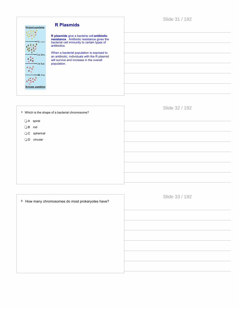
R plasmids give a bacteria cell antibiotic resistance. Antibiotic resistance gives the bacterial cell immunity to certain types of antibiotics.
When a bacterial population is exposed to an antibiotic, individuals with the R plasmid will survive and increase in the overall population.
R Plasmids
Slide 31 / 192
8 Which is the shape of a bacterial chromosome?
A spiral
B rod
C spherical
D circular
Slide 32 / 192
9 How many chromosomes do most prokaryotes have?
Slide 33 / 192
10 The area where a bacterial chromosome is located is called:
A capsule
B flagella
C nucleoid
D ribosome
Slide 34 / 192
11 Bacteria that have R plasmids can cause medical problems in animals because they _____.
A control conjugation in bacteria
B are used as vectors to transfer genes
C make bacteria resistant to antibiotics
D code for DNA polymerase
E protect bacteria against mutations
Slide 35 / 192
12 Antibiotic resistance first arose as the result of...
A increased antibiotic use
B introduction of F factors into the genome
C a genetic mutation
D competition for resources among bacterial colonies
Slide 36 / 192

flagella
fimbriae
nucleoid
bacterial chromosome
cell wallcapsule
cell membraneribosome
Slide 37 / 192
Prokaryotic Reproduction
Prokaryotic cells divide and reproduce by binary fission, the splitting of one cell into two.
In order for each cell to have a complete copy of the DNA, the bacterial chromosome must be replicated prior to cell division.
Slide 38 / 192
Binary Fission
After the chromosome is replicated, the cell divides in half with one copy in each new cell.
Slide 39 / 192

13 At the end of binary fission, there are two prokaryotic cells
A one has all the parent DNA
B both have only parent DNA
C both have only daughter DNA
D both have half parent and half daughter DNA
Slide 40 / 192
Eukaryotes
Return toTable ofContents
Slide 41 / 192
Surface Area to Volume Ratio
At the time when prokaryotic cells were evolving, there were most likely different sizes of cells. A cell's efficiency and ability to survive depended on its surface area to volume ratio.
The volume of the cell determines the amount of chemical activity it can carry out per unit time. The surface area of the cell determines the amount of substances the cell can take in from the environment and the amount of waste it can release.
As a cell grows in size, it's surface area to volume ratio decreases. It performs chemical reactions faster, but it has a harder time getting nutrients in and waste out.
Slide 42 / 192

We know that cells need to be small enough so that they have an increased surface area to volume ratio, but be large enough toperform the chemical reactions of metabolism.
Most Efficient Least Efficient
The smaller the cell, the larger its surface area and the smaller its volume.
Limits of Cell Size
The bigger the cell, the smaller the surface area is compared to its large volume inside.
Slide 43 / 192
Animal Cell (Eukaryote)
Bacterium (Prokaryote)
Eukaryotic cells are, on average, much larger than prokaryotic cells. The average diameter of most prokaryotic cells is between 1 and 10µm. By contrast, most eukaryotic cells are between 5 to 100µm in diameter.
Cell Size
Slide 44 / 192
OrganellesTo increase efficiency in the larger cell, eukaryotes evolved many bacterium-sized parts known as organelles.
Organelles subdivide the cell into specialized compartments.
They play many important roles in the cell. Some transport waste to the cell membrane. Others keep the molecules required for specific chemical reactions located within a certain compartment so they do not need to diffuse long distances to be useful.
Slide 45 / 192

Organelles making up Eukaryotic cells include:
Organelles
· Nucleus
· Lysosomes
· Ribosomes
· Peroxisome
s
· Mitochondria
· Vacuoles
· Smooth Endoplasmic Reticulum
· Rough Endoplasmic Reticulum
· Chloroplasts
· Golgi Apparatus
Slide 46 / 192
14 How did eukaryotes solve the problem of diffusion?
A By remaining the same size as prokaryotes.B By using a nucleus.C Compartmentalization.D They haven't solved the problem.
Slide 47 / 192
15 Which is NOT an advantage of compartmentalization?
A It allows incompatible chemical reactions to be separated.
B It increases the efficiency of chemical reactions.
C It decreases the speed of reactions since reactants have to travel farther.
D Substrates required for particular reactions can be localized and maintained at high concentrations within organelles.
Slide 48 / 192
Multicellular Organisms
Even with organelles, the size of the cell is limited to about 1000µm3. This is why large organisms must consist of many smaller cells.
Slide 49 / 192
Diversity of Eukaryotes
Protists: The first eukaryotic cells. Protists are single-celled eukaryotes. They range from protozoans to algae.
Fungi: These organisms evolved second in time along with plants. Examples include mushrooms, molds, and mildews.
Plants: Plants vary in type from the first plants called mosses to the modern flowering plants.
Animals : Animals were the last eukaryotes to evolve. Animals range from ancient sponges and hydra to primates.
Slide 50 / 192
16 How did eukaryotes solve the problem of small surface area to volume ratio?
A by remaining the same size as prokaryotesB by becoming multicellular organismsC by compartmentalizing functions into organellesD they haven't solved the problem
Slide 51 / 192
17 Identify a single-celled eukaryote.
A Mushroom
B E. coli
C Algae
D Crustacean
Slide 52 / 192
prokaryotes: pro: before karyon: kernel/seed (nucleus)
The Nucleus
eukaryote: eu: truekaryon: kernel/seed (nucleus)
So prokaryote = "before a nucleus"And eukaryote = "true nucleus"
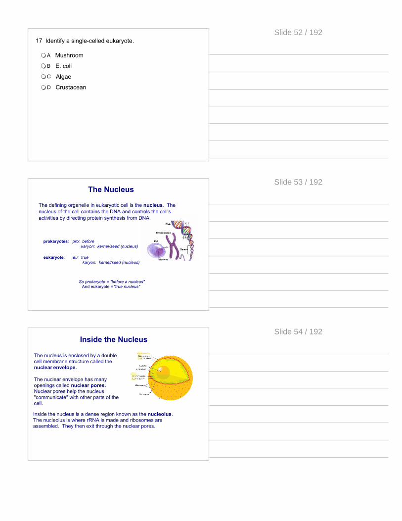
The defining organelle in eukaryotic cell is the nucleus. The nucleus of the cell contains the DNA and controls the cell's activities by directing protein synthesis from DNA.
Slide 53 / 192
Inside the Nucleus
The nucleus is enclosed by a double cell membrane structure called the nuclear envelope.
The nuclear envelope has many openings called nuclear pores. Nuclear pores help the nucleus "communicate" with other parts of the cell.
Inside the nucleus is a dense region known as the nucleolus.The nucleolus is where rRNA is made and ribosomes are assembled. They then exit through the nuclear pores.
Slide 54 / 192
3 Main Functions of the Nucleus
1. To keep and contain a safe copy of all chromosomes (DNA) and pass them on to daughter cells in cell division.
2. To assemble ribosomes (specifically in the nucleolus).
3. To copy DNA instructions into RNA (via transcription).
Slide 55 / 192
18 Cells that contain a "true nucleus" and other membrane bound organelles are _______________.
A archaea.
B bacteria.
C eukaryotes.
D prokaryotes.
Slide 56 / 192
19 Where is the DNA of a eukaryote found?
A Nucleus
B Nucleolus
C Nucleoid region
D Mitochondria
Slide 57 / 192
20 How does the nucleus control the activities of the cell?
A By making DNA.
B By directing protein synthesis.
C By allowing DNA to leave the nucleus to make proteins.
D By sending instructions to the mitochondria.
Slide 58 / 192
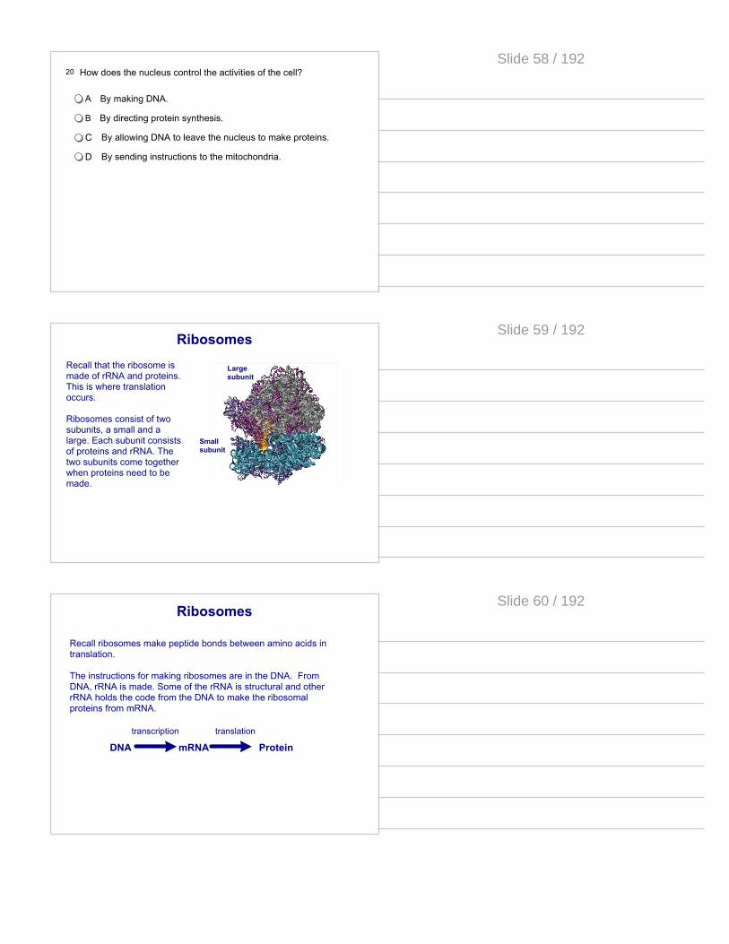
Ribosomes
Large subunit
Small subunit
Recall that the ribosome is made of rRNA and proteins. This is where translation occurs.
Ribosomes consist of two subunits, a small and a large. Each subunit consists of proteins and rRNA. The two subunits come together when proteins need to be made.
Slide 59 / 192
Ribosomes
Recall ribosomes make peptide bonds between amino acids in translation.
The instructions for making ribosomes are in the DNA. From DNA, rRNA is made. Some of the rRNA is structural and other rRNA holds the code from the DNA to make the ribosomal proteins from mRNA.
DNA mRNA Proteintranscription translation
Slide 60 / 192
Ribosomes
Two types of ribosomes exist in eukaryotic cells: free ribosomes and bound ribosomes.
Bound ribosomes are attached to internal membranes in the cell's endomembrane system.
Free ribosomes move freely through the cytoplasm and produce proteins and enzymes used internally by the cell.
Slide 61 / 192
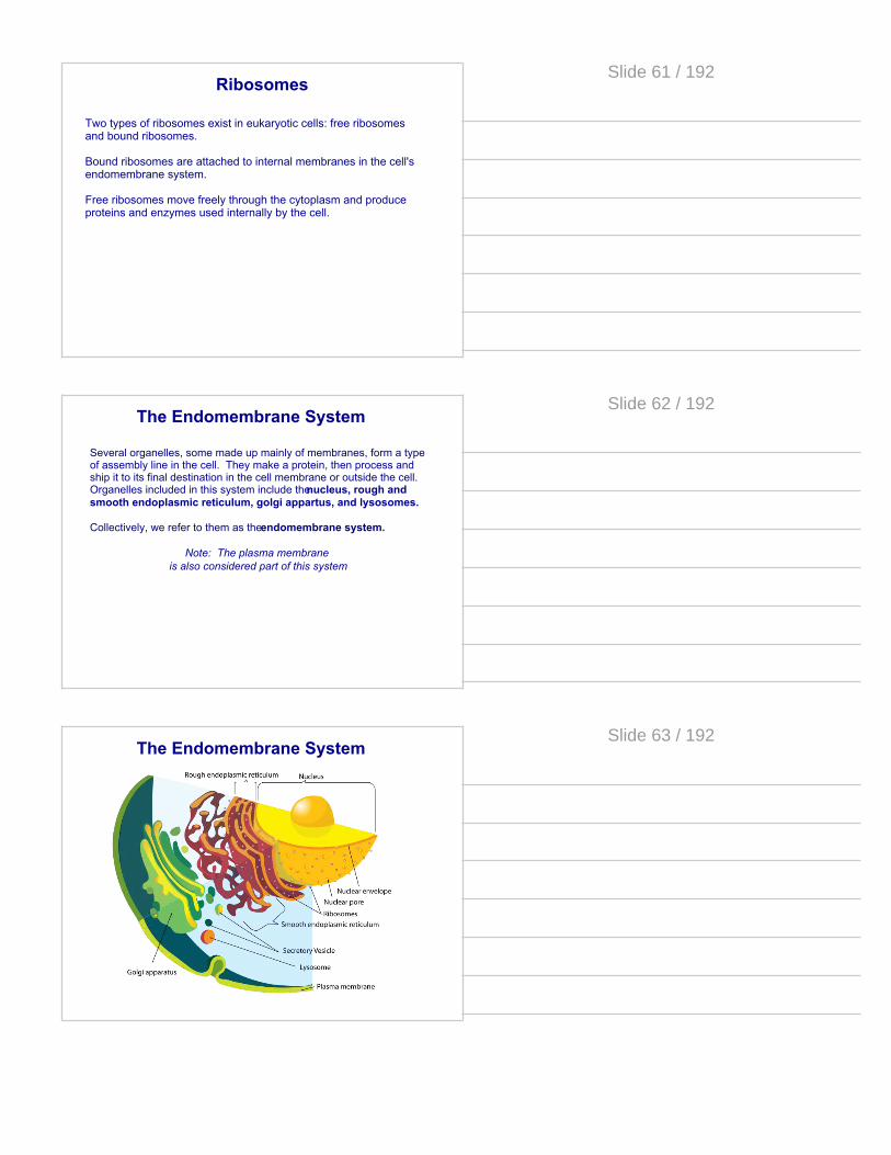
The Endomembrane System
Several organelles, some made up mainly of membranes, form a type of assembly line in the cell. They make a protein, then process and ship it to its final destination in the cell membrane or outside the cell. Organelles included in this system include the nucleus, rough and smooth endoplasmic reticulum, golgi appartus, and lysosomes.
Collectively, we refer to them as the endomembrane system.
Note: The plasma membrane is also considered part of this system
Slide 62 / 192
The Endomembrane System
Slide 63 / 192

Endoplasmic Reticulum
When RNA leaves the nucleus, it enters the endoplasmic reticulum (ER). This organelle is a series of membrane-bound sacs and tubules. It is continuous with the outer membrane of the nuclear envelope (reticulum comes from the latin word for little net). There are two types of endoplasmic reticulum: rough and smooth
Slide 64 / 192
Rough Endoplasmic Reticulum
Rough ER has ribosomes attached to its membrane (thus a rough appearance).
These ribosomes synthesize proteins that will be used in the plasma membrane, secreted outside the cell or shipped to another organelle called a lysosome.
As proteins are made by the ribosomes, they enter the lumen (opening) of the ER where they are folded and processed.
Slide 65 / 192
Smooth Endoplasmic Reticulum
This type of ER is called Smooth because it lacks ribosomes on its surface. (it looks smooth compared to rough ER)
There are a variety of functions of this organelle, which include:· making lipids.· processing certain drugs and poisons absorbed by the cell.· storing calcium ions (for example, in muscle cells).
Note: The liver is an organ that detoxifies substances that are brought into the body. Therefore, liver cells have huge amounts of Smooth ER
Slide 66 / 192

21 Where are ribosomal subunits made in the cell?
A Cytoplasm
B Nucleus
C Nucleolus
D On the Plasma membrane
Slide 67 / 192
22 List all the parts of the endomembrane system.
Arough and smooth endoplasmic reticulum, golgi appartus, lysosomes
B nucleus, rough and smooth endoplasmic reticulum, golgi appartus, lysosomes
C nucleus, rough and smooth endoplasmic reticulum, golgi appartus
Dnucleus, rough and smooth endoplasmic reticulum, golgi appartus, lysosomes, plasma membrane
Slide 68 / 192
23 Where are the proteins of the K+/Na+ pump synthesized?
A free ribsomes
B nucleus
C rough endoplasmic reticulum
D smooth endoplasmic reticulum
Slide 69 / 192

Protein Transport
Once the proteins are processed, short chains of sugars are sometimes linked to these proteins, which are then known as glycoproteins. These glycoproteins serve as "zip codes" that will tell the protein where it will go.
When the molecule is ready to be exported out of the ER, it gets packaged into a transport vesicle. This vesicle is made of membranes from the ER itself. The transport vesicle travels to another organelle known as the Golgi apparatus.
Slide 70 / 192
Golgi Apparatus
The main function of this organelle is to finish, sort, and ship cell products. It works like the postal department of the cell.
Structurally, the golgi consists of stacked flattened sacs (sort of looks like a stack of pita bread).
Slide 71 / 192
The Golgi is located near the cell membrane. The Golgi works closely with the ER of a cell.
It receives and modifies substances manufactured by the ER. Once the substances are modified, they are shipped out to other areas of the cell.
One key difference between the Golgi apparatus and endoplasmic reticulum is that the sacs comprising the Golgi are not interconnected.
Golgi Apparatus
Slide 72 / 192

The Golgi receives transport vesicles that bud off from the ER and contain proteins. It takes the substances contained in these vesicles and modifies them chemically in order to mark them and sort them into different batches depending on their destination.
The finished products are then packaged into new transport vesicles which will then move to lysosomes, or will be inserted into the plasma membrane or dumped out of the cell if the protein is a secretory protein.
The Golgi Apparatus & the ER
Video on Protein Trafficking through the Golgi
Slide 73 / 192
24 Which organelle receives and modifies substances from the endoplasmic reticulum?
A Nucleus
B Ribosomes
C Lysosomes
D Golgi Bodies
Slide 74 / 192
25 A difference between the Golgi Apparatus and the ER is that
A The ER takes the vesicles from the Golgi to transport
B The sacs making the Golgi are not interconnected
C The Golgi has ribosomes, the ER does not
D There is no difference, they are part of the same organelle
Slide 75 / 192
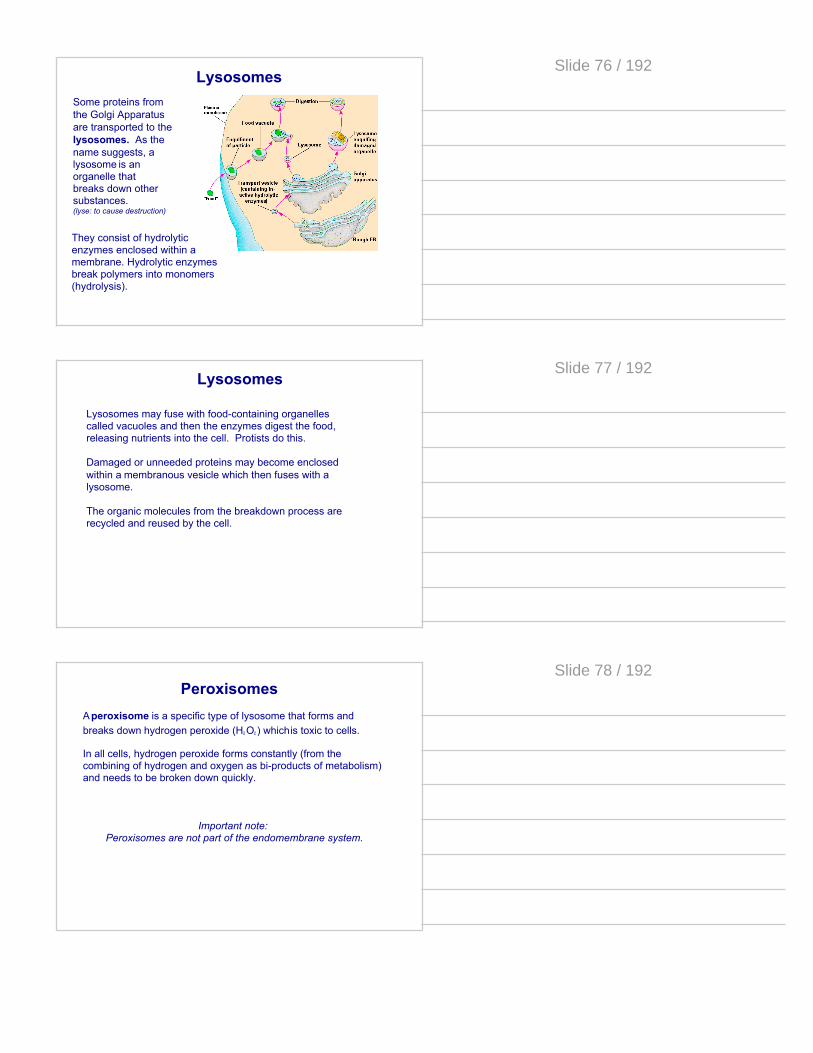
LysosomesSome proteins from the Golgi Apparatus are transported to the lysosomes. As the name suggests, a lysosome is an organelle that breaks down other substances.(lyse: to cause destruction)
They consist of hydrolytic enzymes enclosed within a membrane. Hydrolytic enzymes break polymers into monomers (hydrolysis).
Slide 76 / 192
Lysosomes
Lysosomes may fuse with food-containing organelles called vacuoles and then the enzymes digest the food, releasing nutrients into the cell. Protists do this.
Damaged or unneeded proteins may become enclosed within a membranous vesicle which then fuses with a lysosome.
The organic molecules from the breakdown process are recycled and reused by the cell.
Slide 77 / 192
A peroxisome is a specific type of lysosome that forms and breaks down hydrogen peroxide (H2O2) which is toxic to cells.
In all cells, hydrogen peroxide forms constantly (from the combining of hydrogen and oxygen as bi-products of metabolism) and needs to be broken down quickly.
Important note: Peroxisomes are not part of the endomembrane system.
Peroxisomes
Slide 78 / 192
26 Which organelle contains hydrolytic enzymes that break down other substances?
A Endoplasmic Reticulum
B Golgi Bodies
C Lysosomes
D Vacuoles
Slide 79 / 192
27 Which is not a function of lysosomes?
A aiding the cell in creating ribosomes
B fusing with vacuoles to digest food
C breaking polymers into monomers
D recycling worn out cell parts
Slide 80 / 192
Remember the plasma membrane is a phospholipid bilayer with proteins and other molecules interspersed throughout.
Plasma Membrane
Some proteins from the golgi apparatus become embedded in the membrane. Others are transported through the membrane to the external environment.
Slide 81 / 192
Plasma Membrane
· Selective permeability · Protection· Structural support
The 3 main functions of the plasma membrane:
Slide 82 / 192
28 Which of the following statements about the role of phospholipids in forming membranes is correct?
A they are completely insoluble in water
B they form a single sheet in water
C they form a structure in which the hydrophobic portion faces outward
D they form a selectively permeable structure
Slide 83 / 192
29 Active transport moves molecules
A with their concentration gradients without the use of energy
B with their concentration gradients using energy
C against their concentration gradients without the use of energy
D against their concentration gradients using energy
Slide 84 / 192

30 Which of the following processes includes all others?
A passive transport
B facilitated diffusion
C diffusion of a solute across a membrane
D osmosis
Slide 85 / 192
Many proteins created by the cell are too large to pass through the membrane, even using protein carrier or integral proteins. How do these macromolecules exit the cell?
Then, the substance uses other ways of getting into or out of a cell by fusing with the cell membrane.
Large Molecules and the Plasma Membrane
There are several special functions of the membrane as larger substances enter and exit the cell.
Slide 86 / 192
To excrete a macromolecule from the cell, the vesicles that enclose the proteins fuse with the plasma membrane and the vesicles then open up and spill their contents outside of the cell. This process is known as exocytosis. The vesicle will become part of the cell membrane.
Exocytosis
Exocytosis
This is how secretory proteins from the Golgi exit the cell. This is true for insulin in the pancreas.
Slide 87 / 192

Insulin is a protein hormone made by certain cells of the pancreas that enable cells to take glucose (sugar) in from the blood.
Insulin is a secretory protein made in the rough ER. Specifically, it is secreted out of the pancreas cells into the blood stream.
Insulin - A Secretory Protein
Slide 88 / 192
The opposite of exocytosis is endocytosis.
In this process, the cell takes in macromolecules or other particles by forming vesicles or vacuoles from its plasma membrane.
Endocytosis
This is how many protists ingest food particles
Slide 89 / 192
3 Types of Endocytosis
Slide 90 / 192
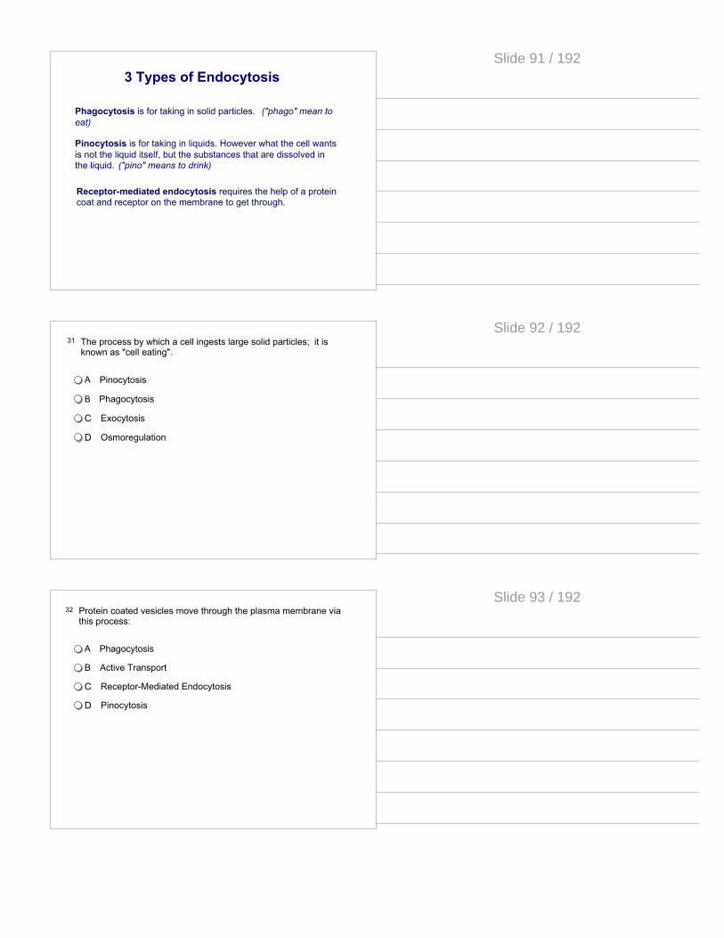
Phagocytosis is for taking in solid particles. ("phago" mean to eat)
Pinocytosis is for taking in liquids. However what the cell wants is not the liquid itself, but the substances that are dissolved in the liquid. ("pino" means to drink)
Receptor-mediated endocytosis requires the help of a protein coat and receptor on the membrane to get through.
3 Types of Endocytosis
Slide 91 / 192
31 The process by which a cell ingests large solid particles; it is known as "cell eating".
A Pinocytosis
B Phagocytosis
C Exocytosis
D Osmoregulation
Slide 92 / 192
32 Protein coated vesicles move through the plasma membrane via this process:
A Phagocytosis
B Active Transport
C Receptor-Mediated Endocytosis
D Pinocytosis
Slide 93 / 192

33 After a vesicle empties its contents outside a cell, the vesicle becomes part of:
A the Golgi
B the plasma membrane
C another vesicle
D the extracellular fluid
Slide 94 / 192
Energy-Converting Organelles
Chloroplasts reside in plant cells and some protists and convert solar radiation into energy stored in the cell for later use.
Mitochondria reside in all eukaryotic cells and convert chemical energy from glucose into ATP.
Interestingly, both chloroplasts and mitochondria have their own DNA, separate from that found in the nucleus of the cell. They also have a double cell membrane.
Slide 95 / 192
eukaryotic chloroplast
Chloroplasts
These organelles convert solar energy to chemical energy through photosynthesis. Chloroplasts are partitioned into three major compartments by internal membranes:
· Thylakoids
· Stroma
· Intermembrane space
Slide 96 / 192

Thylakoids
In prokaryotes, thylakoids are areas of highly folded membranes.
In eukaryotes, they are stacked in the chloroplasts. The fluid outside these stacks of thylakoids is called the stroma; this is where the Calvin cycle takes place.
eukaryotic chloroplast
Remember that during photosynthesis it is on the thylakoid that the Light Dependent Reactions take place.
Slide 97 / 192
Mitochondria
Mitochondria are sometimes referred to as the "powerhouses" of the cell. They convert chemical energy (glucose) into a more usable and regenerative form of chemical energy (ATP).
The mitochondrion is also partitioned like the chloroplast. They only have two compartments as opposed to three in the chloroplast.
· Matrix
· Intermembrane space
Slide 98 / 192
Remember cell respiration must take place near a membrane so that a proton gradient can be built in a "membrane space" that is separate from the rest of the cell. Thus, the membrane would separate the inner volume, with a deficit of protons, from the outside, with an excess.
In prokaryotes, the "inter-membrane space" is between the cell membrane and the cell wall.
In eukaryotes, that membrane is the inter-membrane space of the mitochondria in between the inner membrane and outer membrane.
Mitochondria and Respiration
Slide 99 / 192

The mitochondria and chloroplast are different from other eukaryotic organelles because they have their own DNA, their own ribosomes, and have a double cell membrane.
In 1970, Lynn Margulis published the "Theory of Endosymbiosis" to explain these facts. The theory states that the mitochondria and chloroplast were once free-living prokaryotes that got taken up (or "eaten") by another prokaryote.
The mitochondria was a bacteria that could make its own ATP. The chloroplast was a bacteria that could perform photosynthesis.
The Evolution of Eukaryotes
endo: within sym: together
bio: life sis: condition
endosymbiosis = living together, within
Slide 100 / 192
Since mitochondrial DNA is not in the cell nucleus, it is only passed along from mother to child; animals, including you, inherit your mitochondria from your mother only.
This is because the egg from our mothers contained her organelles. (Dad's sperm only contains the chromosomes, none of his organelles usually).
All of our organelles we inherited from our mothers. Mitochondrial DNA is a way to trace maternal heritage through a family or through a species. The "Mitochondrial Eve" is the first human female that gave rise to all humans. In theory, we can trace all humans back to her through our mitochondrial DNA.
The Mitochondrial Eve
Slide 101 / 192
34 Which organelle converts food energy into chemical energy that the cell can use?
A Nucleus
B Chloroplast
C Mitochondrion
D Golgi
Slide 102 / 192

35 Which organelle converts solar energy into chemical energy in plants and other photosynthetic organisms?
A Nucleus
B Chloroplast
C Mitochondrion
D Golgi
Slide 103 / 192
36 Which of the following is not true of mitochondria and chloroplasts?
A They are present in all eukaryotic cellsB They have their own DNAC They have their own ribosomesD They are surrounded by a double membrane
Slide 104 / 192
37 Which of the following does NOT provide evidence for the endosymbiotic theory?
A Mitochondria and chloroplasts both have their own DNA.
B Mitochondria and chloroplasts both come from pre-existing mitochondria and chloroplasts.
C The DNA of mitochondria and chloroplasts resembles the DNA found in nuclei.
D The DNA of mitochondria and chloroplasts resembles that of bacteria.
Slide 105 / 192

VacuolesVacuoles are membranous sacs and they come in different shapes and sizes and have a variety of functions.
Central Vacuole
PLANTCELL
PROTIST
Slide 106 / 192
Central Vacuoles
Central Vacuoles in plants store water. Absorbing water makes a plant cell more turgid, or having more pressure inside - leading to strength and rigidity.
Central vacuoles that are full will take over most of the cytoplasm and literally push the organelles to the sides of the cell. It can also store vital chemicals, pigments and waste products.
Slide 107 / 192
Increased turgor pressure results from the central vacuole being full with water. It presses out on the cell membrane which then presses out on the cell wall.
Turgor Pressure
The plant cell will not explode or lose its shape like an animal cell would in a hypotonic environment.
When the turgor pressure decreases the cell is limp and droopy. This is associated with wilted, limp lettuce, as well as droopy flowers.
Slide 108 / 192

Contractile Vacuoles
Contractile vacuoles can be found in certain single-celled protists. These act as a pump to expel excess water from the cell. This is especially helpful to those organisms living in a freshwater environment to keep the cell from exploding.
Slide 109 / 192
Food Vacuoles
Food Vacuoles are mainly found in protists.
The protist ingests food particles. The particles then fuse with a lysosome. The lysosome contains hydrolytic enzymes that break the food down. Paramecium fed dyed food showing vacuoles.
Slide 110 / 192
38 An organelle found in plant cells that stores water as well as other important substances is called the ___________.
A Lysosome
B Contractile Vacuole
C Central Vacuole
D Golgi bodies
Slide 111 / 192
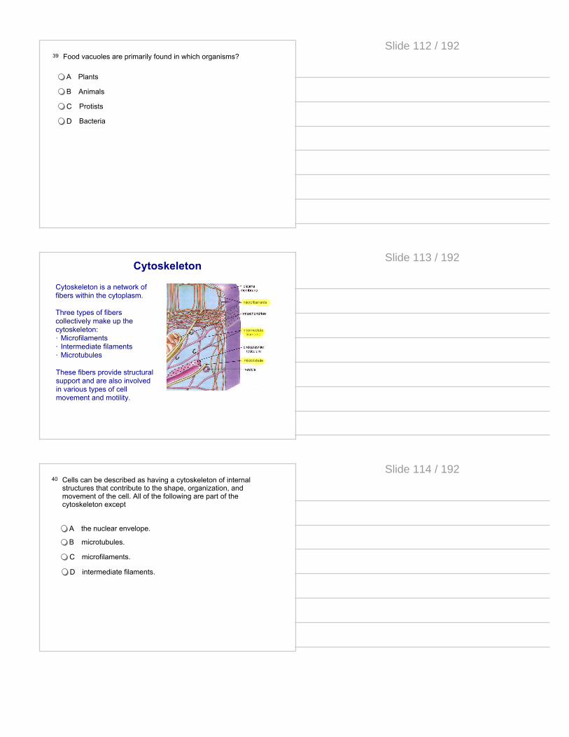
39 Food vacuoles are primarily found in which organisms?
A Plants
B Animals
C Protists
D Bacteria
Slide 112 / 192
Cytoskeleton
Cytoskeleton is a network of fibers within the cytoplasm.
Three types of fibers collectively make up the cytoskeleton:· Microfilaments· Intermediate filaments· Microtubules
These fibers provide structural support and are also involved in various types of cell movement and motility.
Slide 113 / 192
40 Cells can be described as having a cytoskeleton of internal structures that contribute to the shape, organization, and movement of the cell. All of the following are part of the cytoskeleton except
A the nuclear envelope.
B microtubules.
C microfilaments.
D intermediate filaments.
Slide 114 / 192
41 Which of the following is not a known function of the cytoskeleton? A to maintain a critical limit on cell size
B to provide mechanical support to the cell
C to maintain the characteristic shape of the cell
D to hold mitochondria and other organelles in place within the cytosol
Slide 115 / 192
Cell WallThe cell wall is an outer layer in addition to the plasma membrane, found in fungi, algae, and plant cells.
The composition of the cell wall varies among species and even between cells in the same individual. All cell walls have carbohydrate fibers embedded in a stiff matrix of proteins and other carbohydrates.
Plant cell walls are made of the polysaccharide cellulose. Fungal cell walls are made of the polysaccharide chitin.
Slide 116 / 192
Extracellular Matrix
The cells of many multi-cellular animals are surround by a extracellular matrix (ECM). The ECM provides structural support to the cells in addition to providing various other functions such as anchorage, cellular healing, separating tissues from one another and regulating cellular communication.
The ECM is primarily composed of an interlocking mesh of proteins and carbohydrates.
Slide 117 / 192
Plant vs. Animal Cell Organelles
Click here to review the similarities and difference
between plant and animal cells
Slide 118 / 192
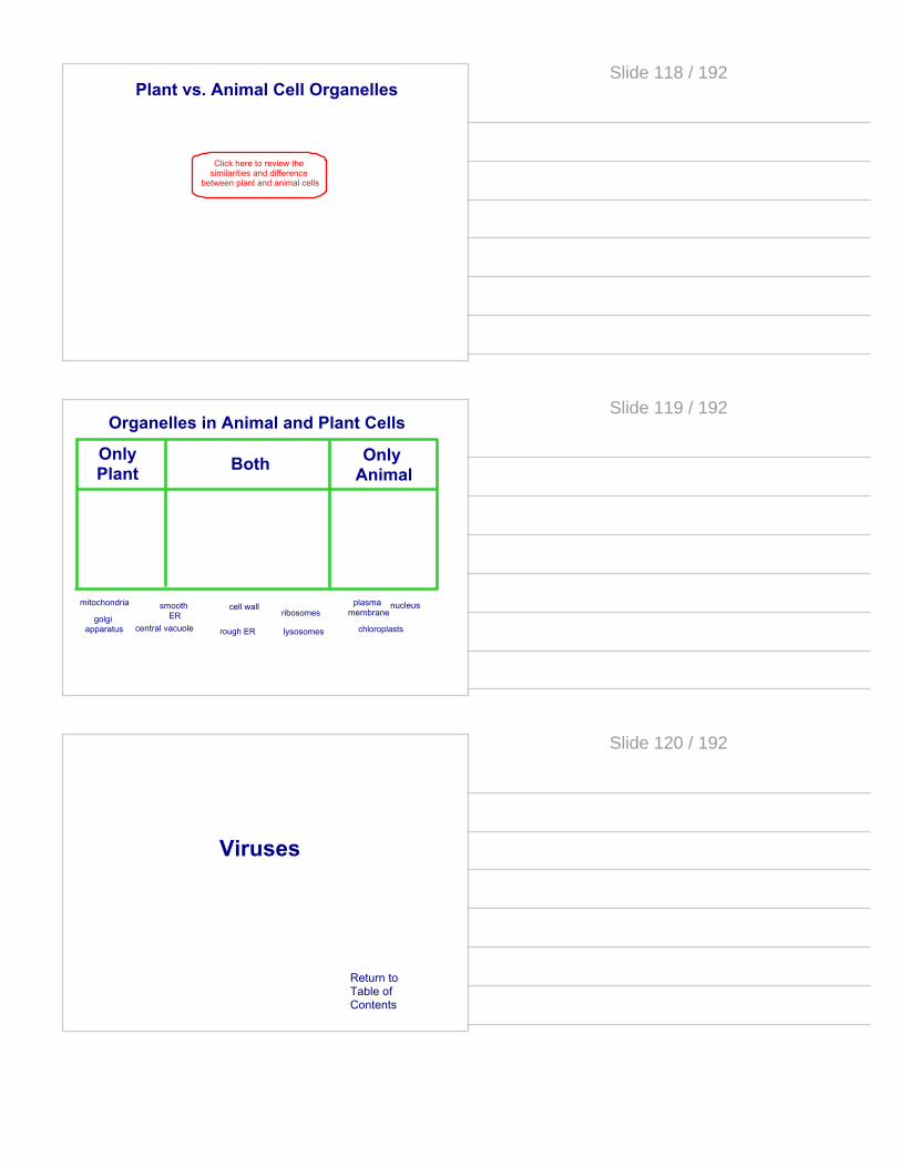
Organelles in Animal and Plant Cells
cell wall
chloroplastscentral vacuole
plasma membrane
mitochondria
Only Animal
Only Plant Both
rough ER
smooth ER
lysosomesgolgi
apparatus
ribosomesnucleus
Slide 119 / 192
Viruses
Return toTable ofContents
Slide 120 / 192

A virus is a small infectious agent that can replicate only inside the living cells of an organism. Viruses can infect all types of organisms, though they are not organisms themselves.
Viruses are particles that are considered non-living because they cannot perform all the functions of living things. However, they share the same genetic code and use the mechanism of host cells to reproduce.
Viruses
A bacterial phage virus
Slide 121 / 192
In biology viruses are important because their genetic and reproductive strategies use the same molecular components but their use is unique. Scientists have learned a lot about genetics by understanding their strategies.
In addition, their infectious nature makes them a threat that requires humans to understand how they work to create defensive technology.
Viruses
Human immunodeficiency virus (HIV) attacking a human lymphocyte cell
Slide 122 / 192
The general mode of operation for all viruses is to · Infect a host cell with its genetic information· Hijack the molecular machinery of the host cell to manufacture the parts needed to build more viruses· Package the parts together to form new viruses for release from the host cell
Viruses
This E. coli cell has been attacked by bacteriophage viruses and they have injected their genetic material into the bacterial cell
Slide 123 / 192
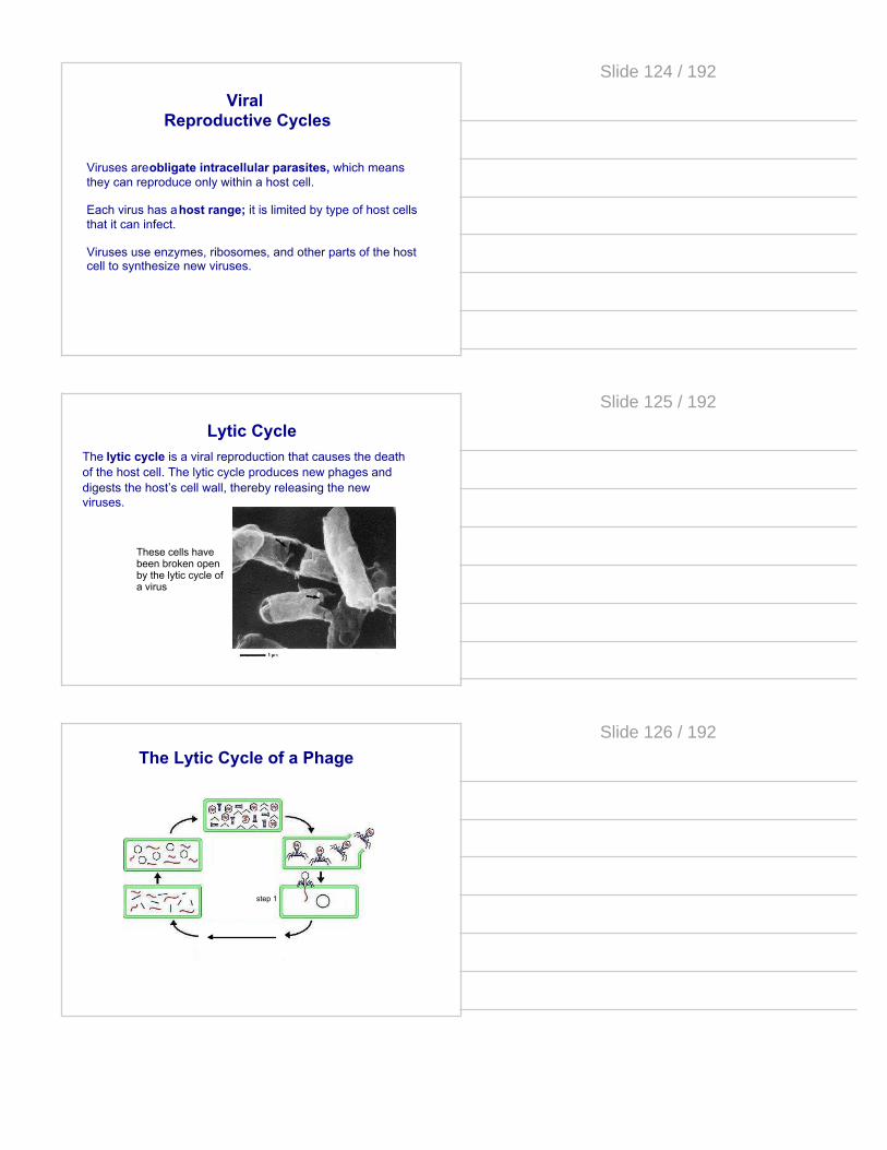
Viruses are obligate intracellular parasites, which means they can reproduce only within a host cell.
Each virus has a host range; it is limited by type of host cells that it can infect.
Viruses use enzymes, ribosomes, and other parts of the host cell to synthesize new viruses.
Viral Reproductive Cycles
Slide 124 / 192
Lytic CycleThe lytic cycle is a viral reproduction that causes the death of the host cell. The lytic cycle produces new phages and digests the host’s cell wall, thereby releasing the new viruses.
These cells have been broken open by the lytic cycle of a virus
Slide 125 / 192
The Lytic Cycle of a Phage
step 1
Slide 126 / 192
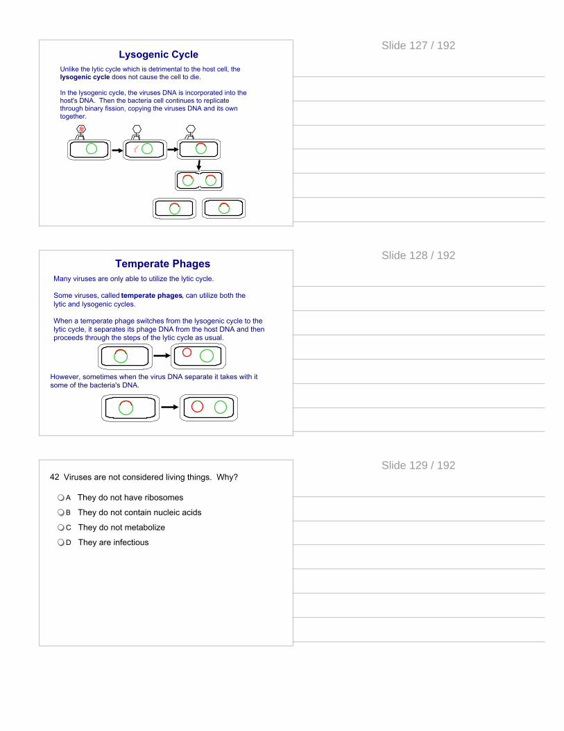
Lysogenic CycleUnlike the lytic cycle which is detrimental to the host cell, the lysogenic cycle does not cause the cell to die.
In the lysogenic cycle, the viruses DNA is incorporated into the host's DNA. Then the bacteria cell continues to replicate through binary fission, copying the viruses DNA and its own together.
Slide 127 / 192
Temperate PhagesMany viruses are only able to utilize the lytic cycle.
Some viruses, called temperate phages, can utilize both the lytic and lysogenic cycles.
When a temperate phage switches from the lysogenic cycle to the lytic cycle, it separates its phage DNA from the host DNA and then proceeds through the steps of the lytic cycle as usual.
However, sometimes when the virus DNA separate it takes with it some of the bacteria's DNA.
Slide 128 / 192
42 Viruses are not considered living things. Why?
A They do not have ribosomes
B They do not contain nucleic acids
C They do not metabolize
D They are infectious
Slide 129 / 192
43 The lytic cycle _______________. The lysogenic cycle _______________.
Aresults in death of the host cell; increases genetic variation in a population of cells
Boccurs only in temperate viruses; incorporates the viral DNA with the host DNA
C is utilized by phages; is utilized by viruses
Dcreates full viral molecules; replicates only the viruses protein coat
Slide 130 / 192
Some viruses use DNA as their genetic material. It does not contain DNA polymerase, so in order for it to reproduce it must inject its DNA into a cell so that it can be copied by the host cell's polymerase.
The bacteriophage example we have seen is a DNA virus.
DNA Viruses
Slide 131 / 192
An RNA virus is a virus that has RNA as its genetic material. This nucleic acid is usually single-stranded RNA (ssRNA), but may be double-stranded RNA (dsRNA). Some human diseases caused by RNA viruses include SARS, influenza, hepatitis C, West Nile fever, polio and measles.
RNA Viruses
Slide 132 / 192
Severe acute respiratory syndrome (SARS) is a viral respiratory disease in humans. An outbreak of SARS in Hong Kong nearly became a pandemic, with 8,273 cases and 775 deaths worldwide. Within weeks, SARS spread from Hong Kong to infect individuals in 37 countries in early 2003.
SARS
They are enveloped RNA viruses that are pathogens of mammals and birds. This group of viruses cause respiratory tract infections in a variety of animals, including humans
Slide 133 / 192
A retrovirus is an RNA virus that replicates in a host cell.
First it uses its own reverse transcriptase enzyme to produce DNA from its RNA genome, reverse of the usual pattern, thus retro.
This new DNA is then incorporated into the host's genome by an integrase enzyme. The cell then treats the viral DNA as part of its own instructions, which it follows blindly, making the proteins required to assemble new copies of the virus.
Retro Viruses
Slide 134 / 192
These viruses are some of the most complex and believed to be the most advanced from an evolutionary perspective. For a virus, their entry system into cells is highly complex. They have systems to bypass the usual defenses of their host cell. HIV is a retro virus.
Retro Viruses
Slide 135 / 192

HIV
continued...
Slide 136 / 192
HIV
Slide 137 / 192
What makes HIV particularly dangerous is that it attacks the human immune system and fools it into treating it is part of the system. No immune attack is offered by the infected cells.
The complex replication system is flawed and many errors are made as the virus replicates its genome. Why would this make treating with medicine difficult?
Each patient has many unique variations of the virus in their system. A treatment that may destroy one version will not destroy the others. In addition any attack on the virus must also be an atack on the immune system.
HIV
Slide 138 / 192

44 When a virus infects an E. coli cell, what part of the virus enters the bacterial cytoplasm?
A the entire virus
B the nucleic acid
C the protein capsid and enclosed nucleic acid
D the tail fibers
Slide 139 / 192
45 RNA viruses mutate more rapidly than DNA viruses, making them more difficult to prevent. Which of the following is a likely cause of this rapid mutation rate?
ADNA viruses contain their own polymerase enzymes, while RNA viruses hijacked cellular machinery
B RNA can be more rapidly copied than DNA
CBoth single and double stranded RNA molecules can be present in a RNA virus
D DNA is structurally more stable than RNA
Slide 140 / 192
46 Which of the following correctly outlines the steps of a retroviral infection?
ARNA insertion - reverse transcription - integration into genome - gene expression
B RNA insertion - integration in genome - gene expression
C RNA insertion - gene expression - integration into genome
D RNA insertion - integration in genome - reverse transcription
Slide 141 / 192
Cellular Defenses
Return toTable ofContents
Slide 142 / 192
Cellular Defenses
All organisms have defenses designed to prevent infection by viruses, bacteria, and other pathogens.
Slide 143 / 192
Slide 144 / 192

Restriction Enzymes
Restriction enzymes are now used by scientists to locate specific genes on a DNA molecules. DNA mixed with these enzymes is called a digest because the enzymes breaks down the fragments of DNA into many smaller pieces.
Gene of interest is somewhere in here
This tube contains many different pieces of DNA
It is important to remember that we are working with molecules. We cannot simply "grab" the piece of DNA we want. We must separate the unique pieces of DNA in the digest and select the fragment we want.
Slide 145 / 192
Gel Electrophoresis
Gel electrophoresis is one way to separate DNA fragments based on length.
The digest is loaded by pipet into a gel, that resembles Jello. The gel is a network of fibers called collagen.
Small pieces of DNA can move through the gel quicker than the longer pieces that get tangled in the collagen fibers.
Slide 146 / 192
Gel Electrophoresis
DNA has a slightly negative charge.
An electrical current is passed through the gel and the DNA fragments move to the positive charge. Small fragments move faster, larger fragments are slowed down by the matrix.
The result is that the small pieces can travel farther than the larger ones. The DNA is separated by size in what is know as a banding pattern.
15k
10k
5k
start
Insulin gene
Slide 147 / 192
Virtual Lab
Gel electrophoresis virtual lab by the University of Utah
The Gel Lab
Slide 148 / 192
Animal DefensesAnimals have multiple defenses against invaders. These are classified into two categories: innate immunity and acquired immunity.
Innate immune defenses are generalized systems, preventing invasion by all pathogens. All animals have some degree of innate immunity.
Acquired immune defenses are developed during the animal's lifetime and respond only to specific invaders, those that have been encountered previously. These defenses are only found in vertebrates.
Slide 149 / 192
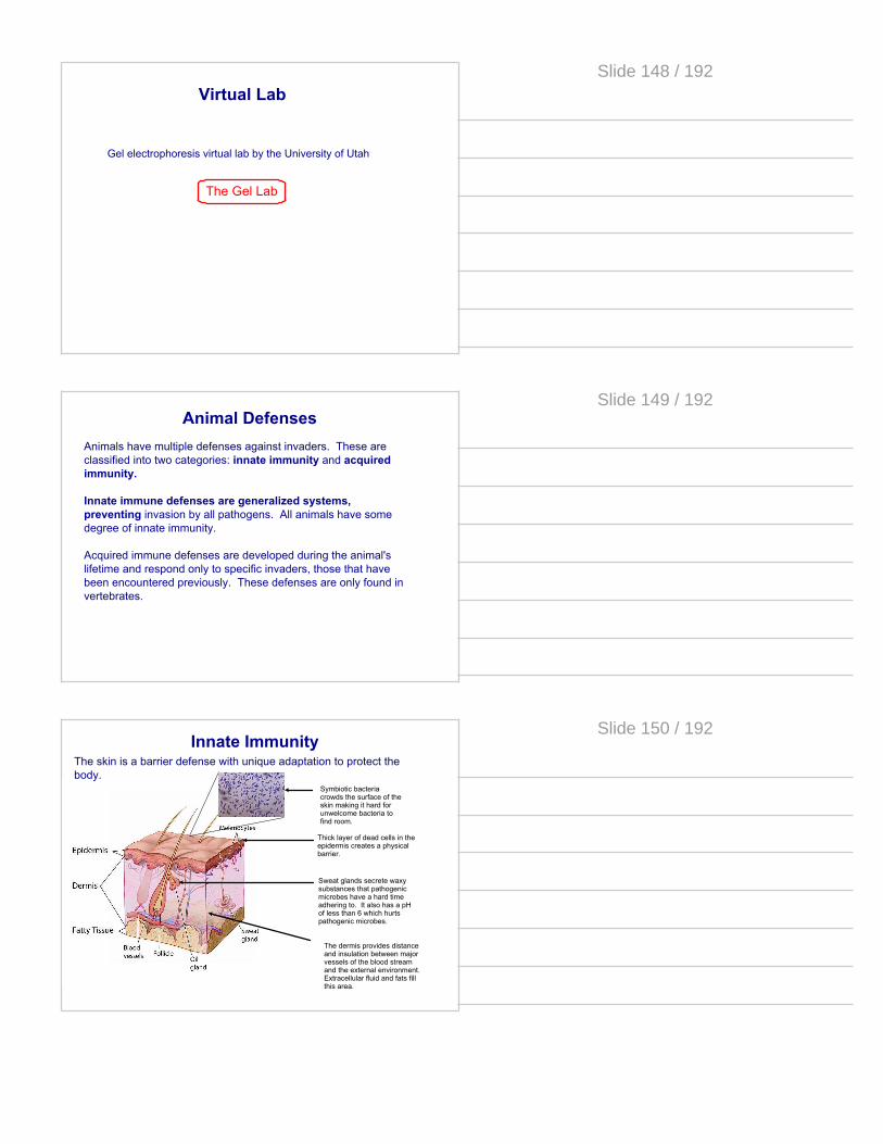
Thick layer of dead cells in the epidermis creates a physical barrier.
Sweat glands secrete waxy substances that pathogenic microbes have a hard time adhering to. It also has a pH of less than 6 which hurts pathogenic microbes.
The dermis provides distance and insulation between major vessels of the blood stream and the external environment. Extracellular fluid and fats fill this area.
Symbiotic bacteria crowds the surface of the skin making it hard for unwelcome bacteria to find room.
Innate ImmunityThe skin is a barrier defense with unique adaptation to protect the body.
Slide 150 / 192
Innate Immunity
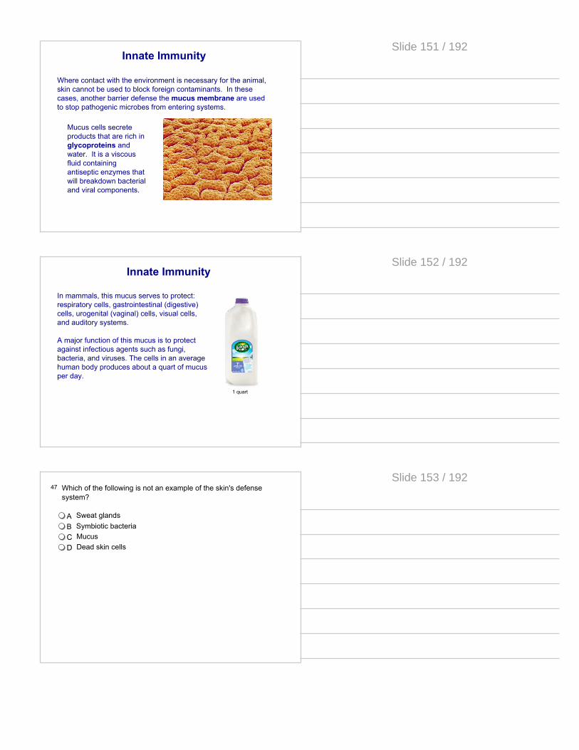
Where contact with the environment is necessary for the animal, skin cannot be used to block foreign contaminants. In these cases, another barrier defense the mucus membrane are used to stop pathogenic microbes from entering systems.
Mucus cells secrete products that are rich in glycoproteins and water. It is a viscous fluid containing antiseptic enzymes that will breakdown bacterial and viral components.
Slide 151 / 192
Innate Immunity
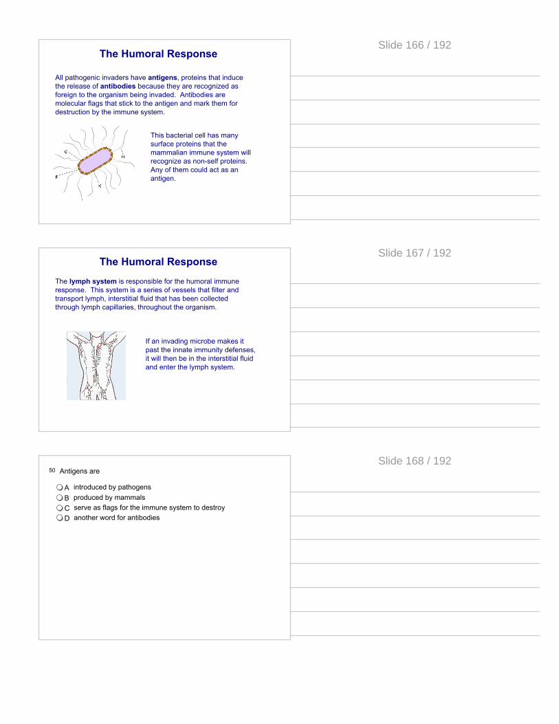
In mammals, this mucus serves to protect: respiratory cells, gastrointestinal (digestive) cells, urogenital (vaginal) cells, visual cells, and auditory systems. A major function of this mucus is to protect against infectious agents such as fungi, bacteria, and viruses. The cells in an average human body produces about a quart of mucus per day.
1 quart
Slide 152 / 192
47 Which of the following is not an example of the skin's defense system?
A Sweat glandsB Symbiotic bacteriaC MucusD Dead skin cells
Slide 153 / 192

Innate Immunity
If a foreign invader makes it past the skin and mucous membrane, the body has specialized cells that can detect and respond.
Slide 154 / 192
Innate Immunity
Consider a laceration (cut) on a part of your body. Immediately foreign cells are entering the break in your bodies barrier. Take a moment and describe to another person what happens in the few minutes after a cut. Make a list of your bodies responses. Can you relate these symptoms to fighting infection?
Slide 155 / 192
Inflammation
Skin
Nearby capilary
Mast Cells
Extracellular fluid
Inflammation is a response triggered by bacteria that enters the skin. A metabolic pathway is initiated by the presence of bacteria under the skin.
Slide 156 / 192

InflammationA splinter enters the skin
Skin
Nearby capilary
Mast Cells
Extracellular fluid
Slide 157 / 192
InflammationBacterial cells that were on the splinter enter the interstitial fluid.
Skin
Nearby capilary
Mast Cells
Extracellular fluid
Slide 158 / 192
InflammationMast cells detect foreign proteins produced by the bacteria and a transduction pathway is triggered. The end result is that histamine is released from the mast cell.
Skin
Nearby capilary
Mast Cells
Extracellular fluid
Slide 159 / 192

InflammationHistamine acts as another signal molecule that causes the cells of the capillary to separate and blood plasma, red blood cells, and phagocytes spill into the area.
Skin
Nearby capilary
Extracellular fluid
Phagocyte
Slide 160 / 192
InflammationBecause of the extra volume of fluid and cells the area becomes hot and swells. This is unfavorable conditions for the bacteria and they cannot reproduce or spread to new areas.
Skin
Nearby capilary
Extracellular fluid
Phagocyte
Slide 161 / 192
InflammationPhagocytes are cells that eat foreign cells. They remove the bacteria, mast cells stop producing histamine and the inflammation is relieved.
Skin
Nearby capilary
Extracellular fluid
Phagocyte
Click here for an animation of inflammation
Slide 162 / 192

48 Which of the following best describes the response to bacteria entering under the surface of the skin?
AMast cells produce histamine, swelling occurs, red blood cells and phagocytes enter the area, phagocytes eat the foreign cells
BMast cells produce histamine, red blood cells and phagocytes enter the area, swelling occurs, phagocytes eat the foreign bacteria
CHistamine produces mast cells, phagocytes eat the mast cells, swelling occurs, new red blood cells enter the area
DSwelling occurs, mast cells produce histamine, red blood cells and phagocytes enter, phagocytes eat the foreign bacteria
Slide 163 / 192
49 Inflammation is
A an example of innate immunity
B an example of acquired immunity
C a response to the presence of phagocytes
D a buildup of fluids in the mucus membranes
Slide 164 / 192
Specific Immunity
The specific immunity of vertebrates includes two types of response: Humoral: Attacking pathogens in the extracellular matrix, prior to entering a body cell Cell-mediated: Destroying body cells that have been infected by pathogens or have become cancerous.
Both responses are derived from white blood cells known as lymphocytes.
Slide 165 / 192
The Humoral Response
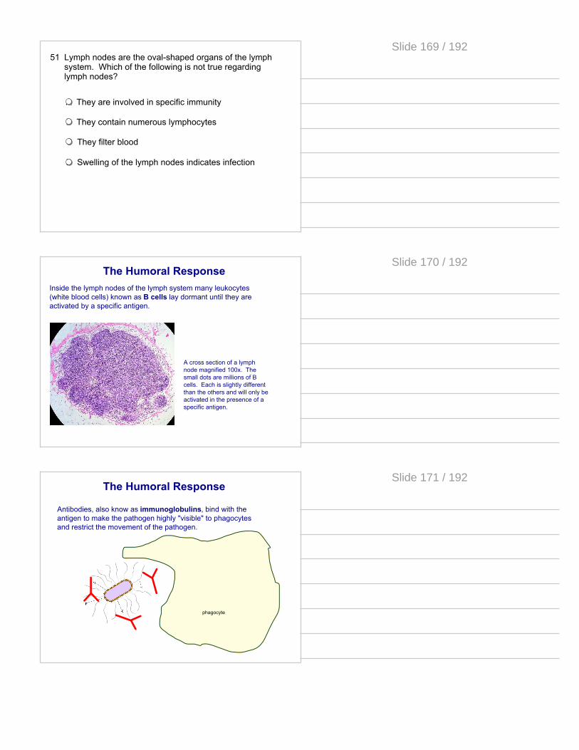
All pathogenic invaders have antigens, proteins that induce the release of antibodies because they are recognized as foreign to the organism being invaded. Antibodies are molecular flags that stick to the antigen and mark them for destruction by the immune system.
This bacterial cell has many surface proteins that the mammalian immune system will recognize as non-self proteins. Any of them could act as an antigen.
Slide 166 / 192
The Humoral Response
The lymph system is responsible for the humoral immune response. This system is a series of vessels that filter and transport lymph, interstitial fluid that has been collected through lymph capillaries, throughout the organism.
If an invading microbe makes it past the innate immunity defenses, it will then be in the interstitial fluid and enter the lymph system.
Slide 167 / 192
50 Antigens are
A introduced by pathogensB produced by mammalsC serve as flags for the immune system to destroyD another word for antibodies
Slide 168 / 192
51 Lymph nodes are the oval-shaped organs of the lymph system. Which of the following is not true regarding lymph nodes?
A They are involved in specific immunity
B They contain numerous lymphocytes
C They filter blood
D Swelling of the lymph nodes indicates infection
Slide 169 / 192
The Humoral ResponseInside the lymph nodes of the lymph system many leukocytes (white blood cells) known as B cells lay dormant until they are activated by a specific antigen.
A cross section of a lymph node magnified 100x. The small dots are millions of B cells. Each is slightly different than the others and will only be activated in the presence of a specific antigen.
Slide 170 / 192
The Humoral Response
Antibodies, also know as immunoglobulins, bind with the antigen to make the pathogen highly "visible" to phagocytes and restrict the movement of the pathogen.
phagocyte
Slide 171 / 192
The Humoral Response
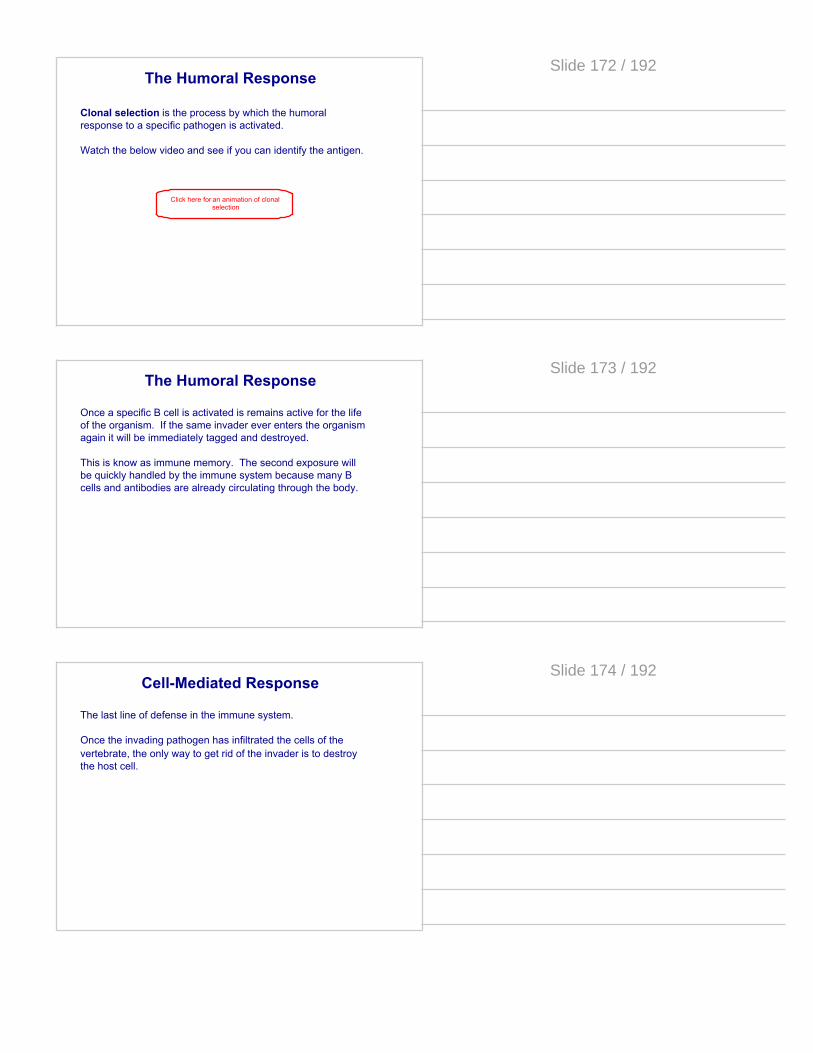
Clonal selection is the process by which the humoral response to a specific pathogen is activated.
Watch the below video and see if you can identify the antigen.
Click here for an animation of clonal selection
Slide 172 / 192
The Humoral Response
Once a specific B cell is activated is remains active for the life of the organism. If the same invader ever enters the organism again it will be immediately tagged and destroyed.
This is know as immune memory. The second exposure will be quickly handled by the immune system because many B cells and antibodies are already circulating through the body.
Slide 173 / 192
Cell-Mediated Response
The last line of defense in the immune system.
Once the invading pathogen has infiltrated the cells of the vertebrate, the only way to get rid of the invader is to destroy the host cell.
Slide 174 / 192
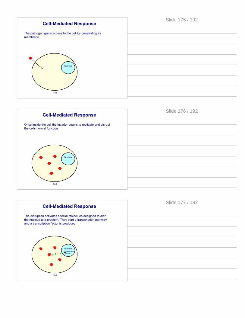
Cell-Mediated Response
The pathogen gains access to the cell by penetrating its membrane.
Nucleus
Cell
Slide 175 / 192
Cell-Mediated Response
Once inside the cell the invader begins to replicate and disrupt the cells normal function.
Nucleus
Cell
Slide 176 / 192
Cell-Mediated Response
The disruption activates special molecules designed to alert the nucleus to a problem. They start a transcription pathway and a transcription factor is produced.
Nucleus
Cell
Transcription Factor
Slide 177 / 192

Cell-Mediated Response
The transcription factor activates a gene that produces a membrane protein that will act as a flag to alert immune system cells that it is infected
Nucleus
Cell
Transcription Factor
Slide 178 / 192
Cell-Mediated Response
This cell is now a dendritic cell or antigen presenting cell. A special leukocyte known as a helper T cell attaches to the antigens of the damaged cell.
Nucleus
Cell
Transcription Factor
Slide 179 / 192
Cell-Mediated Response
The helper T cell activates and releases cytokines, free floating proteins that communicate with other cells of the immune system, into the surrounding fluids.
Nucleus
Cell
Transcription Factor
Slide 180 / 192

Cell-Mediated Response
The cytokines do 2 things: They alert B cells to activate humoral defenses; and they bring cytotoxic T cells that inject hydrolytic enzymes into the diseased cell.
Nucleus
Cell
Transcription Factor
Slide 181 / 192
Cell-Mediated Response
The diseased cell and its invaders are eliminated.
Nucleus
Cell
Transcription Factor
Slide 182 / 192
52 The end result of the cell mediated response is that the
A the T cells are destroyedB the pathogen is destroyedC diseased cell is destroyedD diseased cell and the pathogen is destroyed
Slide 183 / 192

53 An immune memory response is created through
A Humoral responseB Cell-mediated response
Slide 184 / 192
Plant Immunity
Bacteria and viruses are as much a threat to the homeostasis of plants as they are to animals. For this reason plants must also protect themselves and be able to fight foreign pathogens.
Plants only have generalized defenses against pathogens. They do not have specific immunity. Much like invertebrates, the evolution of plants came long before specific immunity evolution on the tree of life.
Slide 185 / 192
Barrier Defenses
As with animals, the first line of defense against pathogens is the outer covering. Plants have varying levels of external defenses that can include waxy coatings, sticky excretions, thick cuticles and others.
Slide 186 / 192

Plant Immunity
Since plants have slow moving circulation systems (or no circulation at all), if a pathogen gets past the external covering individual cells are on their own to defend themselves.
These spongy mesophyll cells each have an internal defense system that is triggered when contacted by a pathogen.
Slide 187 / 192
Plant Immunity
Upon pathogen attack, pathogen-associated molecular patterns (PAMPs) activate receptors in the plant cell, resulting in a signaling cascade that leads to PAMP-triggered immunity (PTI)
Pathogen
Receptor
Nucleus
chemical signal to nucleus
From this point there are 2 possible responses by the cell.
Slide 188 / 192
Plant Immunity
The cell can begin to secrete an antimicrobial agent (a protein designed to disrupt pathogens) that will fill the cytoplasm and secrete from the cell membrane. This will hopefully kill or disable the pathogen.
Pathogen
Receptor
Nucleus
Slide 189 / 192
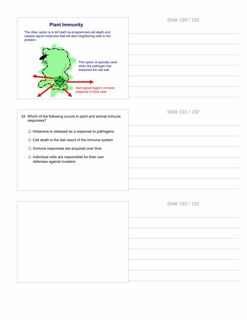
Plant ImmunityThe other option is to kill itself via programmed cell death and release signal molecules that will alert neighboring cells to the problem.
Nucleus
This option is typically used when the pathogen has breached the cell wall.
Alert signal triggers immune response in local cells
Slide 190 / 192
54 Which of the following occurs in plant and animal immune responses?
A Histamine is released as a response to pathogens
B Cell death is the last resort of the immune system
C Immune responses are acquired over time
D Individual cells are responsible for their own defenses against invaders
Slide 191 / 192
Slide 192 / 192