Embed Size (px)
Citation preview
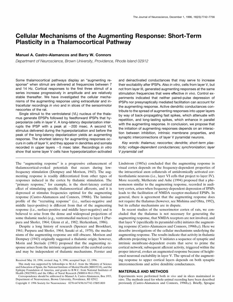
Cellular Mechanisms of the Augmenting Response: Short-TermPlasticity in a Thalamocortical Pathway
Manuel A. Castro-Alamancos and Barry W. Connors
Department of Neuroscience, Brown University, Providence, Rhode Island 02912
Some thalamocortical pathways display an “augmenting re-sponse” when stimuli are delivered at frequencies between 7and 14 Hz. Cortical responses to the first three stimuli of aseries increase progressively in amplitude and are relativelystable thereafter. We have investigated the cellular mecha-nisms of the augmenting response using extracellular and in-tracellular recordings in vivo and in slices of the sensorimotorneocortex of the rat.Single stimuli to the ventrolateral (VL) nucleus of the thala-
mus generate EPSPs followed by feedforward IPSPs that hy-perpolarize cells in layer V. A long-latency depolarization inter-rupts the IPSP with a peak at ;200 msec. A second VLstimulus delivered during the hyperpolarization and before thepeak of the long-latency depolarization yields an augmentingresponse. The shortest latency for augmenting responses oc-curs in cells of layer V, and they appear in dendrites and somatarecorded in upper layers ;5 msec later. Recordings in vitroshow that some layer V cells have hyperpolarization-activated
and deinactivated conductances that may serve to increasetheir excitability after IPSPs. Also in vitro, cells from layer V, butnot from layer III, generated augmenting responses at the samestimulation frequencies that were effective in vivo. Control ex-periments indicated that neither paired-pulse depression ofIPSPs nor presynaptically mediated facilitation can account forthe augmenting response. Active dendritic conductances con-tribute to the spread of augmenting responses into upper layersby way of back-propagating fast spikes, which attenuate withrepetition, and long-lasting spikes, which enhance in parallelwith the augmenting response. In conclusion, we propose thatthe initiation of augmenting responses depends on an interac-tion between inhibition, intrinsic membrane properties, andsynaptic interconnections of layer V pyramidal neurons.
Key words: thalamus; neocortex; dendrite; short-term plas-ticity; voltage-dependent conductances; synchronization; layerV pyramidal cell
The “augmenting response” is a progressive enhancement ofthalamocortical-evoked potentials that occurs during low-frequency stimulation (Dempsey and Morison, 1943). The aug-menting response is readily differentiated from other types ofresponses induced in the cortex by thalamic stimulation. The“primary response,” for example, is the short-latency corticaleffect of stimulating specific thalamocortical afferents, and it isdepressed at stimulus frequencies optimal for the augmentingresponse (Castro-Alamancos and Connors, 1996b). The laminarprofile of the “recruiting response” (i.e., surface-negative andmiddle layer-positive) is different from that of the augmentingresponse (i.e., surface-positive and middle layer-negative) and isbelieved to arise from the dense and widespread projections ofsome thalamic nuclei (e.g., ventromedial nucleus) to layer I (Pur-pura and Shofer, 1964; Glenn et al., 1982; Herkenham, 1986).Despite a long history of research (Spencer and Brookhart,
1961; Purpura and Shofer, 1964; Sasaki et al., 1970), the mecha-nisms of the augmenting response are still obscure. Morison andDempsey (1943) originally suggested a thalamic origin; however,Morin and Steriade (1981) proposed that the augmenting re-sponse arises from the intrinsic organization of the cerebral cortexand may be independent of thalamic mechanisms. Ferster and
Lindstrom (1985a) concluded that the augmenting response invisual cortex depends on the frequency-dependent properties ofthe intracortical axon collaterals of antidromically activated cor-ticothalamic neurons (i.e., layer VI cells that project to layer IV).More recently, Metherate and Ashe (1994) proposed that a phe-nomenon similar to the augmenting response, recorded in audi-tory cortex, arises when frequency-dependent depression of IPSPsleads to the facilitation of NMDA receptor-mediated EPSPs. Ingeneral, there is agreement that the augmenting response doesnot require the thalamus (however, see Mishima and Ohta, 1992),but its cellular mechanisms are in dispute.In recent studies of the sensorimotor cortex of rats, we con-
cluded that the thalamus is not necessary for generating theaugmenting response, that NMDA receptors are not involved, andthat layer V (specifically its pyramidal cells) initiates the augment-ing response (Castro-Alamancos and Connors, 1996b,c). Here wedescribe investigations of the cellular mechanisms underlying theaugmenting response. The results indicate that activity in thalamicafferents projecting to layer V initiates a sequence of synaptic andintrinsic membrane-dependent events that serve to prime thecortical network; subsequent afferent activity, triggered within theproper interval, evokes an augmented response because of height-ened neuronal excitability in layer V. The spread of the augment-ing response to upper cortical layers depends on both synapticinterconnections and active dendritic conductances.
MATERIALS AND METHODSExperiments were performed both in vivo and in slices maintained invitro. The methods used for whole-animal recording have been describedpreviously (Castro-Alamancos and Connors, 1996b,c). Briefly, Sprague
Received May 10, 1996; revised Aug. 9, 1996; accepted Sept. 12, 1996.This study was supported by fellowships to M.A.C. from the Ministry of Science
and Education of Spain, the National Institute of Mental Health (MH19118), and theEpilepsy Foundation of America, and grants to B.W.C. from National Institutes ofHealth (NS25983) and the Office of Naval Research (N00014-90-J-1701).Correspondence should be addressed to Manuel A. Castro-Alamancos, Box 1953,
Department of Neuroscience, Brown University, Providence, RI 02912.Copyright q 1996 Society for Neuroscience 0270-6474/96/167742-15$05.00/0
The Journal of Neuroscience, December 1, 1996, 16(23):7742–7756
Dawley rats (250–350 gm) were anesthetized with ketamine HCl (100mg/kg, i.p.) and supplemented regularly (50 mg/kg, i.m.). After inductionof surgical anesthesia, the animal was placed in a stereotaxic frame. Allskin incisions and frame contacts with the skin were injected with lido-caine (2%). A unilateral craniotomy extended over a large area of theparietofrontal cortex. Small incisions were made in the dura as necessary,at the locations of insertion of the stimulating and recording electrodes.The cortical surface was covered with saline for the duration of theexperiment. Body temperature was monitored and maintained constantwith a heating pad (36–378C). All surgical procedures were reviewed andapproved by the Institutional Animal Care and Use Committee of BrownUniversity.Thalamic-stimulating electrodes were inserted stereotactically (all co-
ordinates are given in millimeters and refer to bregma and the duraaccording to the atlas of Paxinos and Watson, 1982). Coordinates for theventrolateral (VL) nucleus were approximately anterior-posterior 522.0, lateral 5 2.0, and depth 5 5.5. Stimulus current intensity wasselected to induce a stable response (,200 mA), and stimuli weremonophasic and 200 msec in duration. Insulated, twisted, bipolar stainlesssteel electrodes were used for stimulation. Recording electrodes wereTeflon-insulated platinum-iridium wires (0.007 inch diameter, 0.005 inchtip size). Intracellular (conventional sharp-type) recording electrodeswere filled with 3 M potassium acetate (80–120 MV), and recordings weremade from cells of different cortical layers. At the end of each experi-ment, electrolytic marking lesions were placed at the thalamic locationsthat had served as stimulation sites. The animals were given an overdoseof sodium pentobarbital and decapitated, and the brain was extracted andplaced in fixative solution (5% paraformaldehyde in saline). Sections ofthe frontoparietal cortex and thalamus were cut with a vibratome andstained for Nissl.Methods for preparing and recording from slices of sensorimotor
cortex have been described previously in detail (Castro-Alamancos et al.,1995, 1996a). The regions of cortex used in vitro were the same as thosestudied in vivo. In the slice, stimulation (400–800 mm from cell body) wasapplied within layers III–V or, in a few cases, the white matter. Theintracellular recordings that were selected for analyses had overshootingaction potentials and stable membrane potentials more negative than260 mV for at least 15 min. Recordings were identified as regular-spikingcells, intrinsically bursting cells, or dendritic impalements by applyingcurrent pulses of different intensities (Connors and Gutnick, 1990). In thecase of dendritic impalements, action potentials were of low amplitude(,50 mV) and displayed waveform characteristics similar to morpholog-ically confirmed dendritic impalements (Amitai et al., 1993; Kim andConnors, 1993; Stuart and Sakmann, 1994).Electrophysiological responses were sampled at 10 KHz and stored and
analyzed on a computer using Experimenter’s Workbench (Data WaveTechnologies) and Origin (Microcal Software) software.
RESULTSExtracellular and intracellular correlates of theaugmenting response in vivoExtracellular field potential recordings were used to locate theregion of maximal VL-evoked augmenting responses in the pri-mary motor cortex of anesthetized rats (Castro-Alamancos andConnors, 1996b). Intracellular recordings were then obtainedfrom different layers in the same region of cortex. The depth ofthe recording electrode relative to the pia was noted as an indi-cation of the layer in which recorded cells were located. Thesample included regular-spiking cells (n 5 33), intrinsically burst-ing cells (n 5 7) (Connors and Gutnick, 1990), and apparentintradendritic recordings (n 5 5) (Kim and Connors, 1993).Regular-spiking cells were recorded in layers II through VI,whereas bursting cells were found only around layer V (i.e.,1000–1500 mm below the pia). Dendritic recordings were ob-tained only from the upper layers (i.e., 300–800 mm).A single stimulus to VL triggers a characteristic sequence of
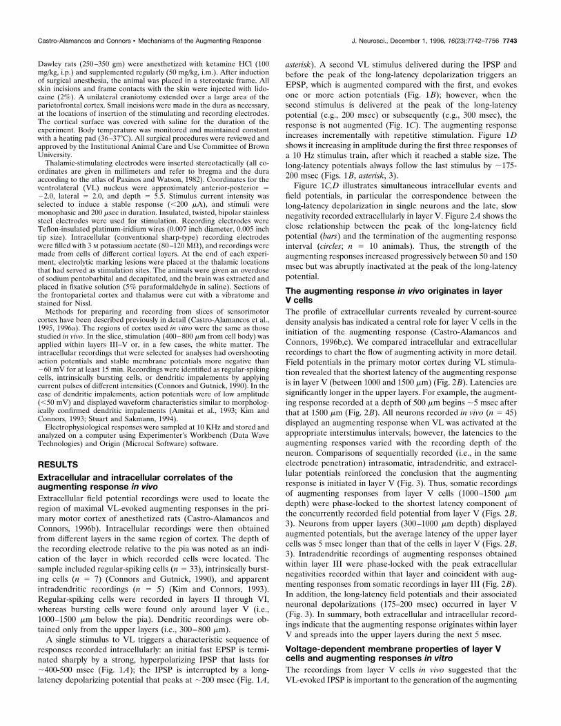
responses recorded intracellularly: an initial fast EPSP is termi-nated sharply by a strong, hyperpolarizing IPSP that lasts for;400-500 msec (Fig. 1A); the IPSP is interrupted by a long-latency depolarizing potential that peaks at ;200 msec (Fig. 1A,
asterisk). A second VL stimulus delivered during the IPSP andbefore the peak of the long-latency depolarization triggers anEPSP, which is augmented compared with the first, and evokesone or more action potentials (Fig. 1B); however, when thesecond stimulus is delivered at the peak of the long-latencypotential (e.g., 200 msec) or subsequently (e.g., 300 msec), theresponse is not augmented (Fig. 1C). The augmenting responseincreases incrementally with repetitive stimulation. Figure 1Dshows it increasing in amplitude during the first three responses ofa 10 Hz stimulus train, after which it reached a stable size. Thelong-latency potentials always follow the last stimulus by ;175-200 msec (Figs. 1B, asterisk, 3).Figure 1C,D illustrates simultaneous intracellular events and
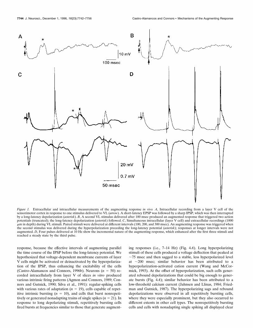
field potentials, in particular the correspondence between thelong-latency depolarization in single neurons and the late, slownegativity recorded extracellularly in layer V. Figure 2A shows theclose relationship between the peak of the long-latency fieldpotential (bars) and the termination of the augmenting responseinterval (circles; n 5 10 animals). Thus, the strength of theaugmenting responses increased progressively between 50 and 150msec but was abruptly inactivated at the peak of the long-latencypotential.
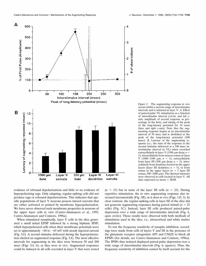
The augmenting response in vivo originates in layerV cellsThe profile of extracellular currents revealed by current-sourcedensity analysis has indicated a central role for layer V cells in theinitiation of the augmenting response (Castro-Alamancos andConnors, 1996b,c). We compared intracellular and extracellularrecordings to chart the flow of augmenting activity in more detail.Field potentials in the primary motor cortex during VL stimula-tion revealed that the shortest latency of the augmenting responseis in layer V (between 1000 and 1500 mm) (Fig. 2B). Latencies aresignificantly longer in the upper layers. For example, the augment-ing response recorded at a depth of 500 mm begins ;5 msec afterthat at 1500 mm (Fig. 2B). All neurons recorded in vivo (n 5 45)displayed an augmenting response when VL was activated at theappropriate interstimulus intervals; however, the latencies to theaugmenting responses varied with the recording depth of theneuron. Comparisons of sequentially recorded (i.e., in the sameelectrode penetration) intrasomatic, intradendritic, and extracel-lular potentials reinforced the conclusion that the augmentingresponse is initiated in layer V (Fig. 3). Thus, somatic recordingsof augmenting responses from layer V cells (1000–1500 mmdepth) were phase-locked to the shortest latency component ofthe concurrently recorded field potential from layer V (Figs. 2B,3). Neurons from upper layers (300–1000 mm depth) displayedaugmented potentials, but the average latency of the upper layercells was 5 msec longer than that of the cells in layer V (Figs. 2B,3). Intradendritic recordings of augmenting responses obtainedwithin layer III were phase-locked with the peak extracellularnegativities recorded within that layer and coincident with aug-menting responses from somatic recordings in layer III (Fig. 2B).In addition, the long-latency field potentials and their associatedneuronal depolarizations (175–200 msec) occurred in layer V(Fig. 3). In summary, both extracellular and intracellular record-ings indicate that the augmenting response originates within layerV and spreads into the upper layers during the next 5 msec.
Voltage-dependent membrane properties of layer Vcells and augmenting responses in vitroThe recordings from layer V cells in vivo suggested that theVL-evoked IPSP is important to the generation of the augmenting
Castro-Alamancos and Connors • Mechanisms of the Augmenting Response J. Neurosci., December 1, 1996, 16(23):7742–7756 7743
response, because the effective intervals of augmenting parallelthe time course of the IPSP before the long-latency potential. Wehypothesized that voltage-dependent membrane currents of layerV cells might be activated or deinactivated by the hyperpolariza-tion of the IPSP, thus enhancing the excitability of the cells(Castro-Alamancos and Connors, 1996b). Neurons (n 5 50) re-corded intracellularly from layer V of slices in vitro producedvarious intrinsic firing patterns (Agmon and Connors, 1989; Con-nors and Gutnick, 1990; Silva et al., 1991): regular-spiking cellswith various rates of adaptation (n 5 19), cells capable of repet-itive intrinsic bursting (n 5 10), and cells that burst nonrepeti-tively or generated nonadapting trains of single spikes (n5 21). Inresponse to long depolarizing stimuli, repetitively bursting cellsfired bursts at frequencies similar to those that generate augment-
ing responses (i.e., 7–14 Hz) (Fig. 4A). Long hyperpolarizingstimuli of these cells produced a voltage deflection that peaked at;75 msec and then sagged to a stable, less hyperpolarized levelat ;200 msec; similar behavior has been attributed to ahyperpolarization-activated cation current (Wang and McCor-mick, 1993). At the offset of hyperpolarization, such cells gener-ated rebound depolarizations that could be big enough to gener-ate bursts (Fig. 4A); similar behavior has been attributed to alow-threshold calcium current (Jahnsen and Llinas, 1984; Fried-man and Gutnick, 1987). The hyperpolarizing sags and rebounddepolarizations were observed in all repetitively bursting cells,where they were especially prominent, but they also occurred todifferent extents in other cell types. The nonrepetitively burstingcells and cells with nonadapting single spiking all displayed clear
Figure 1. Extracellular and intracellular measurements of the augmenting response in vivo. A, Intracellular recording from a layer V cell of thesensorimotor cortex in response to one stimulus delivered to VL (arrow). A short-latency EPSP was followed by a sharp IPSP, which was then interruptedby a long-latency depolarization (asterisk). B, A second VL stimulus delivered after 100 msec produced an augmented response that triggered two actionpotentials (truncated); the long-latency depolarization (asterisk) followed. C, Simultaneous intracellular (layer V cell) and extracellular recordings (1000mm in depth) during VL stimuli. Paired stimuli were delivered at different intervals (100, 200, and 300 msec). An augmenting response was triggered whenthe second stimulus was delivered during the hyperpolarization preceding the long-latency potential (asterisk); responses at longer intervals were notaugmented. D, Four pulses delivered at 10 Hz show the incremental nature of the augmenting response, which enhanced after the first three stimuli andreached a steady state by the third pulse.
7744 J. Neurosci., December 1, 1996, 16(23):7742–7756 Castro-Alamancos and Connors • Mechanisms of the Augmenting Response
evidence of rebound depolarizations and little or no evidence ofhyperpolarizing sags. Only adapting, regular-spiking cells did notproduce sags or rebound depolarizations. This indicates that spe-cific populations of layer V neurons possess inward currents thatare either activated or primed by membrane hyperpolarization.We have never observed such membrane properties in neurons ofthe upper layer cells in vitro (Castro-Alamancos et al., 1995;Castro-Alamancos and Connors, 1996a).When stimulated synaptically, layer V cells in the slice gener-
ated a small initial EPSP followed by a strong biphasic IPSP,which hyperpolarized cells when their membrane potentials wereset to approximately 260 to 265 mV with steady injected current(Fig. 5A). A second stimulus delivered during the hyperpolariza-tion elicited an augmented response (Fig. 5A). The most effectiveintervals for augmenting in the slice were between 50 and 200msec (Figs. 5A, 6), as they were in vivo. Augmented responsescould be induced in all cells recorded in layer V that were tested
(n 5 15) but in none of the layer III cells (n 5 25). Duringrepetitive stimulation, the in vitro augmenting response also in-creased incrementally (Fig. 5B), as it does in vivo (Figs. 1D, 3). Inclear contrast, the regular-spiking cells in layer III of the slice didnot generate augmenting responses during paired stimuli (n 5 25cells) (Fig. 5C). Instead, layer III cells produced paired-pulsedepression over a wide range of interstimulus intervals (Fig. 6,open circles). These results were observed with both methods ofstimulation used in the slice, i.e., intracortical and white matterstimulation.To test the frequency sensitivity of synaptic inhibition, record-
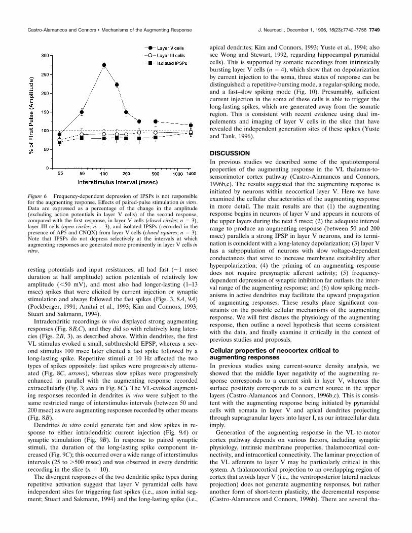
ings were made from cells of layers V and III in the presence ofthe glutamate receptor antagonists AP5 and CNQX to block allEPSPs (for details, see Castro-Alamancos and Connors, 1996a).The IPSPs thus isolated displayed paired-pulse depression over awide range of interstimulus intervals (Fig. 6, squares). Thus, thefrequency sensitivity of inhibition cannot by itself account for the
Figure 2. The augmenting response in vivooccurs within a narrow range of interstimulusintervals and is initiated in layer V. A, Effectof paired-pulse VL stimulation as a functionof interstimulus interval (circles and left y-axis; amplitude of second response as per-centage of the first), and timing of the peakof the long-latency potential for 10 cases(bars and right y-axis). Note that the aug-menting response begins at an interstimulusinterval of 50 msec and is abolished at thepeak of the long-latency potential (200msec). B, Latency of the augmenting re-sponse (i.e., the start of the response to thesecond stimulus delivered at a 100 msec in-terstimulus interval in VL) when recordedextracellularly in layer V (1500 mm deep; n55), intracellularly from neuron somas in layerV (1000–1500 mm; n 5 5), extracellularlyfrom layer III (500 mm deep; n 5 5), intra-cellularly from dendrites located in the upperlayers (layer III dendrites; n 5 5), or fromsomas in the upper layers (n 5 5 layer IIIsomas; 300–1000 mm). The shortest latencieswere observed in cells located in layer V. Alldata expressed as mean 6 SEM.
Castro-Alamancos and Connors • Mechanisms of the Augmenting Response J. Neurosci., December 1, 1996, 16(23):7742–7756 7745
strong augmenting responses observed in drug-free layer V cellsat intervals between 50 and 200 msec in vitro (Fig. 6, filled circles),because depression of IPSPs extended to much longer intervals(.500 msec) and was actually strongest at shorter intervals (i.e.,25 msec) (Fig. 6), when there are no augmenting responses.A primary instigator of the augmenting response could be
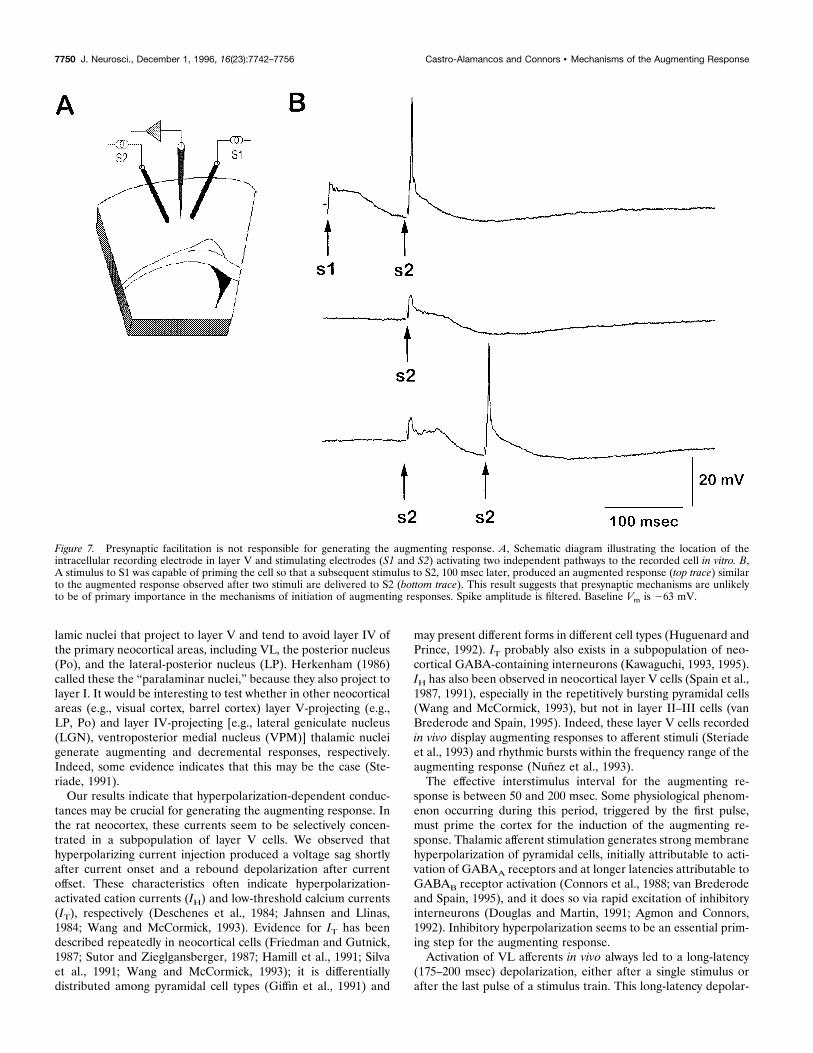
either frequency-sensitive facilitation of excitatory synapses or achange in intrinsic membrane excitability triggered by IPSP-induced hyperpolarization. Paired-pulse facilitation is usually gen-erated by presynaptic mechanisms (Zucker, 1989). If postsynapticproperties are of primary importance in generating the augment-ing response, then it should be possible to induce an augmentingresponse by priming a layer V cell with a single shock to anIPSP-producing pathway and testing with a second, independentexcitatory pathway onto the same cell. We placed two stimulatingelectrodes on opposite sides of an intracellularly recorded layer Vcell, so that each activated an independent horizontal pathway
(Fig. 7A, S1 and S2) (independence was tested by showing that theresponses summed linearly) and stimulated them in differentsequences. A stimulus to either pathway was capable of primingthe response to the other pathway, with an augmenting responseresulting. Paired stimulation of S2 at an interval of 100 msecgenerated an augmenting response, as usual (Fig. 7B). When ashock to S1 was substituted as the first (priming) stimulus, theresponse to an S2 stimulus was also strongly augmented. Thisresult suggests that presynaptic mechanisms are unlikely to be ofprimary importance in the mechanisms of initiation of the aug-menting response.
Intradendritic recordings during augmentingresponses in vivo and in vitroSome intracellular recordings obtained in vivo (n 5 5) (Fig. 8) orin slices (n 5 10) (Fig. 9) displayed the electrophysiologicalcharacteristics of intradendritic recordings. Despite large, stable
Figure 3. Comparison of sequentially recorded intrasomatic, intradendritic, and extracellular potentials indicate that the augmenting response is initiatedin layer V. Recordings were made along a single electrode track as trains of four stimuli (arrows) were delivered to VL at 10 Hz. Intracellular recordingswere from a dendrite located in layer III (400 mm deep; top trace) and a soma in layer V of an intrinsically bursting cell (1400 mm deep; bottom trace).The middle traces show extracellular field potentials at various depths in the cortex (surface, 500, 1000, and 1500 mm). Note that the somatic layer Vrecording was phase-locked to the shortest latency component of the concurrently recorded field potential from layer V, whereas the upper layer dendritewas phase-locked to the longer-latency negativities in those layers. The long-latency depolarization, ;175-200 msec after the last stimulus, occurred firstin the layer V cell (dashed line with arrows).
7746 J. Neurosci., December 1, 1996, 16(23):7742–7756 Castro-Alamancos and Connors • Mechanisms of the Augmenting Response
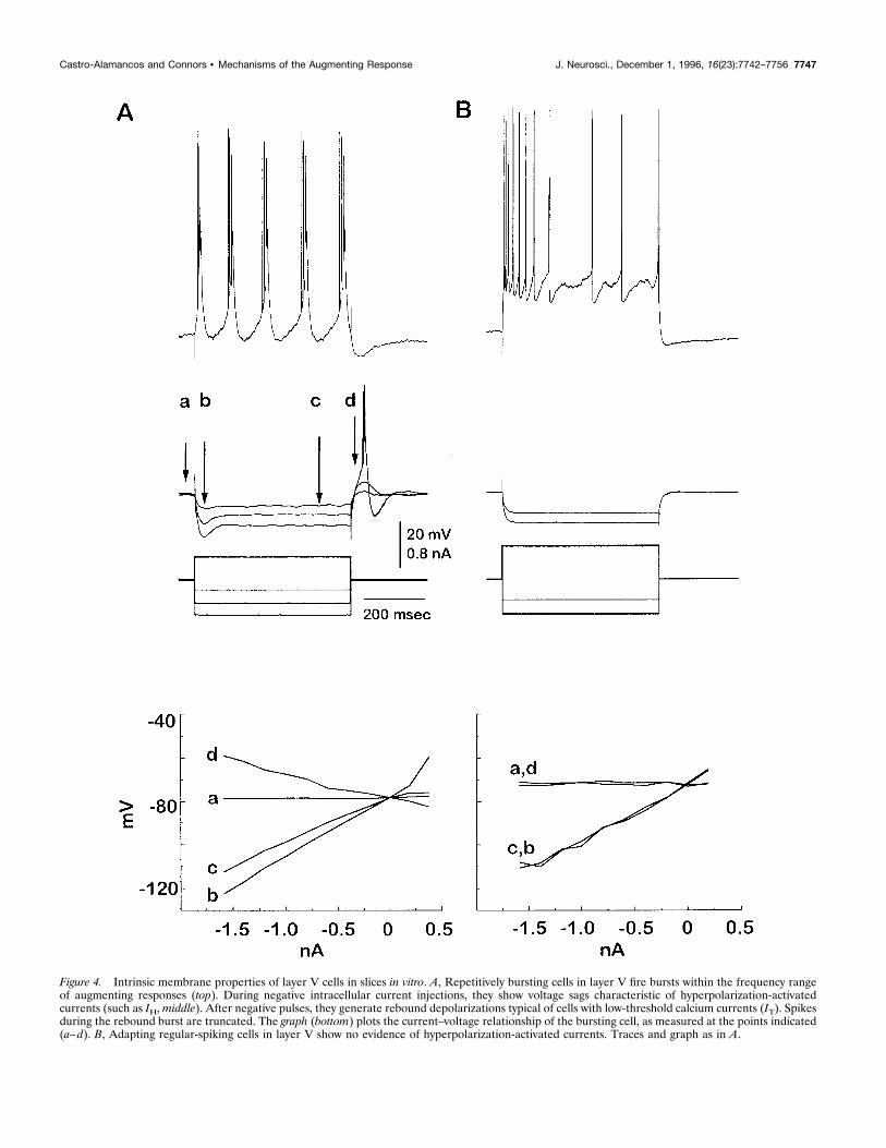
Figure 4. Intrinsic membrane properties of layer V cells in slices in vitro. A, Repetitively bursting cells in layer V fire bursts within the frequency rangeof augmenting responses (top). During negative intracellular current injections, they show voltage sags characteristic of hyperpolarization-activatedcurrents (such as IH,middle). After negative pulses, they generate rebound depolarizations typical of cells with low-threshold calcium currents (IT). Spikesduring the rebound burst are truncated. The graph (bottom) plots the current–voltage relationship of the bursting cell, as measured at the points indicated(a–d). B, Adapting regular-spiking cells in layer V show no evidence of hyperpolarization-activated currents. Traces and graph as in A.
Castro-Alamancos and Connors • Mechanisms of the Augmenting Response J. Neurosci., December 1, 1996, 16(23):7742–7756 7747
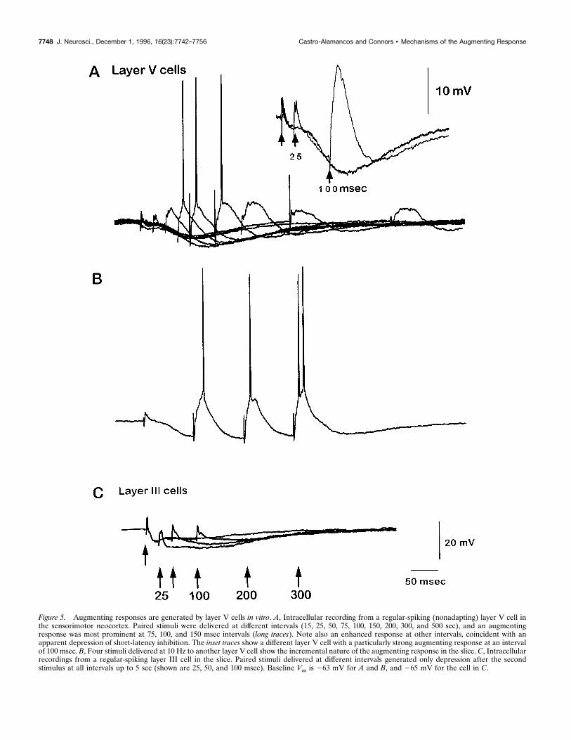
Figure 5. Augmenting responses are generated by layer V cells in vitro. A, Intracellular recording from a regular-spiking (nonadapting) layer V cell inthe sensorimotor neocortex. Paired stimuli were delivered at different intervals (15, 25, 50, 75, 100, 150, 200, 300, and 500 sec), and an augmentingresponse was most prominent at 75, 100, and 150 msec intervals (long traces). Note also an enhanced response at other intervals, coincident with anapparent depression of short-latency inhibition. The inset traces show a different layer V cell with a particularly strong augmenting response at an intervalof 100 msec. B, Four stimuli delivered at 10 Hz to another layer V cell show the incremental nature of the augmenting response in the slice. C, Intracellularrecordings from a regular-spiking layer III cell in the slice. Paired stimuli delivered at different intervals generated only depression after the secondstimulus at all intervals up to 5 sec (shown are 25, 50, and 100 msec). Baseline Vm is 263 mV for A and B, and 265 mV for the cell in C.
7748 J. Neurosci., December 1, 1996, 16(23):7742–7756 Castro-Alamancos and Connors • Mechanisms of the Augmenting Response
resting potentials and input resistances, all had fast (;1 msecduration at half amplitude) action potentials of relatively lowamplitude (,50 mV), and most also had longer-lasting (1–13msec) spikes that were elicited by current injection or synapticstimulation and always followed the fast spikes (Figs. 3, 8A, 9A)(Pockberger, 1991; Amitai et al., 1993; Kim and Connors, 1993;Stuart and Sakmann, 1994).Intradendritic recordings in vivo displayed strong augmenting
responses (Fig. 8B,C), and they did so with relatively long laten-cies (Figs. 2B, 3), as described above. Within dendrites, the firstVL stimulus evoked a small, subthreshold EPSP, whereas a sec-ond stimulus 100 msec later elicited a fast spike followed by along-lasting spike. Repetitive stimuli at 10 Hz affected the twotypes of spikes oppositely: fast spikes were progressively attenu-ated (Fig. 8C, arrows), whereas slow spikes were progressivelyenhanced in parallel with the augmenting response recordedextracellularly (Fig. 3; stars in Fig. 8C). The VL-evoked augment-ing responses recorded in dendrites in vivo were subject to thesame restricted range of interstimulus intervals (between 50 and200 msec) as were augmenting responses recorded by other means(Fig. 8B).Dendrites in vitro could generate fast and slow spikes in re-
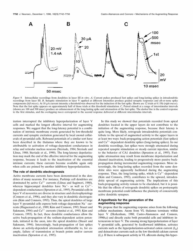
sponse to either intradendritic current injection (Fig. 9A) orsynaptic stimulation (Fig. 9B). In response to paired synapticstimuli, the duration of the long-lasting spike component in-creased (Fig. 9C); this occurred over a wide range of interstimulusintervals (25 to .500 msec) and was observed in every dendriticrecording in the slice (n 5 10).The divergent responses of the two dendritic spike types during
repetitive activation suggest that layer V pyramidal cells haveindependent sites for triggering fast spikes (i.e., axon initial seg-ment; Stuart and Sakmann, 1994) and the long-lasting spike (i.e.,
apical dendrites; Kim and Connors, 1993; Yuste et al., 1994; alsosee Wong and Stewart, 1992, regarding hippocampal pyramidalcells). This is supported by somatic recordings from intrinsicallybursting layer V cells (n 5 4), which show that on depolarizationby current injection to the soma, three states of response can bedistinguished: a repetitive-bursting mode, a regular-spiking mode,and a fast–slow spiking mode (Fig. 10). Presumably, sufficientcurrent injection in the soma of these cells is able to trigger thelong-lasting spikes, which are generated away from the somaticregion. This is consistent with recent evidence using dual im-palements and imaging of layer V cells in the slice that haverevealed the independent generation sites of these spikes (Yusteand Tank, 1996).
DISCUSSIONIn previous studies we described some of the spatiotemporalproperties of the augmenting response in the VL thalamus-to-sensorimotor cortex pathway (Castro-Alamancos and Connors,1996b,c). The results suggested that the augmenting response isinitiated by neurons within neocortical layer V. Here we haveexamined the cellular characteristics of the augmenting responsein more detail. The main results are that (1) the augmentingresponse begins in neurons of layer V and appears in neurons ofthe upper layers during the next 5 msec; (2) the adequate intervalrange to produce an augmenting response (between 50 and 200msec) parallels a strong IPSP in layer V neurons, and its termi-nation is coincident with a long-latency depolarization; (3) layer Vhas a subpopulation of neurons with slow voltage-dependentconductances that serve to increase membrane excitability afterhyperpolarization; (4) the priming of an augmenting responsedoes not require presynaptic afferent activity; (5) frequency-dependent depression of synaptic inhibition far outlasts the inter-val range of the augmenting response; and (6) slow spiking mech-anisms in active dendrites may facilitate the upward propagationof augmenting responses. These results place significant con-straints on the possible cellular mechanisms of the augmentingresponse. We will first discuss the physiology of the augmentingresponse, then outline a novel hypothesis that seems consistentwith the data, and finally examine it critically in the context ofprevious studies and proposals.
Cellular properties of neocortex critical toaugmenting responsesIn previous studies using current-source density analysis, weshowed that the middle layer negativity of the augmenting re-sponse corresponds to a current sink in layer V, whereas thesurface positivity corresponds to a current source in the upperlayers (Castro-Alamancos and Connors, 1996b,c). This is consis-tent with the augmenting response being initiated by pyramidalcells with somata in layer V and apical dendrites projectingthrough supragranular layers into layer I, as our intracellular dataimply.Generation of the augmenting response in the VL-to-motor
cortex pathway depends on various factors, including synapticphysiology, intrinsic membrane properties, thalamocortical con-nectivity, and intracortical connectivity. The laminar projection ofthe VL afferents to layer V may be particularly critical in thissystem. A thalamocortical projection to an overlapping region ofcortex that avoids layer V (i.e., the ventroposterior lateral nucleusprojection) does not generate augmenting responses, but ratheranother form of short-term plasticity, the decremental response(Castro-Alamancos and Connors, 1996b). There are several tha-
Figure 6. Frequency-dependent depression of IPSPs is not responsiblefor the augmenting response. Effects of paired-pulse stimulation in vitro.Data are expressed as a percentage of the change in the amplitude(excluding action potentials in layer V cells) of the second response,compared with the first response, in layer V cells (closed circles; n 5 3),layer III cells (open circles; n 5 3), and isolated IPSPs (recorded in thepresence of AP5 and CNQX) from layer V cells (closed squares; n 5 3).Note that IPSPs do not depress selectively at the intervals at whichaugmenting responses are generated more prominently in layer V cells invitro.
Castro-Alamancos and Connors • Mechanisms of the Augmenting Response J. Neurosci., December 1, 1996, 16(23):7742–7756 7749
lamic nuclei that project to layer V and tend to avoid layer IV ofthe primary neocortical areas, including VL, the posterior nucleus(Po), and the lateral-posterior nucleus (LP). Herkenham (1986)called these the “paralaminar nuclei,” because they also project tolayer I. It would be interesting to test whether in other neocorticalareas (e.g., visual cortex, barrel cortex) layer V-projecting (e.g.,LP, Po) and layer IV-projecting [e.g., lateral geniculate nucleus(LGN), ventroposterior medial nucleus (VPM)] thalamic nucleigenerate augmenting and decremental responses, respectively.Indeed, some evidence indicates that this may be the case (Ste-riade, 1991).Our results indicate that hyperpolarization-dependent conduc-
tances may be crucial for generating the augmenting response. Inthe rat neocortex, these currents seem to be selectively concen-trated in a subpopulation of layer V cells. We observed thathyperpolarizing current injection produced a voltage sag shortlyafter current onset and a rebound depolarization after currentoffset. These characteristics often indicate hyperpolarization-activated cation currents (IH) and low-threshold calcium currents(IT), respectively (Deschenes et al., 1984; Jahnsen and Llinas,1984; Wang and McCormick, 1993). Evidence for IT has beendescribed repeatedly in neocortical cells (Friedman and Gutnick,1987; Sutor and Zieglgansberger, 1987; Hamill et al., 1991; Silvaet al., 1991; Wang and McCormick, 1993); it is differentiallydistributed among pyramidal cell types (Giffin et al., 1991) and
may present different forms in different cell types (Huguenard andPrince, 1992). IT probably also exists in a subpopulation of neo-cortical GABA-containing interneurons (Kawaguchi, 1993, 1995).IH has also been observed in neocortical layer V cells (Spain et al.,1987, 1991), especially in the repetitively bursting pyramidal cells(Wang and McCormick, 1993), but not in layer II–III cells (vanBrederode and Spain, 1995). Indeed, these layer V cells recordedin vivo display augmenting responses to afferent stimuli (Steriadeet al., 1993) and rhythmic bursts within the frequency range of theaugmenting response (Nunez et al., 1993).The effective interstimulus interval for the augmenting re-
sponse is between 50 and 200 msec. Some physiological phenom-enon occurring during this period, triggered by the first pulse,must prime the cortex for the induction of the augmenting re-sponse. Thalamic afferent stimulation generates strong membranehyperpolarization of pyramidal cells, initially attributable to acti-vation of GABAA receptors and at longer latencies attributable toGABAB receptor activation (Connors et al., 1988; van Brederodeand Spain, 1995), and it does so via rapid excitation of inhibitoryinterneurons (Douglas and Martin, 1991; Agmon and Connors,1992). Inhibitory hyperpolarization seems to be an essential prim-ing step for the augmenting response.Activation of VL afferents in vivo always led to a long-latency
(175–200 msec) depolarization, either after a single stimulus orafter the last pulse of a stimulus train. This long-latency depolar-
Figure 7. Presynaptic facilitation is not responsible for generating the augmenting response. A, Schematic diagram illustrating the location of theintracellular recording electrode in layer V and stimulating electrodes (S1 and S2) activating two independent pathways to the recorded cell in vitro. B,A stimulus to S1 was capable of priming the cell so that a subsequent stimulus to S2, 100 msec later, produced an augmented response (top trace) similarto the augmented response observed after two stimuli are delivered to S2 (bottom trace). This result suggests that presynaptic mechanisms are unlikelyto be of primary importance in the mechanisms of initiation of augmenting responses. Spike amplitude is filtered. Baseline Vm is 263 mV.
7750 J. Neurosci., December 1, 1996, 16(23):7742–7756 Castro-Alamancos and Connors • Mechanisms of the Augmenting Response
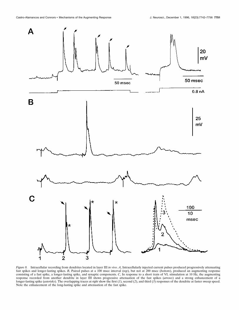
Figure 8. Intracellular recording from dendrites located in layer III in vivo. A, Intracellularly injected current pulses produced progressively attenuatingfast spikes and longer-lasting spikes. B, Paired pulses at a 100 msec interval (top), but not at 200 msec (bottom), produced an augmenting responseconsisting of a fast spike, a longer-lasting spike, and synaptic components. C, In response to a short train of VL stimulation at 10 Hz, the augmentingresponse recorded from another dendrite in layer III shows progressive attenuation of the fast spikes (arrows) and a strong enhancement of alonger-lasting spike (asterisks). The overlapping traces at right show the first (1), second (2), and third (3) responses of the dendrite at faster sweep speed.Note the enhancement of the long-lasting spike and attenuation of the fast spike.
Castro-Alamancos and Connors • Mechanisms of the Augmenting Response J. Neurosci., December 1, 1996, 16(23):7742–7756 7751
ization interrupted the inhibitory hyperpolarization of layer Vcells and marked the longest effective interval for augmentingresponses. We suggest that the long-latency potential is a combi-nation of intrinsic membrane events generated by low-thresholdcurrents and synaptic excitation generated by local axonal collat-erals of pyramidal cells. Rebound potentials of a similar sort havebeen described in the thalamus where they are known to beattributable to activation of voltage-dependent conductances inrelay and reticular nucleus neurons (Steriade, 1984; Steriade andLlinas, 1988; Steriade et al., 1990). The long-latency depolariza-tion may mark the end of the effective interval for the augmentingresponse, because it leads to the inactivation of the essentialintrinsic currents; these currents become available again onlywhen cells are primed by another inhibitory hyperpolarization.
The role of dendritic electrogenesisActive membrane currents have been demonstrated in the den-drites of many neurons. For instance, Purkinje cell dendrites aredominated by active Ca21 currents (Llinas and Sugimori, 1980),whereas hippocampal dendrites have Na1- as well as Ca21-dependent conductances (Spruston et al., 1995). Pyramidal cells inlayer V of neocortex are diverse in structure and electrophysiology(Connors et al., 1996), and they also vary in dendritic electrogen-esis (Kim and Connors, 1993). Thus, the apical dendrites of largelayer V pyramidal cells express both voltage-dependent Na1 cur-rents (Huguenard et al., 1989; Kim and Connors, 1993; Stuart andSakmann, 1994) and Ca21 currents (Amitai et al., 1993; Kim andConnors, 1993). In fact, these dendritic conductances allow theactive back-propagation of the sodium-dependent action poten-tials, initiated in the axon, into the apical dendrites (Stuart andSakmann, 1994). Interestingly, back-propagation of fast spikesshows an activity-dependent attenuation attributable to, for ex-ample, failure of transmission at branch points and/or currentinactivation (Spruston et al., 1995).
In this study we showed that potentials recorded from apicaldendrites located in the upper layers do not contribute to theinitiation of the augmenting response, because their latency isquite long. More likely, retrograde intradendritic potentials con-tribute to the spread of augmented activity to the upper layers inat least two ways: back-propagating action potentials (fast spikes)and Ca21-dependent dendritic spikes (long-lasting spikes). In ourdendritic recordings, fast spikes were strongly attenuated duringrepeated synaptic stimulation or steady current injection, similarto the behavior of CA1 dendrites (Spruston et al., 1995). Fast-spike attenuation may result from membrane depolarization andchannel inactivation, leading to progressively more passive back-propagation during incremental augmenting responses. More in-terestingly, the long-lasting spikes recorded from the same den-dritic sites were apparently facilitated during the augmentingresponse. Thus, the long-lasting spike, which is Ca21-dependent(Kim and Connors, 1993), contributes to the upward, intraden-dritic spread of augmenting activity. The implications of thisback-propagating activity remain to be elucidated, but it is possi-ble that the effects of retrograde dendritic spikes on postsynapticmembrane potential could influence the plasticity of concurrentlyactive dendritic synapses.
A hypothesis for the generation of theaugmenting responseWe propose that the augmenting response arises from the followingcellular mechanisms. Axons from VL thalamus terminate withinlayer V (Herkenham, 1980; Castro-Alamancos and Connors,1996b,c) and directly excite both pyramidal cells and inhibitory in-terneurons in layer V. The ensuing strong hyperpolarization of layerV pyramidal cells, generated by feedforward inhibition, activatescurrents such as the hyperpolarization-activated cation current (IH)and deinactivates currents such as the low threshold calcium current(IT). If there is subsequent activity in VL afferents during this hyper-
Figure 9. Intracellular recordings from dendrites in layer III in vitro. A, Current pulses produced fast spikes and long-lasting spikes in intradendriticrecordings from layer III. B, Synaptic stimulation in layer V applied at different intensities produce graded synaptic responses and all-or-none spikecomponents (left traces). At 10 mA current intensity a threshold was observed for the induction of the fast spike. Shown are 12 trials at 0.1 Hz (right traces).Note that the fast spike appears on approximately half of those trials at this threshold stimulation. C, Paired stimuli at a range of interstimulus intervals(shown are 100 and 500 msec) produce an enhancement of the long-lasting spike and attenuation of the fast spike. The dashed line is the control responseto the first stimulus, and the overlapping traces correspond to the second response delivered at different interstimulus intervals.
7752 J. Neurosci., December 1, 1996, 16(23):7742–7756 Castro-Alamancos and Connors • Mechanisms of the Augmenting Response
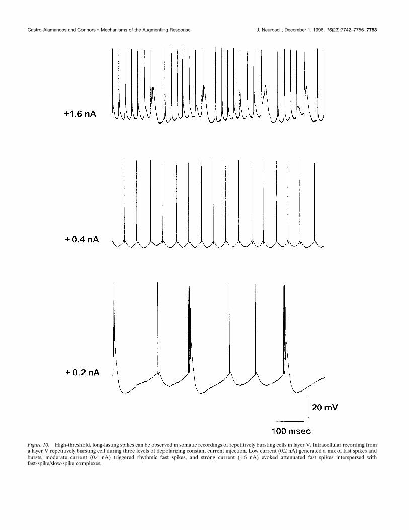
Figure 10. High-threshold, long-lasting spikes can be observed in somatic recordings of repetitively bursting cells in layer V. Intracellular recording froma layer V repetitively bursting cell during three levels of depolarizing constant current injection. Low current (0.2 nA) generated a mix of fast spikes andbursts, moderate current (0.4 nA) triggered rhythmic fast spikes, and strong current (1.6 nA) evoked attenuated fast spikes interspersed withfast-spike/slow-spike complexes.
Castro-Alamancos and Connors • Mechanisms of the Augmenting Response J. Neurosci., December 1, 1996, 16(23):7742–7756 7753
polarization, it yields a larger (augmented) response because of (1)inward IH and the activation of now deinactivated IT-like currents inlayer V pyramidal cells, (2) network-dependent reinforcement byextensive excitatory interconnections between layer V pyramidal cells(Connors and Amitai, 1995; Douglas et al., 1995), (3) spread ofactivity to upper layers via interlaminar excitatory connections andbackpropagating action potentials in the apical dendrites of layer Vcells, and (4) spread to adjacent regions of cortex via horizontalcollaterals.There are several defining characteristics of the augmenting
response that are explained by our hypothesis. Its surface positiv-ity and middle layer negativity in extracellular recordings arisebecause it originates with inward currents (intrinsic and synaptic)in vertically extended layer V pyramidal cells. The onset of theeffective interstimulus interval, at a latency of ;50 msec, is deter-mined by the need for sufficient IPSP-driven hyperpolarization toinfluence intrinsic, voltage-dependent membrane currents. Thetermination of the effective interval is coincident with the long-latency depolarization that follows VL stimulation by ;200 msec.This depolarization is attributable to rebound excitation withinthe network of layer V neurons, which is manifest as a spatiallyand temporally distributed EPSP; it determines the end of theaugmenting interval because its depolarization inactivates theessential intrinsic currents that would otherwise boost VL-generated synaptic currents.
Comparison to previous hypotheses for theaugmenting responseVarious hypotheses have been proposed for the cellular mecha-nisms of the augmenting response. An early proposal, based onintracellular recordings and VL stimulation in the cat, was that theaugmenting response arose from a marked increase in the mag-nitude of long-latency EPSPs, attributable to the stimulus-dependent depression of inhibition (Purpura and Shofer, 1964;Creutzfeldt et al., 1966). A similar and more recent hypothesis isthat long-latency facilitated EPSPs are NMDA receptor-mediatedpotentials (Metherate and Ashe, 1994). Several observations in-dicate that depression of inhibition may contribute to the aug-menting response, but that it is not the primary mechanism. First,the amount of facilitation attributable to depressed inhibitionreported for paired-pulse stimulation is considerably smaller thanthe facilitation displayed by the augmenting response. Thus, al-though augmenting responses increase severalfold over controlresponses, the facilitation attributable to a release from inhibitionby paired pulses is normally ,50% (Metherate and Ashe, 1994).Second, the interstimulus interval range effective in depressinginhibition is much wider than the effective range for generatingthe augmenting response. Metherate and Ashe (1994) reportedthat facilitation of EPSPs attributable to release from inhibitionwas effective for interstimulus intervals from 100 to 1000 msec andwas apparent even after 10 sec. This is consistent with our ownmeasurements of isolated IPSPs (Fig. 6). The augmenting re-sponse, however, occurs during a very narrow time period ofbetween 50 and 200 msec. Third, release from inhibition facilitatesmainly a long-latency potential in the neocortex that is mediatedby NMDA receptor-dependent conductances. This implies thatNMDA receptors are essential for the augmenting response andNMDA receptor antagonists should abolish or strongly depress it;however, this is not the case (Addae and Stone, 1987; Castro-Alamancos and Connors, 1996b). Finally, the IPSP depressionhypothesis does not explain the nature of the long-latency depo-
larization and its coincidence with the end of the effective aug-menting interval or the selective involvement of layer V in theaugmenting process. Cells in upper layers also undergo frequency-dependent enhancement of EPSPs caused by the depression ofIPSPs (Castro-Alamancos and Connors, 1996a), but they do notinitiate augmenting responses (Castro-Alamancos and Connors,1996b).Ferster and Lindstrom (1985a) provided an alternative view of
the augmenting response, based on their investigations of connec-tions between the LGN and primary visual cortex in cats. Theyobserved an incremental cortical response during strong (i.e., 1mA) repetitive LGN stimulation and proposed that it was depen-dent on antidromic firing of layer VI corticothalamic axons andthe singular properties of the synapses on their intracorticalcollaterals. This hypothesis requires that the intracortical synapsesof corticothalamic cells show strong paired-pulse facilitation, aform of short-term plasticity mediated presynaptically (Zucker,1989). Paired-pulse facilitation is usually not observed in excita-tory synapses of neocortex (Thomson et al., 1993; Volgushev etal., 1995), although it is certainly possible that these specificsynapses show it (Thomson et al., 1995). Nevertheless, this hy-pothesis is not consistent with several properties of the augment-ing response. First, paired-pulse facilitation is most prominentbetween 25 and 75 msec (maximal at 50 msec) under normalconditions in the Schaffer-collateral pathway in hippocampal CA1and also in neocortex under low probability of release conditions(i.e., lower than normal calcium concentration; M. Castro-Alamancos and B. Connors, unpublished observations), whereasthe augmenting response is just beginning at 50 msec and peaks insize at 150–175 msec, just before the occurrence of the long-latency potential. Second, previous studies have found no evi-dence for antidromic firing of corticothalamic cells during aug-menting responses elicited with moderate stimulation currents(Castro-Alamancos and Connors, 1996b). Ferster and Lindstrom(1985a,b) used stimulation currents 10-fold higher than we typi-cally use to evoke augmenting responses. Corticothalamic axonstend to be much slower, with higher threshold, than thalamocor-tical axons (Ferster and Lindstrom, 1983; Swadlow, 1994). Third,we found that a presynaptic mechanism is unlikely to account forthe generation of the augmenting response, because two indepen-dent but convergent pathways can prime the augmenting re-sponses to one other. Pure synaptic facilitation is usually attrib-uted to presynaptic processes (Zucker, 1989). Finally, thishypothesis cannot account for the relevance of the inhibitoryhyperpolarization or of the long-latency depolarization and thetiming of the augmenting response.In contrast to the previous two hypotheses, the general proposal
of Morin and Steriade (1981) is consistent with the results pre-sented here. They concluded that the augmenting response de-pends critically on the hyperpolarization of cortical cells.
Implications for behavioral modulation of theaugmenting responseWe recently observed that the generation of the augmentingresponse depends strongly on the awake behavioral state of theanimal (Castro-Alamancos and Connors, 1996c); augmenting re-sponses are robust during periods of awake immobility but areabolished rapidly during states of arousal and movement. Inter-estingly, stimulation of the reticular midbrain inactivates aug-menting responses (Steriade and Morin, 1981) in a manner rem-iniscent of the behavioral inactivation. The behavioral modulation
7754 J. Neurosci., December 1, 1996, 16(23):7742–7756 Castro-Alamancos and Connors • Mechanisms of the Augmenting Response
of the augmenting response may arise from the actions of certainneurotransmitters (i.e., acetylcholine, norepinephrine) that arereleased in neocortex during behaviorally activated states (Aston-Jones et al., 1991; Cooper et al., 1991). These transmitters cantransform or block the firing properties and intrinsic membranecurrents of the layer V cells essential for generating the augment-ing response (Wang and McCormick, 1993). They can also mod-ulate synaptic inhibition presynaptically (Doze et al., 1991). Thus,our hypothesis for the mechanisms of the augmenting responsesuggests critical sites at which modulatory transmitters mightselectively control thalamocortical dynamics during changes ofbehavioral state.
REFERENCESAddae JI, Stone TW (1987) Involvement of N-methyl-D-aspartate recep-tors in the augmenting response in rat neocortex. Neurosci Lett78:323–327.
Agmon R, Connors BW (1989) Repetitive burst-firing neurons in thedeep layers of mouse somatosensory cortex. Neurosci Lett 99:137–141.
Agmon A, Connors BW (1992) Correlation between intrinsic firing pat-terns and thalamocortical synaptic responses of neurons in mouse barrelcortex. J Neurosci 12:319–329.
Amitai Y, Friedman A, Connors BW, Gutnick MJ (1993) Regenerativeactivity in apical dendrites of pyramidal cells in neocortex. Cereb Cortex3:26–38.
Aston-Jones G, Chiang C, Alexinsky T (1991) Discharge of noradrener-gic locus coeruleus neurons in behaving rats and monkeys suggests arole in vigilance. Prog Brain Res 88:501–520.
Castro-Alamancos MA, Connors BW (1996a) Short-term synaptic en-hancement and long-term potentiation in sensorimotor cortex. ProcNatl Acad Sci USA 93:1335–1339.
Castro-Alamancos MA, Connors BW (1996b) Spatiotemporal propertiesof short-term plasticity in sensorimotor thalamocortical pathways of therat. J Neurosci 16:2767–2779.
Castro-Alamancos MA, Connors BW (1996c) Short-term plasticity of athalamocortical pathway dynamically modulated by behavioral state.Science 272:274–277.
Castro-Alamancos MA, Donoghue JP, Connors BW (1995) Differentforms of synaptic plasticity in somatosensory and motor areas of theneocortex. J Neurosci 15:5324–5333.
Connors BW, Amitai Y (1995) Functions of local circuits in neocortex:synchrony and laminae. In: The cortical neuron (Gutnick MJ, Mody I,eds), pp 123–140. Cambridge, UK: Cambridge UP.
Connors BW, Gutnick MJ (1990) Intrinsic firing patterns of diverse neo-cortical neurons. Trends Neurosci 13:99–104.
Connors BW, Malenka RC, Silva LR (1988) Two inhibitory postsynapticpotentials, and GABAA and GABAB receptor-mediated responses inneocortex of rat and cat. J Physiol (Lond) 406:443–468.
Connors BW, Castro-Alamancos MA, Beierlein M (1996) Diverse neu-ronal functions of the cerebral cortex. In: Excitatory amino acids andthe cerebral cortex (Conti F, Hicks P, eds), pp 21–32. Cambridge, MA:MIT.
Cooper JR, Bloom FE, Roth RH (1991) The biochemical basis of neu-ropharmacology. New York: Oxford UP.
Creutzfeldt OD, Watanabe S, Lux HD (1966) Relation between EEGphenomena and potentials of single cortical cells. I. Evoked responsesafter thalamic and epicortical stimulation. Electroencephalogr ClinNeurophysiol 20:1–18.
Dempsey EW, Morison RS (1943) The electrical activity of a thalamo-cortical relay system. Am J Physiol 138:283–296.
Deschenes M, Paradis M, Roy JP, Steriade M (1984) Electrophysiologyof neurons of lateral thalamic nuclei in cat: resting properties and burstdischarges. J Neurophysiol 51:1196–1219.
Douglas RJ, Martin KAC (1991) A functional microcircuit for cat visualcortex. J Physiol (Lond) 440:735–769.
Douglas RJ, Koch C, Mahowald M, Martin KA, Suarez HH (1995)Recurrent excitation in neocortical circuits. Science 269:981–985.
Doze VA, Cohen GA, Madison DV (1991) Synaptic localization of ad-renergic disinhibition in the rat hippocampus. Neuron 6:889–900.
Ferster D, Lindstrom S (1983) An intracellular analysis of geniculo-cortical connectivity in area 17 of the cat. J Physiol (Lond) 342:181–215.
Ferster D, Lindstrom S (1985a) Augmenting responses evoked in area 17of the cat by intracortical axon collaterals of cortico-geniculate cells.J Physiol (Lond) 367:217–232.
Ferster D, Lindstrom S (1985b) Synaptic excitation of neurons in area 17of the cat by intracortical axon collaterals of cortico-geniculate cells.J Physiol (Lond) 367:233–252.
Friedman R, Gutnick MJ (1987) Low-threshold calcium electrogenesis inneocortical neurons. Neurosci Lett 81:117–122.
Giffin K, Solomon JS, Burkhalter A, Nerbonne JM (1991) Differentialexpression of voltage-gated calcium currents in identified visual corticalneurons. Neuron 6:321–332.
Glenn LL, Hada J, Roy JP, Deschenes M, Steriade M (1982) Antero-grade tracer and field potential analysis of the neocortical layer Iprojection from nucleus ventralis medialis of the thalamus in cat.Neuroscience 7:1861–1877.
Hamill OP, Huguenard JR, Prince DA (1991) Patch-clamp studies ofvoltage-gated currents in identified neurons of the rat cerebral cortex.Cereb Cortex 1:48–61.
Herkenham M (1980) Laminar organization of thalamic projections tothe rat neocortex. Science 207:532–535.
Herkenham M (1986) New perspectives on the organization and evolu-tion of nonspecific thalamocortical projections. In: Cerebral cortex, Vol5 (Jones EG, Peters A, eds), pp 403–446. New York: Plenum.
Huguenard JR, Prince DA (1992) A novel T-type current underlies pro-longed Ca(21)-dependent burst firing in GABAergic neurons of ratthalamic reticular nucleus. J Neurosci 12:3804–3817.
Huguenard JR, Hamill OP, Prince DA (1989) Sodium channels in den-drites of rat cortical pyramidal neurons. Proc Natl Acad Sci USA86:2473–2477.
Jahnsen H, Llinas R (1984) Ionic basis for the electro-responsiveness andoscillatory properties of guinea-pig thalamic neurones in vitro. J Physiol(Lond) 349:227–247.
Kawaguchi Y (1993) Groupings of non-pyramidal and pyramidal cellswith specific physiological and morphological characteristics in rat fron-tal cortex. J Neurophysiol 69:416–431.
Kawaguchi Y (1995) Physiological subgroups of nonpyramidal cells withspecific morphological characteristics in layer II/III of rat frontal cortex.J Neurosci 15:2638–2655.
Kim HG, Connors BW (1993) Apical dendrites of the neocortex: corre-lation between sodium- and calcium-dependent spiking and pyramidalcell morphology. J Neurosci 13:5301–5311.
Llinas R, Sugimori M (1980) Electrophysiological properties of in vitroPurkinje cell dendrites in mammalian cerebellar slices. J Physiol (Lond)305:197–213.
Metherate R, Ashe JH (1994) Facilitation of an NMDA receptor medi-ated epsp by paired pulse stimulation in rat neocortex via depression ofGABAergic ipsps. J Physiol (Lond) 481:331–348.
Mishima K, Ohta M (1992) Reappraisal of the corticothalamic andthalamocortical interactions that contribute to the augmenting responsein the rat. Jpn J Physiol 4:211–221.
Morin D, Steriade M (1981) Development from primary to augmentingresponses in the somatosensory system. Brain Res 205:49–66.
Morison RS, Dempsey EW (1943) Mechanism of thalamocortical aug-mentation and repetition. Am J Physiol 138:297–308.
Nunez A, Amzica F, Steriade M (1993) Electrophysiology of cat associ-ation cortical cells in vivo: intrinsic properties and synaptic responses.J Neurophysiol 70:418–430.
Paxinos G, Watson C (1982) The rat brain in stereotaxic coordinates.New York: Academic.
Pockberger H (1991) Electrophysiological and morphological propertiesof rat motor cortex neurons in vivo. Brain Res 539:181–190.
Purpura DP, Shofer RJ (1964) Cortical intracellular potentials duringaugmenting and recruiting responses I. Effects of injected hyperpolar-izing currents on evoked membrane potential changes. J Neurophysiol27:117–132.
Sasaki K, Staunton HP, Dieckmann G (1970) Characteristic features ofaugmenting and recruiting responses in the cerebral cortex. Exp Neurol26:369–392.
Silva LR, Amitai Y, Connors BW (1991) Intrinsic oscillations of neocor-tex generated by layer 5 pyramidal neurons. Science 251:432–435.
Spain WJ, Schwindt PC, Crill WE (1987) Anomalous rectification inneurons from cat sensorimotor cortex in vitro. J Neurophysiol57:1555–1576.
Castro-Alamancos and Connors • Mechanisms of the Augmenting Response J. Neurosci., December 1, 1996, 16(23):7742–7756 7755
Spain WJ, Schwindt PC, Crill WE (1991) Post-inhibitory excitation andinhibition in layer V pyramidal neurones from cat sensorimotor cortex.J Physiol (Lond) 434:609–626.
Spencer WA, Brookhart JM (1961) Electrical patterns of augmentingand recruiting waves in depths of sensorimotor cortex of the rat.J Neurophysiol 24:26–49.
Spruston N, Schiller Y, Stuart G, Sakmann B (1995) Activity-dependentaction potential invasion and calcium influx into hippocampal CA1dendrites. Science 297–300.
Steriade M (1984) The excitatory-inhibitory response sequence in tha-lamic and neocortical cells: state-related changes and regulatory sys-tems. In: Dynamic aspects of neocortical function (Edelman GM, GallWE, Cowan WM, eds), pp 107–158. New York: Wiley.
Steriade M (1991) Alertness, quiet sleep, dreaming. In: Cerebral cortex,Vol 9 (Peters A, Jones EG, eds), pp 279–358. New York: Plenum.
Steriade M, Llinas RR (1988) The functional states of the thalamus andthe associated neuronal interplay. Physiol Rev 68:649–742.
Steriade M, Morin D (1981) Reticular influences on primary and aug-menting responses in the somatosensory cortex. Brain Res 205:67–80.
Steriade M, Jones GJ, Llinas RR (1990) Thalamic oscillations and sig-naling. New York: Wiley.
Steriade M, Nunez A, Amzica F (1993) Intracellular analysis of relationsbetween the slow (,1 Hz) neocortical oscillation and other sleeprhythms of the electroencephalogram. J Neurosci 13:3266–3283.
Stuart GJ, Sakmann B (1994) Active propagation of somatic action po-tentials into neocortical pyramidal cell dendrites. Nature 367:69–72.
Sutor B, Zieglgansberger W (1987) A low-threshold activated transientcalcium current is responsible for the time-dependent depolarizinginward rectification of rat neocortical neurons in vitro. Eur J Physiol410:102–111.
Swadlow HA (1994) Efferent neurons and suspected interneurons in
motor cortex of the awake rabbit: axonal properties, sensory receptivefields, and subthreshold synaptic inputs. J Neurophysiol 71:437–453.
Thomson AM, Deuchars J, West DC (1993) Large, deep layer pyramid-pyramid single axon EPSPs in slices of rat motor cortex display pairedpulse and frequency-dependent depression, mediated presynapticallyand self-facilitation, mediated postsynaptically. J Neurophysiol70:2354–2369.
Thomson AM, West DC, Deuchars J (1995) Properties of single axonexcitatory postsynaptic potentials elicited in spiny interneurons by ac-tion potentials in pyramidal neurons in slices of rat neocortex. Neuro-science 69:727–738.
van Brederode JFM, Spain WJ (1995) Differences in inhibitory synapticinput between layer II–III and layer V neurons of the cat neocortex.J Neurophysiol 74:1149–1166.
Volgushev M, Voronin LL, Chistiakova M, Artola A, Singer W (1995)All-or-none excitatory postsynaptic potentials in the rat visual cortex.Eur J Neurosci 7:1751–1760.
Wang Z, McCormick DA (1993) Control of firing mode of corticotectaland corticopontine layer V burst-generating neurons by norepineph-rine, acetylcholine, and 1S,3R-ACPD. J Neurosci 13:2199–2216.
Wong RK, Stewart M (1992) Different firing patterns generated in den-drites and somata of CA1 pyramidal neurones in guinea-pig hippocam-pus. J Physiol (Lond) 457:675–687.
Yuste R, Tank DW (1996) Dendritic integration in mammalian neurons,a century after Cajal. Neuron 16:701–716.
Yuste R, Gutnick MJ, Saar D, Delaney KR, Tank DW (1994) Ca21
accumulations in dendrites of neocortical pyramidal neurons: anapical band and evidence for two functional compartments. Neuron13:23–43.
Zucker RS (1989) Short-term synaptic plasticity. Annu Rev Neurosci12:13–31.
7756 J. Neurosci., December 1, 1996, 16(23):7742–7756 Castro-Alamancos and Connors • Mechanisms of the Augmenting Response