Embed Size (px)
Citation preview
I ANDROLOGIA 23,4143 ( 1 9 9 1 ) ACCEPTED: AUGUST 15, 1990
Cellular sensitization against sperm and seminal antigens in women
P. Mallmann, W. Schroder, H. van der Ven, K. Diedrich and K. Krebs
Key words. Spermatozoa - seminal antigens - women - immune response.
Summary. In 13 healthy women and 6 virgins the cellular sensitization against sperm and seminal plasma antigens was demonstrated by an indirect lymphokin assay, the leucocyte migration inhibition test (LMI-test) using the following preparations: “washed” spermatozoa, seminal plasma and sper- matozoa of the supernatant prepared with the “swim-up” technique. In both groups of women a cellular sensitization against sperm and seminal plasma antigens could be observed. Further, a dose dependent correlation was found in that way, that increasing concentrations of spermatozoa lead to an increased inhibition of macrophage migration. In virgins cellular sensitization against seminal plasma proteins did not differ from non-virgins, only the percentage of significant reactions in the LMI-Test was reduced. As low sperm concentrations (1 million ml-I), which represent best the physio- logical situation in the uterus, induced an enhanced migrations of macrophages the enhancement of macrophage migration is considered as physiologi- cal cellular sensitization of females against sperm- associated antigens.
Introduction
Since 1954 when Rumke could proof the existence ofsperm-associated antibodies there is no doubt that spermatozoa and also seminal plasma are provided with antigenic components which are able to acti- vate the humoral and the cellular immune system by repeated exposure as it occurs during normal intercourse. Since most of the studies published so far are dealing with the influence of sperm antibod- ies on male and female reproduction the aim of this
Department of Gynecology and Obstetrics, Bonn, Germany.
Correspondence: Dr P. Mallmann, Universitats-Frauenklinik, Sigmund-Freud-StraBe 25, D-W-5300 Bonn 1 , Germany.
study was to investigate the cellular sensitization of healthy females against sperm and seminal plasma antigens.
Material and method
The cellular sensitization against sperm and seminal antigens was demonstrated by an indirect lym- phokin assay, the leucocyte migration inhibition test (LMI-test). The principle of this test is based on the fact that after repetitive contact to antigens lym- phocytes secrete lymphokins which can be function- ally demonstrated in vitro by their effect on the migration of macrophages. Peripheral blood lym- phocytes were isolated and incubated together with the preparations of sperm and seminal antigens. Aft- er an incubation of 18 hours the migration area of macrophages in antigen containing culture medium was compared to controls without antigen. The cell- mediated reactions against sperm and seminal an- tigens were quantified using the migration index (M.I.) (M.I. = migration area with antigen/mi- gration area without antigen x 100) and could be differentiated as a reduced or as an increased mi- gration area.
The immunological response was considered as significant reaction when it was beyond the twofold standard deviation of the control without antigen.
In the LMI-test the following preparations were used as antigen in the LMI-test: (1) “washed” spermatozoa (2 x 500 g for 5 min.
in Ham’s F10 medium) (2) seminal plasma (sperm free suspension) (3) spermatozoa of the supernatant prepared with
the “swim-up” technique ( > 90% motile sper- matozoa)
Following semen preparation the concentration was determined and adjusted to 1 ,5 and 10 million sper- matozoa ml- * culture medium, the seminal plasma to 5, 10 and 50 p1 ml-’ culture medium.
42 P. MALLMANN ET AL.
m--
13 healthy women aged 27.8 years on average provided blood samples for the LMI-test. Further- more a group of 6 virgins (healthy women without prior sperm exposure) aged 2 1.6 years on average was investigated.
: + >
Results
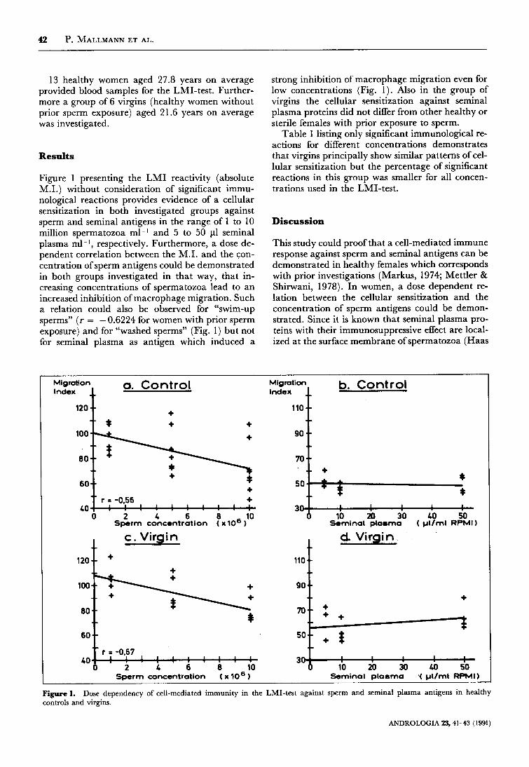
Figure 1 presenting the LMI reactivity (absolute M.I.) without consideration of significant immu- nological reactions provides evidence of a cellular sensitization in both investigated groups against sperm and seminal antigens in the range of 1 to 10 million spermatozoa ml-l and 5 to 50 p1 seminal plasma ml- I , respectively. Furthermore, a dose de- pendent correlation between the M.I. and the con- centration of sperm antigens could be demonstrated in both groups investigated in that way, that in- creasing concentrations of spermatozoa lead to an increased inhibition of macrophage migration. Such a relation could also be observed for “swim-up sperms” ( r = - 0.6224 for women with prior sperm exposure) and for “washed sperms” (Fig. 1) but not for seminal plasma as antigen which induced a
strong inhibition of macrophage migration even for low concentrations (Fig. 1). Also in the group of virgins the cellular sensitization against seminal plasma proteins did not differ from other healthy or sterile females with prior exposure to sperm.
Table 1 listing only significant immunological re- actions for different concentrations demonstrates that virgins principally show similar patterns of cel- lular sensitization but the percentage of significant reactions in this group was smaller for all concen- trations used in the LMI-test.
Discussion
This study could proof that a cell-mediated immune response against sperm and seminal antigens can be demonstrated in healthy females which corresponds with prior investigations (Markus, 1974; Mettler & Shirwani, 1978). In women, a dose dependent re- lation between the cellular sensitization and the concentration of sperm antigens could be demon- strated. Since it is known that seminal plasma pro- teins with their immunosuppressive effect are local- ized at the surface membrane of spermatozoa (Haas
Migration a. Cont ro l Index
+ * + +
l o o h 80 + r = -0.56 +
0 2 L 6 8 10 Sperm concentration ( x106 )
LO 1 1 1 1 1 1 1
c. Virgin
+ + 1
l o o k + 8ol f
LO
Migration b. Cont ro l Index
90
l ’ i 70
So[[*
d. Virgin
90 +
1 I 1 I
10 20 30 Lo 50 Sperm concentration ( x lo6 I Saminal plasma f pVml RPMI)
Figure 1. controls and virgins.
Dose dependency of cell-mediated immunity in the LMI-test against sperm and seminal plasma antigens in healthy
ANDROLOGIA 23, 41-43 (1991)
CELLULAR SENSITIZATION 43
Table 1. “washed” and “swim-up” spermatozoa
Percentage of healthy women and virgins with different immunologic reactions against three concentrations of
Spermatozoa-concentration 1 x loG ml-I 5 x 10⁶ ml-’ 10 x lo6 ml-’
1. Control “washed” spermatozoa:
Enhancement 2/3 (28.6%) no reaction 417 (57.1%)
Inhibition 1/7 (14.3%) “swim-up”-Spermatozoa: Enhancement 3/7 (42.9%) no reaction 4/7 (57.1%) Inhibition 017 (0%)
0/8 (0%) 4/8 (50.0%) 4/8 (50.0%)
017 (0%) 3/7 (42.9%) 417 (57.1%)
018 (0%) 2/8 (25.0%) 6/8 (75.0%)
0/5 (0%)
5/5 (100%) 015 (w0)
2. Virgins “washed” spermatozoa:
Enhancement 1/5 (20.0%) no reaction 4/5 (80.0%)
Inhibition 0/5 (0%)
0/6 (0%) 4/6 (66.7%) 2/6 (33.3%)
016 (0%) 2/6 (33.3%) 416 (66.7%)
& Beer, 1986) and since in this study seminal plasma proteins are proofed to have a strong inhibitory ef- fect on the migration of macrophages it is necessary to take these proteins into consideration when com- menting on the sensitization against spermatozoa.
I t seems to be remarkable that in virgins also an inhibitory immune response could be demonstrated which has to take place independently from any sexual activity and therefore could be related to unspecific antigenic components. The fact that sem- inal plasma proteins cannot be removed by several washing procedures (Roberts & Boettcher, 1969) might explain the cellular sensitisation of virgins against sperm antigen which is still at a lower im- munological level in comparison to women with prior sperm exposure.
Furthermore, it is noticeable that a low sperm concentration of 1 million ml-I “swim-up” and “washed” sperms is able to induce a significant en- hanced migration of macrophages. This type of re- action not found in infertile women (McShane et al., 1985) is changing when higher sperm concen- trations (5 and 10 million ml-l) are used in the LMI-test. Since low concentrations of spermatozoa best represent the physiological situation in the uter- us where the sensitization after intercourse takes place we finally consider the enhancement of mac- rophage migration as physiological cellular sensiti-
zation of females against sperm-associated antigens. The induction of an macrophage-activating-factor (MAF) which is known to be responsible for the phenomenon of the enhancement of macrophages could possibly explain the normally undisturbed mi- gration of spermatozoa to the fallopian tube where fertilization takes place as MAF probably prevents macrophages from staying at the place of immu- nological confrontation and therefore from phago- cytosing spermatozoa.
References
Haas, G. G. & A. E. Beer (1986): Immunologic influences on reproductive biology: sperm gametogenesis and maturation in the male and female genital tract. Fertil. Steril. 46:753-766.
Markus, Z. H. (1974): Leucocyte migration inhibition test in human beings with problems of infertility. WHO workshop on sperm immunology, Aarhus.
Mc Shane, P. M., J. Schiff & D. E. Trentham (1985): Cellular immunity in infertile women. JAMA 253:3555-3562.
Mettler, L. & D. Shirwani (1978): Macrophage migration in- hibitory factor in female sterility. Am. J. Obstet. Gynecol. 121:
Roberts, T. K. & B. Boettcher (1969): Identification of human sperm coating antigens. J. Reprod. Fertil. 18:347-354.
Riimke, P. (1954): The presence ofsperm antibodies in the serum of two patients with oligozoospermia. VOX. Sang. 4 135-143.
1 1 7-1 20.
ANDROLOGIA 23, 41-43 (1991)


![Journal Article Sensitization[1]](https://img.pdfslide.net/doc/110x75/56d6bea91a28ab3016930fdf/journal-article-sensitization1.jpg)