Embed Size (px)
Citation preview
Cellular/Molecular
The Antiaging Protein Klotho Enhances OligodendrocyteMaturation and Myelination of the CNS
Ci-Di Chen,1 Jacob A. Sloane,2* Hu Li,3* Nurgul Aytan,4* Eustathia L. Giannaris,5* Ella Zeldich,1 Jason D. Hinman,6
Alpaslan Dedeoglu,4 Douglas L. Rosene,5 Rashmi Bansal,7 Jennifer I. Luebke,5 Makoto Kuro-o,8 andCarmela R. Abraham1
1Department of Biochemistry, Boston University School of Medicine, Boston, Massachusetts 02118, 2Department of Neurology, Beth Israel DeaconessMedical Center, Boston, Massachusetts 02215, 3Department of Biomedical Engineering, Boston University, Boston, Massachusetts 02215, 4Department ofNeurology, Boston University School of Medicine and Veterans Administration Boston Healthcare System, Boston, Massachusetts 02130, 5Department ofAnatomy & Neurobiology, Boston University School of Medicine, Boston, Massachusetts 02118, 6Department of Neurology, University of California LosAngeles, Los Angeles, California 90095, 7Department of Neuroscience, University of Connecticut Medical School, Farmington, Connecticut 06030, and8University of Texas Southwestern Medical Center, Dallas, Texas 75390
We have previously shown that myelin abnormalities characterize the normal aging process of the brain and that an age-associatedreduction in Klotho is conserved across species. Predominantly generated in brain and kidney, Klotho overexpression extends life span,whereas loss of Klotho accelerates the development of aging-like phenotypes. Although the function of Klotho in brain is unknown, lossof Klotho expression leads to cognitive deficits. We found significant effects of Klotho on oligodendrocyte functions, including inducedmaturation of rat primary oligodendrocytic progenitor cells (OPCs) in vitro and myelination. Phosphoprotein analysis indicated thatKlotho’s downstream effects involve Akt and ERK signal pathways. Klotho increased OPC maturation, and inhibition of Akt or ERKfunction blocked this effect on OPCs. In vivo studies of Klotho knock-out mice and control littermates revealed that knock-out mice havea significant reduction in major myelin protein and gene expression. By immunohistochemistry, the number of total and matureoligodendrocytes was significantly lower in Klotho knock-out mice. Strikingly, at the ultrastructural level, Klotho knock-out mice exhib-ited significantly impaired myelination of the optic nerve and corpus callosum. These mice also displayed severe abnormalities at thenodes of Ranvier. To decipher the mechanisms by which Klotho affects oligodendrocytes, we used luciferase pathway reporters to identifythe transcription factors involved. Together, these studies provide novel evidence for Klotho as a key player in myelin biology, which maythus be a useful therapeutic target in efforts to protect brain myelin against age-dependent changes and promote repair in multiplesclerosis.
IntroductionKlotho is an antiaging protein named after the mythical Greekgoddess who “spins the thread of life” (Kuro-o et al., 1997).Klotho knock-out mice exhibit many changes that also frequently
occur during human aging, including arteriosclerosis, osteopo-rosis, and cognitive decline. The mice develop normally but dieprematurely, with an average life span of �61 d (Kuro-o et al.,1997), whereas mice overexpressing Klotho live 30% longer thanwild-type mice (Kurosu et al., 2005). In early adulthood, Klothoknock-out mice show memory retention deficits (Nagai et al.,2003), a reduction in synapses in the hippocampus (Li et al.,2004), perturbations in axonal transport, and a neurodegenera-tive phenotype in the hippocampus (Shiozaki et al., 2008). Inter-estingly, humans carrying the Klotho-VS polymorphism exhibitreduced cognitive abilities (Nagai et al., 2003; Deary et al., 2005),as well as reduced life span (Nagai et al., 2000; Arking et al., 2003;Arking et al., 2005). The basis of cognitive deficits in Klothoknock-out mice or in humans with Klotho-VS polymorphism isnot known, and a more thorough assessment of neuropathologyis needed.
Klotho is highly expressed in the choroid plexus and neurons,especially in the hippocampus, as well as in the kidney and repro-ductive organs (Kuro-o et al., 1997), but its function in the brainis not clear. Klotho is a type I transmembrane protein cleaved byADAM10 and 17 from the cell membrane (Chen et al., 2007;
Received April 30, 2012; revised Nov. 19, 2012; accepted Dec. 4, 2012.Author contributions: C.-D.C., J.A.S., R.B., and C.R.A. designed research; C.-D.C., J.A.S., N.A., E.L.G., E.Z., J.D.H.,
and J.I.L. performed research; A.D. and M.K-o. contributed unpublished reagents/analytic tools; C.-D.C., J.A.S., H.L.,N.A., E.L.G., J.D.H., and C.R.A. analyzed data; C.-D.C., J.A.S., J.D.H., D.L.R., J.I.L., and C.R.A. wrote the paper.
This work was supported by National Institutes of Health NIA Grant AG-00001, an ADDF Award to C.R.A., NationalInstitutes of Health NINDS R25 Training Grant to J.D.H., a National Multiple Sclerosis Society Research Grant to J.A.S.,and an Ellison Foundation Award to C.-D.C. We thank Dr. Alan Peters for help in interpretation of the electronmicroscopy data; Drs. Yuriy Alekseyev and Marc Lenburg for help with the microarray analysis; Drs. Sha Mi, PaulRosenberg, and Jianlin Wang for help with the early rat OPC preparations; Dr. Cindy Lemere and Jeffrey Frost for themice samples and help in isolating mouse brain tissues; Dr. Rong Fan for help in preparing Klotho mice brain tissuehomogenates; Chun-Tsin Hsu for assistance with the qRT-PCR experiments; Dr. Matt Rasband for sharing of antibodyreagents; and Drs. Gwendalyn King, James J. Collins, Ji-Hye Paik, Ido Wolf, and Tami Rubinek for critical review of andsuggestions for this manuscript.
The authors declare no competing financial interests.*J.A.S., H.L., N.A., and E.L.G. contributed equally to this work.Correspondence should be addressed to Dr. Carmela R. Abraham, Department of Biochemistry, Boston University
School of Medicine, 72 E. Concord Street, K304, Boston, MA 02118. E-mail: [email protected]:10.1523/JNEUROSCI.2080-12.2013
Copyright © 2013 the authors 0270-6474/13/331927-13$15.00/0
The Journal of Neuroscience, January 30, 2013 • 33(5):1927–1939 • 1927
Bloch et al., 2009) and is detectable in serum and CSF (Imura etal., 2004). Klotho functions include regulation of FGF23 signal-ing, suppression of the insulin/IGF1 and Wnt signaling pathways,and regulation of calcium and phosphate homeostasis (Kuro-o,2010). In contrast to other organ systems, none of the down-stream effects of Klotho has been studied for the CNS and little isknown regarding the downstream signal transduction machineryrequired for Klotho’s CNS effects. Considering the distributionof Klotho expression and processing within the CNS and theeffects on cognition cited above, Klotho is positioned to influencea variety of CNS structures and functions in development andaging.
As part of our studies of age-associated cognitive decline in therhesus monkey, we discovered using microarray analysis thatKlotho expression was decreased in the aged corpus callosum(Duce et al., 2008), probably because of the hypermethylation ofits promoter (King et al., 2012). This is of interest as damage tomyelin is ubiquitous in aging monkey brain (Kohama et al., 2011)at biochemical (Sloane et al., 2003), ultrastructural (Bowley et al.,2010), and macroscopic levels (Makris et al., 2007; Wisco et al.,2008), and these abnormalities are often associated with cogni-tive decline (Hinman and Abraham, 2007; Peters, 2009). To de-termine whether there is a connection between age-relatedalterations in Klotho expression (Duce et al., 2008) and CNSdysmyelination (Sloane et al., 2003; Hinman and Abraham,2007), and to understand the functions of Klotho in the brain,we assessed how Klotho influences oligodendrocyte functionand developmental myelination and describe here the impor-tant and novel role Klotho plays in oligodendrocyte biologyand myelination.
Materials and MethodsMaterials. The recombinant mouse Klotho protein containing the extra-cellular domain of mouse Klotho (Ala 35-Lys 982) was from R&D Sys-tems. Akt inhibitor LY294002 and ERK inhibitor UO126 were from CellSignaling Technology. Growth factors for oligodendrocytes were fromPeprotech. All other chemicals were from Sigma-Aldrich.
Animal tissue and protein sample preparation. Klotho knock-out(Kuro-o et al., 1997) and control mice were perfused with 0.1 M PBS, pH7.4, at 4°C and hemibrains homogenized in 5� (v/w) ice-cold RIPAbuffer with protease and phosphatase inhibitor cocktails (Roche) (Kinget al., 2012). Samples were then centrifuged at 16,000 � g for 15 min, andthe supernatant was collected for SDS-PAGE and Western blot analysis.
Oligodendrocyte progenitor cell (OPC) cultures. OPCs were isolatedfrom female Sprague Dawley postnatal day 2 rat pups as described pre-viously (Mi et al., 2005; Sloane and Vartanian, 2007). Cultures weremaintained in high-glucose DMEM OPC culture medium (4 mM
L-glutamine, 1 mM sodium pyruvate, 0.1% BSA, 50 �g/ml insulin, 30 nM
sodium selenite, 10 nM D-biotin, and 10 nM hydrocortisone) containingbFGF/PDGF (10 ng/ml) for 1–3 d and replaced with either DMEM me-dium with CNTF (10 ng/ml), T3 (15 nM), and NT3 (10 ng/ml), orDMEM with CNTF and NT3 alone, with or without recombinant mouseKlotho at a concentration of 0.4 �g/ml at 37°C for 3– 8 d, as indicated inthe figures. Half of the medium bathing the cells was replaced with freshmedium with or without Klotho every other day. The same concentra-tion of Klotho was used in all experiments. The cell lysates were collectedin RIPA buffer as described previously, and protein samples were ana-lyzed by SDS-PAGE and Western blotting.
Western blotting. Protein concentrations were measured using the Mi-cro BCA Protein Assay Reagent Kit (Pierce) according to the manufac-turer’s protocol. For SDS-PAGE, cell lysates containing the same amountof total protein were boiled for 5 min and loaded on 4 –20% precastTris-Glycine gels (Bio-Rad). Proteins were transferred to nitrocellulosemembranes (Millipore). All antibodies were diluted in TBST (50 mM
Tris, pH 8.0, 150 mM NaCl, and 0.1% (v/v) Tween 20) containing 5%(w/v) nonfat dry milk. Secondary antibodies were horseradish peroxidase-
conjugated goat anti-mouse, anti-rat, or anti-rabbit (1:5000, Kirkegaard& Perry Laboratories). Enhanced chemiluminescence was detected usingImmobilon Western Chemiluminescent Substrate (Millipore). Autora-diography was done using Kodak Scientific Imaging Film X-OMAT AR(Eastman Kodak).
The primary antibodies used were as follows: anti-myelin basic protein(MBP) mouse monoclonal antibody (1:1000, Covance), anti-myelin-associated glycoprotein (MAG) mouse monoclonal antibody (cloneB11F7, 1:1000), anti-2�, 3�cyclic nucleotide-3�-phosphodiesterase mousemonoclonal antibody (1:1000, Sigma), anti-FGF receptor substrate(FRS) Y196 (1:1000, Cell Signaling Technology), anti-OSP rabbit poly-clonal antibody (1:1000, Abcam), anti-proteolipid protein (PLP) mousemonoclonal antibody (1:1000, Millipore), anti-PCNA clone PC10 (1:1000, Millipore), and anti-�-tubulin monoclonal antibody (1:1000, In-vitrogen). The antibodies in the Akt and ERK pathways were fromphospho Akt and ERK pathway kit (Cell Signaling Technology) and wereused according to the manufacturer’s protocol.
qRT-PCR. Total RNA was isolated using QIAGEN�s RNeasy kit (QIA-GEN). A reverse transcription was performed with 2 �g of total RNAfrom each sample. Primers for selected mouse myelin-related genes (seeTable 1) and corresponding rat oligodendrocyte maturation-enrichedgenes as published previously (Cahoy et al., 2008) were designed byRealTimePrimers. Controls included � actin, �-2 microglobulin, phos-phoglycerate kinase 1, hypoxanthine phosphoribosyltransferase 1, andGAPDH. The qRT-PCR experiments were performed with iQ SYBRGreen Supermix (Bio-Rad) with detection on a Bio-Rad C1000 ThermalCycler. The whole cDNA from 2 �g of total RNA was used for one 96-wellplate. Triplicates of 20 �l reactions containing primer and cDNA tem-plate were used for quantitation. A PCR was carried out as follows: 1 cycleof 95°C for 3 min followed by 40 cycles of 95°C for 10 s, 55°C for 20 s, and72°C for 30 s. This was followed by a dissociation curve beginning at 55°Cand increasing by 0.2°C every 3 s, with SYBR Green fluorescence mea-sured at every interval. Relative quantitation of the difference betweenthe control and Klotho-treated samples was done using RT 2 Profiler PCRArray Data Analysis Program (QIAGEN). Genes were tested for statisti-cal significance ( p � 0.05), relative to the control, by Student’s t test.
Luciferase assay. Paired homeodomain protein 3 (Pax3) reporter plas-mid (pluc-TKCD-19) was kindly provided by Dr. Frank Rauscher III(Wistar Institute, Philadelphia, Pennsylvania). Signal Transduction 45pathway Reporter Array (see Tables 2 and 3) was from SABiosciences(QIAGEN). OPCs were plated into 96-well plates at a density of 0.65 �10 6 cells/plate in OPC culture medium containing bFGF/PDGF for 3 dbefore transfection. A total of 200 ng of Luciferase reporter or controlempty-vector DNA (with 5 ng Renilla luciferase) was transfected into
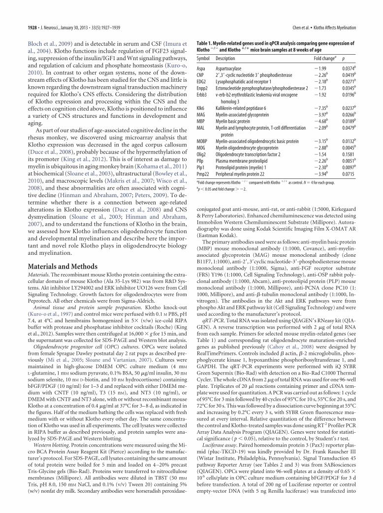
Table 1. Myelin-related genes used in qPCR analysis comparing gene expression ofKlotho �/� and Klotho �/� mice brain samples at 8 weeks of age
Symbol Description Fold changea p
Aspa Aspartoacylase �1.99 0.0374b
CNP 2�,3�-cyclic nucleotide 3� phosphodiesterase �2.26b 0.0419b
EDG2 Lysophosphatidic acid receptor 1 �2.18b 0.0271b
Enpp2 Ectonucleotide pyrophosphatase/phosphodiesterase 2 �1.73 0.0345b
Erbb3 v-erb-b2 erythroblastic leukemia viral oncogenehomolog 3
�1.92 0.0196b
Klk6 Kallikrein-related peptidase 6 �7.35b 0.0237b
MAG Myelin-associated glycoprotein �3.97b 0.0266b
MBP Myelin basic protein �4.68b 0.0189b
MAL Myelin and lymphocyte protein, T-cell differentiationprotein
�2.09b 0.0479b
MOBP Myelin-associated oligodendrocytic basic protein �3.15b 0.0132b
MOG Myelin oligodendrocyte glycoprotein �2.88b 0.0045b
Olig2 Oligodendrocyte transcription factor 2 �1.54 0.1581Pllp Plasma membrane proteolipid �2.26b 0.0051b
Plp1 Proteolipid protein (myelin) 1 �2.30b 0.0097b
Pmp22 Peripheral myelin protein 22 �3.94b 0.0715aFold change represents Klotho �/� compared with Klotho �/� as control. N � 4 for each group.bp � 0.05 and fold change � �2.
1928 • J. Neurosci., January 30, 2013 • 33(5):1927–1939 Chen et al. • Klotho Affects Myelination
cells using Lipofectamine 2000 (Invitrogen), and cells were treated withor without recombinant mouse Klotho in OPC culture medium as de-scribed above. Twenty-four hours after transfection, cells were washedonce with PBS and assayed for luciferase activity using the Dual-luciferase system (Promega) as described previously (Oh et al., 2010).
Immunofluorescence. Cells were fixed in 4% paraformaldehyde in PBSat room temperature, rinsed with PBS, and treated for 1 h with blockingsolution (PBS supplemented with 0.1% Triton X-100 and 1% BSA). Cellswere incubated overnight at 4°C with the primary antibody diluted inblocking solution. Cells were stained with antibodies to Olig2 (1:10,000;Millipore), CC-1 (1:200; Millipore), or O1 (1:2; ATCC). Subsequently,cells were rinsed and incubated with the relevant secondary antibodies(Cy3 or Alexa-488; Jackson ImmunoResearch Laboratories) for 1 h atroom temperature. Immunofluorescence images were obtained by aNikon Eclipse 660 microscope and a SPOT-cooled CCD digital camera(Diagnostic Instruments). For every condition, we identified the matu-ration state of each OL for the �30 – 60 cells within a given field for allfields acquired (N � 450 –900 cells/condition) from 3 independent ex-periments. We quantified numbers of mature OLs (O1 � or CC1 �) andtotal OL-lineage cells (Olig2 �) and determined percentage mature OLsof total OL � lineage cells (O1 �/Olig2 � or CC1 �/Olig2 �) (becauseOlig2 labels OPCs as well as OLs).
Cell number determination, cell viability assay, and LDH release. Cellnumber was determined colorimetrically by crystal violet staining as de-scribed previously (Zeldich et al., 2007). Cells were plated at 50,000 cells/well in 24-well plates in triplicates and allowed to attach and spread inOPC culture medium containing bFGF/PDGF for 1–3 d and then re-placed with medium containing CNTF and NT3, with or without Klothofor the indicated time periods. At the end of each time interval, the cellswere washed with PBS, fixed in 70% ethanol, and kept at 4°C until thestaining with 1% crystal violet. Unincorporated stain was removed bywashing, cells were air-dried, and the dye was extracted with 70%ethanol and optical density (absorbance 550 nm and baseline refer-ence absorbance 750 nm) was measured by Microplate Reader (Glo-max Multi Detection System, Promega). The data analysis was doneby subtracting the baseline readings (750 nm) from the absorbancereadings (560 nm).
For cell viability assay, cells were plated in 96-well plates in six repeatsand allowed to attach and spread in OPC culture medium containingbFGF/PDGF for 3 d and then replaced with medium containing CNTFand NT3, with or without Klotho for the indicated time periods. At theend of each time interval, cell number was assessed using CellTiter-GloLuminescent Cell Viability Assay (Promega) according to the manufac-turer’s instructions. This assay signals the presence of metabolically ac-tive cells. Briefly, 100 �l of CellTiter Glo Reagent was added to the equalvolume of cell culture medium present in each cell culture well. Thecontents were mixed for 2 min and after stabilization of the signal atroom temperature for 10 min, the luminescence was recorded on a Mi-croplate Reader (Glomax Multi Detection System, Promega). Cell deathwas assessed by using the CytoTox 96 nonradioactive cytotoxicity assay(Promega), which measures the LDH release into the medium.
Immunohistochemistry and cell counting in wild-type and Klotho knock-out mice. Five-week-old Klotho �/� and Klotho �/� mice were perfusedthrough the heart with 0.1 M PBS (4°C, pH 7.4). The brains were re-moved, and one hemisphere was immersion fixed in 4% paraformalde-hyde overnight while the other was left unfixed and flash frozen withpulverized dry ice and stored at �80°C. The fixed hemisphere was re-moved from fixative the next day and cryoprotected in successive solu-tions of first 10%, then 20% glycerol in PBS with 2% DMSO (Rosene etal., 1986). The frozen hemibrains were cut in the coronal plane into30-�m-thick sections with a sliding microtome. The sections were di-vided into six interrupted series and stored at 4°C in 0.1 M PBS with 1%sodium azide as a preservative until they were processed.
For Olig2, CC-1, and GST-Pi immunohistochemistry, one series fromeach subject was selected and all sections were processed together in asingle batch at the same time in the same reagents to eliminate anyprocedural variance. All steps were performed at room temperature un-less otherwise indicated. Free-floating sections were washed, quenchedwith 3% hydrogen peroxide, and then blocked with 10% normal goatserum in PBS with 0.4% Triton X-100 for 1 h. Next, sections were incu-bated in a stock solution (1% normal goat serum, 0.2% Triton X-100 in0.05 M PBS) containing rabbit polyclonal anti-Olig2 antibody (Milliporeab9610, 1:10,000), mouse mAb against CC-1 (Millipore OP-80, 1:40) oranti-GST Pi (Abcam, 1:500) for 48 h at 4°C. Sections were then washed in
Table 2. Signal transduction 45-pathway reporter array
Reporter Pathway Transcription factor
AARE Amino acid deprivation response ATF4/ATF3/ATF2AR Androgen receptor Androgen receptorARE Antioxidant response Nrf2 and Nrf1ATF6 ATF6 ATF6C/EBP C/EBP C/EBPCRE cAMP/PKA CREBE2F Cell cycle E2F/DP1p53 p53/DNA damage p53EGR1 EGR1 EGR1ERSE Endoplasmic reticulum stress CBF/NF-Y/YY1ERE Estrogen receptor Estrogen receptorGATA GATA GATAGRE Glucocorticoid receptor Glucocorticoid receptorHSR Heat shock response HSFMTF1 Heavy metal stress MTF1GLI Hedgehog GLIHNF4 Hepatocyte nuclear factor 4 HNF4HIF Hypoxia HIF-1IRF1 Interferon regulation IRF1ISRE Type I interferon STAT1/STAT2GAS Interferon � STAT1/STAT1KLF4 KLF4 KLF4LXR Liver X receptor LXRaSRE MAPK/ERK Elk-1/SRFAP1 MAPK/JNK AP-1MEF2 MEF2 MEF2Myc c-myc Myc/MaxNanog Nanog NanogRBP-Jk Notch RBP-JkNF�B NF�B NF�BOct4 Oct4 Oct4Pax6 Pax6 Pax6FOXO PI3K/AKT FOXONFAT PKC/Ca 2� NFATPPAR PPAR PPARPR Progesterone receptor Progesterone receptorRARE Retinoic acid receptor Retinoic acid receptorRXR Retinoid X receptor Retinoid X receptorSox2 Sox2 Sox2SP1 SP1 SP1STAT3 STAT3 STAT3SMAD TGF� SMAD2/SMAD3/SMAD4VDR Vitamin D receptor Vitamin D receptorTCF/LEF Wnt TCF/LEFXRE Xenobiotic AhR
Table 3. Transcriptional regulatory element sequence information of the luciferasereporters involved in Klotho-dependent responses in OPCs
Reporter Pathway Transcriptional regulatory element sequence
AP1 MAPK/JNK TGAGTCAGARE Antioxidant response AACATTGCATCATCCCCGCC/EBP C/EBP ATTGCGCAATLXR Liver X receptor TGAATGACCAGCAGTAACCTCAGCNF�B NF�B GGGACTTTCCPR Progesterone receptor GGGACATGGTGTTCTSRE MAPK/ERK GGATGTCCATATTAGGASTAT3 STAT3 GTCGACATTTCCCGTAAATCGTCGA
Chen et al. • Klotho Affects Myelination J. Neurosci., January 30, 2013 • 33(5):1927–1939 • 1929
stock solution and treated with biotinylatedgoat anti-rabbit or anti-mouse secondary anti-body (1:600, BA5000, Vector Laboratories) for1 h. After washing, sections were incubatedwith an avidin-biotinylated horseradish perox-idase enzyme complex (PK6100, VectastainElite ABC kit, Vector Laboratories) for 1 h,rinsed in KPBS, then sodium acetate buffer(0.175 M), and processed with a solution ofnickel sulfate hexahydrate (0.095 M), 3,3 di-aminobenzidine tetrahydrochloride (DAB)(0.55 mM), and hydrogen peroxide (0.0025%)in 0.175 M sodium acetate buffer for 15 min.The immunostained sections were mountedon gelatin-coated slides and air-dried. Thensections were defatted in chloroform:ethanol(1:1), rehydrated, counterstained with Neu-tral Red (1%), dehydrated in a series of etha-nols, cleared in xylenes, and coverslippedwith Permount (Fisher Scientific).
To quantify Olig2 � and GST-Pi � cells, thefractionator method was used to obtain a pop-ulation estimate within the fimbria. The fim-bria was chosen because it is an easily defined and circumscribed bundleof white matter. The fimbria was outlined in six 30 �m sections spaced180 �m apart, starting rostrally at the level of the anterior end of thehabenula and spanning �1.08 mm caudally using StereoInvestigatorsystem (Version 9.14.3, MBF Bioscience) and a 4� Nikon Plan objectiveon a Nikon Eclipse E600 microscope. Cell counts were made with a 20�Nikon Plan Fluor objective. A 120 �m � 120 �m grid was overlaid on theregion of interest, and Olig2 � cells were counted within 40 �m � 40 �mcounting frames placed at the grid intersections that fell within the out-line of the fimbria. The criterion for counting cells was the appearance ofthe black Ni-DAB immunoproduct in the nucleus. In an adjacent seriesof six 30-�m-thick tissue sections, GST-Pi � cells within the fimbria werequantified. The fimbria was outlined with a 4� objective as describedabove. A 120 �m � 120 �m sampling grid was superimposed over theregion of interest, and GST-Pi � cells were counted within a 40 �m � 40�m counting frame and 5 �m disector using a 60� Nikon Plan Fluor oilobjective. A guard volume of 1 �m above the disector box was used and2– 4 �m below, depending on the thickness of the tissue section (�8 –10�m total). GST-Pi � cells had brown DAB immunoproduct in the cyto-plasm and nucleolus, yet the nucleus was unstained.
Nodal and paranodal immunofluorescence. Free-floating 30 �m sec-tions were stained for nodes and paranodes with rabbit anti-�-IV spec-trin (1:400) or rabbit anti-NaV1.6 (1:200) (gifts from M. Rasband, BaylorCollege of Medicine) (Rasband et al., 2003; Ogawa et al., 2006), andmouse anti-caspr antibodies (1:500), using the following protocol withall steps performed at room temperature unless otherwise stated. Themonoclonal antibody mouse anti-caspr (clone K65/35) was developed byand obtained from the UC Davis/National Institutes of Health Neuro-mab Facility. Sections were washed in PBS, transferred to prewarmed 10mM sodium citrate, pH 8.5, and incubated at 80°C for 30 min for antigenretrieval. After cooling to room temperature, sections were washed 3times in PBS, then blocked and permeabilized in PBS with 0.3% TX-100(Sigma) and 10% normal donkey serum (Jackson ImmunoResearch Lab-oratories) for 30 min. Sections were then placed directly in MOM Block(Vector Laboratories) diluted in PBS for 1 h, washed 3 times in PBS, andthen incubated overnight in primary antibody cocktails. The next day,after 3 washes in PBS, sections were incubated in donkey anti-mouse Cy2(Jackson ImmunoResearch Laboratories; 1:300) and donkey anti-rabbitDyLight 549 (Jackson ImmunoResearch Laboratories; 1:300) for 1 h.Sections were then mounted on gelatin subbed slides, air dried, anddehydrated with 70 –100% EtOH and xylene and coverslipped with DPXmounting medium.
Image acquisition was performed on a Nikon Eclipse Ti C2 � laserscanning confocal microscope. Two-micron optical sections at 100�optical objective with 2� digital zoom were obtained from five differentimmunolabeled sections from each of two wild-type and two Klotho-null
mice. The imaged white matter included corpus callosum overlying stria-tum to that overlying hippocampus. Maximum intensity projectionswere obtained and images were postprocessed in NIS Elements software(Nikon). Nodal and paranodal segments were measured using the two-point length measurement tool. For measurement, only complete para-nodal pairs (both paranodes and an immunolabeled node) were selectedfor measurement. A total of 30 paranodes per section (150 per animal)were measured, and a total of 10 nodes per section (50 per animal) weremeasured. Statistical significance was determined using a Student’s t testassuming one tail and equal variance. For presentation, images werefurther postprocessed and cropped in Photoshop CS5 (Adobe).
Electron microscopy. Thirty-five-day-old Klotho �/�, Klotho �/�, andKlotho �/� mice, and 25-d-old Klotho �/� mice were perfused transcar-dially with 1% paraformaldehyde and 2.5% glutaraldehyde fixative in 0.1M cacodylate buffer, pH 7.2–7.4. Optic nerve, corpus callosum, and cer-vical spinal cord were postfixed with 1% osmium oxide, dehydrated inalcohol series and propylene oxide before embedding in Epon. One-micrometer-thick sections were cut and stained with toluidine blue. Thinsections were contrasted with uranyl acetate and lead citrate and imagedwith a JEOL 1200EX electron microscope.
Gene set enrichment analysis. Gene set enrichment analysis was per-formed according to Subramanian et al. (2005) with 1186 chemical andgenetic perturbation gene sets. Klotho �/� mice versus Wild-type �/�
mice gene expression profiles were ranked based on p value. The rankedgene list was used as input for the chemical and genetic perturbation genesets. The significant gene sets were selected based on the normalizedenrichment score and false discovery rate, accordingly.
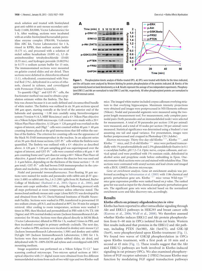
ResultsKlotho effects on primary oligodendrocytes in vitroKlotho has been reported to affect intracellular signaling throughAkt and ERK1/2 pathways in HEK293 and breast cancer cells(Kurosu et al., 2006; Wolf et al., 2008). We therefore assessedwhether Klotho induces ERK1/2 and Akt protein phosphoryla-tion from 0 – 60 min in OPCs isolated from rat brain. Westernblot results indicated that proteins in the ERK1/2 and Akt path-way, including PTEN (Ser380), Akt (Ser473), and GSK-3�(Ser9), were phosphorylated upon Klotho treatment (Fig. 1).We found two waves of GSK3� phosphorylation in OPCsupon Klotho treatment, with a first peak at 10 min and asecond at 45 min (Fig. 1). These results suggest that the Aktand ERK/12 pathways are both involved in Klotho-inducedsignaling in rat primary OPCs. We also examined phosphory-lation of FGF receptor substrate 2 (FRS2) because Klotho mayfunction by modulating FGF signal transduction pathways
Figure 1. Phosphorylation kinetic analysis of Klotho-treated OPCs. A, OPCs were treated with Klotho for the times indicated,and the cell lysates were analyzed by Western blotting for protein phosphorylation of the proteins indicated. B, Kinetics of thesignal intensity based on band densitometry as in A. Results represent the average of two independent experiments. Phosphory-lated ERK1/2 and Akt are normalized to total ERK1/2 and Akt, respectively. All other phosphorylated proteins are normalized toGAPDH internal control.
1930 • J. Neurosci., January 30, 2013 • 33(5):1927–1939 Chen et al. • Klotho Affects Myelination
(Kurosu et al., 2006; Urakawa et al., 2006). Western blot re-sults indicated that FRS2 was phosphorylated at Y196 uponKlotho treatment (Fig. 1), suggesting that the FGF signalingpathways may be involved in Klotho-induced signaling in ratOPCs. However, it is also possible that Klotho affects FRS2phosphorylation via Klotho’s interaction with the TrkA recep-tor (Meakin et al., 1999).
To examine the effects of Klotho on the OPC phenotype, oli-godendrocyte maturation was assessed by immunohistochemis-try for mature oligodendrocytes (O1), and pan-oligodendrocytes(Olig2) after Klotho treatment. The OPCs were allowed to attachand spread in OPC culture medium containing bFGF/PDGF for
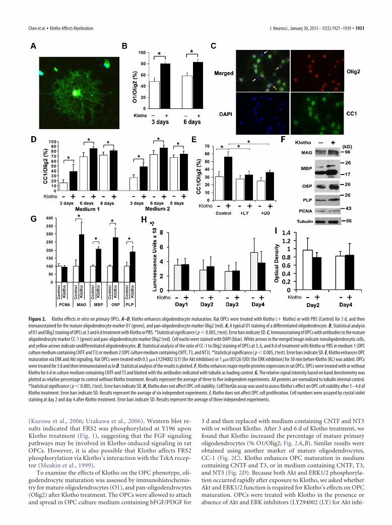
3 d and then replaced with medium containing CNTF and NT3with or without Klotho. After 3 and 6 d of Klotho treatment, wefound that Klotho increased the percentage of mature primaryoligodendrocytes (% O1/Olig2; Fig. 2A,B). Similar results wereobtained using another marker of mature oligodendrocytes,CC-1 (Fig. 2C). Klotho enhances OPC maturation in mediumcontaining CNTF and T3, or in medium containing CNTF, T3,and NT3 (Fig. 2D). Because both Akt and ERK1/2 phosphoryla-tion occurred rapidly after exposure to Klotho, we asked whetherAkt and ERK1/2 function is required for Klotho’s effects on OPCmaturation. OPCs were treated with Klotho in the presence orabsence of Akt and ERK inhibitors (LY294002 (LY) for Akt inhi-
Figure 2. Klotho effects in vitro on primary OPCs. A–D, Klotho enhances oligodendrocyte maturation. Rat OPCs were treated with Klotho (� Klotho) or with PBS (Control) for 3 d, and thenimmunostained for the mature oligodendrocyte marker O1 (green), and pan-oligodendrocyte marker Olig2 (red). A, A typical O1 staining of a differentiated oligodendrocyte. B, Statistical analysisof O1 and Olig2 staining of OPCs at 3 and 6 d treatment with Klotho or PBS. *Statistical significance ( p�0.005, t test). Error bars indicate SD. C, Immunostaining of OPCs with antibodies to the matureoligodendrocyte marker CC-1 (green) and pan-oligodendrocyte marker Olig2 (red). Cell nuclei were stained with DAPI (blue). White arrows in the merged image indicate nonoligodendrocytic cells,and yellow arrows indicate undifferentiated oligodendrocytes. D, Statistical analysis of the ratio of CC-1 to Olig2 staining of OPCs at 3, 6, and 8 d of treatment with Klotho or PBS in medium 1 (OPCculture medium containing CNTF and T3) or medium 2 (OPC culture medium containing CNTF, T3, and NT3). *Statistical significance ( p � 0.005, t test). Error bars indicate SD. E, Klotho enhances OPCmaturation via ERK and Akt signaling. Rat OPCs were treated with 0.5 �M LY294002 (LY) (for Akt inhibition) or 1 �M UO126 (UO) (for ERK inhibition) for 30 min before Klotho (KL) was added. OPCswere treated for 3 d and then immunostained as in D. Statistical analysis of the results is plotted. F, Klotho enhances major myelin proteins expression in rat OPCs. OPCs were treated with or withoutKlotho for 6 d in culture medium containing CNTF and T3 and blotted with the antibodies indicated with tubulin as loading control. G, The relative signal intensity based on band densitometry wasplotted as relative percentage to control without Klotho treatment. Results represent the average of three to five independent experiments. All proteins are normalized to tubulin internal control.*Statistical significance ( p � 0.005, t test). Error bars indicate SD. H, Klotho does not affect OPC cell viability. CellTiterGlo assay was used to assess Klotho’s effect on OPC cell viability after 1– 4 d ofKlotho treatment. Error bars indicate SD. Results represent the average of six independent experiments. I, Klotho does not affect OPC cell proliferation. Cell numbers were assayed by crystal violetstaining at day 2 and day 4 after Klotho treatment. Error bars indicate SD. Results represent the average of three independent experiments.
Chen et al. • Klotho Affects Myelination J. Neurosci., January 30, 2013 • 33(5):1927–1939 • 1931
bition or UO126 (UO) for ERK inhibi-tion). Inhibition of ERK functionreduced, while Akt functional inhibitioncompletely abolished, the effects ofKlotho on OPC maturation (Fig. 2E), sug-gesting that Klotho enhances OPC matu-ration primarily via signaling requiringAkt but also, to a more limited degree,through ERK1/2. Western blot analysis ofKlotho-treated OPCs revealed thatKlotho enhanced the expression of themajor myelin proteins, including MAG,MBP, oligodendrocyte-specific protein(OSP/Claudin11), and PLP (Fig. 2F,G),confirming that Klotho enhances OPCmaturation in vitro. Western blotting forthe cell proliferation marker, PCNA, re-vealed no difference with Klotho treat-ment (Fig. 2F,G), suggesting that Klothodoes not enhance OPC proliferation. Wealso examined cell number and cell viabil-ity by crystal violet staining and CellTiter-Glo assay, which reflects the amount ofthe ATP present, and we found no differ-ence in Klotho-treated and untreatedOPCs from day 1 to day 4 in the cell via-bility assay and for day 2 and day 4 forcrystal violet staining (Fig. 2H, I). BecauseOPCs were allowed to attach and spreadfor 3 d in the presence of basic FGF(bFGF) and PDGF, cell proliferation oc-curred during this period. After we treatedthe OPCs with Klotho in culture mediumcontaining CNTF and T3, OPCs started todifferentiate but cell numbers remained constant as shown inFigure 2H, I. We also observed no difference with a cell deathassay by measuring LDH release to the media with or withoutKlotho. These results suggest that Klotho affects oligodendrocytedifferentiation and maturation, but not their proliferation andcell death.
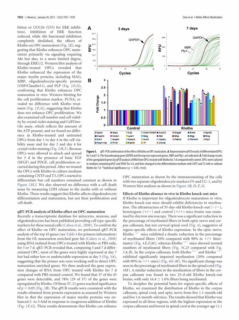
qRT-PCR analysis of Klotho effect on OPC maturationRecently a transcriptome database for astrocytes, neurons, andoligodendrocytes has been published providing cell type-specificmarkers for these neural cells (Cahoy et al., 2008). To confirm theeffect of Klotho on OPC maturation, we performed qRT-PCRanalysis of the top 45 genes (see Table 4 for primers information)from the OL maturation enriched gene list (Cahoy et al., 2008)using RNA isolated from OPCs treated with Klotho or PBS only,for 3 or 7 d. qRT-PCR revealed that, comparing 3 and 7 d differ-entiated OPC, most of the genes were highly expressed at day 7but had either low or undetectable expression at day 3 (Fig. 3A),suggesting that the primer sets were working well to detect OPCmaturation-enriched genes. We then analyzed the gene expres-sion changes of RNA from OPC treated with Klotho for 7 dcompared with PBS-treated control. We found that 37 of the 45genes were detectable, and 78% (29 of 37) of the genes wereupregulated by Klotho. Of those 37, 21 genes reached significanceof p � 0.05 (Fig. 3B). The qPCR results were consistent with theresults obtained from protein expression as assessed by Westernblot in that the expression of major myelin proteins was en-hanced 2- to 3-fold in response to exogenous addition of Klotho(Fig. 2F,G). These results demonstrate that Klotho can enhance
OPC maturation as shown by the immunostaining of the cellswith two separate oligodendrocyte markers O1 and CC-1, and byWestern blot analyses as shown in Figure 2B, D, F, G.
Effects of Klotho absence in vivo in Klotho knock-out miceIf Klotho is important for oligodendrocyte maturation in vitro,Klotho knock-out mice should exhibit deficiencies in myelina-tion. The ultrastructure of 35-day-old Klotho knock-out (�/�),hemizygous (�/�) and control (�/�) mice brains was exam-ined by electron microscopy. There was a significant reduction inthe percentage of myelinated fibers in both optic nerve and cor-pus callosum, but not cervical spinal cord (Fig. 4), suggestive ofregion-specific effects of Klotho expression. In the optic nerve,Klotho�/� mice exhibited a drastic reduction in the percentageof myelinated fibers (10% compared with 90% in �/� litter-mates) (Fig. 4E,F,M), whereas Klotho�/� mice showed normalnumbers of myelinated fibers (Fig. 5C,D compared with Fig.5A,B). In the corpus callosum, both Klotho�/� and Klotho�/�
exhibited significantly impaired myelination (20% comparedwith 90% in �/� mice) (Fig. 4G–M). No significant change wasseen in the percentage of myelinated fibers in the spinal cord (Fig.4M). A similar reduction in the myelination of fibers in the cor-pus callosum was found in two 25-d-old Klotho knock-outbrains, with only 16.4 1.6% fibers being myelinated.
To decipher the potential basis for region-specific effects ofKlotho, we examined the distribution of Klotho in the corpuscallosum, spinal cord, and optic nerve from five 1.1-month-oldand five 1.6-month-old mice. The results showed that Klotho wasexpressed in all three regions, with the highest expression in thecorpus callosum and lowest in spinal cord at the younger age (1.1
Figure 3. qRT-PCR confirmation of the effect of Klotho on OPC maturation. A, Representative qPCR results of differentiated OPCsfor 3 and 7 d. The housekeeping gene GAPDH and the top two expressed genes, MBP and Plp1, are indicated. B, Fold change resultsof the upregulated genes by qPCR analysis of RNA from OPCs treated with Klotho for 7 d compared with control. OPCs were culturedin medium containing bFGF and PDGF for 3 d, and then changed to the differentiation medium with CNTF and T3 with or withoutKlotho for 7 d. *Statistical significance ( p � 0.05, t test).
1932 • J. Neurosci., January 30, 2013 • 33(5):1927–1939 Chen et al. • Klotho Affects Myelination
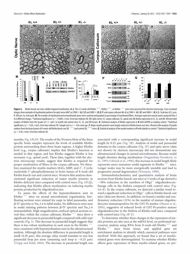
months, Fig. 4N,O). The results of the Western blots of the threespecific brain samples represent the levels of available Klothoprotein surrounding these three brain regions. A higher Klotholevel (e.g., corpus callosum) implies that Klotho’s function isneeded in that region, and less Klotho suggests Klotho is lessnecessary (e.g., spinal cord). These data, together with the elec-tron microscopy results, suggest that Klotho is required forproper myelination of fibers in the corpus callosum. We there-fore examined the myelin markers MAG, MBP, and 2�, 3�cyclicnucleotide-3�-phosphodiesterase in brain tissues of 8-week-oldKlotho knock-out and control mice. Western blot analyses dem-onstrated significant reduction of major myelin proteins inKlotho-deficient mice compared with control mice (Fig. 4P,Q),indicating that Klotho affects myelination via inducing myelinprotein production by oligodendrocytes.
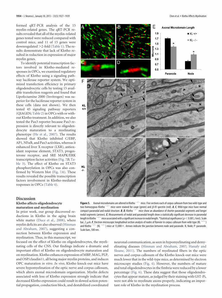
To assess the effects of the hypomyelination seen inKlotho�/� mice on axonal microdomain organization, free-floating sections were stained for caspr to label paranodes, and�-IV spectrin or NaV1.6 to label nodes. No differences were seenin nodal staining patterns between �-IV spectrin and NaV1.6immunolabels. High-magnification confocal optical sections re-veal that, within the corpus callosum, Klotho�/� mice show asignificant decrease in paranodal length compared with wild-typecontrols (Fig. 5). This decrease in paranodal length suggests thatthere is less robust myelination in each axon in the Klotho�/�
mice consistent with hypomyelination seen in the ultrastructuralanalysis. Although the absolute difference in paranodal length issmall (0.30 �m), this average value would translate to one lessparanodal loop per axon (assuming each loop is �0.25 �m)(Trapp and Kidd, 2004). The decrease in paranodal length was
associated with a corresponding significant increase in nodallength by 0.25 �m (Fig. 5E). Analysis of nodal and paranodaldomains in the corpus callosum (Fig. 5F) and optic nerve (datanot shown) by electron microscopy did not demonstrate anyultrastructural changes in axonal microdomains. Because nodallength shortens during myelination (Dugandzija-Novakovic etal., 1995; Vabnick et al., 1996), this increase in nodal length likelyrepresents more immature nodal segments in Klotho�/� mice.Longer nodes may be more energetically unstable and lead toprogressive axonal degeneration (Waxman, 1998).
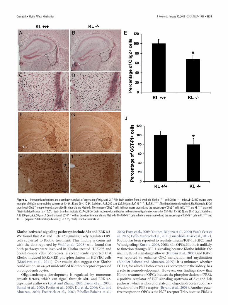
Immunohistochemistry and quantitative analysis of brainsections from Klotho knock-out mice at 5 weeks of age showed a�30% reduction in the numbers of Olig2� oligodendrocyte-lineage cells in the fimbria compared with control mice (Fig.6A–E). In the corpus callosum, we detected a similar trend to-ward a significant reduction in Olig2� cells in Klotho knock-outmice (data not shown). In addition, we found a similar and con-firmatory reduction (21%) in the number of mature oligoden-drocytes immunopositive for the GST-Pi marker (Mason et al.,2004), suggestive of reduced levels of GST-Pi-positive matureoligodendrocytes in the fimbria of Klotho-null mice comparedwith control mice (Fig. 6F–J).
To determine whether these changes in the expression of my-elin proteins are also seen at the mRNA level, we performed mi-croarray analysis using RNA from 8-week-old Klotho�/� andKlotho�/� mice brain tissue, and applied gene setenrichment analysis to identify which canonical pathways wereinvolved. With this approach, we found that a set of myelin-related genes were downregulated. To examine whether Klothoaffects gene expression of these myelin-related genes, we per-
Figure 4. Klotho knock-out mice exhibit impaired myelination. A–L, The 4.5-week-old Klotho �/�, Klotho �/�, or Klotho �/� mice were processed for electron microscopy. Cross-sectionalimages show examples of myelination patterns for optic nerve (A-F ) at 2900� (A, C, E) and 5800� (B, D, F ) and corpus callosum (G–L) at 2900� (G, I, K ) and 5800� (H, J, L). Scale bars: E, 2 �m;F, 500 nm. As, Astrocyte. M, The number of myelinated and unmyelinated axons were counted and graphed as percentage of myelinated fibers. Averages represent axonal counts analyzed from 3to 6 different images. *Statistical significance ( p � 0.0001, t test). Error bars indicate SD. ON, Optic nerve; CC, corpus callosum; SC, spinal cord. N, Klotho expression in CC, SC, and ON. Western blotanalysis of Klotho from the lysates of 1.1- and 1.6-month-old control mice CC, SC, and ON tissues. O, Statistical analysis of Klotho expression in N with GAPDH as loading control. *Statisticalsignificance ( p � 0.05, t test). Error bars indicate SD. Sample size is n � 4 for each age. P, Major myelin proteins were largely reduced in Klotho knock-out mice. Western blot analysis of myelinmarkers from the brain lysates of 8-week-old Klotho knock-out (KL �/�) and control (KL �/�) mice. Q, Statistical analysis of the myelin markers in P with tubulin as control. *Statistical significance( p � 0.05, t test). Error bars indicate SD.
Chen et al. • Klotho Affects Myelination J. Neurosci., January 30, 2013 • 33(5):1927–1939 • 1933
formed qRT-PCR analysis of the 15myelin-related genes. The qRT-PCR re-sults revealed that all of the myelin-relatedgenes tested were reduced compared withcontrol mice, and 11 of 15 genes weredownregulated �2-fold (Table 1). The re-sults demonstrate that lack of Klotho re-sulted in reduction in expression of majormyelin genes.
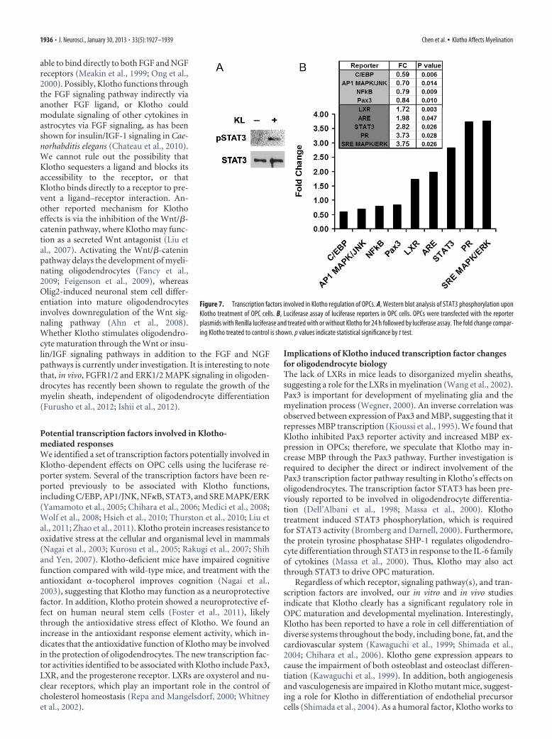
To identify potential transcription fac-tors involved in Klotho-mediated re-sponses in OPCs, we examined regulatoryeffects of Klotho using a signaling path-way luciferase reporter system. We opti-mized transfection efficiency in primaryoligodendrocytic cells by testing 15 avail-able transfection reagents and found thatLipofectamine 2000 (Invitrogen) was su-perior for the luciferase reporter system inthese cells (data not shown). We thentested 45 signaling pathway reporters(QIAGEN; Table 2) in OPCs with or with-out Klotho treatment. In addition, we alsotested the Pax3 reporter because Pax3 re-pression is directly relevant to oligoden-drocyte maturation to a myelinatingphenotype (He et al., 2007). The resultsshowed that Klotho inhibited C/EBP,AP1, NF�B, and Pax3 activities, whereas itenhanced liver X receptor (LXR), antiox-idant response element, STAT3, proges-terone receptor, and SRE MAPK/ERKtranscription factor activities (Fig. 7B; Ta-ble 3). The effect of Klotho on STAT3phosphorylation in OPCs was also con-firmed by Western blot (Fig. 7A). Theseresults revealed the possible transcriptionfactors involvement in Klotho-mediatedresponses in OPCs (Table 4).
DiscussionKlotho affects oligodendrocytematuration and myelinationIn prior work, our group discovered re-ductions in Klotho in the aging brainwhite matter (Duce et al., 2008), wheremyelin deficits are also observed (Hinmanand Abraham, 2007), suggesting a con-nection between Klotho expression andmyelination. Thus, in this manuscript, wefocused on the effect of Klotho on oligodendrocytes, the myeli-nating cells of the CNS. Our findings indicate a dramatic andimportant effect of Klotho on oligodendrocyte maturation andon myelination. Klotho enhances expression of MBP, MAG, PLP,and OSP/claudin11, all being major myelin proteins, and inducesOPC maturation in vitro. In vivo, Klotho knock-out mice havesevere hypomyelination of the optic nerve and corpus callosum,which alters axonal microdomain organization. Myelin defectsassociated with loss of Klotho expression strongly indicate thatdecreased Klotho expression could result in slowed action poten-tial propagation, conduction block, and destabilized coordinated
neuronal communication, as seen in hypomyelinating and demy-elinating diseases (Hinman and Abraham, 2007; Hanafy andSloane, 2011). The numbers of myelinated fibers in the opticnerves and corpus callosum of the Klotho knock-out mice weremuch lower that in the wild-type mice, as determined by electronmicroscopy studies (Fig. 4). However, the numbers of matureand total oligodendrocytes in the fimbria were reduced by a lowerpercentage (Fig. 6). These data suggest that those oligodendro-cytes that appear mature, as judged by their staining with GST-Pi,were not able to myelinate axons properly, indicating an impor-tant role of Klotho in the myelination process.
Figure 5. Axonal microdomains are altered in Klotho �/� mice. Five sections each of corpus callosum from two wild-type andtwo homozygous Klotho �/� mice were stained for caspr (green) and �-IV spectrin (red). A, C, Wild-type mice have normalcompact paranodal and nodal structure. B, D, Klotho �/� mice show an abundance of shorter paranodal segments and longernodal segments (arrows). E, Measurements of nodal and paranodal length show a statistically significant decrease in paranodallength in Klotho �/� mice associated with a significant increase in nodal length. *Statistical significance ( p � 0.001, t test). Scalebar, 2 �m. F, Electron microscopic longitudinal section analysis of nodes of Ranvier in corpus callosum from wild-type (KL �/�)and Klotho �/� (KL �/�) mice at 13,000�. Arrows indicate the junction between node and paranode. N, Node; P: paranode.Scale bars, 500 nm.
1934 • J. Neurosci., January 30, 2013 • 33(5):1927–1939 Chen et al. • Klotho Affects Myelination
Klotho-activated signaling pathways include Akt and ERK1/2We found that Akt and ERK1/2 signaling likely regulates OPCcells subjected to Klotho treatment. This finding is consistentwith the data reported by Wolf et al. (2008) who found thatboth pathways were involved in Klotho-treated HEK293 andbreast cancer cells. Moreover, a recent study reported thatKlotho induced ERK/MEK phosphorylation in HUVEC cells(Maekawa et al., 2011). Our results also suggest that Klothocould act on an as-yet unidentified Klotho receptor expressedon oligodendrocytes.
Oligodendrocyte development is regulated by numerousgrowth factors, which can signal through Akt- and ERK1/2-dependent pathways (Bhat and Zhang, 1996; Baron et al., 2000;Bansal et al., 2003; Fortin et al., 2005; Du et al., 2006; Cui andAlmazan, 2007; Frederick et al., 2007; Bibollet-Bahena et al.,
2009; Frost et al., 2009; Younes-Rapozo et al., 2009; Van’t Veer etal., 2009; Fyffe-Maricich et al., 2011; Guardiola-Diaz et al., 2012).Klotho has been reported to regulate insulin/IGF-1, FGF23, andWnt signaling (Kuro-o, 2006, 2008a). In OPCs, Klotho is unlikelyto function through IGF-1 signaling because Klotho inhibits theinsulin/IGF-1 signaling pathway (Kurosu et al., 2005) and IGF-1was reported to enhance OPC maturation and myelination(Bibollet-Bahena and Almazan, 2009). It is unknown whetherFGF23, for which Klotho serves as a coreceptor in the kidney, hasa role in neurodevelopment. However, our findings show thatKlotho treatment of OPCs induces the phosphorylation of FRS2,a positive regulator of FGF signaling upstream of Akt and Erkpathway, which is phosphorylated in oligodendrocytes upon ac-tivation of the FGF receptor (Bryant et al., 2009). Another puta-tive receptor on OPCs is the NGF receptor TrkA because FRS2 is
Figure 6. Immunohistochemistry and quantitative analysis of expression of Olig2 and GST-Pi in brain sections from 5-week-old Klotho �/� and Klotho �/� mice. A–D, IHC images showexamples of Olig2 nuclear staining patterns at 4� (A, B) and 20� (C, D). Scale bars: A, B, 200 �m; C, D, 50 �m. A, C, KL �/�. B, D, KL �/�. The fimbria region is outlined. Hb, Habenula. E, Cellcounting of Olig2 � was performed as described in Materials and Methods. The number of Olig2 � cells in fimbria were counted and the percentage of Olig2 � cells in KL �/� and KL �/� graphed.*Statistical significance ( p � 0.01, t test). Error bars indicate SD. F–I, IHC of brain sections with antibodies to the mature oligodendrocyte marker GST-Pi at 4� (F, G) and 20� (H, I ). Scale bars:F, G, 200 �m; H, I, 50 �m. J, Quantitation of GST-Pi � cells as described in Materials and Methods. The GST-Pi � cells in fimbria were counted and the percentage of GST-Pi � cells in KL �/� andKL �/� graphed. *Statistical significance ( p � 0.05, t test). Error bars indicate SD.
Chen et al. • Klotho Affects Myelination J. Neurosci., January 30, 2013 • 33(5):1927–1939 • 1935
able to bind directly to both FGF and NGFreceptors (Meakin et al., 1999; Ong et al.,2000). Possibly, Klotho functions throughthe FGF signaling pathway indirectly viaanother FGF ligand, or Klotho couldmodulate signaling of other cytokines inastrocytes via FGF signaling, as has beenshown for insulin/IGF-1 signaling in Cae-norhabditis elegans (Chateau et al., 2010).We cannot rule out the possibility thatKlotho sequesters a ligand and blocks itsaccessibility to the receptor, or thatKlotho binds directly to a receptor to pre-vent a ligand–receptor interaction. An-other reported mechanism for Klothoeffects is via the inhibition of the Wnt/�-catenin pathway, where Klotho may func-tion as a secreted Wnt antagonist (Liu etal., 2007). Activating the Wnt/�-cateninpathway delays the development of myeli-nating oligodendrocytes (Fancy et al.,2009; Feigenson et al., 2009), whereasOlig2-induced neuronal stem cell differ-entiation into mature oligodendrocytesinvolves downregulation of the Wnt sig-naling pathway (Ahn et al., 2008).Whether Klotho stimulates oligodendro-cyte maturation through the Wnt or insu-lin/IGF signaling pathways in addition to the FGF and NGFpathways is currently under investigation. It is interesting to notethat, in vivo, FGFR1/2 and ERK1/2 MAPK signaling in oligoden-drocytes has recently been shown to regulate the growth of themyelin sheath, independent of oligodendrocyte differentiation(Furusho et al., 2012; Ishii et al., 2012).
Potential transcription factors involved in Klotho-mediated responsesWe identified a set of transcription factors potentially involved inKlotho-dependent effects on OPC cells using the luciferase re-porter system. Several of the transcription factors have been re-ported previously to be associated with Klotho functions,including C/EBP, AP1/JNK, NF�B, STAT3, and SRE MAPK/ERK(Yamamoto et al., 2005; Chihara et al., 2006; Medici et al., 2008;Wolf et al., 2008; Hsieh et al., 2010; Thurston et al., 2010; Liu etal., 2011; Zhao et al., 2011). Klotho protein increases resistance tooxidative stress at the cellular and organismal level in mammals(Nagai et al., 2003; Kurosu et al., 2005; Rakugi et al., 2007; Shihand Yen, 2007). Klotho-deficient mice have impaired cognitivefunction compared with wild-type mice, and treatment with theantioxidant �-tocopherol improves cognition (Nagai et al.,2003), suggesting that Klotho may function as a neuroprotectivefactor. In addition, Klotho protein showed a neuroprotective ef-fect on human neural stem cells (Foster et al., 2011), likelythrough the antioxidative stress effect of Klotho. We found anincrease in the antioxidant response element activity, which in-dicates that the antioxidative function of Klotho may be involvedin the protection of oligodendrocytes. The new transcription fac-tor activities identified to be associated with Klotho include Pax3,LXR, and the progesterone receptor. LXRs are oxysterol and nu-clear receptors, which play an important role in the control ofcholesterol homeostasis (Repa and Mangelsdorf, 2000; Whitneyet al., 2002).
Implications of Klotho induced transcription factor changesfor oligodendrocyte biologyThe lack of LXRs in mice leads to disorganized myelin sheaths,suggesting a role for the LXRs in myelination (Wang et al., 2002).Pax3 is important for development of myelinating glia and themyelination process (Wegner, 2000). An inverse correlation wasobserved between expression of Pax3 and MBP, suggesting that itrepresses MBP transcription (Kioussi et al., 1995). We found thatKlotho inhibited Pax3 reporter activity and increased MBP ex-pression in OPCs; therefore, we speculate that Klotho may in-crease MBP through the Pax3 pathway. Further investigation isrequired to decipher the direct or indirect involvement of thePax3 transcription factor pathway resulting in Klotho’s effects onoligodendrocytes. The transcription factor STAT3 has been pre-viously reported to be involved in oligodendrocyte differentia-tion (Dell’Albani et al., 1998; Massa et al., 2000). Klothotreatment induced STAT3 phosphorylation, which is requiredfor STAT3 activity (Bromberg and Darnell, 2000). Furthermore,the protein tyrosine phosphatase SHP-1 regulates oligodendro-cyte differentiation through STAT3 in response to the IL-6 familyof cytokines (Massa et al., 2000). Thus, Klotho may also actthrough STAT3 to drive OPC maturation.
Regardless of which receptor, signaling pathway(s), and tran-scription factors are involved, our in vitro and in vivo studiesindicate that Klotho clearly has a significant regulatory role inOPC maturation and developmental myelination. Interestingly,Klotho has been reported to have a role in cell differentiation ofdiverse systems throughout the body, including bone, fat, and thecardiovascular system (Kawaguchi et al., 1999; Shimada et al.,2004; Chihara et al., 2006). Klotho gene expression appears tocause the impairment of both osteoblast and osteoclast differen-tiation (Kawaguchi et al., 1999). In addition, both angiogenesisand vasculogenesis are impaired in Klotho mutant mice, suggest-ing a role for Klotho in differentiation of endothelial precursorcells (Shimada et al., 2004). As a humoral factor, Klotho works to
Figure 7. Transcription factors involved in Klotho regulation of OPCs. A, Western blot analysis of STAT3 phosphorylation uponKlotho treatment of OPC cells. B, Luciferase assay of luciferase reporters in OPC cells. OPCs were transfected with the reporterplasmids with Renilla luciferase and treated with or without Klotho for 24 h followed by luciferase assay. The fold change compar-ing Klotho treated to control is shown. p values indicate statistical significance by t test.
1936 • J. Neurosci., January 30, 2013 • 33(5):1927–1939 Chen et al. • Klotho Affects Myelination
promote expression of differentiation markers in 3T3-L1 cells,indicating that Klotho may play an essential role in adipocytedifferentiation (Chihara et al., 2006).
In conclusion, we demonstrate a novel function of Klotho inoligodendrocyte maturation and developmental myelination ofthe CNS. This role is in addition to Klotho’s function as a neuro-protective factor through preventing neurons from oxidativedamage as proposed previously (Kuro-o, 2008b). Klotho mayfunction as a humoral factor secreted by neurons or choroidplexus to promote myelination in neurodevelopment. It is possi-ble that Klotho plays a regulatory role in maintaining or support-ing oligodendrocyte and OPC function in the adult CNS, oncethe development has plateaued. Because we observed downregu-lation of Klotho in aged brain white matter, it is plausible thatreduced Klotho level may account for damage to myelin and
age-associated cognitive decline, and increasing Klotho level mayprotect myelin integrity and prevent myelin degeneration in theaged brain. Klotho is thus a new member of the large family ofproteins that are crucial to neuron-oligodendrocyte communica-tion, and studies on the functions of Klotho are likely to providenew therapeutic approaches for diseases in which myelin abnor-malities play important pathogenic roles, such as multiple scle-rosis and schizophrenia (Edgar et al., 2004; Taveggia et al., 2010).
ReferencesAhn SM, Byun K, Kim D, Lee K, Yoo JS, Kim SU, Jho EH, Simpson RJ, Lee B
(2008) Olig2-induced neural stem cell differentiation involves down-regulation of Wnt signaling and induction of Dickkopf-1 expression.PLoS One 3:e3917. CrossRef Medline
Arking DE, Becker DM, Yanek LR, Fallin D, Judge DP, Moy TF, Becker LC,Dietz HC (2003) KLOTHO allele status and the risk of early-onset oc-cult coronary artery disease. Am J Hum Genet 72:1154 –1161. CrossRefMedline
Arking DE, Atzmon G, Arking A, Barzilai N, Dietz HC (2005) Associationbetween a functional variant of the KLOTHO gene and high-density li-poprotein cholesterol, blood pressure, stroke, and longevity. Circ Res96:412– 418. CrossRef Medline
Bansal R, Magge S, Winkler S (2003) Specific inhibitor of FGF receptorsignaling: FGF-2-mediated effects on proliferation, differentiation, andMAPK activation are inhibited by PD173074 in oligodendrocyte-lineagecells. J Neurosci Res 74:486 – 493. CrossRef Medline
Baron W, Metz B, Bansal R, Hoekstra D, de Vries H (2000) PDGF andFGF-2 signaling in oligodendrocyte progenitor cells: regulation of prolif-eration and differentiation by multiple intracellular signaling pathways.Mol Cell Neurosci 15:314 –329. CrossRef Medline
Bhat NR, Zhang P (1996) Activation of mitogen-activated protein kinases inoligodendrocytes. J Neurochem 66:1986 –1994. CrossRef Medline
Bibollet-Bahena O, Almazan G (2009) IGF-1-stimulated protein synthesisin oligodendrocyte progenitors requires PI3K/mTOR/Akt and MEK/ERKpathways. J Neurochem 109:1440 –1451. CrossRef Medline
Bibollet-Bahena O, Cui QL, Almazan G (2009) The insulin-like growthfactor-1 axis and its potential as a therapeutic target in central nervoussystem (CNS) disorders. Cent Nerv Syst Agents Med Chem 9:95–109.Medline
Bloch L, Sineshchekova O, Reichenbach D, Reiss K, Saftig P, Kuro-o M,Kaether C (2009) Klotho is a substrate for �-, �- and �-secretase. FEBSLett 583:3221–3224. CrossRef Medline
Bowley MP, Cabral H, Rosene DL, Peters A (2010) Age changes in myelin-ated nerve fibers of the cingulate bundle and corpus callosum in therhesus monkey. J Comp Neurol 518:3046 –3064. CrossRef Medline
Bromberg J, Darnell JE Jr (2000) The role of STATs in transcriptional con-trol and their impact on cellular function. Oncogene 19:2468 –2473.CrossRef Medline
Bryant MR, Marta CB, Kim FS, Bansal R (2009) Phosphorylation and lipidraft association of fibroblast growth factor receptor-2 in oligodendro-cytes. Glia 57:935–946. CrossRef Medline
Cahoy JD, Emery B, Kaushal A, Foo LC, Zamanian JL, Christopherson KS,Xing Y, Lubischer JL, Krieg PA, Krupenko SA, Thompson WJ, Barres BA(2008) A transcriptome database for astrocytes, neurons, and oligoden-drocytes: a new resource for understanding brain development and func-tion. J Neurosci 28:264 –278. CrossRef Medline
Chateau MT, Araiz C, Descamps S, Galas S (2010) Klotho interferes with anovel FGF-signalling pathway and insulin/IGF-like signalling to improvelongevity and stress resistance in Caenorhabditis elegans. Aging (AlbanyNY) 2:567–581. Medline
Chen CD, Podvin S, Gillespie E, Leeman SE, Abraham CR (2007) Insulinstimulates the cleavage and release of the extracellular domain of Klothoby ADAM10 and ADAM17. Proc Natl Acad Sci U S A 104:19796 –19801.CrossRef Medline
Chihara Y, Rakugi H, Ishikawa K, Ikushima M, Maekawa Y, Ohta J, Kida I,Ogihara T (2006) Klotho protein promotes adipocyte differentiation.Endocrinology 147:3835–3842. CrossRef Medline
Cui QL, Almazan G (2007) IGF-I-induced oligodendrocyte progenitor pro-liferation requires PI3K/Akt, MEK/ERK, and Src-like tyrosine kinases.J Neurochem 100:1480 –1493. CrossRef Medline
Deary IJ, Harris SE, Fox HC, Hayward C, Wright AF, Starr JM, Whalley LJ
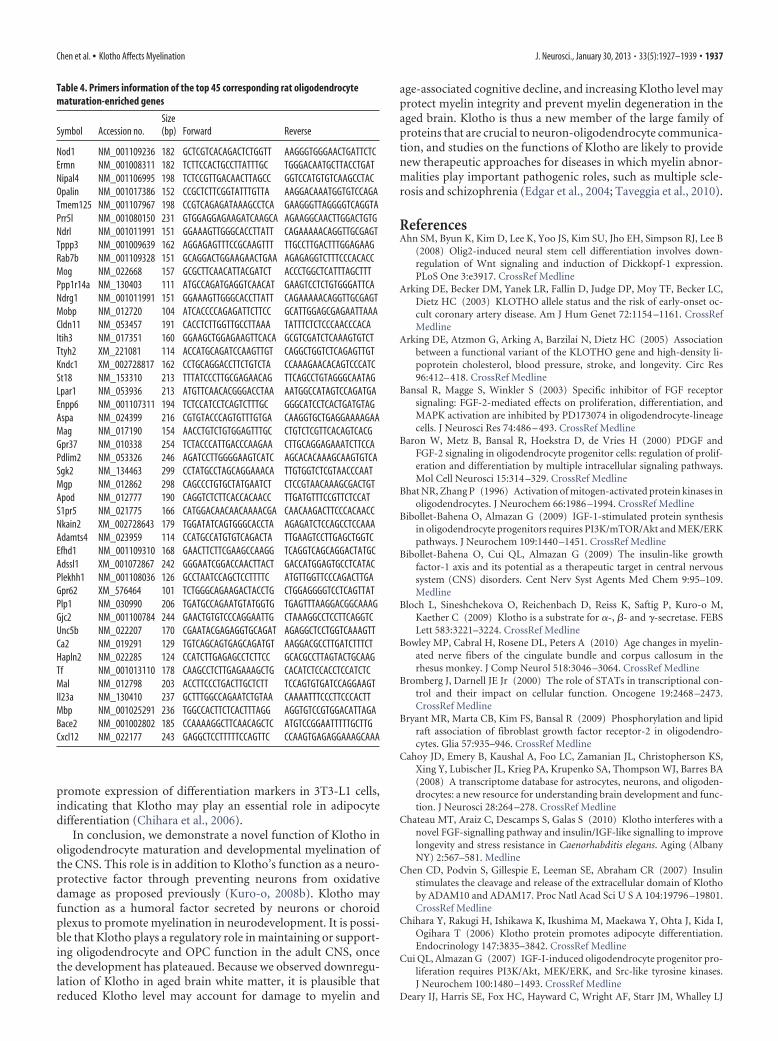
Table 4. Primers information of the top 45 corresponding rat oligodendrocytematuration-enriched genes
Symbol Accession no.Size(bp) Forward Reverse
Nod1 NM_001109236 182 GCTCGTCACAGACTCTGGTT AAGGGTGGGAACTGATTCTCErmn NM_001008311 182 TCTTCCACTGCCTTATTTGC TGGGACAATGCTTACCTGATNipal4 NM_001106995 198 TCTCCGTTGACAACTTAGCC GGTCCATGTGTCAAGCCTACOpalin NM_001017386 152 CCGCTCTTCGGTATTTGTTA AAGGACAAATGGTGTCCAGATmem125 NM_001107967 198 CCGTCAGAGATAAAGCCTCA GAAGGGTTAGGGGTCAGGTAPrr5l NM_001080150 231 GTGGAGGAGAAGATCAAGCA AGAAGGCAACTTGGACTGTGNdrl NM_001011991 151 GGAAAGTTGGGCACCTTATT CAGAAAAACAGGTTGCGAGTTppp3 NM_001009639 162 AGGAGAGTTTCCGCAAGTTT TTGCCTTGACTTTGGAGAAGRab7b NM_001109328 151 GCAGGACTGGAAGAACTGAA AGAGAGGTCTTTCCCACACCMog NM_022668 157 GCGCTTCAACATTACGATCT ACCCTGGCTCATTTAGCTTTPpp1r14a NM_130403 111 ATGCCAGATGAGGTCAACAT GAAGTCCTCTGTGGGATTCANdrg1 NM_001011991 151 GGAAAGTTGGGCACCTTATT CAGAAAAACAGGTTGCGAGTMobp NM_012720 104 ATCACCCCAGAGATTCTTCC GCATTGGAGCGAGAATTAAACldn11 NM_053457 191 CACCTCTTGGTTGCCTTAAA TATTTCTCTCCCAACCCACAItih3 NM_017351 160 GGAAGCTGGAGAAGTTCACA GCGTCGATCTCAAAGTGTCTTtyh2 XM_221081 114 ACCATGCAGATCCAAGTTGT CAGGCTGGTCTCAGAGTTGTKndc1 XM_002728817 162 CCTGCAGGACCTTCTGTCTA CCAAAGAACACAGTCCCATCSt18 NM_153310 213 TTTATCCCTTGCGAGAACAG TTCAGCCTGTAGGGCAATAGLpar1 NM_053936 213 ATGTTCAACACGGGACCTAA AATGGCCATAGTCCAGATGAEnpp6 NM_001107311 194 TCTCCATCCTCAGTCTTTGC GGGCATCCTCACTGATGTAGAspa NM_024399 216 CGTGTACCCAGTGTTTGTGA CAAGGTGCTGAGGAAAAGAAMag NM_017190 154 AACCTGTCTGTGGAGTTTGC CTGTCTCGTTCACAGTCACGGpr37 NM_010338 254 TCTACCCATTGACCCAAGAA CTTGCAGGAGAAATCTTCCAPdlim2 NM_053326 246 AGATCCTTGGGGAAGTCATC AGCACACAAAGCAAGTGTCASgk2 NM_134463 299 CCTATGCCTAGCAGGAAACA TTGTGGTCTCGTAACCCAATMgp NM_012862 298 CAGCCCTGTGCTATGAATCT CTCCGTAACAAAGCGACTGTApod NM_012777 190 CAGGTCTCTTCACCACAACC TTGATGTTTCCGTTCTCCATS1pr5 NM_021775 166 CATGGACAACAACAAAACGA CAACAAGACTTCCCACAACCNkain2 XM_002728643 179 TGGATATCAGTGGGCACCTA AGAGATCTCCAGCCTCCAAAAdamts4 NM_023959 114 CCATGCCATGTGTCAGACTA TTGAAGTCCTTGAGCTGGTCEfhd1 NM_001109310 168 GAACTTCTTCGAAGCCAAGG TCAGGTCAGCAGGACTATGCAdssl1 XM_001072867 242 GGGAATCGGACCAACTTACT GACCATGGAGTGCCTCATACPlekhh1 NM_001108036 126 GCCTAATCCAGCTCCTTTTC ATGTTGGTTCCCAGACTTGAGpr62 XM_576464 101 TCTGGGCAGAAGACTACCTG CTGGAGGGGTCCTCAGTTATPlp1 NM_030990 206 TGATGCCAGAATGTATGGTG TGAGTTTAAGGACGGCAAAGGjc2 NM_001100784 244 GAACTGTGTCCCAGGAATTG CTAAAGGCCTCCTTCAGGTCUnc5b NM_022207 170 CGAATACGAGAGGTGCAGAT AGAGGCTCCTGGTCAAAGTTCa2 NM_019291 129 TGTCAGCAGTGAGCAGATGT AAGGACGCCTTGATCTTTCTHapln2 NM_022285 124 CCATCTTGAGAGCCTCTTCC GCACGCCTTAGTACTGCAAGTf NM_001013110 178 CAAGCCTCTTGAGAAAGCTG CACATCTCCACCTCCATCTCMal NM_012798 203 ACCTTCCCTGACTTGCTCTT TCCAGTGTGATCCAGGAAGTIl23a NM_130410 237 GCTTTGGCCAGAATCTGTAA CAAAATTTCCCTTCCCACTTMbp NM_001025291 236 TGGCCACTTCTCACTTTAGG AGGTGTCCGTGGACATTAGABace2 NM_001002802 185 CCAAAAGGCTTCAACAGCTC ATGTCCGGAATTTTTGCTTGCxcl12 NM_022177 243 GAGGCTCCTTTTTCCAGTTC CCAAGTGAGAGGAAAGCAAA
Chen et al. • Klotho Affects Myelination J. Neurosci., January 30, 2013 • 33(5):1927–1939 • 1937
(2005) KLOTHO genotype and cognitive ability in childhood and oldage in the same individuals. Neurosci Lett 378:22–27. CrossRef Medline
Dell’Albani P, Kahn MA, Cole R, Condorelli DF, Giuffrida-Stella AM, deVellis J (1998) Oligodendroglial survival factors, PDGF-AA and CNTF,activate similar JAK/STAT signaling pathways. J Neurosci Res 54:191–205. CrossRef Medline
Du Y, Lercher LD, Zhou R, Dreyfus CF (2006) Mitogen-activated proteinkinase pathway mediates effects of brain-derived neurotrophic factor ondifferentiation of basal forebrain oligodendrocytes. J Neurosci Res 84:1692–1702. CrossRef Medline
Duce JA, Podvin S, Hollander W, Kipling D, Rosene DL, Abraham CR (2008)Gene profile analysis implicates Klotho as an important contributor toaging changes in brain white matter of the rhesus monkey. Glia 56:106 –117. CrossRef Medline
Dugandzija-Novakoviæ S, Koszowski AG, Levinson SR, Shrager P (1995)Clustering of Na � channels and node of Ranvier formation in remyeli-nating axons. J Neurosci 15:492–503. Medline
Edgar JM, McLaughlin M, Barrie JA, McCulloch MC, Garbern J, Griffiths IR(2004) Age-related axonal and myelin changes in the rumpshaker muta-tion of the Plp gene. Acta Neuropathol 107:331–335. CrossRef Medline
Fancy SP, Baranzini SE, Zhao C, Yuk DI, Irvine KA, Kaing S, Sanai N, FranklinRJ, Rowitch DH (2009) Dysregulation of the Wnt pathway inhibitstimely myelination and remyelination in the mammalian CNS. GenesDev 23:1571–1585. CrossRef Medline
Feigenson K, Reid M, See J, Crenshaw EB 3rd, Grinspan JB (2009) Wntsignaling is sufficient to perturb oligodendrocyte maturation. Mol CellNeurosci 42:255–265. CrossRef Medline
Fortin D, Rom E, Sun H, Yayon A, Bansal R (2005) Distinct fibroblastgrowth factor (FGF)/FGF receptor signaling pairs initiate diverse cellularresponses in the oligodendrocyte lineage. J Neurosci 25:7470 –7479.CrossRef Medline
Foster PP, Rosenblatt KP, Kuljis RO (2011) Exercise-induced cognitiveplasticity, implications for mild cognitive impairment and Alzheimer’sdisease. Front Neurol 2:28. CrossRef Medline
Frederick TJ, Min J, Altieri SC, Mitchell NE, Wood TL (2007) Synergisticinduction of cyclin D1 in oligodendrocyte progenitor cells by IGF-I andFGF-2 requires differential stimulation of multiple signaling pathways.Glia 55:1011–1022. CrossRef Medline
Frost EE, Zhou Z, Krasnesky K, Armstrong RC (2009) Initiation of oligo-dendrocyte progenitor cell migration by a PDGF-A activated extracellularregulated kinase (ERK) signaling pathway. Neurochem Res 34:169 –181.CrossRef Medline
Furusho M, Dupree JL, Nave KA, Bansal R (2012) Fibroblast growth factorreceptor signaling in oligodendrocytes regulates myelin sheath thickness.J Neurosci 32:6631– 6641. CrossRef Medline
Fyffe-Maricich SL, Karlo JC, Landreth GE, Miller RH (2011) The ERK2mitogen-activated protein kinase regulates the timing of oligodendrocytedifferentiation. J Neurosci 31:843– 850. CrossRef Medline
Guardiola-Diaz HM, Ishii A, Bansal R (2012) Erk1/2 MAPK and mTORsignaling sequentially regulates progression through distinct stages of oli-godendrocyte differentiation. Glia 60:476 – 486. CrossRef Medline
Hanafy KA, Sloane JA (2011) Regulation of remyelination in multiple scle-rosis. FEBS Lett 585:3821–3828. CrossRef Medline
He Y, Dupree J, Wang J, Sandoval J, Li J, Liu H, Shi Y, Nave KA, Casaccia-Bonnefil P (2007) The transcription factor Yin Yang 1 is essential foroligodendrocyte progenitor differentiation. Neuron 55:217–230.CrossRef Medline
Hinman JD, Abraham CR (2007) What’s behind the decline? The role ofwhite matter in brain aging. Neurochem Res 32:2023–2031. CrossRefMedline
Hsieh CC, Kuro-o M, Rosenblatt KP, Brobey R, Papaconstantinou J (2010)The ASK1-Signalosome regulates p38 MAPK activity in response to levelsof endogenous oxidative stress in the Klotho mouse models of aging.Aging (Albany NY) 2:597– 611. Medline
Imura A, Iwano A, Tohyama O, Tsuji Y, Nozaki K, Hashimoto N, Fujimori T,Nabeshima Y (2004) Secreted Klotho protein in sera and CSF: implica-tion for post-translational cleavage in release of Klotho protein from cellmembrane. FEBS Lett 565:143–147. CrossRef Medline
Ishii A, Fyffe-Maricich SL, Furusho M, Miller RH, Bansal R (2012) ERK1/ERK2 MAPK signaling is required to increase myelin thickness indepen-dent of oligodendrocyte differentiation and initiation of myelination.J Neurosci 32:8855– 8864. CrossRef Medline
Kawaguchi H, Manabe N, Miyaura C, Chikuda H, Nakamura K, Kuro-o M(1999) Independent impairment of osteoblast and osteoclast differenti-ation in klotho mouse exhibiting low-turnover osteopenia. J Clin Invest104:229 –237. CrossRef Medline
King GD, Rosene DL, Abraham CR (2012) Promoter methylation and age-related downregulation of Klotho in rhesus monkey. Age (Dordr) 34:1405–1419. CrossRef Medline
Kioussi C, Gross MK, Gruss P (1995) Pax3: a paired domain gene as a reg-ulator in PNS myelination. Neuron 15:553–562. CrossRef Medline
Kohama SG, Rosene DL, Sherman LS (2012) Age-related changes in humanand non-human primate white matter: from myelination disturbances tocognitive decline. Age (Dordr) 34:1093–1110. CrossRef Medline
Kuro-o M (2006) Klotho as a regulator of fibroblast growth factor signalingand phosphate/calcium metabolism. Curr Opin Nephrol Hypertens 15:437– 441. CrossRef Medline
Kuro-o M (2008a) Endocrine FGFs and Klothos: emerging concepts.Trends Endocrinol Metab 19:239 –245. CrossRef Medline
Kuro-o M (2008b) Klotho as a regulator of oxidative stress and senescence.Biol Chem 389:233–241. CrossRef Medline
Kuro-o M (2010) Klotho. Pflugers Arch 459:333–343. CrossRef MedlineKuro-o M, Matsumura Y, Aizawa H, Kawaguchi H, Suga T, Utsugi T,
Ohyama Y, Kurabayashi M, Kaname T, Kume E, Iwasaki H, Iida A,Shiraki-Iida T, Nishikawa S, Nagai R, Nabeshima YI (1997) Mutation ofthe mouse klotho gene leads to a syndrome resembling ageing. Nature390:45–51. CrossRef Medline
Kurosu H, Yamamoto M, Clark JD, Pastor JV, Nandi A, Gurnani P, McGuin-ness OP, Chikuda H, Yamaguchi M, Kawaguchi H, Shimomura I, Ta-kayama Y, Herz J, Kahn CR, Rosenblatt KP, Kuro-o M (2005)Suppression of aging in mice by the hormone Klotho. Science 309:1829 –1833. CrossRef Medline
Kurosu H, Ogawa Y, Miyoshi M, Yamamoto M, Nandi A, Rosenblatt KP,Baum MG, Schiavi S, Hu MC, Moe OW, Kuro-o M (2006) Regulation offibroblast growth factor-23 signaling by klotho. J Biol Chem 281:6120 –6123. CrossRef Medline
Li SA, Watanabe M, Yamada H, Nagai A, Kinuta M, Takei K (2004) Immu-nohistochemical localization of Klotho protein in brain, kidney, and re-productive organs of mice. Cell Struct Funct 29:91–99. CrossRef Medline
Liu F, Wu S, Ren H, Gu J (2011) Klotho suppresses RIG-I-mediatedsenescence-associated inflammation. Nat Cell Biol 13:254 –262. CrossRefMedline
Liu H, Fergusson MM, Castilho RM, Liu J, Cao L, Chen J, Malide D, Rovira II,Schimel D, Kuo CJ, Gutkind JS, Hwang PM, Finkel T (2007) Aug-mented Wnt signaling in a mammalian model of accelerated aging. Sci-ence 317:803– 806. CrossRef Medline
Maekawa Y, Ohishi M, Ikushima M, Yamamoto K, Yasuda O, Oguro R,Yamamoto-Hanasaki H, Tatara Y, Takeya Y, Rakugi H (2011) Klothoprotein diminishes endothelial apoptosis and senescence via a mitogen-activated kinase pathway. Geriatr Gerontol Int 11:510 –516. CrossRefMedline
Makris N, Papadimitriou GM, van der Kouwe A, Kennedy DN, Hodge SM,Dale AM, Benner T, Wald LL, Wu O, Tuch DS, Caviness VS, Moore TL,Killiany RJ, Moss MB, Rosene DL (2007) Frontal connections and cog-nitive changes in normal aging rhesus monkeys: a DTI study. NeurobiolAging 28:1556 –1567. CrossRef Medline
Mason JL, Toews A, Hostettler JD, Morell P, Suzuki K, Goldman JE, Matsu-shima GK (2004) Oligodendrocytes and progenitors become progres-sively depleted within chronically demyelinated lesions. J Pathol 164:1673–1682. Medline
Massa PT, Saha S, Wu C, Jarosinski KW (2000) Expression and function ofthe protein tyrosine phosphatase SHP-1 in oligodendrocytes. Glia 29:376 –385. CrossRef Medline
Meakin SO, MacDonald JI, Gryz EA, Kubu CJ, Verdi JM (1999) The signal-ing adapter FRS-2 competes with Shc for binding to the nerve growthfactor receptor TrkA: a model for discriminating proliferation and differ-entiation. J Biol Chem 274:9861–9870. CrossRef Medline
Medici D, Razzaque MS, Deluca S, Rector TL, Hou B, Kang K, Goetz R,Mohammadi M, Kuro-o M, Olsen BR, Lanske B (2008) FGF-23-Klothosignaling stimulates proliferation and prevents vitamin D-induced apo-ptosis. J Cell Biol 182:459 – 465. CrossRef Medline
Mi S, Miller RH, Lee X, Scott ML, Shulag-Morskaya S, Shao Z, Chang J, ThillG, Levesque M, Zhang M, Hession C, Sah D, Trapp B, He Z, Jung V,
1938 • J. Neurosci., January 30, 2013 • 33(5):1927–1939 Chen et al. • Klotho Affects Myelination

McCoy JM, Pepinsky RB (2005) LINGO-1 negatively regulates myelina-tion by oligodendrocytes. Nat Neurosci 8:745–751. CrossRef Medline
Nagai R, Saito Y, Ohyama Y, Aizawa H, Suga T, Nakamura T, Kurabayashi M,Kuroo M (2000) Endothelial dysfunction in the klotho mouse anddownregulation of klotho gene expression in various animal models ofvascular and metabolic diseases. Cell Mol Life Sci 57:738 –746. CrossRefMedline
Nagai T, Yamada K, Kim HC, Kim YS, Noda Y, Imura A, Nabeshima Y,Nabeshima T (2003) Cognition impairment in the genetic model of ag-ing klotho gene mutant mice: a role of oxidative stress. FASEB J 17:50 –52.CrossRef Medline
Ogawa Y, Schafer DP, Horresh I, Bar V, Hales K, Yang Y, Susuki K, Peles E,Stankewich MC, Rasband MN (2006) Spectrins and ankyrinB constitutea specialized paranodal cytoskeleton. J Neurosci 26:5230 –5239. CrossRefMedline
Oh SY, Chen CD, Abraham CR (2010) Cell-type dependent modulation ofNotch signaling by the amyloid precursor protein. J Neurochem 113:262–274. CrossRef Medline
Ong SH, Guy GR, Hadari YR, Laks S, Gotoh N, Schlessinger J, Lax I (2000)FRS2 proteins recruit intracellular signaling pathways by binding to di-verse targets on fibroblast growth factor and nerve growth factor recep-tors. Mol Cell Biol 20:979 –989. CrossRef Medline
Peters A (2009) The effects of normal aging on myelinated nerve fibers inmonkey central nervous system. Front Neuroanat 3:11. CrossRef Medline
Rakugi H, Matsukawa N, Ishikawa K, Yang J, Imai M, Ikushima M, MaekawaY, Kida I, Miyazaki J, Ogihara T (2007) Anti-oxidative effect of Klothoon endothelial cells through cAMP activation. Endocrine 31:82– 87.CrossRef Medline
Rasband MN, Kagawa T, Park EW, Ikenaka K, Trimmer JS (2003) Dysregu-lation of axonal sodium channel isoforms after adult-onset chronic de-myelination. J Neurosci Res 73:465– 470. CrossRef Medline
Repa JJ, Mangelsdorf DJ (2000) The role of orphan nuclear receptors in theregulation of cholesterol homeostasis. Annu Rev Cell Dev Biol 16:459 –481. CrossRef Medline
Rosene DL, Roy NJ, Davis BJ (1986) A cryoprotection method that facili-tates cutting frozen sections of whole monkey brains for histological andhistochemical processing without freezing artifact. J Histochem Cy-tochem 34:1301–1315. CrossRef Medline
Shih PH, Yen GC (2007) Differential expressions of antioxidant status inaging rats: the role of transcriptional factor Nrf2 and MAPK signalingpathway. Biogerontology 8:71– 80. CrossRef Medline
Shimada T, Takeshita Y, Murohara T, Sasaki K, Egami K, Shintani S, KatsudaY, Ikeda H, Nabeshima Y, Imaizumi T (2004) Angiogenesis and vascu-logenesis are impaired in the precocious-aging klotho mouse. Circulation110:1148 –1155. CrossRef Medline
Shiozaki M, Yoshimura K, Shibata M, Koike M, Matsuura N, Uchiyama Y,Gotow T (2008) Morphological and biochemical signs of age-relatedneurodegenerative changes in klotho mutant mice. Neuroscience 152:924 –941. CrossRef Medline
Sloane JA, Vartanian TK (2007) Myosin Va controls oligodendrocyte mor-phogenesis and myelination. J Neurosci 27:11366 –11375. CrossRefMedline
Sloane JA, Hinman JD, Lubonia M, Hollander W, Abraham CR (2003) Age-dependent myelin degeneration and proteolysis of oligodendrocyte pro-teins is associated with the activation of calpain-1 in the rhesus monkey.J Neurochem 84:157–168. CrossRef Medline
Subramanian A, Tamayo P, Mootha VK, Mukherjee S, Ebert BL, Gillette MA,Paulovich A, Pomeroy SL, Golub TT, Lander ES, Mesirov JP (2005)Gene set enrichment analysis: a knowledge-based approach for interpret-
ing genome-wide expression profiles. Proc Natl Acad Sci U S A 102:15545–15550. CrossRef Medline
Taveggia C, Feltri ML, Wrabetz L (2010) Signals to promote myelin forma-tion and repair. Nat Rev Neurol 6:276 –287. CrossRef Medline
Thurston RD, Larmonier CB, Majewski PM, Ramalingam R, Midura-KielaM, Laubitz D, Vandewalle A, Besselsen DG, Muhlbauer M, Jobin C, KielaPR, Ghishan FK (2010) Tumor necrosis factor and interferon-gammadown-regulate Klotho in mice with colitis. Gastroenterology 138:1384 –1394; 1394.e1–1394.e2. CrossRef Medline
Trapp BD, Kidd GJ (2004) Structure of the myelinated axon. In: Myelinbiology and disorders (Lazzarini RA, ed), pp 3–22. London: ElsevierAcademic.
Urakawa I, Yamazaki Y, Shimada T, Iijima K, Hasegawa H, Okawa K, Fujita T,Fukumoto S, Yamashita T (2006) Klotho converts canonical FGF recep-tor into a specific receptor for FGF23. Nature 444:770 –774. CrossRefMedline
Vabnick I, Novakoviæ SD, Levinson SR, Schachner M, Shrager P (1996) Theclustering of axonal sodium channels during development of the periph-eral nervous system. J Neurosci 16:4914 – 4922. Medline
Van’t Veer A, Du Y, Fischer TZ, Boetig DR, Wood MR, Dreyfus CF (2009)Brain-derived neurotrophic factor effects on oligodendrocyte progenitorsof the basal forebrain are mediated through trkB and the MAP kinasepathway. J Neurosci Res 87:69 –78. CrossRef Medline
Wang L, Schuster GU, Hultenby K, Zhang Q, Andersson S, Gustafsson JA(2002) Liver � receptors in the central nervous system: from lipid ho-meostasis to neuronal degeneration. Proc Natl Acad Sci U S A 99:13878 –13883. CrossRef Medline
Waxman SG (1998) Demyelinating diseases: new pathological insights, newtherapeutic targets. N Engl J Med 338:323–325. CrossRef Medline
Wegner M (2000) Transcriptional control in myelinating glia: the basic rec-ipe. Glia 29:118 –123. CrossRef Medline
Whitney KD, Watson MA, Collins JL, Benson WG, Stone TM, Numerick MJ,Tippin TK, Wilson JG, Winegar DA, Kliewer SA (2002) Regulation ofcholesterol homeostasis by the liver � receptors in the central nervoussystem. Mol Endocrinol 16:1378 –1385. CrossRef Medline
Wisco JJ, Killiany RJ, Guttmann CR, Warfield SK, Moss MB, Rosene DL(2008) An MRI study of age-related white and gray matter volumechanges in the rhesus monkey. Neurobiol Aging 29:1563–1575. CrossRefMedline
Wolf I, Levanon-Cohen S, Bose S, Ligumsky H, Sredni B, Kanety H, Kuro-oM, Karlan B, Kaufman B, Koeffler HP, Rubinek T (2008) Klotho: a tu-mor suppressor and a modulator of the IGF-1 and FGF pathways inhuman breast cancer. Oncogene 27:7094 –7105. CrossRef Medline
Yamamoto M, Clark JD, Pastor JV, Gurnani P, Nandi A, Kurosu H, MiyoshiM, Ogawa Y, Castrillon DH, Rosenblatt KP, Kuro-o M (2005) Regula-tion of oxidative stress by the anti-aging hormone klotho. J Biol Chem280:38029 –38034. CrossRef Medline
Younes-Rapozo V, Felgueiras LO, Viana NL, Fierro IM, Barja-Fidalgo C,Manhaes AC, Barradas PC (2009) A role for the MAPK/ERK pathway inoligodendroglial differentiation in vitro: stage specific effects on cellbranching. Int J Dev Neurosci 27:757–768. CrossRef Medline
Zeldich E, Koren R, Nemcovsky C, Weinreb M (2007) Enamel matrix de-rivative stimulates human gingival fibroblast proliferation via ERK. JDent Res 86:41– 46. CrossRef Medline
Zhao Y, Banerjee S, Dey N, LeJeune WS, Sarkar PS, Brobey R, Rosenblatt KP,Tilton RG, Choudhary S (2011) Klotho depletion contributes to in-creased inflammation in kidney of the db/db mouse model of diabetes viaRelA (serine)536 phosphorylation. Diabetes 60:1907–1916. CrossRefMedline
Chen et al. • Klotho Affects Myelination J. Neurosci., January 30, 2013 • 33(5):1927–1939 • 1939