Embed Size (px)
Citation preview
Cellular/Molecular
Endophilin Promotes a Late Step in Endocytosis at GlialInvaginations in Drosophila Photoreceptor Terminals
Ruth Fabian-Fine,1,2* Patrik Verstreken,3* P. Robin Hiesinger,4 Jane Anne Horne,2 Rita Kostyleva,2 Yi Zhou,5
Hugo J. Bellen,3,4,5,6 and Ian A. Meinertzhagen1,2
1Neuroscience Institute, 2Department of Psychology, Life Sciences Centre, Dalhousie University, Halifax, Nova Scotia, Canada B3H 4J1, and 3Program inDevelopmental Biology, 4Howard Hughes Medical Institute, 5Department of Molecular and Human Genetics, and 6Division of Neuroscience, Baylor Collegeof Medicine, Houston, Texas 77030
Retrieval of synaptic vesicles from the membrane of neurons is crucial to maintain normal rates of neurotransmitter release. Photore-ceptor terminals of the fly’s eye release neurotransmitter in a tonic manner. They therefore rely heavily on vesicle regeneration. Nullmutations in endophilin (endo) block clathrin-mediated endocytosis at the Drosophila neuromuscular junction, where previous analysisof hypomorphic mutations has suggested a function for Endophilin (Endo) before vesicle fission, during membrane bending. Here, at flyphotoreceptor synapses, we show that Endo is localized to synaptic vesicles at sites of endocytosis that are glial invaginations calledcapitate projections, and that when the photoreceptor synapses lack Endo they are impaired in their ability to release neurotransmitter.Detailed ultrastructural analysis of endo null mutant photoreceptor synapses fails to reveal a defect at early stages of vesicle reformationbut, instead, reveals an accumulation of clusters of electron-dense, apparently nonfunctional, late endocytotic vesicles. Using dynamin;endo double-mutant photoreceptors, we provide further evidence that ultimately the function of Endophilin is required late in endocy-tosis, allowing vesicles to progress through the synaptic vesicle cycle.
Key words: clathrin; endophilin; endocytosis; capitate projection; glia; vesicle cycle
IntroductionIn neurons, evidence exists for at least two pathways of synapticvesicle regeneration (Slepnev and De Camilli, 2000; Jarousse andKelly, 2001; Kjaerulff et al., 2002; Aravanis et al., 2003; Gandhiand Stevens, 2003). In so-called “kiss-and-run,” synaptic vesiclesexpel their contents but do not collapse into the presynapticmembrane (Ceccarelli et al., 1973; Fesce and Meldolesi, 1999;Verstreken et al., 2002). In clathrin-mediated endocytosis, syn-aptic vesicles collapse completely in the presynaptic membraneand reform during events that dimple and shape the membrane.Kinetic studies of both pathways in different neuron types suggestthat kiss-and-run prevails under low-frequency stimulation, aug-mented by clathrin-mediated endocytosis during high-frequencystimulation (Ceccarelli et al., 1973; Heuser and Reese, 1973; Pal-frey and Artalejo, 1998; Sun et al., 2002; Verstreken et al., 2002).Neurons are also reported to modify their release characteristicsand mode of vesicle regeneration under altered stimulation con-ditions (Zakharenko et al., 2002).
The molecular requirements for each of these pathways in-clude the action of the GTPase dynamin, which is required toseparate synaptic vesicles attached to the presynaptic membraneand is implicated in both kiss-and-run and clathrin-mediatedendocytosis (Artalejo et al., 1995; Verstreken et al., 2002). Incontrast, numerous proteins have been proposed to function ex-clusively in the clathrin-mediated pathway (Zhang and Ra-maswami, 1999). Despite evidence for the cooperative action ofthese proteins, further dissection of their functions in vivo is re-quired to gain a better understanding of how synaptic vesiclesform and eventually reenter the functional vesicle pool within anintact nerve terminal.
The formation of new vesicles in clathrin-mediated endocy-tosis comprises various stages. During the “early stages,” the pla-nar membrane bends to form a shallow, hemispherical, clathrin-coated pit. Subsequently, in “mid-endocytotic stages,” a deeplyinvaginating coated pit forms, constricted at its neck. Finally dur-ing “late stages” of endocytosis, the newly formed vesicle sepa-rates from the membrane and is uncoated before reentering thefunctional vesicle pool. As used in this study, endocytosis is takento include all of these events, and in vivo and in vitro studiesimplicate requirements for specific proteins at each of thesestages (Gonzalez-Gaitan and Jackle, 1997; Cremona et al., 1999;Simpson et al., 1999; Hill et al., 2001; Guichet et al., 2002).
One protein required for clathrin-mediated endocytosis is En-dophilin A (Endo), an SH3 domain protein that harbors lyso-phosphatidic acid acyl transferase (LPAAT) activity (Schmidt etal., 1999; Guichet et al., 2002). By virtue of its enzymatic activity,
Received July 18, 2003; revised Sept. 26, 2003; accepted Oct. 3, 2003.R.F-F. was supported by a postdoctoral fellowship from the Killam Trust of Dalhousie University, P.V. is supported
by the Belgian American Educational Foundation, H.J.B. is a Howard Hughes Medical Institute (HHMI) investigator,and P.R.H. was supported by the HHMI and a European Molecular Biology Organization fellowship. I.A.M. wassupported by Natural Sciences and Engineering Research Council Genomics Grant GENPJ 239772 and NationalInstitutes of Health Grant EY-03592. We thank the Bloomington Stock Center for flies and Elaine Seto and Tong-WeyKoh for comments. We also thank members of the Meinertzhagen and Bellen labs for valuable discussions.
*R.F.-F. and P.V. contributed equally to this work.Correspondence should be addressed to I. A. Meinertzhagen, Neuroscience Institute, Life Sciences Centre, Dal-
housie University, Halifax, Nova Scotia, Canada B3H 4J1. E-mail: [email protected] © 2003 Society for Neuroscience 0270-6474/03/2310732-13$15.00/0
10732 • The Journal of Neuroscience, November 19, 2003 • 23(33):10732–10744

Endo has been proposed to act during early- and mid-endocytotic stages of endocytosis. By converting lysophospha-tidic acid into the more bulky phosphatidic acid, LPAAT has beenproposed to induce inward negative membrane curvature toform a new vesicle (Scales and Scheller, 1999; Schmidt et al.,1999). In vivo analysis of endophilin (endo) hypomorphic muta-tions in Drosophila supports this idea, because mid-stage endo-cytotic shallow pits accumulate at the membrane of neuromus-cular junctions (NMJs) (Guichet et al., 2002). Similarly, injectingEndo-SH3 antibodies into lamprey synapses leads to an accumu-lation of early and mid-stage endocytotic structures, as well as ofsome deeply invaginating coated pits (Ringstad et al., 1999).Hence, Endo may aid in the transit from early to mid stages ofsynaptic vesicle endocytosis.
Here we present evidence that, in Drosophila photoreceptors,Endo plays no obvious role during membrane bending duringearly endocytosis, but rather that loss of Endo function leads to anaccumulation of late endocytotic vesicles. In addition, our anal-ysis reveals that clathrin-mediated endocytosis in Drosophilaphotoreceptor terminals occurs at specific sites of glial invagina-tion, which possibly couple endocytosis with neurotransmitterreuptake.
Materials and MethodsDrosophila strains and genetics. For this study, we used the w; endo1 nullallele, the w; endo2 hypomorphic allele, and the temperature-sensitiveshits1 allele (Verstreken et al., 2002). yw; FRT82B endo1 was reportedpreviously (Verstreken et al., 2002). yw; eyFLP2 glas-lacZ; FRT82Bw�cl3R/TM3Sb was crossed to yw; FRT82B endo1 to create flies with eyeshomozygous for endo1 (Newsome et al., 2000). Expression of wild-typeEndo in homozygous mutant endo1 eyes was achieved by crossing yw;eyGAL4 UAS-FLP; FRT82 GMR-hid 3R CL3R to yw; FRT82B endo1
(Stower and Schwarz, 1999). The endo1 P-element insertion is an EPP-element with upstream activating sequence (UAS) sites in the correctorientation for expression of wild-type endo (Verstreken et al., 2002). ywshits1/Y; eyFLP5; FRT82B endo1/FRT82B w�cl3R male flies with double-mutant eyes were generated by crossing yw shits1; FRT82B w�cl3R/TM3Sbvirgins to yw/Y; eyFLP5; FRT82B endo1/TM3Sb males. These two stocks weregenerated by standard genetics. The control used for experiments performedwith homozygous endo1 eyes was yw eyFLP2 glas-lacZ; FRT82B w�cl3R/FRT82B. For experiments on flies with double-mutant eyes, similar flies atthe permissive temperature were used as controls as well as shits1 flies treatedidentically to flies with double-mutant eyes (see below).
Immunocytochemistry and adult retina preparation. Immunocyto-chemistry on adult whole-mounted brains was performed as described(Hiesinger et al., 2001). Anti-Endo (GP69) (Verstreken et al., 2002) wasused at 1:500, anti-cysteine string protein (CSP) (Zinsmaier et al., 1994)at 1:200, anti-synaptobrevin (Syb) (Wu et al., 1999) at 1:200, anti-Rasopposite (ROP) (Harrison et al., 1994) at 1:500, and 24B10 (Zipursky etal., 1984) at 1:50. Fluorescent secondary antibodies were from JacksonImmunoResearch (West Grove, PA) and were used at 1:250. Images werecaptured with a Zeiss 510 confocal microscope and processed with Amira2.2 or 2.3 (TGS) and Photoshop 7 (Adobe).
Electrophysiology and phototaxis assay. Electroretinograms (ERGs)from flies immobilized with nail polish (Top Speed, Revlon) were re-corded with a fine glass pipette filled with 3 M NaCl placed on the cornealsurface of the fly’s eye. The reference electrode was inserted into thethorax. Light flashes of 1 sec were delivered from a 150 W halogen lamp(Volpi), and field potential recordings were digitized and stored on a PCusing Clampex software. Ten flies with mutant eyes and 10 with controleyes were used to record 30 ERG traces, and the amplitudes of the “on”and “off” transients as well as the sustained negative component wereanalyzed using Clampfit.
A countercurrent phototaxis apparatus (Benzer, 1973) equipped withseven vials was used to determine whether flies exhibited phototaxis. Thelight source was a 60 W fluorescent light bulb (GE Lighting). Flies, 10groups of 20 with mutant eyes and 10 groups of 20 with control eyes (hdc
as a negative control and yw eyFLP; FRT82B w� cl3R/FRT82B as a posi-tive control) were loaded in the apparatus and allowed to walk sevenconsecutive times toward light during 10 sec.
We should point out that in this work we used the eyFLP technique tocreate homozygous endo1 eyes (Newsome et al., 2000), whereas Rikhy etal. (2002) used a modified version of the EGUFhid technique (Stower andSchwarz, 1999). The technique used by Rikhy et al. (2002) eliminatesnonhomozygous mutant cells after differentiation of the cell types in theeye disc (Stower and Schwarz, 1999) and leads to homozygous eyes withdisrupted morphology. This roughening may sensitize the genetic back-ground leading to reduced phototaxis reported in Rikhy et al. (2002),whereas we report here that flies with endo1 mutant eyes, created with theeyFLP technique, display normal phototaxis.
Fly head fractionation and Western blot analysis. Canton-S fly headhomogenates were prepared as described previously (Schulze et al.,1995). Homogenates were fractionated by 0.2 M/0.4 M sucrose step cen-trifugation (Schulze et al., 1995). In Figure 2, “input” was fly head ho-mogenate, “cytosol” was the supernatant material that failed to enter the0.2 M sucrose layer, and “vesicles” was the material at the interface be-tween the 0.2 and 0.4 M layers (Schulze et al., 1995). Proteins in thefractions were separated by SDS-PAGE and transferred to nitrocellulose.Fifteen micrograms of total protein were loaded per lane. To detect an-tigens on the blots, primary antibodies were used as follows: anti-Endo1:5000, anti-Drab3 1:1000 (Schulze et al., 1995), and anti-Syb (R29)1:5000. Secondary HRP-conjugated antibodies (Jackson ImmunoRe-search) were used at 1:5000; bands were visualized by enhanced chemi-luminescence (NEN).
Conventional electron microscopy. Twenty-four-hours-old flies withendo1 mutant eyes and control flies were fixed under constant Ganzfeldillumination comprising an array of light-emitting diodes within a spher-ical chamber (Kral and Meinertzhagen, 1989). Fixation was for 2 hr at23°C in 2.5% of glutaraldehyde and paraformaldehyde in 0.1 M cacody-late buffer, pH 7.3, containing 7 mM CaCl2, followed by postfixation in2% OsO4 (1 hr at 4°C), all as described previously (Meinertzhagen,1996). Preparations were examined at 60 or 80 kV using Philips 201C orTecnai 12 electron microscopes.
Postembedding immunogold labeling for electron microscopy. Canton-Sflies were dissected in 0.1 M phosphate buffer (PB), pH 7.4, with 4%paraformaldehyde, 0.05% glutaraldehyde, and 0.2% picric acid (Somo-gyi and Takagi, 1982), soaked in PB for 15 min, and then immersed in0.125 M triethanolamine hydrochloride in PB for 30 min to quench un-reacted aldehydes. Heads were cryoprotected for �2 hr in increasingconcentrations of glycerol in PB up to 30% and then impact frozen on apolished copper mirror at �193°C (Leica MM80) and, using a Leicaautomatic freeze-substitution system (AFS), freeze-substituted in meth-anol with 0.5% uranyl acetate for 24 hr at �85°C. Specimens were sub-sequently rinsed in methanol, and the temperature was then raised to�50°C before infiltration and embedment in Lowicryl HM 20 (AgarScientific) as described previously (Fabian-Fine et al., 2001). The resinwas polymerized at �50°C using UV light, and ultrathin sections (50 nm)were collected on Pioloform-coated single-slot nickel grids. For uniformtreatment, all sections were mounted on grids using a support plate(Leica Microsystems). Sections were soaked in PB for 30 min and incu-bated in blocking medium (BM) (1% BSA, 10% fetal calf serum in PB)for 30 min at 23°C. Anti-Endo (GP69) was used at 1:100 overnight at 4°C,1 hr at 37°C, and overnight again at 4°C. Polyclonal anti-clathrin (Sigma,St. Louis, MO) was used at 1:50 overnight at 4°C. Sections were thenrinsed thoroughly in PB and incubated in BM at 37°C for 30 min. Sec-ondary antibodies conjugated to 10 nm gold particles (Jackson Immu-noResearch) applied for 4 hr at 37°C were used at 1:100 for Endophilinlabeling and at 1:50 for clathrin labeling. Preparations were washed inBM, in PB, and finally in double-distilled water. Sections were contrastedwith uranyl acetate (4 min) and Reynold’s lead citrate (2 min) as de-scribed previously (Meinertzhagen, 1996).
Specificity of labeling for Endo was confirmed by the absence of im-munolabeling in preparations from which primary anti-Endo was omit-ted. The specificity of anti-Endo (GP69) was demonstrated previously(Verstreken et al., 2002). The specificity of anti-clathrin (Sigma) wastested by Western blot analysis and labeling of tissue. At the NMJ, label-
Fabian-Fine et al. • Endophilin Promotes Endocytosis at Glial Invaginations J. Neurosci., November 19, 2003 • 23(33):10732–10744 • 10733

ing with this anti-clathrin antibody reveals pre-synaptic enrichment (Zhang et al., 1998). Todemonstrate the specificity of this antibodymore directly, anti-clathrin antibodies werefirst preadsorbed overnight at 4°C against a 178kDa protein corresponding to clathrin heavychain, from fly head extract. Subsequently con-trol preparations were incubated with this pre-absorbed antibody. Specific labeling was absentin these preparations.
Quantitative analysis of electron microscopicimages. We made single-blind counts of synap-tic organelles in single-section profiles of pho-toreceptor terminals in all genotypes examined(Meinertzhagen and O’Neil, 1991). We nextcounted the following organelle profiles: 30 nmsynaptic vesicles, T-shaped presynaptic ribbonsat tetrad synapses, and capitate projections. Us-ing previous criteria (Pyza and Meinertzhagen,1997; Rybak and Meinertzhagen, 1997), the lat-ter were counted as shallow (with a head invag-inating not more than half its diameter), single(penetrating, with a single head profile; see Fig.7E), and multiple (penetrating, with more thanone head; see Fig. 7F ). The perimeters andcross-sectional areas of terminal profiles weremeasured with software (NIH Image). Tests forstatistical significance in organelle counts werefirst made by an unweighted means ANOVA,followed by a Tukey honestly significant differ-ence (HSD) test, using software (Systat 5.2.1).
We evaluated the distribution of 207 (anti-clathrin) and 89 (anti-Endophilin) immuno-gold particles from 10 and 16 micrographs, re-spectively, containing both particle clusters andwide areas with only occasional particles. Wecounted particles in two categories, either asso-ciated with membrane, for which we desig-nated a border of 60 nm, two synaptic vesiclediameters (Meinertzhagen and O’Neil, 1991),or in the cytoplasm. Membrane-associated par-ticles were either close to capitate projections orto the plasmalemma. Those in the cytoplasmoccurred at lower densities, which we calcu-lated from measurements of the membrane pe-rimeter and cross-sectional area of the photo-receptor terminals. The correct attribution ofparticles to capitate projection or plasma-lemma was made difficult by the complex ge-ometry of the former, because in any one sec-tion capitate projection heads are rarelyconnected to the plasmalemma by a stalk. Thussome plasmalemma-associated particles prob-ably belong to capitate projection stalks lyingjust in front of or behind the section plane. Totest the significance of differences in distribu-tion, we used an unweighted means ANOVA, followed by a Tukey HSDtest, as above. Comparisons were made on the subcounts of particles ineach category made on individual micrographs.
Synaptic vesicle depletion-recovery assay. The functional vesicle pool inendo1 mutant eyes was depleted by exposing flies with double-mutanteyes (shits1; endo1) to the restrictive temperature (29°C) for shits1 to blocksynaptic vesicle recovery. Returning flies to the permissive temperature(18°C) restores the function of dynamin but not Endo. We allowed fliesto recover for various times at the permissive temperature and thenprocessed their eyes for electron microscopy (EM) (see Fig. 8 A).
We performed the following set of experiments on flies with shits1;endo1 eyes as well as on shits1 flies (as a control). Flies were exposed for 15min to 29°C in normal room light and then (1) fixed at 29°C before
processing for EM, (2) shifted to 18°C for an additional 15 min andfinally fixed at 18°C for EM, and (3) shifted to 18°C for 30 min and finallyfixed at 18°C for EM. As another control, (4) shits1;endo1 flies were held at18°C and fixed at 18°C for EM.
ResultsEndophilin is present in photoreceptor terminalsTo study the role of Endo in endocytosis we have used the pho-toreceptors of the fly’s eye. The Drosophila eye consists of �800ommatidia, each containing eight photoreceptors, R1–R8. Six ofthese, R1–R6, emit axons that terminate and synapse in the firstoptic neuropile, the lamina (Fig. 1A). The axons of R7 and R8synapse in the second optic neuropile, the medulla. In the lamina,
Figure 1. Localization of Endo in photoreceptor terminals. A, Schematic representation of the optic lobe neuropiles in Drosoph-ila. R, Photoreceptor cell; La, lamina; Me, medulla; Lo, lobula; Lp, lobular plate. An array of photoreceptors (gray) project axons thatterminate in the lamina, R1–R6, or medulla, R7 and 8. B, Three-dimensional volume rendering of half a lamina labeled withanti-Endophilin (green) and anti-Synaptobrevin (red). Cartridges comprising R1–R6 terminals that innervate lamina monopolarcells are immunoreactive for both Endo and Syb (overlap: orange/yellow). Note, however, that Endo is also present in unidentifiedcells of the lamina cortex surrounding the cartridges. C, Schematic of a single cartridge, box in B, containing six photoreceptorterminals (R1–R6; orange), surrounding lamina monopolar cells L1 and L2 (blue). In endo1 mutant eyes created by the ey-FLPtechnique, almost all R1–R6 are mutant. D–F, Colocalization of synaptic markers (magenta) with Endo in lamina cartridges, shownas single confocal images from deconvolved three-dimensional datasets. D, CSP and Endo extensively colocalize; however, Endo-positive CSP negative puncta exist (arrowhead). E, Double-labeling for ROP and Endo; F, double-labeling for Syb and Endo. Endoand Syb overlap, although not entirely (F, arrowhead).
10734 • J. Neurosci., November 19, 2003 • 23(33):10732–10744 Fabian-Fine et al. • Endophilin Promotes Endocytosis at Glial Invaginations

the terminals of photoreceptors R1–R6 sort precisely into laminacartridges (Fig. 1B) so that each cartridge receives six terminalsthat originate from different ommatidia (Fig. 1C) (Braitenberg,1967). Each cartridge is surrounded by three epithelial glial cells(Meinertzhagen and O’Neil, 1991); hence the fly’s eye harbors ahighly organized set of synaptic contacts allowing us to assessreadily morphological and ultrastructural defects in mutant pho-toreceptor terminals.
To determine whether Endo is present in the terminals ofR1–R6, we dissected laminas from control flies and labeled themwith anti-Endo (Verstreken et al., 2002) and monoclonal anti-body (mAb) 24B10, which is specific to photoreceptor cell mem-branes (Zipursky et al., 1984). Double labeling with 24B10 andEndo antibodies indicated that Endo colocalized extensively with24B10-positive regions and is associated with lamina cartridges(data not shown), suggesting that Endo is present at photorecep-tor terminals.
To determine more precisely the localization of Endo within
the lamina cartridges, we double labeled laminas with anti-Endoand various synaptic markers. Confocal stacks of such immuno-labeled preparations were scanned, and the three-dimensionaldatasets were deconvolved using previously established algo-rithms (Hiesinger et al., 2001). Endophilin was enriched in lam-ina cartridges and, along with synaptic markers such as thevesicle-associated proteins neuronal Syb (Fig. 1B,F) (Wu et al.,1999) and CSP (Fig. 1D) (Zinsmaier et al., 1994), localized to thephotoreceptor synaptic terminals. Endo did not entirely colocal-ize with CSP and Syb and was also found in compartments within
Figure 2. Subcellular distribution of Endo. A–C, Protein extracts collected from various sub-cellular fractions from fly heads (see Materials and Methods) were resolved on polyacrylamidegels and transferred to nitrocellulose and probed for the presence of Endo ( A), Syb ( B), andDRab3 ( C). Details on fractions loaded in lanes 1–3 (input, cytosol, vesicles) are in Materials andMethods. Endo appears in the vesicle and membrane fraction but is almost absent from thecytosolic fraction.
Figure 3. EM immunogold labeling of Endo in wild-type photoreceptor terminals. Immuno-gold particles labeling Endo localize to capitate projections, invaginations from the surroundingepithelial glial cells. A–J, Gold particles marked by arrows are located at the heads of capitateprojections (A, CP). Particles marked by arrowheads are on vesicles close to capitate projections.K–N, Particles are also detected along the penetrating shafts of capitate projections (doublearrowhead). Scale bars, 0.2 �m.
Fabian-Fine et al. • Endophilin Promotes Endocytosis at Glial Invaginations J. Neurosci., November 19, 2003 • 23(33):10732–10744 • 10735

the terminal that were devoid of either of these vesicle markers(Fig. 1). Likewise, Endo mostly colocalized with ROP, a cytosolicprotein at the nerve terminal (Fig. 1E) (Harrison et al., 1994).Hence, these data suggest that Endo is localized to specific com-partments within the photoreceptor terminals.
Endophilin is present on synaptic vesiclesTo further assess the subcellular localization of Endo, we frac-tionated fly heads to enrich the synaptic vesicle and cytosolicproteins (Schulze et al., 1995). The fractions were separated bySDS-PAGE and transferred to nitrocellulose. To test the purity ofthe fractions, we probed the blots with antibodies that recognizevarious neuronal markers (Fig. 2A–C). Immunoreactivity to thesynaptic vesicle marker Syb (Deitcher et al., 1998) was found inthe synaptic vesicle-enriched fraction but not in the cytosolicfraction (Fig. 2B), whereas the synaptic vesicle cycle regulatoryprotein DRab3 (Schulze et al., 1995) was detected both in thecytosol fraction and in the vesicle-enriched fractions (Fig. 2C).When a blot was probed with anti-Endo antibody, an antigen at41 kDa was recognized (Fig. 2A, input, lane 1) that correspondedto the predicted molecular weight of EndoA (Guichet et al., 2002;Rikhy et al., 2002; Verstreken et al., 2002). Endo was present inthe vesicle-enriched fraction (Fig. 2, vesicles, lane 3) and was notprominent in the cytosolic fraction (Fig. 2, cytosol, lane 2). Thesynaptic vesicle-enriched fraction probably contained somemembrane contaminants (Schulze et al., 1995), so that althoughwe cannot exclude Endo as having some membrane association,our data nevertheless indicate that at the synapse Endo is associ-ated with vesicles.
Endo and clathrin colocalize near sites of glial invaginationTo determine the ultrastructural localization of Endo withinphotoreceptor terminals in the lamina, we performed immuno-gold labeling (Fig. 3). Localization of Endo within the presynapticphotoreceptor terminal profiles was confined to areas on or nearsites at which the photoreceptor synaptic terminals are invagi-nated by epithelial glial cells (Fig. 3). Such invaginations formcharacteristic stalked organelles called capitate projections (Fig.3A), but they are still of unknown function (Trujillo-Cenoz,1965; Stark and Carlson, 1986). Endo labeling was detected nearthe heads of the capitate projections (Fig. 3A–I), possibly associ-ated with synaptic vesicles, and was present at the membrane ofthe capitate projection (Fig. 3A–I), where it was particularly con-vincing at the actual head (Fig. 3E–G). Label was seen at the headsof both types of capitate projection head: shallow (Fig. 3B) andinvaginating (Fig. 3C). Finally, gold labeling was detected alongthe shaft of the capitate projection (Fig. 3K–N) as well as close tothe surrounding plasma membrane (Fig. 3A). The distribution ofgold particles was assessed from their density over or near thecapitate projections, the plasma membrane, and the cytoplasm(see Materials and Methods). The density of particles was 21.9 �18.0/�m 2 for the capitate projections, 8.8 � 6.2/�m 2 for theplasma membrane, and 1.85.� 1.43/�m 2 for the cytoplasm(mean � SD; n � 16). The density over the capitate projectionswas significantly greater than that over the membrane ( p �0.0114) and the cytoplasm ( p � 0.0013). Hence, in photorecep-tor terminals, our data from both fractionation assays and immu-nogold labeling indicate that Endo is localized primarily to syn-aptic vesicles that are close to capitate projections or localized tothe membrane covering the capitate projections themselves.
Endophilin is essential for clathrin-mediated endocytosis(Ringstad et al., 1999; Gad et al., 2000; Verstreken et al., 2002). To
correlate the ultrastructural localization of Endo with that ofclathrin in photoreceptor terminals, we also immunolabeledlamina ultrathin sections with anti-clathrin heavy-chain antibod-ies (Fig. 4). Interestingly, labeling with anti-clathrin antibodiesshowed a pattern similar to that of Endo. clathrin labeling wasapparent at and near capitate projections (Fig. 4A–I); however,unlike Endo, clathrin immunolabeling was more widespread inthe terminal and more prominent along the plasma membrane ofthe photoreceptor terminal profile (Fig. 4A,B). The distributionof gold particles assessed as for anti-clathrin immunolabelingrevealed that the density of particles was 62 � 25.5/�m 2 for thecapitate projections, 17.7 � 13.7/�m 2 for the plasma membrane,and 11.0 � 5.6/�m 2 for the cytoplasm (mean � SD; n � 10). Thedensity over the capitate projections was significantly greaterthan the other two ( p � 0.001). The localization of these twoendocytotic markers at sites of glial invagination into capitateprojections suggests a role for capitate projections in clathrin-mediated retrieval of synaptic vesicles and indicates the existenceof specific endocytotic zones in the photoreceptor terminals ofDrosophila.
Figure 4. EM immunogold labeling of clathrin heavy chain in wild-type photoreceptor ter-minals. A–I, Anti-clathrin immunogold particles often distribute close to capitate projections(arrowheads). Particles are also found in association with capitate projections themselves (arrows),and some also localize to the plasma membrane (A, B, open arrowheads). Scale bars, 0.2 �m.
10736 • J. Neurosci., November 19, 2003 • 23(33):10732–10744 Fabian-Fine et al. • Endophilin Promotes Endocytosis at Glial Invaginations

endo mutations impair the synaptic functionof photoreceptorsendo null mutations cause homozygous lethality. To study thefunction of Endo in the adult fly photoreceptor cells, we haveused the eyFLP technique (Newsome et al., 2000) to create ho-mozygous mutant endo1 photoreceptor cells in heterozygousflies. Eyes that lack Endo develop normally (Fig. 5A,B). Sectionsthrough the retina show a normal organization of the rhab-domeres, the photosensitive organelles in Drosophila photore-ceptors (Fig. 5C,D). These data demonstrate that Endo A, unlikeother proteins involved in clathrin-mediated endocytosis, such as�-adaptin (Berdnik et al., 2002; P. Verstreken and H. J. Bellen,unpublished observations) or dynamin (Poodry et al., 1973), isnot required for development of the Drosophila eye.
Within their cartridges in the lamina, the terminals of R1–R6provide synaptic input to a set of lamina cells, in particular thelarge lamina monopolar cells L1 and L2, which they contact in astereotyped manner (Meinertzhagen and O’Neil, 1991). The ax-ons of photoreceptors R7 and R8, on the other hand, project tothe distal layers of the medulla (Fischbach and Dittrich, 1989)(Fig. 1A–C). To investigate whether Endophilin is involved inaxon growth and pathfinding, target recognition, or gross synap-tic development, we have labeled photoreceptor cell membraneswith mAb 24B10, a photoreceptor-specific membrane marker,and visualized the axon projection patterns and synaptic contactsin the lamina and medulla of control and endo1 mutant eyes.Mutant R8 and R7 photoreceptors form normal synapses in thecorrect layers of the medulla (Fig. 5E,F). Mutant R1–R6 alsoform morphologically normal synaptic contacts in the lamina. Todetermine whether the sorting of the photoreceptors R1–R6 intolamina cartridges is normal in endo1 mutants, we counted thenumber of their terminals per cartridge in EM images of controland endo1 mutant laminas. We did not find a difference in thenumber of terminals per cartridge in endo1 mutant eyes com-pared with controls (Fig. 5G,H). Hence, Endo is not involved inaxon pathfinding or in growth cone target recognition in thevisual system of Drosophila.
Previous work has shown that at the NMJ of endo null larvae(endo1 and endo�4), synaptic function is impaired (Verstreken etal., 2002). We tested whether Endophilin is also involved in thesynaptic vesicle cycle in photoreceptors by recording ERGs fromcontrol and endo1 mutant eyes and by testing the ability of the flyto exhibit phototaxis. The ERG records the light-evoked depolar-ization and subsequent hyperpolarization of the photoreceptormembrane as a sustained negative response (Heisenberg, 1971),thus indicating the presence of a normal phototransduction cas-cade. In addition, at the beginning and end of the light flash, theon and off transients of the ERG also report the responses ofpostsynaptic cells during photoreceptor neurotransmission inthe lamina that have been attributed to the main targets of R1–R6, the monopolar lamina cells L1 and L2 (Coombe, 1986). Inresponse to a light flash, the photoreceptors in control and endo1
mutant eyes depolarize in a similar manner; however, the on andoff transients are severely reduced or absent in endo1 mutants(Fig. 6A,B), in agreement with data reported by Rikhy et al.(2002). This defect is specific to endo because expression of wild-type endo in endo1 mutant photoreceptors leads to complete res-toration of the on and off transients (Fig. 6A, EGUF; FRT82Bendo1). The fact that several mutations that impair synaptic en-docytosis in the Drosophila eye also lack light-evoked on and offtransients, for example stn (Homyk and Pye, 1989), shi (Kelly,1974; Chen and Stark, 1993; Sanyal et al., 2001), and syt (Littletonet al., 2001), is consistent with the ERG defect in endo1 mutants
Figure 5. Endo does not affect eye development. A, B, External morphology of compoundeyes that are almost entirely homozygous for either a control chromosome arm ( A) or mutantfor endo1 ( B). Control yw eyFLP; FRT82B/FRT82B cl w� ( A) and endo1 mutant yw eyFLP; FRT82Bendo1/FRT82B cl w� ( B) have an indistinguishable external appearance to their eyes. Redpatches are remaining heterozygous or homozygous nonmutant tissue, whereas homozygousendo1 eye tissue is white. C, D, Semithin plastic sections through the retinas of control ( C) andendo1 mutant ( D) eyes show normal numbers of photoreceptors in each ommatidium in mutanteyes. Six outer rhabdomeres and one of the two central rhabdomeres are visible in each omma-tidial cross section. Rarely a photoreceptor is missing from ommatidia in endo1 mutant eyes (D,arrowhead). E, F, Projection patterns of the R7 and R8 control ( E) and endo1 mutant ( F) photo-receptors into the optic lobe revealed by 24B10. The terminals of R7 and R8 beneath endo1
mutant eyes ( F) appear as ordered as those beneath control ( E) and wild-type (Hiesinger et al.,1999) eyes. La, Lamina; Me, medulla. G, H, EM cross-sections of control ( G) and endo1 mutant( H ) lamina cartridges. The organization of the mutant cartridge shows no obvious differencesfrom that of a control cartridge. The profiles of R1–R6 as well as of their chief lamina cell targets,L1 and L2, are indicated in H. Scale bar, 1 �m.
Fabian-Fine et al. • Endophilin Promotes Endocytosis at Glial Invaginations J. Neurosci., November 19, 2003 • 23(33):10732–10744 • 10737

also arising from a defect in the synapticvesicle cycle, likely synaptic vesicle endo-cytosis. On the other hand, the transientsof the ERGs not only result from light-evoked transmission in the lamina, butshould also reflect the synchrony of thattransmission; it is not clear how reliablythe transients can be expected to revealgreatly reduced or desynchronized trans-mission, especially in mutants such asendo1 from which the complete lack oftransients is not invariable.
To test whether flies with endo1 mutanteyes have normal vision, we examinedphototaxis using a countercurrent photo-taxis assay (Benzer, 1973). Flies are loadedinto a tube with a light source on one sideand allowed to walk toward the light for 10sec. Flies that exhibited positive photo-taxis were separated from those that didnot, and all flies were retested in sevenconsecutive trials. In this assay, controlflies preferentially walk toward the light(Fig. 6C); however, flies that lack histidinedecarboxylase and are therefore deficientin the production of histamine (Burg etal., 1993), a neurotransmitter of fly photo-receptors (Hardie, 1987), fail to exhibitnormal phototaxis (Fig. 6C). Flies withendo1 mutant eyes behave like control fliesin this phototaxis assay. A phototaxis assayreported by Rikhy et al. (2002) also on flieswith endo1 mutant eyes showed, by con-trast, that endo1 partially impairs photo-taxic behavior. Although both studies usethe Benzer (1973) countercurrent assaymethod, there are many reasons why theconditions of the two assays could havediffered, for example in the light intensitychosen. Furthermore, the genetic back-ground differs between our flies (see Mate-rials and Methods). Moreover, we report thenumber of individual positive phototaxischoices, whereas Rikhy et al. (2002) report asingle combined phototaxis index. Even so,our data and those of Rikhy et al. (2002) sug-gest that despite the almost complete lack ofon and off transients, neurotransmitter re-lease in tonically active photoreceptors is notblocked totally (Fig. 6A), in agreement withprevious findings at the NMJ (Verstreken etal., 2002).
Synaptic vesicles in endo mutant terminals cluster and appeardarker than in nonmutant controlsTo study the function of Endo in greater detail, we investigatedthe synaptic ultrastructure of R1–R6 photoreceptor terminals inthe lamina by EM and quantified several features (Table 1). Inendo1 eyes, homozygous mutant photoreceptors innervate lam-ina cells to form a cartridge of normal composition (Fig. 5G,H).Synaptic vesicles in control nonmutant terminals of R1–R6 werepale and dispersed (Fig. 7A,B, Table 1). Compared with thesecontrols, the most striking feature of mutant photoreceptors was
that synaptic vesicles in endo1 terminals appeared more electrondense on their surface (Fig. 7C,D), were mostly found in clusters(Fig. 7C–F), and appeared to be spaced more closely within theclusters. These features were all borne out quantitatively (Fig.7G,H) and were visible to naive observers. To be confident thatclustered vesicles in mutant terminals did not appear to have anincreased electron density through a simple overall difference inthe contrast of the micrograph, we normalized their density tothat of vesicles at the active zone (Fig. 7G). Interestingly, suchvesicle clusters in endo mutant photoreceptor terminals appeared
Figure 6. Endo is required for normal synaptic function of Drosophila photoreceptors but not for vision. A, B, ERGs recorded fromflies with control eyes ( yw eyFLP; FRT82B/FRT82B cl w� labeled “FRT82B”) show a normal sustained negative component (�10mV) in response to a light flash and transients at lights on and lights off, respectively (B, open arrowheads; first recording in theright panel). Likewise, when wild-type Endo protein is expressed in endo1 mutant eyes (EGUF; FRT82B endo1/FRT82B cl GMR-hidw� labeled “EGUF; FRT82B endo1”), on and off transients are present, as normal (B, open arrowheads, second recording in the rightpanel). Expression of wild-type Endo is achieved by expression of GAL4 from the eye promoter (EGUF). This GAL4 can bind to theUAS sites at the 3 end of the endo1 EP P element and activate transcription of endo (Verstreken et al., 2002). Illustrated in the thirdrecording of the right panel in B, however, ERGs from endo1 mutant eyes ( yw eyFLP; FRT82B endo1/FRT82B cl w� labeled “FRT82Bendo1”) either have severely reduced on and off transients in a few recordings or lack them in most others. The light-evokeddepolarization of the photoreceptor cell bodies, the sustained negative response to a light flash (Heisenberg, 1971), by contrast, isnormal. Amplitudes for the ERG transients shown in A, n � 10 for each genotype, show statistically significant differences ( p �0.001; t test). C, Countercurrent phototaxis assays performed on control FRT82B flies, on hdcJK910 flies lacking neurotransmitter,and on flies with endo1 mutant eyes. Values reveal the mean proportion of positive phototaxis in five groups each of 20 flies for eachgenotype. Flies with endo1 mutant eyes exhibit phototaxis identical to controls, unlike flies that lack neurotransmitter (hdcJK910).(Error bars represent SEM; n � 5.)
10738 • J. Neurosci., November 19, 2003 • 23(33):10732–10744 Fabian-Fine et al. • Endophilin Promotes Endocytosis at Glial Invaginations

mostly to coincide with the immunogold localization of Endo inwild-type photoreceptor terminals, at capitate projections (Figs.3, 7C,D). The size of the cluster and the normal proximity ofcapitate projections to the photoreceptor terminal synapsesmeant that all three were closely grouped. Although the distribu-tion of vesicles in endo1 mutant terminals was altered, neithertheir total number nor their overall packing density in the crosssection differed significantly from that in control terminals.Hence the distribution and appearance of synaptic vesicles isskewed in endo1 mutant photoreceptor terminals.
To test whether ultrastructural defects can also be observedwhen Endo protein levels are severely reduced but not absent, wecreated flies with homozygous mutant eyes for an endo hypomor-phic allele, endo2 (Verstreken et al., 2002). Although we observeddifferences in the appearance and distribution of synaptic vesiclesin endo1 null mutant photoreceptors, when we analyzed the ul-trastructure of R1–R6 terminals in flies with homozygous mutantendo2 eyes no obvious vesicle phenotype was apparent, nor did wedetect any other obvious ultrastructural defect. Similarly, hypo-morphic alleles such as endo10 (Guichet et al., 2002) or endo2
(Verstreken et al., 2002) or partial rescue of the endo1 null alleleby expressing wild-type Endo protein (Rikhy et al., 2002) all re-veal much weaker phenotypes than endo null alleles at the NMJ.These data indicate that the complete removal of Endo is requiredto reveal clearly its function in vivo.
Given that Endo has been localized by immunogold in pho-toreceptor terminals to the capitate projections, we next consid-ered the possibility that Endo may play a role in the biogenesis orinvagination of capitate projections. From counts of all capitateprojection profiles in control and endo1 mutant terminals, how-ever, we found no statistically significant differences in either thenumber or size of capitate projections between control and endo1
mutant terminals (Table 1). Moreover, counts of active zones, thetetrad synapses (Meinertzhagen and O’Neil, 1991; Meinertzha-gen and Sorra, 2001), in control and mutant terminals, also didnot show a difference (Table 1). Thus, the loss of Endo functionseems not to directly affect either the sites of exocytosis or thecapitate projections.
Epistatic relations between dynamin and EndophilinDepletion–reformation assayTo study the function of Endo during synaptic vesicle recovery,we next created flies with endo1 mutant photoreceptors in whichwe could block the synaptic vesicle cycle temporarily, allowing usto deplete the synaptic terminals of functional, releasable vesicles.
To do this, we created eyes that were double-mutant for endo1
and shits1. shits1 encodes a temperature-sensitive mutation in dy-namin, which is essential for vesicle fission from the plasmamembrane. At the restrictive temperature (29°C) all synaptic ves-icle recycling is blocked in shits1, whereas at the permissive tem-perature (18°C) endocytosis can proceed (Kosaka and Ikeda,1983; Chen et al., 1991). Hence, when shits1; endo1 double-mutantterminals were brought to the restrictive temperature in the light(15 min at 29°C), functional vesicles were depleted. When thesedepleted double-mutant shits1; endo1 terminals were then broughtback to the permissive temperature, we could follow the effects of thelack of Endo function during the recovery of the vesicles from endo-cytotic blockade (Fig. 8A). We monitored the recovery of these dou-ble-mutant terminals by EM after various times (light: 0, 15, or 30min) at the permissive temperature (Figs. 8–10). Data from double-mutant photoreceptor terminals were then compared with datafrom shits1 single-mutant terminals subjected to the same paradigm.
Capitate projections proliferate during recovery from anendocytotic blockAs shits1;endo1 mutant photoreceptors recovered from exposureto the restrictive temperature, they exhibited altered numbers ofcapitate projections compared with shits1 photoreceptor termi-nals. At the restrictive temperature, the number of capitate pro-jections in double-mutant terminals was unchanged comparedwith those in shits1;endo1 control animals at the permissive tem-perature or with those in shits1 flies at the restrictive temperature(Fig. 9A). During a 30 min recovery phase at 18°C, however, thenumber of capitate projections in shits1;endo1 terminals increasedcompared with shits1 or shits1;endo1 terminals at the permissivetemperature (Fig. 9A, Table 1). Thus, it appears that when endo-cytosis is induced by the release of functional vesicles in shits1
terminals recovering from endocytotic blockade, the number ofcapitate projections also increases, either by an increased rate ofinvagination or by a decreased rate of evagination, thus support-ing a role for capitate projections in synaptic endocytosis.
Membrane uptake at synapses requires components ofclathrin-mediated endocytosisAt least two morphologically distinct pathways for presynapticmembrane recovery have been observed at photoreceptor termi-nals in Drosophila (Koenig and Ikeda, 1996). One occurs awayfrom the active zone and likely represents classical clathrin-mediated endocytosis. The other occurs close to the active zoneand is marked morphologically by the appearance of membrane
Table 1. Organelle counts in single endo1 and double-mutant shits1;endo1 photoreceptor terminals
18°CControl
18°Cendo1
18°Cshits1;endo1
29°Cshits1
29°Cshits1;endo1
15 minat 18°C shits1
15 minat 18°Cshits1;endo1
30 minat 18°Cshits1
30 minat 18°Cshits1;endo1
Vesicles/area (�m�2) 40.24 � 3.03 52.36 � 24.69 41.55 � 4.82 2.49 � 1.90 10.96 � 3.49 37.80 � 9.44 48.52 � 22.69 42.32 � 11.92 43.13 � 12.90Vesicle diameter (�m) 0.033 � 0.0034 0.030 � 0.0013 0.030 � 0.0013 0.045 � 0.0029 0.039 � 0.0018Tetrad active zones/perimeter
(�m�1) 0.077 � 0.02 0.077 � 0.04 0.033 � 0.06 0.077 � 0.02 0.075 � 0.03 0.077 � 0.04 0.098 � 0.04 0.086 � 0.04 0.040 � 0.04Capitate projections/perimeter
Single head (�m�1) 0.69 � 0.19 0.79 � 0.10 0.52 � 0.19 0.78 � 0.05 0.34 � 0.11 0.72 � 0.19 0.40 � 0.21 0.76 � 0.24 0.75 � 0.08Shallow invaginations (�m�1) 0.03 � 0.04 0.04 � 0.01 0.02 � 0.02 0.11 � 0.05 0.04 � 0.01 0.09 � 0.05 0.03 � 0.02 0.10 � 0.05 0.03 � 0.01Multiple heads (�m�1) 0.02 � 0.02 0.01 � 0.02 0.00 � 0.01 0.04 � 0.02 0.01 � 0.01 0.05 � 0.02 0.02 � 0.02 0.03 � 0.02 0.04 � 0.05
Membrane sheets/perimeter(�m�1) 0.12 � 0.21 0.83 � 0.07 1.00 � 0.00 0.00 � 0.00 0.60 � 0.26 0.00 � 0.00 0.03 � 0.05
Endocytisis on capitates/perimeter(�m�1) 0.08 � 0.08 0.06 � 0.08 0.10 � 0.11 0.07 � 0.07 0.27 � 0.18 0.06 � 0.06 0.33 � 0.11
Fabian-Fine et al. • Endophilin Promotes Endocytosis at Glial Invaginations J. Neurosci., November 19, 2003 • 23(33):10732–10744 • 10739
sheets that arise from the base of the T-shaped presynaptic rib-bon. These represent the first sites of reinternalization of vesiclemembrane, endocytotic invaginations of the plasmalemma (“cis-ternae”) (Koenig and Ikeda, 1996) that fail to pinch off in shits1
held at the restrictive temperature. To determine the possible roleof Endo in the uptake of membrane associated with the activezone, we counted the number of membrane sheets (Figs. 8D,inset, 9B) emerging from active zones in shits1;endo1 and shits1
control photoreceptor terminals after exposure to the restrictivetemperature. In shits1;endo1 double-mutant terminals, somemembrane sheets already existed during exposure to the restric-tive temperature (data not shown), and they persisted for a pe-riod of up to 30 min during the recovery at 18°C (Fig. 9B, Table1). These data suggest that membrane uptake and vesicle refor-
mation from such sheets at the active zone depend on the func-tion of Endo and likely occur via a clathrin-mediated pathway.
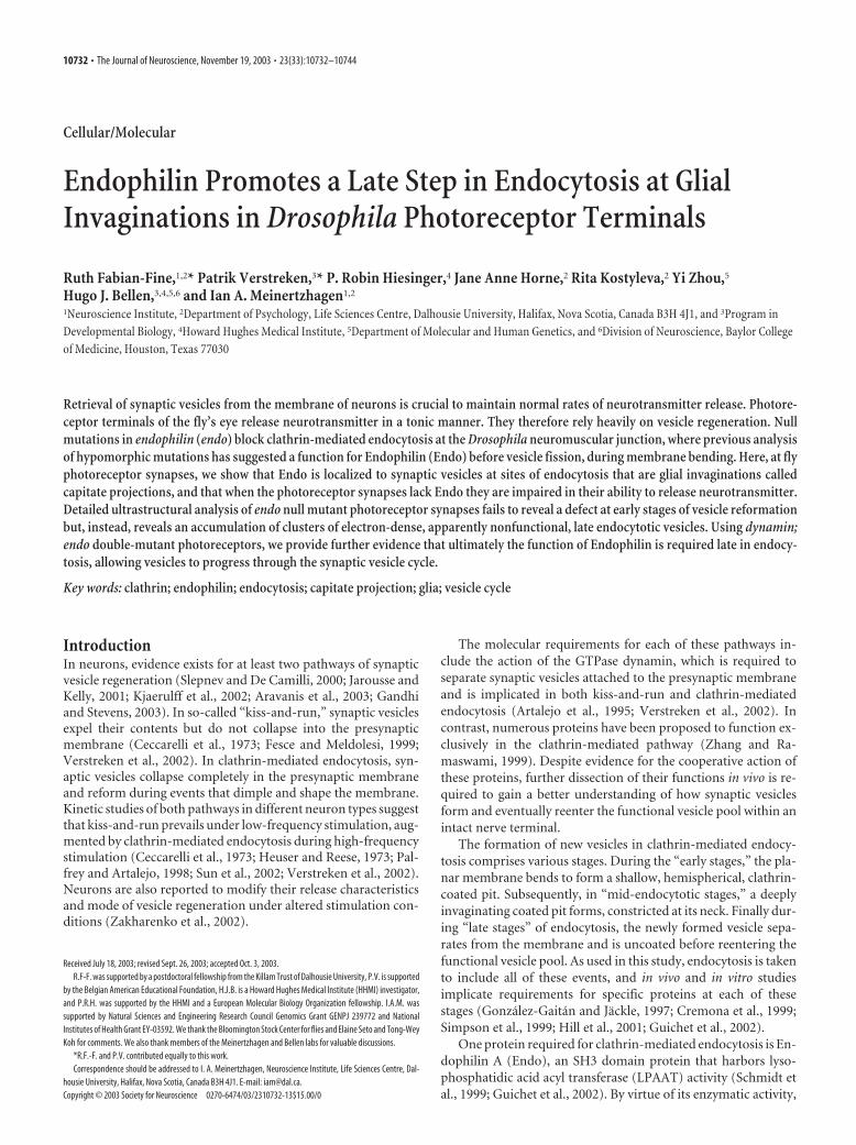
Lack of Endophilin leads to accumulation of postfission vesiclesTo determine which fraction of the vesicle pool in shits1;endo1
mutant R1–R6 terminals is functional, we exposed flies to 29°Cfor 15 min and assessed the extent of vesicle depletion (Table 1).Although shits1 terminals at the restrictive temperature were al-most completely depleted of synaptic vesicles (Figs. 8B, 9C), theirdepletion in shits1;endo1 terminals was much less severe, with sur-viving vesicles forming more densely packed clusters than in shits1
single-mutant terminals (Figs. 8D, 9C). Their packing density inshits1;endo1 terminals at the restrictive temperature was in turnapproximately four times less than in control terminals. The re-maining vesicles in shits1;endo1 double-mutant terminals exposedto high temperature, in addition to being clustered, also appearedelectron dense (Fig. 8C), reminiscent of the clustered vesicles inendo1 single-mutant terminals (Figs. 7C–F, 8D,E). Such dark ves-icles were not observed in shits at the restrictive temperature (Fig.8B,C) (data not shown) (Koenig and Ikeda, 1996). Hence, thelack of Endo in double-mutant terminals created a pool of vesi-cles that would normally be depleted by loss of shi function atrestrictive temperatures. Apparently the vesicles that persist inshits1;endo1 terminals differed in some qualitative way from nor-mal synaptic vesicles, not allowing them to reenter the synapticvesicle cycle and be released at restrictive temperatures (Fig.8D,E).
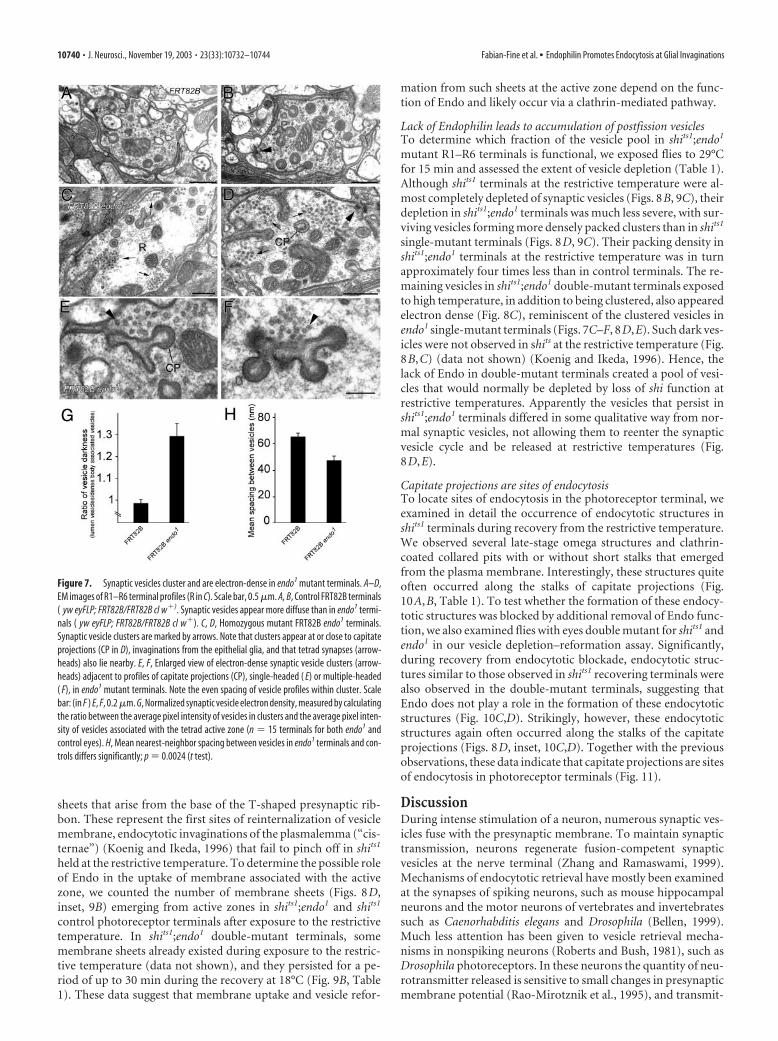
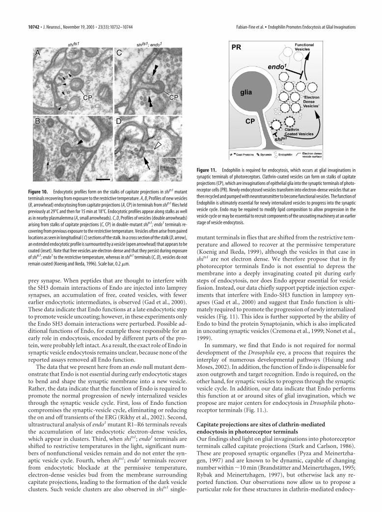
Capitate projections are sites of endocytosisTo locate sites of endocytosis in the photoreceptor terminal, weexamined in detail the occurrence of endocytotic structures inshits1 terminals during recovery from the restrictive temperature.We observed several late-stage omega structures and clathrin-coated collared pits with or without short stalks that emergedfrom the plasma membrane. Interestingly, these structures quiteoften occurred along the stalks of capitate projections (Fig.10A,B, Table 1). To test whether the formation of these endocy-totic structures was blocked by additional removal of Endo func-tion, we also examined flies with eyes double mutant for shits1 andendo1 in our vesicle depletion–reformation assay. Significantly,during recovery from endocytotic blockade, endocytotic struc-tures similar to those observed in shits1 recovering terminals werealso observed in the double-mutant terminals, suggesting thatEndo does not play a role in the formation of these endocytoticstructures (Fig. 10C,D). Strikingly, however, these endocytoticstructures again often occurred along the stalks of the capitateprojections (Figs. 8D, inset, 10C,D). Together with the previousobservations, these data indicate that capitate projections are sitesof endocytosis in photoreceptor terminals (Fig. 11).
DiscussionDuring intense stimulation of a neuron, numerous synaptic ves-icles fuse with the presynaptic membrane. To maintain synaptictransmission, neurons regenerate fusion-competent synapticvesicles at the nerve terminal (Zhang and Ramaswami, 1999).Mechanisms of endocytotic retrieval have mostly been examinedat the synapses of spiking neurons, such as mouse hippocampalneurons and the motor neurons of vertebrates and invertebratessuch as Caenorhabditis elegans and Drosophila (Bellen, 1999).Much less attention has been given to vesicle retrieval mecha-nisms in nonspiking neurons (Roberts and Bush, 1981), such asDrosophila photoreceptors. In these neurons the quantity of neu-rotransmitter released is sensitive to small changes in presynapticmembrane potential (Rao-Mirotznik et al., 1995), and transmit-
Figure 7. Synaptic vesicles cluster and are electron-dense in endo1 mutant terminals. A–D,EM images of R1–R6 terminal profiles (R in C). Scale bar, 0.5 �m. A, B, Control FRT82B terminals( yw eyFLP; FRT82B/FRT82B cl w�). Synaptic vesicles appear more diffuse than in endo1 termi-nals ( yw eyFLP; FRT82B/FRT82B cl w�). C, D, Homozygous mutant FRT82B endo1 terminals.Synaptic vesicle clusters are marked by arrows. Note that clusters appear at or close to capitateprojections (CP in D), invaginations from the epithelial glia, and that tetrad synapses (arrow-heads) also lie nearby. E, F, Enlarged view of electron-dense synaptic vesicle clusters (arrow-heads) adjacent to profiles of capitate projections (CP), single-headed ( E) or multiple-headed( F), in endo1 mutant terminals. Note the even spacing of vesicle profiles within cluster. Scalebar: (in F ) E, F, 0.2 �m. G, Normalized synaptic vesicle electron density, measured by calculatingthe ratio between the average pixel intensity of vesicles in clusters and the average pixel inten-sity of vesicles associated with the tetrad active zone (n � 15 terminals for both endo1 andcontrol eyes). H, Mean nearest-neighbor spacing between vesicles in endo1 terminals and con-trols differs significantly; p � 0.0024 (t test).
10740 • J. Neurosci., November 19, 2003 • 23(33):10732–10744 Fabian-Fine et al. • Endophilin Promotes Endocytosis at Glial Invaginations

ter is released in a tonic manner (Juusolaet al., 1996). Although the molecularmechanisms of endocytosis in the synapticterminals of Drosophila photoreceptorshave been studied in only a few mutants(Koenig and Ikeda, 1996; Littleton et al.,2001; Chang et al., 2002), the visual systemin Drosophila is ideally suited to study suchquestions. Not only is it amenable to ge-netic manipulation through the creationof whole-eye mosaics (Stower andSchwarz, 1999; Newsome et al., 2000), butits modular organization is also well suitedto detailed ultrastructural analyses (Mein-ertzhagen and O’Neil, 1991; Koenig andIkeda, 1996; Meinertzhagen, 1996).
Endo function is required to promotethe progression of newly internalizedvesicles in the synaptic vesicle cycleInterfering with Endo function at live syn-apses has demonstrated the essential roleof Endo during endocytosis of synapticvesicles. Even so, the exact function ofEndo remains controversial. Biochemicalanalyses and vesicle reformation assayshave suggested a role for the protein dur-ing endocytosis via LPAAT activity, in thetransition from shallow to deeply invagi-nated pits, possibly by modifying lipidstructure (Scales and Scheller, 1999;Schmidt et al., 1999; Hill et al., 2001). Fur-ther support for a role of Endo duringmembrane bending has been suggestedfrom analyses of endo hypomorphic allelesat the Drosophila NMJ and from injectionexperiments in the lamprey. At the Dro-sophila NMJ, although partial loss of endofunction in hypomorphic alleles such asendo10 does not dramatically reduce totalvesicle number, shallow pits accumulatealong the plasma membrane (Guichet etal., 2002). Furthermore, when Endo anti-bodies, which probably disrupt some ofthe functions of Endo, are injected in ton-ically stimulated lamprey synapses, accu-mulations of shallow and more deeply in-vaginating coated pits are observed(Ringstad et al., 1999). These data are con-sistent with an early role of Endo inclathrin-mediated endocytosis. It is un-likely, however, that all endo function iscompletely removed in any of these assays,which may therefore not necessarily revealthe exact function of Endo during vesicleretrieval. The importance of LPAAT activ-ity, in particular, remains controversial.Although Endo binds and tubulates mem-branes in vitro, this property is indepen-dent of the enzymatic activity of Endo(Farsad et al., 2001).
Other injection experiments suggest anadditional function for Endo at the lam-
Figure 8. Vesicle reformation in shits1; endo1 double-mutant terminals. A, Experimental plan. Flies with shits1; endo1 double-mutant and shits1 single-mutant eyes were shifted to the restrictive temperature for 15 min (�15– 0) to deplete synaptic vesicles(see Results for details and Materials and Methods for genotypes). They were then shifted back to the permissive temperature,revealing at 0, 15, and 30 min the surviving defects in endocytosis in the absence of Endo function, for comparison with terminalsof shits1; endo1 flies kept at the permissive temperature. B–E, Examples of terminals during recovery from exposure to therestrictive temperature in single-mutant shits1 either dark-exposed at 29°C ( B) or after 30 min recovery at 18°C from previousexposure to 29°C ( C), and in double-mutant shits1; endo1 either at 29°C ( D) or 18°C after 30 min recovery from 29°C ( E). Membranesheet in B, enlarged in the top right-hand panel (arrow), arises from the pedestal of a presynaptic T-bar ribbon (arrowhead).Synaptic vesicle profiles (arrow) are enlarged from recovering shits1 terminal ( C) in the middle right-hand panel, and an endocy-totic profile (arrowhead) is enlarged from recovering shits1; endo1 terminal ( E) in the bottom right-hand panel.
Figure 9. Time course of recovery from exposure to the restrictive temperature among organelle populations in shits1 andshits1;endo1 terminals. A, During recovery from exposure to the restrictive temperature, the number of capitate projections in shits1;endo1 terminals increases; differences are significant after 15 min ( p � 0.036; t test) of recovery, but lose significance after 30 min( p �0.072; t test). We counted different morphological forms of capitate profiles (those with shallow, single, and multiple heads,which are thought to constitute the sequence for organelle formation and regression). B, Membrane sheets emerging from nearthe pedestal of the T-bar ribbon, probably representing endocytosis from that site (Koenig and Ikeda, 1996), persisted longer inshits1; endo1 exposed to the restrictive temperature than in shits1 single-mutant terminals. C, More synaptic vesicles survive inshits1; endo1 mutant terminals that are exposed to the restrictive temperature than in shits1 single-mutant terminals.
Fabian-Fine et al. • Endophilin Promotes Endocytosis at Glial Invaginations J. Neurosci., November 19, 2003 • 23(33):10732–10744 • 10741
prey synapse. When peptides that are thought to interfere withthe SH3 domain interactions of Endo are injected into lampreysynapses, an accumulation of free, coated vesicles, with fewerearlier endocytotic intermediates, is observed (Gad et al., 2000).These data indicate that Endo functions at a late endocytotic stepto promote vesicle uncoating; however, in these experiments onlythe Endo SH3 domain interactions were perturbed. Possible ad-ditional functions of Endo, for example those responsible for anearly role in endocytosis, encoded by different parts of the pro-tein, were probably left intact. As a result, the exact role of Endo insynaptic vesicle endocytosis remains unclear, because none of thereported assays removed all Endo function.
The data that we present here from an endo null mutant dem-onstrate that Endo is not essential during early endocytotic stagesto bend and shape the synaptic membrane into a new vesicle.Rather, the data indicate that the function of Endo is required topromote the normal progression of newly internalized vesiclesthrough the synaptic vesicle cycle. First, loss of Endo functioncompromises the synaptic-vesicle cycle, eliminating or reducingthe on and off transients of the ERG (Rikhy et al., 2002). Second,ultrastructural analysis of endo1 mutant R1–R6 terminals revealsthe accumulation of late endocytotic electron-dense vesicles,which appear in clusters. Third, when shits1; endo1 terminals areshifted to restrictive temperatures in the light, significant num-bers of nonfunctional vesicles remain and do not enter the syn-aptic vesicle cycle. Fourth, when shits1; endo1 terminals recoverfrom endocytotic blockade at the permissive temperature,electron-dense vesicles bud from the membrane surroundingcapitate projections, leading to the formation of the dark vesicleclusters. Such vesicle clusters are also observed in shits1 single-
mutant terminals in flies that are shifted from the restrictive tem-perature and allowed to recover at the permissive temperature(Koenig and Ikeda, 1999), although the vesicles in that case inshits1 are not electron dense. We therefore propose that in flyphotoreceptor terminals Endo is not essential to depress themembrane into a deeply invaginating coated pit during earlysteps of endocytosis, nor does Endo appear essential for vesiclefission. Instead, our data chiefly support peptide injection exper-iments that interfere with Endo-SH3 function in lamprey syn-apses (Gad et al., 2000) and suggest that Endo function is ulti-mately required to promote the progression of newly internalizedvesicles (Fig. 11). This idea is further supported by the ability ofEndo to bind the protein Synaptojanin, which is also implicatedin uncoating synaptic vesicles (Cremona et al., 1999; Nonet et al.,1999).
In summary, we find that Endo is not required for normaldevelopment of the Drosophila eye, a process that requires theinterplay of numerous developmental pathways (Hsiung andMoses, 2002). In addition, the function of Endo is dispensable foraxon outgrowth and target recognition. Endo is required, on theother hand, for synaptic vesicles to progress through the synapticvesicle cycle. In addition, our data indicate that Endo performsthis function at or around sites of glial invagination, which wepropose are major centers for endocytosis in Drosophila photo-receptor terminals (Fig. 11.).
Capitate projections are sites of clathrin-mediatedendocytosis in photoreceptor terminalsOur findings shed light on glial invaginations into photoreceptorterminals called capitate projections (Stark and Carlson, 1986).These are proposed synaptic organelles (Pyza and Meinertzha-gen, 1997) and are known to be dynamic, capable of changingnumber within �10 min (Brandstatter and Meinertzhagen, 1995;Rybak and Meinertzhagen, 1997), but otherwise lack any re-ported function. Our observations now allow us to propose aparticular role for these structures in clathrin-mediated endocy-
Figure 11. Endophilin is required for endocytosis, which occurs at glial invaginations insynaptic terminals of photoreceptors. Clathrin-coated vesicles can form on stalks of capitateprojections (CP), which are invaginations of epithelial glia into the synaptic terminals of photo-receptor cells (PR). Newly endocytosed vesicles transform into electron-dense vesicles that arethen recycled and pumped with neurotransmitter to become functional vesicles. The function ofEndophilin is ultimately essential for newly internalized vesicles to progress into the synapticvesicle cycle. Endo may be required to modify lipid composition to allow progression in thevesicle cycle or may be essential to recruit components of the uncoating machinery at an earlierstage of vesicle endocytosis.
Figure 10. Endocytotic profiles form on the stalks of capitate projections in shits1 mutantterminals recovering from exposure to the restrictive temperature. A, B, Profiles of new vesicles(B, arrowhead) endocytosing from capitate projections (A, CP) in terminals from shits1 flies heldpreviously at 29°C and then for 15 min at 18°C. Endocytotic profiles appear along stalks as wellas in nearby plasmalemma (A, small arrowheads). C, D, Profiles of vesicles (double arrowheads)arising from stalks of capitate projections (C, CP) in double-mutant shits1; endo1 terminals re-covering from previous exposure to the restrictive temperature. Vesicles often arise from pairedlocations as seen in longitudinal ( C) sections of the stalk. In a cross section of the stalk (D, arrow),an extended endocytotic profile is surmounted by a vesicle (open arrowhead) that appears to becoated (inset). Note that free vesicles are electron-dense and that they persist during exposureof shits1; endo1 to the restrictive temperature, whereas in shits1 terminals (C, D), vesicles do notremain coated (Koenig and Ikeda, 1996). Scale bar, 0.2 �m.
10742 • J. Neurosci., November 19, 2003 • 23(33):10732–10744 Fabian-Fine et al. • Endophilin Promotes Endocytosis at Glial Invaginations
tosis. First, immuno-EM with anti-Endo and anti-clathrin showsthat both of these markers of clathrin-mediated endocytosis areenriched at vesicles and membranes associated with capitate pro-jections. Second, in endo1 mutant photoreceptor terminals,electron-dense vesicles cluster near the capitate projections.Third, when clathrin-mediated endocytosis is blocked, the num-ber of capitate projections per terminal increases. Finally, duringrecovery from endocytotic blockade in shits1; endo1 eyes, deeplyinvaginating coated pits are observed on the stalks of capitateprojections. Hence, we propose that these glial invaginations, thecapitate projections, play a prominent role in clathrin-mediatedendocytotic vesicle retrieval (Fig. 11). It is therefore interestingthat at the frog NMJ, endocytotic intermediates at the presynapticmembrane have also been observed in association with glial cellprocesses extending into the motor nerve terminal (Heuser andReese, 1973).
Glia are also implicated in the clearance of transmitter fromthe synaptic cleft and in transmitter reuptake by the presynapticterminal (Schousboe and Hertz, 1981; Steinhauser and Gallo,1996; Palmada and Centelles, 1998). Likewise, epithelial glia sur-rounding photoreceptor terminals in the lamina could be involvedin transmitter re-uptake into the terminals. The epithelial glia ex-press Ebony (Richardt et al., 2002), a major enzyme for the metab-olism of released histamine (Borycz et al., 2002), which thus mayclear transmitter from the cleft. We propose that in addition to theirrole in endocytosis, capitate projections may also aid in the transportof neurotransmitter back into the photoreceptor terminal, therebylinking at a single site the function of vesicle recovery from the mem-brane with transmitter recycling. In this context, it may be relevantthat mutants of ebony and tan, which together regulate histaminemetabolism (Borycz et al., 2002), have altered numbers of capitateprojections (Meinertzhagen and Wang, 1997).
Flies with endo1 mutant eyes can still seeAlthough their on and off ERG transients are essentially absent,flies with endo1 mutant eyes are still not blind. Our evidence does notallow us to suggest that their vision is normal, only that phototaxischoice behavior under the conditions of our assay does not differfrom controls. We have not examined the light intensity thresholdfor such behavior or the many other tests of visual behavior (Heisen-berg and Wolf, 1984) that could be examined. It is therefore inter-esting that under different conditions and using a different geneticbackground, Rikhy et al. (2002) report that flies with endo1 mutanteyes, although not behaving as well as controls in a light-choice test,were also not entirely blind. Thus, our data as well as theirs indicatethat at least some neurotransmitter release from endo1 photorecep-tors must persist to give rise to signals in the lamina; even if these maybe amplified at more central synapses in the visual pathway, they aresufficient to endow the flies with phototactic behavior. This persis-tent release must occur despite the loss of the ERG transients andimplies that some regeneration of fusion-competent vesicles stillproceeds in the absence of Endo function. Two mechanisms mayexist to achieve this in endo1 mutant photoreceptors. Either residualendocytosis and minute rates of regenerating fusion-competent ves-icles, sufficient for the fly to detect light, or alternative modes ofsynaptic vesicle regeneration that do not require Endo persist inendo1 mutant terminals. Given previous findings on endo1 mutantNMJs (Kjaerulff et al., 2002; Marte, 2002; Verstreken et al., 2002), itseems likely that in the fly’s eye, too, a fast vesicle retrieval pathwayoperates to ensure the regeneration of fusion-competent vesicles.
ReferencesAravanis AM, Pyle JL, Tsien RW (2003) Single synaptic vesicles fusing tran-
siently and successively without loss of identity. Nature 423:643– 647.
Artalejo CR, Henley JR, McNiven MA, Palfrey HC (1995) Rapid endocyto-sis coupled to exocytosis in adrenal chromaffin cells involves Ca 2�, GTP,and dynamin but not clathrin. Proc Natl Acad Sci USA 92:8328 – 8332.
Bellen HJ (1999) Neurotransmitter release. In: Frontiers in molecular biol-ogy, Vol 23 (Hames BD, Glover DM, eds), p 436. Oxford: Oxford UP.
Benzer S (1973) Genetic dissection of behavior. Sci Am 229:24 –37.Berdnik D, Torok T, Gonzalez-Gaitan M, Knoblich JA (2002) The endocytic
protein alpha-Adaptin is required for numb-mediated asymmetric celldivision in Drosophila. Dev Cell 3:221–231.
Borycz J, Borycz JA, Loubani M, Meinertzhagen IA (2002) tan and ebonygenes regulate a novel pathway for transmitter metabolism at fly photo-receptor terminals. J Neurosci 22:10549 –10557.
Braitenberg V (1967) Patterns of projection in the visual system of the fly. I.Retina-lamina projections. Exp Brain Res 3:271–298.
Brandstatter JH, Meinertzhagen IA (1995) The rapid assembly of synapticsites in photoreceptor terminals of the fly’s optic lobe recovering fromcold shock. Proc Natl Acad Sci USA 92:2677–2681.
Burg MG, Sarthy PV, Koliantz G, Pak WL (1993) Genetic and molecularindentification of a Drosophila histidine decarboxylase gene required inphotoreceptor transmitter synthesis. EMBO J 12:911–919.
Ceccarelli B, Hurlbut WP, Mauro A (1973) Turnover of transmitter and synap-tic vesicles at the frog neuromuscular junction. J Cell Biol 57:499–524.
Chang HC, Newmyer SL, Hull MJ, Ebersold M, Schmid SL, Mellman I(2002) Hsc70 is required for endocytosis and clathrin function in Dro-sophila. J Cell Biol 159:477– 487.
Chen D, Stark WS (1993) Effects of temperature on visual receptors intemperature-sensitive paralytic shibire (shits) mutants of Drosophila. J In-sect Physiol 39:385–392.
Chen MS, Obar RA, Schroeder CC, Austin TW, Poodry CA, Wadsworth SC,Vallee RB (1991) Multiple forms of dynamin are encoded by shibire, aDrosophila gene involved in endocytosis. Nature 351:583–586.
Coombe PE (1986) The large monopolar cells L1 and L2 are responsible forERG transients in Drosophila. J Comp Physiol [A] 159:655– 665.
Cremona O, Di Paolo G, Wenk MR, Luthi A, Kim WT, Takei K, Daniell L,Nemoto Y, Shears SB, Flavell RA, McCormick DA, De Camilli P (1999)Essential role of phosphoinositide metabolism in synaptic vesicle recy-cling. Cell 99:179 –188.
Deitcher DL, Ueda A, Stewart BA, Burgess RW, Kidokoro Y, Schwarz TL(1998) Distinct requirements for evoked and spontaneous release of neu-rotransmitter are revealed by mutations in the Drosophila gene neuronal-synaptobrevin. J Neurosci 18:2028 –2039.
Fabian-Fine R, Skehel P, Errington ML, Davies HA, Sher E, Stewart MG, FineA (2001) Ultrastructural distribution of the �7 nicotinic acetylcholinereceptor subunit in rat hippocampus. J Neurosci 21:7993– 8003.
Farsad K, Ringstad N, Takei K, Floyd SR, Rose K, De Camilli P (2001) Gen-eration of high curvature membranes mediated by direct endophilin bi-layer interactions. J Cell Biol 155:193–200.
Fesce R, Meldolesi J (1999) Peeping at the vesicle kiss. Nat Cell Biol 1:E3–E4.Fischbach K-F, Dittrich APM (1989) The optic lobe of Drosophila melano-
gaster. I. A Golgi analysis of wild-type structure. Cell Tissue Res258:441– 475.
Gad H, Ringstad N, Low P, Kjaerulff O, Gustafsson J, Wenk M, Di Paolo G,Nemoto Y, Crum J, Ellisman MH, De Camilli P, Shupliakov O, Brodin L(2000) Fission and uncoating of synaptic clathrin-coated vesicles are per-turbed by disruption of interactions with the SH3 domain of endophilin.Neuron 27:301–312.
Gandhi SP, Stevens CF (2003) Three modes of synaptic vesicular recyclingrevealed by single-vesicle imaging. Nature 423:607– 613.
Gonzalez-Gaitan M, Jackle H (1997) Role of Drosophila �-adaptin in pre-synaptic vesicle recycling. Cell 88:767–776.
Guichet A, Wucherpfennig T, Dudu V, Etter S, Wilsch-Brauniger M, HellwigA, Gonzalez-Gaitan M, Huttner WB, Schmidt AA (2002) Essential roleof endophilin A in synaptic vesicle budding at the Drosophila neuromus-cular junction. EMBO J 21:1661–1672.
Hardie RC (1987) Is histamine a neurotransmitter in insect photoreceptors?J Comp Physiol [A] 161:201–213.
Harrison SD, Broadie K, van de Goor J, Rubin GM (1994) Mutations in theDrosophila Rop gene suggest a function in general secretion and synaptictransmission. Neuron 13:555–566.
Heisenberg M (1971) Separation of receptor and lamina potentials in the elec-troretinogram of normal and mutant Drosophila. J Exp Biol 55:85–100.
Heisenberg M, Wolf R (1984) Vision in Drosophila. Berlin: Springer.
Fabian-Fine et al. • Endophilin Promotes Endocytosis at Glial Invaginations J. Neurosci., November 19, 2003 • 23(33):10732–10744 • 10743

Heuser JE, Reese TS (1973) Evidence for recycling of synaptic vesicle mem-brane during transmitter release at the frog neuromuscular junction.J Cell Biol 57:315–344.
Hiesinger PR, Reiter C, Schau H, Fischbach K-F (1999) Neuropil patternformation and regulation of cell adhesion molecules in Drosophila opticlobe development depend on synaptobrevin. J Neurosci 19:7548 –7556.
Hiesinger PR, Scholz M, Meinertzhagen IA, Fischbach K-F, Obermayer K(2001) Visualization of synaptic markers in the optic neuropils of Dro-sophila using a new constrained deconvolution method. J Comp Neurol429:277–288.
Hill E, van Der Kaay J, Downes CP, Smythe E (2001) The role of dynaminand its binding partners in coated pit invagination and scission. J Cell Biol152:309 –324.
Homyk Jr T, Pye Q (1989) Some mutations affecting neural or musculartissues alter the physiological components of the electroretinogram inDrosophila. J Neurogenet 5:37– 48.
Hsiung F, Moses K (2002) Retinal development in Drosophila: specifyingthe first neuron. Hum Mol Genet 11:1207–1214.
Jarousse N, Kelly RB (2001) Endocytotic mechanisms in synapses. CurrOpin Cell Biol 13:461– 469.
Juusola M, French AS, Uusitalo RO, Weckstrom M (1996) Informationprocessing by graded-potential transmission through tonically active syn-apses. Trends Neurosci 19:292–297.
Kelly LE (1974) Temperature-sensitive mutations affecting the regenerativesodium channel in Drosophila melanogaster. Nature 248:166 –168.
Kjaerulff O, Verstreken P, Bellen HJ (2002) Synaptic vesicle retrieval: stilltime for a kiss. Nat Cell Biol 4:E245–248.
Koenig JH, Ikeda K (1996) Synaptic vesicles have two distinct recyclingpathways. J Cell Biol 135:797– 808.
Koenig JH, Ikeda K (1999) Contribution of active zone subpopulation ofvesicles to evoked and spontaneous release. J Neurophysiol81:1495–1505.
Kosaka T, Ikeda K (1983) Possible temperature-dependent blockage of syn-aptic vesicle recycling induced by a single gene mutation in Drosophila.J Neurobiol 14:207–225.
Kral K, Meinertzhagen IA (1989) Anatomical plasticity of synapses in thelamina of the optic lobe of the fly. Philos Trans R Soc Lond B Biol Sci323:155–183.
Littleton JT, Bai J, Vyas B, Desai R, Baltus AE, Garment MB, Carlson SD,Ganetzky B, Chapman ER (2001) synaptotagmin mutants reveal essen-tial functions for the C2B domain in Ca 2�-triggered fusion and recyclingof synaptic vesicles in vivo. J Neurosci 21:1421–1433.
Marte B (2002) An encore for kiss and run? Nat Cell Biol 4:E123.Meinertzhagen IA (1996) Ultrastructure and quantification of synapses in
the insect nervous system. J Neurosci Methods 69:59 –73.Meinertzhagen IA, O’Neil SD (1991) Synaptic organization of columnar
elements in the lamina of the wild type in Drosophila melanogaster.J Comp Neurol 305:232–263.
Meinertzhagen IA, Sorra KE (2001) Synaptic organization in the fly’s opticlamina: few cells, many synapses and divergent microcircuits. Prog BrainRes 131:53– 69.
Meinertzhagen IA, Wang Y (1997) Drosophila mutants tan and ebony havealtered numbers of capitate projections, glial invaginations into photore-ceptor terminals. In: Neurobiology: from membrane to mind, Vol II (El-sner N, Wassle H, eds), p 457. Stuttgart: Georg Thieme Verlag.
Newsome TP, Asling B, Dickson BJ (2000) Analysis of Drosophila photore-ceptor axon guidance in eye-specific mosaics. Development 127:851– 860.
Nonet ML, Holgado AM, Brewer F, Serpe CJ, Norbeck BA, Holleran J, Wei L,Hartwieg E, Jorgensen EM, Alfonso A (1999) UNC-11, a Caenorhabditiselegans AP180 homologue, regulates the size and protein composition ofsynaptic vesicles. Mol Biol Cell 10:2343–2360.
Palfrey HC, Artalejo CR (1998) Vesicle recycling revisited: rapid endocyto-sis may be the first step. Neuroscience 83:969 –989.
Palmada M, Centelles JJ (1998) Excitatory amino acid neurotransmission.Pathways for metabolism, storage and reuptake of glutamate in brain.Front Biosci 3:D701–718.
Poodry CA, Hall L, Suzuki DT (1973) Developmental properties of Shibi-rets1: a pleiotropic mutation affecting larval and adult locomotion anddevelopment. Dev Biol 32:373–386.
Pyza E, Meinertzhagen IA (1997) Circadian rhythms in screening pigmentand invaginating organelles in photoreceptor terminals of the housefly’sfirst optic neuropile. J Neurobiol 32:517–529.
Rao-Mirotznik R, Harkins AB, Buchsbaum G, Sterling P (1995) Mamma-lian rod terminal: architecture of a binary synapse. Neuron 14:561–569.
Richardt A, Rybak J, Stortkuhl KF, Meinertzhagen IA, Hovemann BT (2002)Ebony protein in the Drosophila nervous system: optic neuropile expres-sion in glial cells. J Comp Neurol 452:93–102.
Rikhy R, Kumar V, Mittal R, Krishnan KS (2002) Endophilin is criticallyrequired for synapse formation and function in Drosophila melanogaster.J Neurosci 22:7478 –7484.
Ringstad N, Gad H, Low P, Di Paolo G, Brodin L, Shupliakov O, De Camilli P(1999) Endophilin/SH3p4 is required for the transition from early to latestages in clathrin-mediated synaptic vesicle endocytosis. Neuron 24:143–154.
Roberts A, Bush BMH (1981) Neurones without impulses: their signifi-cance for vertebrate and invertebrate nervous systems. Cambridge, UK:Cambridge UP.
Rybak J, Meinertzhagen IA (1997) The effects of light reversals on photorecep-tor synaptogenesis in the fly Musca domestica. Eur J Neurosci 9:319–333.
Sanyal S, Tolar LA, Pallanck L, Krishnan KS (2001) Genetic interaction be-tween shibire and comatose mutations in Drosophila suggest a role forsnap-receptor complex assembly and disassembly for maintenance ofsynaptic vesicle cycling. Neurosci Lett 311:21–24.
Scales SJ, Scheller RH (1999) Lipid membranes shape up. Nature401:123–124.
Schmidt A, Wolde M, Thiele C, Fest W, Kratzin H, Podtelejnikov AV, WitkeW, Huttner WB, Soling H-D (1999) Endophilin I mediates synaptic ves-icle formation by transfer of arachidonate to lysophosphatidic acid. Na-ture 401:133–141.
Schousboe A, Hertz L (1981) Role of astroglial cells in glutamate homeosta-sis. Adv Biochem Psychopharmacol 27:103–113.
Schulze KL, Broadie K, Perin MS, Bellen HJ (1995) Genetic and electrophys-iological studies of Drosophila syntaxin-1A demonstrate its role in non-neuronal secretion and neurotransmission. Cell 80:311–320.
Simpson F, Hussain NK, Qualmann B, Kelly RB, Kay BK, McPherson PS,Schmid SL (1999) SH3-domain-containing proteins function at distinctsteps in clathrin-coated vesicle formation. Nat Cell Biol 1:119 –124.
Slepnev VI, De Camilli P (2000) Accessory factors in clathrin-dependentsynaptic vesicle endocytosis. Nat Rev Neurosci 1:161–172.
Somogyi P, Takagi H (1982) A note on the use of picric acid-paraformaldehyde-glutaraldehyde fixative for correlated light and elec-tron microscopic immunocytochemistry. Neuroscience 7:1779 –1783.
Stark WS, Carlson SD (1986) Ultrastructure of capitate projections in theoptic neuropil of Diptera. Cell Tissue Res 246:481– 486.
Steinhauser C, Gallo V (1996) News on glutamate receptors in glial cells.Trends Neurosci 19:339 –345.
Stower RS, Schwarz TL (1999) A genetic method for generating Drosophilaeyes composed exclusively of mitotic clones of a single genotype. Genetics152:1631–1639.
Sun J-Y, Wu X-S, Wu L-G (2002) Single and multiple vesicle fusion inducedifferent rates of endocytosis at a central synapse. Nature 417:555–559.
Trujillo-Cenoz O (1965) Some aspects of the structural organization of theintermediate retina of dipterans. J Ultrastruct Res 13:1–33.
Verstreken P, Kjaerulff O, Lloyd TE, Atkinson R, Zhou Y, Meinertzhagen IA,Bellen HJ (2002) Endophilin mutations block clathrin-mediated endo-cytosis but not neurotransmitter release. Cell 109:101–112.
Wu MN, Fergestad T, Lloyd TE, He Y, Broadie K, Bellen HJ (1999) Syntaxin1A interacts with multiple exocytic proteins to regulate neurotransmitterrelease in vivo. Neuron 23:593– 605.
Zakharenko SS, Zablow L, Siegelbaum SA (2002) Altered presynaptic vesiclerelease and cycling during mGluR-dependent LTD. Neuron 35:1099–1110.
Zhang B, Ramaswami M (1999) Synaptic vesicle endocytosis and recycling.In: Neurotransmitter release, Vol 23 (Bellen HJ, ed), p 436. Oxford, UK:Oxford UP.
Zhang B, Koh YH, Beckstead RB, Budnik V, Ganetzky B, Bellen HJ (1998)Synaptic vesicle size and number are regulated by a clathrin adaptor pro-tein required for endocytosis. Neuron 21:1465–1475.
Zinsmaier KE, Eberle KK, Buchner E, Walter N, Benzer S (1994) Paralysisand early death in cysteine string protein mutants of Drosophila. Science263:977–980.
Zipursky SL, Venkatesh TR, Teplow DB, Benzer S (1984) Neuronal devel-opment in the Drosophila retina: monoclonal antibodies as molecularprobes. Cell 36:15–26.
10744 • J. Neurosci., November 19, 2003 • 23(33):10732–10744 Fabian-Fine et al. • Endophilin Promotes Endocytosis at Glial Invaginations