Embed Size (px)
Citation preview

Cellular/Molecular
Active Dendrites and Differential Distribution of CalciumChannels Enable Functional Compartmentalization of GolgiCells
Stephanie Rudolph,1 Court Hull,2 and X Wade G. Regehr1
1Department of Neurobiology, Harvard Medical School, Boston, Massachusetts 02115, and 2Department of Neurobiology, Duke University, Durham, NorthCarolina 27708
Interneurons are essential to controlling excitability, timing, and synaptic integration in neuronal networks. Golgi cells (GoCs) servethese roles at the input layer of the cerebellar cortex by releasing GABA to inhibit granule cells (grcs). GoCs are excited by mossy fibers(MFs) and grcs and provide feedforward and feedback inhibition to grcs. Here we investigate two important aspects of GoC physiology:the properties of GoC dendrites and the role of calcium signaling in regulating GoC spontaneous activity. Although GoC dendrites areextensive, previous studies concluded they are devoid of voltage-gated ion channels. Hence, the current view holds that somatic voltagesignals decay passively within GoC dendrites, and grc synapses onto distal dendrites are not amplified and are therefore ineffective atfiring GoCs because of strong passive attenuation. Using whole-cell recording and calcium imaging in rat slices, we find that dendriticvoltage-gated sodium channels allow somatic action potentials to activate voltage-gated calcium channels (VGCCs) along the entiredendritic length, with R-type and T-type VGCCs preferentially located distally. We show that R- and T-type VGCCs located in the dendritescan boost distal synaptic inputs and promote burst firing. Active dendrites are thus critical to the regulation of GoC activity, andconsequently, to the processing of input to the cerebellar cortex. In contrast, we find that N-type channels are preferentially located nearthe soma, and control the frequency and pattern of spontaneous firing through their close association with calcium-activated potassium(KCa ) channels. Thus, VGCC types are differentially distributed and serve specialized functions within GoCs.
Key words: calcium buffering; calcium channels; calcium-activated potassium channels; cerebellum; dendritic excitability;interneuron
IntroductionThroughout the brain, diverse types of interneurons provide in-hibition to modulate excitability, temporal precision, and synap-tic integration within neural circuits (Pouille and Scanziani,
2001; Mittmann et al., 2005; Isaacson and Scanziani, 2011). Thefunction of different interneuron types is determined by theiranatomy, connectivity, and activity patterns (Somogyi andKlausberger, 2005). At the input layer of the cerebellar cortex,only a single type of interneuron, the Golgi cell (GoC), servesthese roles (Eccles et al., 1964; Brickley et al., 1996; Hamann et al.,
Received Aug. 19, 2015; revised Sept. 16, 2015; accepted Sept. 25, 2015.Author contributions: S.R., C.H., and W.G.R. designed research; S.R. and C.H. performed research; S.R., C.H., and
W.G.R. analyzed data; S.R. and W.G.R. wrote the paper.This work was supported by the National Institutes of Health Grant R01 NS032405 to W.G.R., Ruth L. Kirschstein
Awards F32 NS087708 to S.R. and F32 NS060585 to C.H., and Nancy Lurie Marks Foundation and Lefler Foundationgrants to W.G.R. We thank the members of the W.G.R. laboratory for comments on this manuscript and Bruce Beanfor helpful discussions.
The authors declare no competing financial interests.Correspondence should be addressed to Dr. Wade G. Regehr, Harvard Medical School, 220 Longwood Avenue,
Goldenson 308, Boston, MA 02115. E-mail: [email protected]:10.1523/JNEUROSCI.3132-15.2015
Copyright © 2015 the authors 0270-6474/15/3515492-13$15.00/0
Significance Statement
Interneurons are essential to neural processing because they modulate excitability, timing, and synaptic integration withincircuits. At the input layer of the cerebellar cortex, a single type of interneuron, the Golgi cell (GoC), carries these functions. Theextent of inhibition depends on both spontaneous activity of GoCs and the excitatory synaptic input they receive. In this study, wefind that different types of calcium channels are differentially distributed, with dendritic calcium channels being activated bysomatic activity, boosting synaptic inputs and enabling bursting, and somatic calcium cannels promoting regular firing. Wetherefore challenge the current view that GoC dendrites are passive and identify the mechanisms that contribute to GoCs regulat-ing the flow of sensory information in the cerebellar cortex.
15492 • The Journal of Neuroscience, November 25, 2015 • 35(47):15492–15504

2002; Mitchell and Silver, 2003; Crowley et al., 2009; Duguid etal., 2012). Within the cerebellar circuit, mossy fibers (MFs) con-vey multimodal sensory information to the cerebellar cortex byexciting granule cells (grcs). Grcs in turn regulate the output ofthe cerebellar cortex by exciting its sole output neurons, the Pur-kinje cells. Because GoCs inhibit hundreds of grcs via their elab-orate axons (Palkovits et al., 1971), they powerfully regulate inputlayer excitability and control the flow of sensory information(Eccles et al., 1964; Rossi and Hamann, 1998; Vos et al., 1999;Chadderton et al., 2004; Barmack and Yakhnitsa, 2008; Duguid etal., 2012). MFs also excite GoCs at their basal dendrites, allowingthem to mediate feedforward inhibition (MF ¡ GoC ¡ grc),whereas grcs synapse onto basal and apical dendrites enablingfeedback inhibition (grc ¡ GoC ¡ grc) (Chan-Palay and Palay,1971a,b; Pellionisz and Szentagothai, 1973; Dieudonne, 1998;Kanichay and Silver, 2008; Cesana et al., 2013; Yaeger and Trus-sell, 2015). It has been established that GoCs are crucial to cere-bellar function, as eliminating GoCs severely impairs motorbehavior and leads to ataxia (Watanabe et al., 1998).
A central function of GoCs is to dynamically adjust input layerexcitability to the volume and pattern of inputs. It is thereforenecessary to clarify the intrinsic properties that enable GoCs toefficiently respond to network activity. In particular, dendriticexcitability determines both whether somatic activity is relayed tothe dendrites and how synaptic inputs are transferred to thesoma. Because gap junctions between dendrites are thought topromote synchronous firing and network oscillations (Dugue etal., 2009; Vervaeke et al., 2010), dendritic properties may be a keydeterminant of circuit level activity. Moreover, because GoCsreceive thousands of excitatory synapses (Pellionisz and Szen-tagothai, 1973), dendritic processing of these inputs determineshow they drive GoC firing and hence feedback inhibition. In thecurrent view, GoC dendrites lack voltage-gated conductances(Vervaeke et al., 2012); and in consequence, grc synapses areineffective at activating GoCs to produce feedback inhibition be-cause inputs onto apical dendrites are strongly attenuated.
GoCs fire spontaneously at 5–20 Hz (Midtgaard, 1992; Forti etal., 2006) and consequently provide ongoing inhibition to grcs.Different neuron types use specific ion channels [e.g., calciumand calcium-activated potassium (KCa) channels] (Wolfart et al.,2001; Swensen and Bean, 2003; Womack and Khodakhah, 2003,2004; Deignan et al., 2012; Benton et al., 2013) to control actionpotential firing. It is therefore important to identify the ion chan-nels that regulate spontaneous activity in GoCs.
Here we investigate two critical aspects of GoC physiology: theproperties of GoC dendrites and the role of calcium signaling inregulating GoC spontaneous activity. Using calcium imaging andpharmacology, we find that GoC dendrites express voltage-gatedsodium and calcium channels (VGCCs). Dendritic sodium chan-nels amplify depolarization provided by somatic action poten-tials to promote the opening of dendritic VGCCs. We also findthat calcium channel types are differentially distributed andfunctionally specialized: R-type and T-type calcium channels arelocated in distal apical dendrites but are absent near the soma. Wefind that dendritic calcium channels can boost synaptic inputsonto distal dendrites and promote burst firing. Conversely,N-type calcium channels are present near the soma and absentfrom distal dendrites, and regulate spontaneous firing throughtheir close association with KCa channels. Our data challenge theassumption that GoC dendrites are passive, and suggest that dif-ferentially distributed VGCCs can either boost synaptic inputs orregulate spontaneous firing.
Materials and MethodsSlice preparation. Acute parasagittal slices of 200 –270 �m thickness wereprepared from Sprague Dawley rats aged P17–P25 of either sex. Sagittalslices were used for all experiments, except for those requiring PF elec-trical stimulation, where transverse slices were cut (see Fig. 9). Slices werecut in an ice-cold solution containing the following (in mM): 130K-gluconate, 15 KCl, 0.05 EGTA, 20 HEPES, and 25 mM glucose, 0.02(R)-CPP, pH 7.4 with NaOH. After cutting, slices were rinsed in ACSFand incubated in a submerged chamber at 32°C for 30 min in ACSFequilibrated with 95% O2 and 5% CO2, containing the following (inmM): 125 NaCl, 26 NaHCO3, 1.25 NaH2PO4, 2.5 KCl, 1 MgCl2, 2 CaCl2,and 25 glucose. Slices were then kept at room temperature until record-ing for no more than 4 h. The gluconate-based cutting solution dimin-ishes calcium entry into GoCs and preserves spontaneous activity(Dugue et al., 2005). We found that, when other cutting solutions wereused, such as ACSF, sucrose (Vervaeke et al., 2010), or choline chloride,GoCs were more hyperpolarized and did not fire spontaneously. In ad-dition, although we found that cerebellar slices from mice containedspontaneously active GoCs when cut in gluconate, slices from rats were ofsuperior health. A previous study, in which it was concluded that GoCdendrites are passive, was performed with mouse slices cut using non-gluconate cutting solutions (Vervaeke et al., 2012). In that study, GoCswere sufficiently hyperpolarized that they were not spontaneously active,which is consistent with elevated levels of intracellular calcium activatingKCa channels.
Electrophysiology. Visually guided whole-cell recordings were obtainedwith patch pipettes of 2– 4 MOhm resistances pulled from borosilicatecapillary glass (Sutter Instrument) with a Sutter P-97 horizontal puller.Glass electrodes were washed with acetic acid before use to remove cal-cium ions. Electrophysiological recordings were performed at 31°C–33C°. For current-clamp recordings and calcium imaging, internalsolution contained the following (in mM): 150 K-gluconate, 3 KCl, 10HEPES, 3 MgATP, 0.5 NaGTP, 5 phosphocreatine-tris2, and 5 phos-phocreatine-Na2, 0.05– 0.1 Fluo-5F, and 0.01 Alexa 594, with pH ad-justed to 7.2 with KOH. When calcium indicators were not necessary forthe experiment, 0.5 mM EGTA was included in the internal solution. In asubset of experiments (see Fig. 8), we used 10 mM EGTA, 1 or 10 mM
BAPTA instead of Fluo-5F as indicated. The osmolarity was adjusted to310 mOsm for all conditions. Electrophysiology data were acquired usinga Multiclamp 700B amplifier (Molecular Devices), digitized at 20 kHzwith a National Instruments USB-6229, a National Instruments PCI-MIO 16E-4 board, or an ITC-18 (Instrutech), and filtered at 4 kHz.Drugs were purchased from Abcam and Tocris Bioscience. SNX-482 waspurchased from Alomone Labs. All ion channel blocker peptides (SNX-482, �-agatoxin IVA, �-conotoxin GVIA, apamin, and TTX) were usedin conjunction with cytochrome C (1 mg/ml) to prevent nonspecificbinding. Drugs were either bath-applied or delivered locally with a pneu-matic picopump (World Precision Instruments). All peptide toxins wererecirculated when bath-applied. For extracellular stimulation of parallelfibers, theta glass electrodes were filled with ACSF and a current pulsewas applied with a stimulus isolation unit (A360, World PrecisionInstruments).
Imaging and analysis. Neurons were filled with a red dye (10 �M Alexa594) and a green calcium-sensitive dye (50 –100 �M Fluo-5F). For exper-iments measuring calcium buffer capacity, 200 �M Fluo-5F were used.Cells were allowed to fill for 20 –30 min to allow equilibration of the dyes.We used a custom two-photon laser-scanning microscope with a 40�,0.8 numerical aperture (NA) objective (Olympus Optical) and a pulsedtwo-photon laser (Chameleon, Coherent). Simultaneous excitation ofAlexa 594 and Fluo-5F was achieved with 800 nm excitation. Line scanswere performed over the dendrite at 500 Hz for 256 ms. We convertedacquired fluorescence signals to calcium concentrations using values ofRmin and Rmax (Grynkiewicz et al., 1985). Rmin and Rmax refer to theminimum ratio of green fluorescence to red fluorescence (G/R) obtainedunder conditions of zero calcium (0 mM Ca 2�/3 mM EGTA) and themaximum ratio measured at saturating calcium (3 mM Ca 2�) as de-scribed previously (Brenowitz and Regehr, 2007). Stacks of GoCs wereacquired at the end of each experiment, and locations of line scans were
Rudolph et al. • Functional Compartmentalization of Golgi Cells J. Neurosci., November 25, 2015 • 35(47):15492–15504 • 15493
marked. Acquisition of calcium imaging and electrophysiology data werecontrolled with custom software written in MATLAB (generously pro-vided by Bernardo Sabatini, Harvard Medical School). Data were ana-lyzed using custom routines written in IgorPro, AxoGraphX, Prism, andExcel. Images were further processed with ImageJ by adjusting the con-trast, brightness, and image noise. Data are reported as mean � SEM, andstatistical analysis was performed using the two-tailed Student’s t test,Wilcoxon signed rank test, or the Kruskal–Wallis test where indicatedwhen Gaussian distribution of the data could not be assumed (as deter-mined using the D’Agostino and Pearson omnibus normality test). Sta-tistical significance was assumed at p � 0.05.
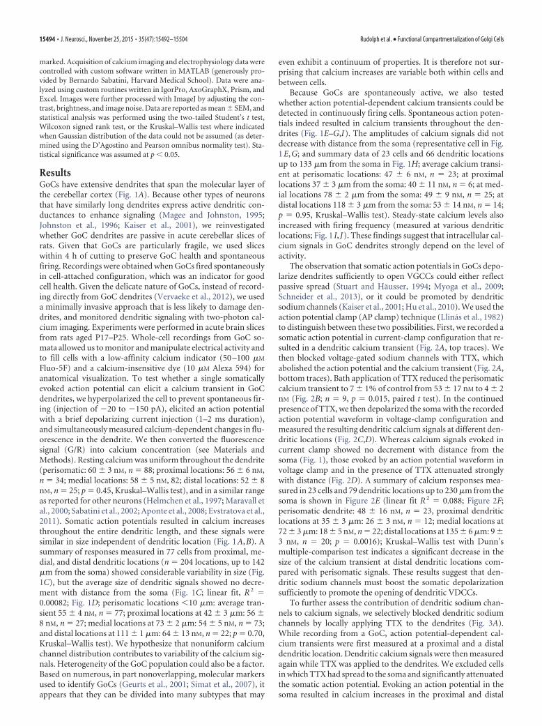
ResultsGoCs have extensive dendrites that span the molecular layer ofthe cerebellar cortex (Fig. 1A). Because other types of neuronsthat have similarly long dendrites express active dendritic con-ductances to enhance signaling (Magee and Johnston, 1995;Johnston et al., 1996; Kaiser et al., 2001), we reinvestigatedwhether GoC dendrites are passive in acute cerebellar slices ofrats. Given that GoCs are particularly fragile, we used sliceswithin 4 h of cutting to preserve GoC health and spontaneousfiring. Recordings were obtained when GoCs fired spontaneouslyin cell-attached configuration, which was an indicator for goodcell health. Given the delicate nature of GoCs, instead of record-ing directly from GoC dendrites (Vervaeke et al., 2012), we useda minimally invasive approach that is less likely to damage den-drites, and monitored dendritic signaling with two-photon cal-cium imaging. Experiments were performed in acute brain slicesfrom rats aged P17–P25. Whole-cell recordings from GoC so-mata allowed us to monitor and manipulate electrical activity andto fill cells with a low-affinity calcium indicator (50 –100 �M
Fluo-5F) and a calcium-insensitive dye (10 �M Alexa 594) foranatomical visualization. To test whether a single somaticallyevoked action potential can elicit a calcium transient in GoCdendrites, we hyperpolarized the cell to prevent spontaneous fir-ing (injection of �20 to �150 pA), elicited an action potentialwith a brief depolarizing current injection (1–2 ms duration),and simultaneously measured calcium-dependent changes in flu-orescence in the dendrite. We then converted the fluorescencesignal (G/R) into calcium concentration (see Materials andMethods). Resting calcium was uniform throughout the dendrite(perisomatic: 60 � 3 nM, n � 88; proximal locations: 56 � 6 nM,n � 34; medial locations: 58 � 5 nM, 82; distal locations: 52 � 8nM, n � 25; p � 0.45, Kruskal–Wallis test), and in a similar rangeas reported for other neurons (Helmchen et al., 1997; Maravall etal., 2000; Sabatini et al., 2002; Aponte et al., 2008; Evstratova et al.,2011). Somatic action potentials resulted in calcium increasesthroughout the entire dendritic length, and these signals weresimilar in size independent of dendritic location (Fig. 1A,B). Asummary of responses measured in 77 cells from proximal, me-dial, and distal dendritic locations (n � 204 locations, up to 142�m from the soma) showed considerable variability in size (Fig.1C), but the average size of dendritic signals showed no decre-ment with distance from the soma (Fig. 1C; linear fit, R 2 �0.00082; Fig. 1D; perisomatic locations �10 �m: average tran-sient 55 � 4 nM, n � 77; proximal locations at 42 � 3 �m: 56 �8 nM, n � 27; medial locations at 73 � 2 �m: 54 � 5 nM, n � 73;and distal locations at 111 � 1 �m: 64 � 13 nM, n � 22; p � 0.70,Kruskal–Wallis test). We hypothesize that nonuniform calciumchannel distribution contributes to variability of the calcium sig-nals. Heterogeneity of the GoC population could also be a factor.Based on numerous, in part nonoverlapping, molecular markersused to identify GoCs (Geurts et al., 2001; Simat et al., 2007), itappears that they can be divided into many subtypes that may
even exhibit a continuum of properties. It is therefore not sur-prising that calcium increases are variable both within cells andbetween cells.
Because GoCs are spontaneously active, we also testedwhether action potential-dependent calcium transients could bedetected in continuously firing cells. Spontaneous action poten-tials indeed resulted in calcium transients throughout the den-drites (Fig. 1E–G,I). The amplitudes of calcium signals did notdecrease with distance from the soma (representative cell in Fig.1E,G; and summary data of 23 cells and 66 dendritic locationsup to 133 �m from the soma in Fig. 1H; average calcium transi-ent at perisomatic locations: 47 � 6 nM, n � 23; at proximallocations 37 � 3 �m from the soma: 40 � 11 nM, n � 6; at med-ial locations 78 � 2 �m from the soma: 49 � 9 nM, n � 25; atdistal locations 118 � 3 �m from the soma: 53 � 14 nM, n � 14;p � 0.95, Kruskal–Wallis test). Steady-state calcium levels alsoincreased with firing frequency (measured at various dendriticlocations; Fig. 1 I, J). These findings suggest that intracellular cal-cium signals in GoC dendrites strongly depend on the level ofactivity.
The observation that somatic action potentials in GoCs depo-larize dendrites sufficiently to open VGCCs could either reflectpassive spread (Stuart and Hausser, 1994; Myoga et al., 2009;Schneider et al., 2013), or it could be promoted by dendriticsodium channels (Kaiser et al., 2001; Hu et al., 2010). We used theaction potential clamp (AP clamp) technique (Llinas et al., 1982)to distinguish between these two possibilities. First, we recorded asomatic action potential in current-clamp configuration that re-sulted in a dendritic calcium transient (Fig. 2A, top traces). Wethen blocked voltage-gated sodium channels with TTX, whichabolished the action potential and the calcium transient (Fig. 2A,bottom traces). Bath application of TTX reduced the perisomaticcalcium transient to 7 � 1% of control from 53 � 17 nM to 4 � 2nM (Fig. 2B; n � 9, p � 0.015, paired t test). In the continuedpresence of TTX, we then depolarized the soma with the recordedaction potential waveform in voltage-clamp configuration andmeasured the resulting dendritic calcium signals at different den-dritic locations (Fig. 2C,D). Whereas calcium signals evoked incurrent clamp showed no decrement with distance from thesoma (Fig. 1), those evoked by an action potential waveform involtage clamp and in the presence of TTX attenuated stronglywith distance (Fig. 2D). A summary of calcium responses mea-sured in 23 cells and 79 dendritic locations up to 230 �m from thesoma is shown in Figure 2E (linear fit R 2 � 0.088; Figure 2F;perisomatic dendrite: 48 � 16 nM, n � 23, proximal dendriticlocations at 35 � 3 �m: 26 � 3 nM, n � 12; medial locations at72 � 3 �m: 18 � 5 nM, n � 22; distal locations at 135 � 6 �m: 9 �3 nM, n � 20; p � 0.0016); Kruskal–Wallis test with Dunn’smultiple-comparison test indicates a significant decrease in thesize of the calcium transient at distal dendritic locations com-pared with perisomatic signals. These results suggest that den-dritic sodium channels must boost the somatic depolarizationsufficiently to promote the opening of dendritic VDCCs.
To further assess the contribution of dendritic sodium chan-nels to calcium signals, we selectively blocked dendritic sodiumchannels by locally applying TTX to the dendrites (Fig. 3A).While recording from a GoC, action potential-dependent cal-cium transients were first measured at a proximal and a distaldendritic location. Dendritic calcium signals were then measuredagain while TTX was applied to the dendrites. We excluded cellsin which TTX had spread to the soma and significantly attenuatedthe somatic action potential. Evoking an action potential in thesoma resulted in calcium increases in the proximal and distal
15494 • J. Neurosci., November 25, 2015 • 35(47):15492–15504 Rudolph et al. • Functional Compartmentalization of Golgi Cells

dendrites (Fig. 3B, top traces; 3C, �Ca 2� � 36 � 6 nM, n � 12and 30 � 3 nM, n � 16; p 0.05, Kruskal–Wallis test with Dunn’smultiple-comparison post test). Dendritic TTX application de-creased the amplitude of distal dendritic calcium signals (Fig.3C,D; to 50 � 5% of control to 15 � 2 nM, n � 16; p � 0.05,Kruskal–Wallis test) without affecting calcium signals in proxi-mal dendrites (Fig. 3B, bottom traces, and 3D; 92 � 7% of con-trol, 31 � 5 nM, n � 12; p 0.05, Kruskal–Wallis test). The distalTTX-dependent decrease in calcium influx recovered within5–10 min (to 115 � 14% of control, 35 � 6 nM, n � 16; p 0.05compared with control, Kruskal–Wallis test, summary data inFig. 3D). These results suggest that local inhibition of voltage-gated sodium channels prevents the propagation of the somaticaction potential and the subsequent activation of VGCCs. To-gether, these experiments establish that dendritic sodium chan-nels promote the activation of VGCCs in GoC dendrites.
Different types of VGCCs can serve distinct functional rolesand can be differentially distributed within neurons (Johnston etal., 1996). To determine which types of VGCCs are present inGoC dendrites, we evaluated the effect of specific calcium chan-nel antagonists on calcium signals evoked by short bursts of ac-tion potentials in GoCs that were hyperpolarized to suppressspontaneous firing. VGCC inhibitors were applied locally to ei-ther proximal or distal regions, and calcium responses were mea-sured in the corresponding dendritic region (Fig. 4A). To identifypotential VGCC types in GoC dendrites, we started out with amixture of VGCC blockers. We chose a combination of L-, N-,and P-type blockers for several reasons: (1) these calcium chan-nels are the most common high-voltage activated calcium chan-nels in neurons; (2) the blockers are well characterized; and (3)the conditions for activation of these channel types are similar.The blockade of P-type (�-agatoxin IVA, 2 �M), N-type (�-conotoxin GVIA, 5 �M), and L-type (nimodipine, 100 �M)VGCCs decreased the proximal calcium signal to 44 � 8% ofcontrol (Fig. 4B, left traces, and 4E, n � 7; p � 0.016, Wilcoxonsigned-rank test), as well as broadening the action potential anddecreasing the afterhyperpolarization following the action poten-tial (see below). However, blocking P-, N-, and L-type calciumchannels did not significantly alter distal calcium signals (Fig. 4B,right traces, and 4E; 85 � 9% of control, p � 0.44, n � 8) and hadlittle effect on the action potential waveforms measured at thesoma (see below). In contrast, local blockade of either R-type(SNX-482, 5 �M) or T-type (TTA-P2, 10 �M) calcium channelsdid not alter proximal calcium signals (Fig. 4C and 4D, respec-tively, left traces, and 4E; for SNX-482: 103 � 5% of control, n �7, p � 0.73; for TTA-P2: 97 � 13% of control, p � 1.0), but bothdecreased distal calcium signals by approximately half (Fig. 4C,right traces, and 4E; for SNX-482 to 55 � 9% of control, n � 8,p � 0.016; for TTA-P2 to 60 � 8% of control, n � 9, p � 0.004).These findings establish that R-type and T-type channels localizeselectively to the distal dendrites, whereas some combination of
B
C D
dist
med
prox
0 50 100
300
Distance from Soma (μm)
∆ [C
a2+ ]
(nM
)
50 nM
20 μm
A
prox
10 - 4
9 μm
50 - 9
9 μm
> 100
μm
80
∆ [C
a2+ ]
(nM
)
40 nM 20 μm
20 nM
dist
med
prox
100 ms
80
prox
10 - 4
9 μm
50 - 9
9 μm
> 100
μm
∆ [C
a2+ ]
(nM
)
E F
G H
I J
0 10 20 30
600
[Ca2
+ ]ba
selin
e (n
M)
frequency (Hz)
100 ms
0
0
0 0
0 nM
0 nM
100 nM200 ms
1 s
-600
-60
0
-60
0
-600
Figure 1. Action potential-dependent calcium transients in Golgi cell dendrites. A, Two-photon image of a GoC filled with Alexa 594 and the calcium-sensitive dye Fluo-5F. Horizontalbars represent the locations of line scans along the dendrite. B, Top trace, Action potentialevoked by a somatic current injection. Bottom traces, Average action potential-dependentcalcium transients recorded at the locations indicated in A. C, Amplitudes of dendritic calciumtransients dependent on distance from the soma. D, Summary data of 77 cells with calcium
4
transient amplitudes binned according to distance from soma. E, Two-photon image of a spon-taneously active GoC with locations of line scans indicated. F, Top trace, Spontaneous actionpotentials recorded at the soma. Bracket indicates 0 mV and �60 mV. Bottom trace, Calciumsignals recorded at a dendritic location indicated in E. Broken line indicates 0 nM baselinecalcium. G, Top traces, Aligned spontaneous action potentials (gray traces) and average mem-brane potential (black trace). Bottom, Average calcium transients in response to spontaneousaction potentials recorded at locations indicated in E. H, Summary data of 23 cells. Binnedamplitudes of calcium transients. I, Example traces of a GoC firing at different frequencies andassociated dendritic calcium signals. Broken line indicates 0 nM baseline calcium. J, Amplitudesof dendritic baseline calcium concentration for different firing frequencies.
Rudolph et al. • Functional Compartmentalization of Golgi Cells J. Neurosci., November 25, 2015 • 35(47):15492–15504 • 15495

P-, N-, and/or L-type calcium channels is present in proximalregions.
Based on the observation that both SNX-482 and TTA-P2decrease distal calcium signals by approximately half, we hypoth-esized that coapplication of both blockers would completelyabolish the signal. Because R- and T-type calcium channels local-ize selectively to the distal dendrites and have only minor effectson the somatic action potential waveforms (also see below), webath-applied SNX-482 (1 �M) and TTA-P2 (4 �M). The coappli-cation of these antagonists did not alter the proximal calciumsignal (Fig. 5A, left traces, and 5D; 96 � 12% of control, n � 5,p � 0.62, Wilcoxon signed rank test) but reduced the distal cal-cium signal (Fig. 5A, right traces, and 5D; to 18 � 4% of control,n � 10, p � 0.002). In contrast, blocking either L- or P-typecalcium channels with bath-applied nimodipine (20 �M) or�-agatoxin IVA (1 �M), respectively, had no effect on proximalor distal calcium transients (Fig. 5B,D; nimodipine: proximal106 � 11% of control, n � 5, p � 0.40, distal 97 � 6% of control,n � 8, p � 0,47, Fig. 5C,D; �-agatoxin IVA: proximal 111 � 10%of control, n � 7, p � 0.55; distal 103 � 11% of control, n � 14,p � 0.99). These findings confirm that calcium signals in distaldendrites rely mainly on the activation of dendritic R- and T-typecalcium channels, whereas L- and P-type calcium channels do notcontribute to calcium signals in either proximal or distal den-drites.
In GoCs, calcium regulates the frequency and regularity ofspontaneous firing by activating KCa channels (Solinas et al.,2007; Hull et al., 2013). GoCs are typically spontaneously active,and application of the SK antagonist apamin (200 nM) increasedthe firing frequency to 242 � 59% of control (n � 6, p � 0.031,
measured in regularly firing episodes; Table 1) and, in some cases,changed the regular firing pattern into a burst firing pattern thateventually resulted in depolarization block (3 of 6 cells). Apaminalso decreased the amplitude of the afterhyperpolarization (25 �1% of control, n � 6, p � 0.03, measured at 50 ms after the actionpotential peak; Fig. 6A; Table 1), but not the action potentialwidth (98 � 4% of control, n � 6, p � 0.68). The BK channelantagonist paxilline (10 �M) also significantly increased the firingfrequency (to 213 � 39% of control, n � 9, p � 0.019; Table 1)and decreased the afterhyperpolarization (83 � 9% of control,n � 9; p � 0.008 Fig. 6B; Table 1). In addition, paxilline increasedthe width of the action potential (119 � 13% of control, n � 9,p � 0.027; Table 1). KCa channels are sometimes opened byclosely associated calcium channels (Lancaster and Nicoll, 1987;Berkefeld et al., 2006; Engbers et al., 2012) and, in other instances,by VGCCs that are more loosely coupled (Alle et al., 2011; Jonesand Stuart, 2013). To determine the roles of different types ofVGCCs in spontaneous firing by regulation of KCa channels, weapplied specific calcium channel blockers. Inhibition of N-typecalcium channels doubled firing frequency (to 200 � 33% ofcontrol, n � 12, p � 0.0005; Fig. 6G; Table 1), and in a subset ofcells resulted in a transition to burst firing and/or depolarizationblock (7 of 12 cells; Fig. 6C). Importantly, N-type channel inhi-bition also increased action potential width (123 � 13% of con-trol, n � 11, p � 0.001; Fig. 6H; Table 1) and strongly decreasedafterhyperpolarization (53 � 6% of control, n � 11, p � 0.001;Fig. 6I). The effect of �-conotoxin GVIA (2 �M) on action po-tential width resembled the effect on BK channel inhibition withpaxilline, whereas the effect on afterhyperpolarization and firingpattern resembled inhibition of SK channels with apamin. Con-
100 ms
20 nM
current clamp
TTX AP-clamp
prox
prox
med
dist0
80
10 - 4
9 μm
50 - 9
9 μm
> 100
μm prox
TTX AP-C
A
D
E
20 nM
current clamp
TTX current clamp
100 ms
*
F
0
80
∆ [C
a2+ ]
(nM
)
prox
TTX prox
CC
*
B0
0
200
∆ [C
a2+ ]
(nM
)
Distance from Soma (μm)100 200
C
∆ [C
a2+ ]
(nM
)
-60
0
-60
0
-60-30
Figure 2. Dendritic sodium channels promote dendritic calcium signals. A, Top traces, The somatic action potential evokes a calcium transient in the proximal dendrite. Bottom, TTX abolishes boththe action potential and the calcium signal. B, Summary data of calcium signals in the presence and in the absence of TTX. C, Waveform of the somatically evoked action potential in current-clampconfiguration and the resulting calcium transient in the proximal dendrite. D, Top, Action potential waveform fed back to the GoC in voltage-clamp configuration in the presence of TTX. Bottom,Calcium transients measured at proximal (gray), medial (42 �m, blue), and distal (112 �m, red) dendritic locations. E, The amplitude of calcium transients in dependence on distance from the somaevoked in AP-clamp configuration. F, Binned summary data of calcium transient amplitudes. *Denotes statistical significance.
15496 • J. Neurosci., November 25, 2015 • 35(47):15492–15504 Rudolph et al. • Functional Compartmentalization of Golgi Cells

versely, the blockade of either L-type (Fig. 6D) or P-type calciumchannels (Fig. 6E) or the blockade of P-type (Fig. 6E) or R-typeand T-type calcium channels (Fig. 6F) had little, if any, effect onthe firing frequency (L-type: 114.8 � 10.8% of control, n � 7, p �0.23; P-type: 94.2 � 7.8% of control, n � 7, p � 0.47; R/T-type:128.3 � 13.1%, n � 16, p � 0.01; Table 1), the action potentialwidth (L-type: 101 � 1% of control, n � 7, p � 0.81; P-type:103 � 2% of control, n � 7, p � 0.22, R/T-type: 108 � 4% ofcontrol, n � 16, p � 0.073; Table 1), or the afterhyperpolarization(L-type: 95 � 3% of control, n � 7, p � 0.16; P-type: 99 � 6% ofcontrol, n � 7, p � 0.69; R/T-type: 85 � 5% of control, n � 16,p � 0.02; Fig. 6G,H; Table 1). These findings suggest that N-typecalcium channels located near the soma are the primary calciumsource that controls action potential width, afterhyperpolariza-tion, and the frequency and regularity of spontaneous firing,whereas the effect of R- and T-type channel blockade on actionpotential firing and afterhyperpolarization is small. The lack ofeffect on action potential width suggests that T- and/or R-typechannels do not signal to low-affinity BK channels in GoCs.
Because endogenous calcium-binding proteins control cal-cium signaling and calcium-activated processes in cells(Schwaller, 2010; Eggermann et al., 2012; Matthews and Dietrich,2015), including the interaction of VGCCs with target proteins,we next investigated calcium dynamics in GoCs. The types andconcentrations of calcium binding proteins in GoCs are notknown, although it is recognized that they lack the classic calciumbinding proteins parvalbumin, calbindin, and calretinin that arepresent at high concentrations in many types of interneurons(Geurts et al., 2001; Simat et al., 2007). Because the level ofcalcium-binding proteins can spatially restrict the spread of cal-cium within a cell (Goldberg et al., 2003; Soler-Llavina and Saba-
tini, 2006) and can decrease the amplitudeand/or prolong the decay of bulk calciumlevels (Lee et al., 2000; Aponte et al.,2008), it is important to determine the en-dogenous buffer capacity in GoCs. Wetook advantage of a widely used method(Neher and Augustine, 1992; Helmchen etal., 1996; Maravall et al., 2000; Sabatini etal., 2002; Brenowitz and Regehr, 2007) inwhich action potential-evoked calciumincreases are monitored while a calciumindicator (200 �M Fluo-5F) along with ared calcium-insensitive dye (10 �M Alexa594) fills the cell to increase the total buf-fer capacity (Fig. 7A). Dye loading wasmonitored at the proximal dendriteshortly after break-in (within 30 – 45 s)until full equilibration of the dye wasachieved (typically 40 min). Added buf-fer capacity, �ind, was calculated as previ-ously described (Brenowitz and Regehr,2007), with �ind � [Fluo-5F](t)/Kd,[Fluo-5F](t) � (R(t)/Rmax)[Fluo 5F]pi-
pette, where [Fluo-5F](t) and R(t) are theindicator concentration and the Alexa 594fluorescence intensity at time t, respec-tively, Kd is the dissociation constant ofthe indicator, and [Fluo-5F]pipette(t) andRpipette(t) are the indicator concentrationand the intensity of Alexa 594 at time Rmax
the intensity at steady state. Figure 7Bshows a typical experiment in which �ind
continuously increases, resulting in a decrease in the actionpotential-evoked calcium signal �[Ca 2�]. As �ind increases, theaction potential evoked calcium transients that become smallerand slower (Fig. 7C). A plot of 1/�[Ca 2�] versus �ind is fit with aline and extrapolated to determine the peak calcium transient inthe absence of added buffer (�[Ca 2�]0, the y-intercept, which is183 nM for this cell), and the endogenous buffer capacity in theabsence of added buffer (�0, the x-intercept, which is 44 for thiscell; Fig. 7D). The average �[Ca 2�]0 was 143 � 23 nM in GoCs(range 55–295 nM, n � 10; Fig. 7E) and �0 was 66 � 9 (range38 –117; Fig. 7E), which is low compared with many other in-terneurons (see Discussion). Because intracellular calcium buf-fers bind free calcium and thereby can decrease the peak of thecalcium transient, this low buffer capacity allows even a modestdensity of VGCCs to produce large increases in calcium (see Dis-cussion). Importantly, to ensure that calcium influx remains con-stant over the course of the experiment, we also determinedaction potential width shortly after and 20 min after breaking intothe cell. Action potential width did not change after introductionof Fluo-5F (0.62 � 0.05 ms and 0.66 � 0.06 ms, n � 10; p � 0.074,paired t test).
Another important aspect of calcium signaling in GoCs is theproximity of calcium channels to KCa channels. The ability ofGoCs to fire spontaneously and regularly requires calcium entrythrough N-type calcium channels to activate KCa channels (Fig.6). In other types of neurons, the interaction between VGCCs andcalcium-dependent activation of potassium channels requires aclose spatial relationship, so-called nanodomains or microdo-mains (Lancaster and Nicoll, 1987; Marrion and Tavalin, 1998;Ngo-Anh et al., 2005; Fakler and Adelman, 2008; Jones and Stu-art, 2013). One way of probing the distance between N-type cal-
50 nM
proximal dendrite
100 ms
proximal dendrite+ dendritic TTX
25 nM
distal dendrite distal dendrite+ dendritic TTX
B C
D20 μm
dendriticTTXAproximal AP proximal AP
+ dendritic TTX
contr
ol
dend
ritic
TTXco
ntrol
recov
ery
∆ [C
a2+ ]
(nM
)
*
0
proximal distal
dend
ritic
TTX
80
-60
0
-60
0
Figure 3. Local inhibition of dendritic sodium channels attenuates dendritic calcium signals. A, Schematic of the experimental setup.Action potentials are evoked somatically, whereas calcium transients are recorded at a proximal and a distal dendritic location. The puffpipette (white lines) is placed near the distal dendrite, and TTX is applied locally (cyan dashed circle). B, Somatically evoked action potential,proximal calcium transient (black), and distal calcium transient (recorded 72 �m from soma, blue). C, Somatic action potential duringdendritic TTX application, proximal calcium transient (gray), and distal calcium transient (cyan). D, Summary data of calcium signals.*Denotes statistical significance.
Rudolph et al. • Functional Compartmentalization of Golgi Cells J. Neurosci., November 25, 2015 • 35(47):15492–15504 • 15497

cium channels and KCa channels is to introduce exogenousbuffers. If high concentrations of fast calcium buffers, such asBAPTA, are required to prevent calcium activation of channels, thenthe coupling distance between N-type calcium channels and KCa
channels is predicted to be short (Adler et al., 1991). Surprisingly, wefound that low BAPTA concentrations (1 mM) had no effect onaction potential frequency (5.2 � 0.4 Hz to 5.5 � 0.7 Hz, n � 11, p �0.23; Fig. 8A,D; Table 2), and only minor effects on action potentialwidth (113 � 5% of control, n � 11, p � 0.013; Fig. 8A,E; Table 2)and afterhyperpolarization (87 � 4% of control, n � 11, p � 0.009;Fig. 8A,F), whereas higher concentrations (10 mM) resulted in apronounced increase in action potential frequency (273 � 28% ofcontrol, n � 4, p � 0.002; Fig. 8B,D; Table 2), bursting, an increasein action potential width (156 � 8%, n � 4, p � 0.004, Fig. 8B,E)and a decrease in afterhyperpolarization (reduced to 14 � 6% ofcontrol, n � 4, p � 0.003; Fig. 8B,F). High concentrations (10 mM)of the slow buffer EGTA, however, had no significant effect on actionpotential frequency (5.2 � 0.6 Hz to 6.7 � 1.3 Hz, n � 9, p � 0.11;Fig. 8C,D), and only a minor effect on afterhyperpolarization (81 �9%, n � 9, p � 0.02; Fig. 8C,E) and width (118 � 4%, n � 9, p �0.013; Fig. 8C,F), suggesting that the majority of VGCCs are closelycoupled to KCa channels (Fig. 6).
It is possible to estimate the distance between VGCCs and KCa
channels as distance � (DCa/ko[buffer]) 1/2, where [buffer] is the
buffer concentration, DCa is the diffusion coefficient for calcium(2.2 � 10�10 m 2s�1), and k0 is the binding rate of the addedbuffer, which is 4 � 10 8 M�1s�1 for BAPTA and 1 � 10 7
M�1s�1 for EGTA. We estimate that the distance betweenN-type calcium channels and KCa channels is �20 nm (predicteddistance for 1 mM BAPTA � 23 nm), given the small effect of 1mM BAPTA and 10 mM EGTA, the lack of effect of 200 �M
Fluo-5F (Fig. 7), and the strong effect of 10 �M BAPTA on spon-taneous firing and action potential waveform.
The observation that VGCC types in GoCs are segregated sug-gests that they carry location-specific functional roles. We haveestablished that N-type calcium channels near the soma are es-sential to regulating GoC output. We next examined the func-tional role of VGCCs expressed in GoC dendrites. T-type calciumchannels, which we found are localized to distal GoC dendrites(Figs. 4, 5), have been implicated in burst firing in many types ofneurons (Huguenard and Prince, 1992; Molineux et al., 2006;Pressler et al., 2013). To test whether T-type channels in GoCs cancontribute to rebound bursting, we allowed GoCs to fire spontane-ous action potentials, and then hyperpolarized the cells for 500 ms to�84 � 3 mV (n � 19) to relieve inactivation of T-type calciumchannels. Returning the cells to their resting membrane potential led
A B
prox
0
% o
f con
trol
R-type
200 ms
400 nM
200nM
100 nM
200 nM
100 nM
100 nM
R-type
T-type
P, N, L-type
P, N, L-type
distalapplication
T-type
C
prox
prox
proximalapplication
100
proximal distalapply + measure
D
distal
distal
distal
E
** *
Figure 4. Different calcium channels mediate proximal and distal calcium transients. A,Schematic of experiment. Bursts of action potentials are evoked somatically. Calcium transientsare recorded at a proximal and a distal location; specific calcium channel antagonists are appliedlocally. B–D, Calcium transients in the presence and absence of calcium channel antagonistsapplied to the proximal and distal dendrites. B, Antagonists of P-type (�-agatoxin IVA), N-type(�-conotoxin GVIA), and L-type (nimodipine) channels were coapplied. C, Calcium transients inthe presence and absence of the R-type channel antagonist SNX-482. D, Application of theT-type channel antagonist TTA-P2. E, Summary data of calcium channel pharmacology.*Denotes statistical significance.
100 nM100 nM100 ms
R-type + T-type channel antagonistsA
C
proximal distal
B
100 nM 100 nM
50 nM
0
100
% o
f con
trol
proxdista
lprox
distal
proxdista
l
R/T-type
L-type P-typeD
L-type channel antagonist
P-type channel antagonist
100 nM
*
Figure 5. Bath application of R-type and T-type but not L-type or P-type channel blockerssuppresses dendritic calcium signals. A, Coapplication of R-type and T-type channel antagonistsdoes not affect the proximal calcium transient but diminishes the distal dendritic calcium tran-sient. B, Block of L-type calcium channels or (C) P-type calcium channels has no effect on theamplitude of the proximal and distal calcium transients. D, Summary data of the effect ofcalcium channel antagonists on the amplitude of dendritic calcium signals. *Denotes statisticalsignificance.
15498 • J. Neurosci., November 25, 2015 • 35(47):15492–15504 Rudolph et al. • Functional Compartmentalization of Golgi Cells

to a transient increase in firing frequency (to 166 � 14% of control,range 106%–344%, from 6.5 � 0.9 Hz at baseline measured forseveral seconds before the hyperpolarizing step to 9.7 � 1.2 Hz mea-sured during the 500 ms following the pulse, n � 19, p � 0.0001; Fig.9A). TTA-P2 strongly attenuated this increase in firing frequency(112 � 6%, range 99%–131%, from 6.1 � 1.1 Hz at baseline, to6.7 � 1.1 Hz, n � 9, p � 0.04; Fig. 9B), indicating that it was medi-ated primarily by T-type calcium channels. The variability in theextent of bursting we observed could be explained by differences inT-type channel availability and some heterogeneity in VGCC ex-pression within the GoC population. Figure 9C shows the cumula-tive probability histogram of the ratio of the posthyperpolarizationfiring frequency (frequencyburst) divided by the baseline firing fre-quency before hyperpolarization (frequencypre, n � 19; black trace)and in the presence of TTA-P2 (n � 9, blue trace, p � 0.001, Kolm-ogorov–Smirnov test). We also tested whether dendritic calciumchannels influence the integration of grc inputs onto distal GoCdendrites. Because transmitter release at parallel fiber terminals par-
tially depends on R-type calcium channels (Mintz et al., 1995), welimited our investigation to T-type channels. We found that TTA-P2did not affect synaptic responses (parallel fiber EPSC in TTA-P2 was107 � 13% of control, n � 6, p � 0.84, paired-pulse ratio was 97 �10% of control, n � 6, p � 0.57, paired t test, data not shown). Incurrent clamp, we hyperpolarized GoCs to �65 mV to preventspontaneous spiking and then evoked parallel fiber EPSPs by placinga stimulus electrode in the molecular layer of the cerebellar cortex.We found that, when no inhibitor was applied, the EPSP amplituderemained constant over time (102.2 � 6.2% of control, n � 10, p �0.64; Fig. 9D). Conversely, inhibition of T-type calcium channelsreduced the EPSP amplitude to 80.7 � 7.8% of control (n � 19, p �0.007; Fig. 9E; cumulative probability histogram of the ratio of EPSPamplitudes at the indicated time points, p � 0.001, Kolmogorov–Smirnov test; Fig. 9F). These data suggest that T-type calcium chan-nels located in distal GoC dendrites can boost synaptic inputs fromgrcs onto GoCs.
Table 1. Action potential properties: potassium channel and calcium channel antagonists
Control/SK-type Control/BK-type Control/N-type Control/L-type Control/R/T-type Control/P-type
Frequency (Hz) 6.4 � 0.8 3.6 � 0.6 5.7 � 0.7 7.4 � 1.2 6.4 � 0.5 7.1 � 1.515.5 � 3.7 6.2 � 0.6 10.9 � 1.7 8.1 � 1.1 7.8 � 0.6 6.1 � 1.0
AP width (ms) 0.60 � 0.04 0.81 � 0.02 0.60 � 0.03 0.51 � 0.04 0.70 � 0.06 0.60 � 0.050.60 � 0.05 0.96 � 0.05 0.76 � 0.07 0.51 � 0.05 0.77 � 0.07 0.60 � 0.05
AHP (mV) 8.8 � 1.0 9.1 � 0.6 11.8 � 0.7 11.2 � 1.1 13.5 � 0.7 11.8 � 0.82.2 � 0.6 8.0 � 0.7 6.3 � 0.8 10.6 � 1.1 11.2 � 0.6 11.5 � 0.8
N-type antagonist
L-type antagonist
R- and T-type antagonist
control SK-type antagonist
P-type antagonist
0
1
2
3 *
freq
uenc
y (n
orm
)
0.0
1.0
2.0
AP
wid
th (n
orm
)
0.0
0.5
1.0
*
*
* AH
P (n
orm
)
SK-type
N-type
R/T-type
P-type
L-typ
e
A
B
C F
G H
250 ms 20 ms
**
D
EBK-type antagonist
BK-type
* *
SK-type
N-type
R/T-type
P-type
L-typ
e
BK-type
SK-type
N-type
R/T-type
P-type
L-typ
e
BK-type
I
*
-60
0
-60
0
-60
0
-60
0
-60
0
-60
0
Figure 6. The effect of calcium-dependent potassium channel and calcium channel inhibition on Golgi cell firing. A, Spontaneously firing GoC in the absence (black trace, left) or in the presenceof the SK-type potassium channel blocker apamin (blue trace, right). B, Same experiment but blue trace in the presence of the BK-type potassium channel blocker paxilline. C, The N-type channelblocker �-conotoxin GVIA. D, The L-type calcium channel antagonist nimodipine. E, The P-type calcium channel blocker �-agatoxin IVA. F, The R-type and T-type channel blockers SNX-482 andTTA-P2, respectively. G, Summary data of action potential frequency. H, Action potential (AP) width. I, Afterhyperpolarization (AHP). *Denotes statistical significance.
Rudolph et al. • Functional Compartmentalization of Golgi Cells J. Neurosci., November 25, 2015 • 35(47):15492–15504 • 15499

DiscussionHere we report that differential distribution of various types ofVGCCs within GoCs leads to functional compartmentalization.To our surprise, we found that GoC dendrites are electricallyactive and that voltage-gated sodium channels help to activate theR- and T-type calcium channels that are present in distal den-drites but absent near the soma. Dendritic T-type channels me-diate rebound bursting and boost synaptic inputs from grcs,thereby amplifying feedback inhibition (grc ¡ GoC ¡ grc). Incontrast, N-type calcium channels are absent from distal den-drites but are present near the soma where they control the open-ing of KCa channels and thereby regulate spontaneous firing.
Active dendrites in GoCsCounter to previous results, we find that that GoC dendrites areactive and contain both voltage-gated sodium and calcium chan-nels that allow somatic depolarization to propagate along theentire length of GoC dendrites to enable calcium influx (Fig. 1). Itis likely that the fragile nature of GoCs, coupled with low calciumchannel density, hampered the detection of active dendriticproperties. For example, even modest dendritic depolarizationcan inactivate low voltage-activated T-type calcium channels.Moreover, low channel density complicated the optical detectionof dendritic calcium signals, which were strongly attenuatedwhen using typical concentrations of calcium indicator (100 –200 �M). We thus limited our experiments to spontaneously ac-tive GoCs and used low calcium indicator concentrations.
Active dendrites provide diverse functions in many neuronal celltypes, including interneurons (Goldberg and Yuste, 2005), and wehave begun to examine some of the possible roles of dendritic ion
channels in GoCs. GoC activity depends on both the intrinsic prop-erties of GoCs and the excitation they receive from synaptic inputonto their dendrites. However, assuming GoC dendrites are passive,synaptic input to distal dendrites would be ineffective at drivingfiring due to passive attenuation. In pyramidal neurons, differen-tially distributed voltage-dependent conductances produce activedendrites that compensate for this spatial handicap and selectivelystrengthen distal synaptic inputs (Branco and Hausser, 2011), butlittle is known whether interneurons use similar strategies. In GoCdendrites, T-type calcium channels might enhance parallel fiber in-puts to distal apical dendrites, allowing distal voltage signals to prop-agate to the soma and increasing the likelihood of evoking an actionpotential. T-type calcium channels in GoCs also promote reboundburst firing. Similar burst firing occurs in other types of neurons andcontributes to synaptic plasticity and rhythmic activity during cer-tain behavioral states (Aizenman et al., 1998; Williams and Stuart,1999; Cueni et al., 2008; Coulon et al., 2009; Errington et al., 2010).GoCs exhibit bursts of activity in vivo during certain behaviors(Heine et al., 2010), but the mechanism underlying this bursting isunknown. In order for T-type calcium channels to be available topromote bursting, neurons must be hyperpolarized. In GoCs, this
-200 0 200 4000
4
8
0
0
200
400
0 20 400
100
40 nM100 ms
0
100
01 min
10 min
45 min
D E
A B
C
0
300
∆[C
a]0
(nM
)
∆[C
a]-1
(μM
)
∆[C
a] (n
M)
adde
d
time (min)
added
Figure 7. Low endogenous calcium buffer capacity in Golgi cells. A, Schematic showingexperimental setup for measuring calcium buffer capacity. Action potential-evoked calciumtransients are measured continuously at a proximal dendritic site until the calcium-insensitivered dye has equilibrated. B, Top, Added buffer capacity. Bottom, Peak calcium concentration inresponse to a single action potential during loading of the cell with Alexa 594 and Fluo-5F. C,Calcium transients recorded at time points indicated. D, Relationship between �[Ca 2�] �1
and added buffer capacity � during dye loading. Arrow indicates endogenous buffer capacity. E,Summary data of endogenous buffer capacity and amplitude of the calcium signal in the ab-sence of exogenous buffer.
10 mM BAPTA
1 mM BAPTA
0
1
2
3
AP
freq
uenc
y (n
orm
)
0.0
1.0
2.0*
* *
AP
wid
th (n
orm
)
0.0
0.5
1.0
*
*
AH
P (n
orm
)
10 BAPTA
1 BAPTA
10 EGTA
A
10 BAPTA
1 BAPTA
10 EGTA
10 BAPTA
1 BAPTA
10 EGTA
10 mM EGTA
250 ms
B
C
D*
*
E F
0-60
0
-60
0
-60
20 ms
Figure 8. The effect of exogenous calcium buffers on firing pattern and action potentialwaveform in Golgi cells. A–C. Left, Spontaneous action potentials directly after break-in (blacktraces) and after introduction of the indicated concentrations of exogenous buffer (blue traces).Right, Average action potentials on an expanded time scale. D, Normalized action potentialfiring rate (left). E, Action potential width. F, Afterhyperpolarization after introduction of exog-enous buffers. *Denotes statistical significance.
Table 2. Action potential properties: calcium buffers
Control/10 BAPTA Control/1 BAPTA Control/10 EGTA
Frequency (Hz) 7.4 � 0.9 5.2 � 0.4 5.2 � 0.619.8 � 1.5 5.5 � 0.7 6.7 � 1.3
AP width (ms) 0.73 � 0.06 0.89 � 0.03 0.74 � 0.021.13 � 0.10 1.00 � 0.03 0.87 � 0.04
AHP (mV) 7.8 � 1.0 13.8 � 0.5 12.5 � 1.40.2 � 0.9 11.9 � 0.4 9.8 � 1.2
15500 • J. Neurosci., November 25, 2015 • 35(47):15492–15504 Rudolph et al. • Functional Compartmentalization of Golgi Cells

could be accomplished by inhibition from other GoCs (Hull andRegehr, 2012) and Lugaro cells (Dieudonne and Dumoulin, 2000),or by activation of metabotropic receptors such as mGluR2 recep-tors following parallel fiber activity (Watanabe and Nakanishi,2003). Therefore, T-type channel activation in GoC dendrites coulddepend strongly on network state.
Active dendrites in electrically coupled neurons can contribute tosynchronous activity (Bennett and Zukin, 2004; Connors and Long,2004). GoC dendrites express gap junctions and display synchro-nous spiking (Dugue et al., 2009). We speculate that synchrony ofGoCs could be facilitated by (1) propagation of somatic action po-tentials to the gap junctions on dendrites, and (2) calcium-dependent local boosting of afterhyperpolarization by activation ofdendritic KCa channels, as described in other neurons (Cueni et al.,2008; Ohtsuki et al., 2012; Jones and Stuart, 2013). We also proposethat boosting of distal synaptic input by T-type channels could fur-ther enhance depolarization of neighboring coupled GoC dendrites.
There are many additional potential roles of active dendritesin GoCs that have yet to be explored. More specifically, the func-tion of R-type calcium channels in GoC dendrites is unknown.Because of their distinct properties, the conditions for activationand the functions of R-type and T-type calcium channels arepredicted to differ considerably. As observed in other neurons,dendritic R-type calcium channels could locally regulatecalcium-dependent processes, such as control of excitability, syn-aptic strength, and dendritic release of chemical messengers(Bloodgood and Sabatini, 2007; Jones and Stuart, 2013). It willtherefore be interesting to investigate the physiological signifi-cance of active GoC dendrites in more detail.
A comparison of GoC dendrites to other interneuronsMuch is known about active dendrites in large excitatory neurons(Johnston et al., 1996; Major et al., 2013), but functional and
anatomical heterogeneity has precluded ageneral conclusion on dendrite propertiesin interneurons. A uniformly high densityof sodium channels was found in den-drites of hippocampal somatostatin in-terneurons (Martina et al., 2000), whereasa gradient of sodium and potassium chan-nel expression prevailed in fast spikingparvalbumin interneurons (Hu et al.,2010). Additional studies reported activedendrites in other cortical (Goldberg etal., 2004; Kaiser et al., 2004), hippocampal(Rozsa et al., 2004; Evstratova et al., 2011),and thalamic (Acuna-Goycolea et al.,2008; Casale and McCormick, 2011) in-terneurons. GoC dendrites are thereforenot unusual in that they express diversevoltage-dependention channels. Indeed,small electrically compact interneurons ofthe cerebellar cortex are some of the fewinterneuron types that promote dendriticaction potential-dependent calcium sig-nals despite the lack of dendritic sodiumchannels (Myoga et al., 2009) but inte-grate distal synaptic inputs with consider-able sublinearity (Abrahamsson et al.,2012).
A comparison of calcium signaling inGoCs and other interneurons also revealssignificant differences. We find that ac-
tion potentials elevate calcium by 140 nM in the absence ofexogenous buffers (Fig. 7) in GoCs, compared with 40 nM forhippocampal fast spiking interneurons (Aponte et al., 2008) and140 nM for cortical bitufted interneurons (Kaiser et al., 2001).The reasonably large calcium increases in GoCs are in part aconsequence of their low buffer capacity (66), which is substan-tially lower than that of hippocampal fast spiking interneurons(200), and cortical bitufted interneurons (285). For dendritesof comparable diameters, an estimate of the relative calcium in-flux per action potential (Cainflux) is proportional to (�[Ca 2�]0/�0). Thus, single action potentials evoke calcium signals in GoCsthat are comparable in size to those in cortical bitufted interneu-rons, but �0 and Cainflux (and calcium channel density) aresmaller by a factor of 4 in GoCs. This suggests that, in GoCs, amodest density of calcium channels can produce large calciumincreases because their buffer capacity is low. We hypothesizethat the low calcium buffer capacity of GoCs likely contributes totheir fragility. GoCs are exceptionally sensitive to slice prepara-tion and are either not viable, or their spontaneous activity iseliminated under suboptimal conditions. GoCs might thereforebe particularly susceptible to damage in vivo, e.g., to acidosis andcalcium overload during ischemia (Huang et al., 2010). Given theimportance of GoCs to cerebellar function, it is tempting to spec-ulate GoC damage could contribute to cerebellar disorders suchas ataxia.
Calcium and potassium channels near the soma controlaction potential firing in GoCsWe find that the characteristic spontaneous GoC firing requiresan interplay between calcium influx through N-type calciumchannels and KCa channels near the soma. Two types of KCa chan-nels are involved. Calcium and voltage-sensitive BK-type chan-nels rapidly help to terminate the action potential, whereas
0250 ms
A
B
control
T-type antagonist
10 ms
C
D
T-type antagonist
-5 0 5 10 150
10
20
time (min)-5 0 5 10 15
0
10
20
EP
SP
(m
V)5 mV 1
2
1 2
1
2
12
1
0210
EPSP2/EPSP1
E
fract
ion
EP
SP
(m
V)
1
01 2 3
fract
ion
frequencyburst/frequencypre
F
control - ACSF
5 mV0
-60
0
-60
Figure 9. Dendritic T-type calcium channels promote burst firing and boost parallel fiber input to Golgi cells. A, Rebound burstfiring following hyperpolarization of a spontaneously active GoC (black trace). B, The T-type channel antagonist TTA-P2 decreasesrebound bursting (blue trace). C, Cumulative probability histogram of the ratio of burst and prehyperpolarization action potentialfrequency (frequburst/frequpre) before (black trace) and after TTA-P2 application (blue trace). D, PF-EPSPs (left, black and graytrace) recorded at the time points indicated (right graphs) in the absence of the T-type channel antagonist. E, PF-EPSPs before (left,black trace) and after TTA-P2 application (blue trace). Inhibition of T-type calcium channels decreases PF-EPSP amplitude in arepresentative experiment (right). F, Cumulative probability histogram of the EPSP2/EPSP1 ratio: gray trace represents ACSFcontrol; blue trace represents TTA-P2 application.
Rudolph et al. • Functional Compartmentalization of Golgi Cells J. Neurosci., November 25, 2015 • 35(47):15492–15504 • 15501

calcium-sensitive SK channels contribute to afterhyperpolariza-tion in GoCs. Blocking N-type calcium channels recapitulatesinhibition of KCa channels, whereas the blockade of the othercalcium channel types (R-type, T-type, L-type, and P-type) doesnot have a major effect on action potential properties or theregularity of firing. N-type calcium channels thus activate bothSK and BK channels. Strikingly, introduction of exogenous buf-fers with different binding rates revealed that N-type channels aretightly coupled to both BK and SK channels. Both types of KCa
channels have been described to interact with various VGCCsubtypes (Marrion and Tavalin, 1998; Wolfart and Roeper, 2002;Womack et al., 2004; Jones and Stuart, 2013), but only BKchannels form tight complexes with VGCCs, ensuring local cal-cium concentrations are sufficiently high for reliable activation(Berkefeld et al., 2006). In GoCs, a tight spatial relationship of BKand N-type channels permits narrow spikes and rapid repolariza-tion. Because of their high affinity to calcium, SK channels oftencouple loosely and promiscuously to VGCCs, although nanodo-main coupling can occur (Fakler and Adelman, 2008; Jones andStuart, 2013). In GoCs, where the overall density of calciumchannels, and therefore bulk calcium entry, is low the short in-termolecular distance between N-type and SK channels ensurestheir rapid and specific activation required for regular spiking.We propose that nanodomain coupling between N-type calciumchannels and KCa channels allows for precise spatiotemporal con-trol of membrane potential and enables GoCs to fire spontane-ously with great regularity and reliability over a range of differentfrequencies.
ReferencesAbrahamsson T, Cathala L, Matsui K, Shigemoto R, Digregorio DA (2012)
Thin dendrites of cerebellar interneurons confer sublinear synaptic inte-gration and a gradient of short-term plasticity. Neuron 73:1159 –1172.CrossRef Medline
Acuna-Goycolea C, Brenowitz SD, Regehr WG (2008) Active dendritic con-ductances dynamically regulate GABA release from thalamic interneu-rons. Neuron 57:420 – 431. CrossRef Medline
Adler EM, Augustine GJ, Duffy SN, Charlton MP (1991) Alien intracellularcalcium chelators attenuate neurotransmitter release at the squid giantsynapse. J Neurosci 11:1496 –1507. Medline
Aizenman CD, Manis PB, Linden DJ (1998) Polarity of long-term synapticgain change is related to postsynaptic spike firing at a cerebellar inhibitorysynapse. Neuron 21:827– 835. CrossRef Medline
Alle H, Kubota H, Geiger JR (2011) Sparse but highly efficient Kv3 outpaceBKCa channels in action potential repolarization at hippocampal mossyfiber boutons. J Neurosci 31:8001– 8012. CrossRef Medline
Aponte Y, Bischofberger J, Jonas P (2008) Efficient Ca 2� buffering in fast-spiking basket cells of rat hippocampus. J Physiol 586:2061–2075.CrossRef Medline
Barmack NH, Yakhnitsa V (2008) Functions of interneurons in mouse cer-ebellum. J Neurosci 28:1140 –1152. CrossRef Medline
Bennett MV, Zukin RS (2004) Electrical coupling and neuronal synchroni-zation in the Mammalian brain. Neuron 41:495–511. CrossRef Medline
Benton MD, Lewis AH, Bant JS, Raman IM (2013) Iberiotoxin-sensitive and-insensitive BK currents in Purkinje neuron somata. J Neurophysiol 109:2528 –2541. CrossRef Medline
Berkefeld H, Sailer CA, Bildl W, Rohde V, Thumfart JO, Eble S, Klugbauer N,Reisinger E, Bischofberger J, Oliver D, Knaus HG, Schulte U, Fakler B(2006) BKCa-Cav channel complexes mediate rapid and localized Ca 2�-activated K� signaling. Science 314:615– 620. CrossRef Medline
Bloodgood BL, Sabatini BL (2007) Nonlinear regulation of unitary synapticsignals by CaV(2.3) voltage-sensitive calcium channels located in den-dritic spines. Neuron 53:249 –260. CrossRef Medline
Branco T, Hausser M (2011) Synaptic integration gradients in single corticalpyramidal cell dendrites. Neuron 69:885– 892. CrossRef Medline
Brenowitz SD, Regehr WG (2007) Reliability and heterogeneity of calciumsignaling at single presynaptic boutons of cerebellar granule cells. J Neu-rosci 27:7888 –7898. CrossRef Medline
Brickley SG, Cull-Candy SG, Farrant M (1996) Development of a tonicform of synaptic inhibition in rat cerebellar granule cells resulting frompersistent activation of GABAA receptors. J Physiol 497:753–759.CrossRef Medline
Casale AE, McCormick DA (2011) Active action potential propagation butnot initiation in thalamic interneuron dendrites. J Neurosci 31:18289 –18302. CrossRef Medline
Cesana E, Pietrajtis K, Bidoret C, Isope P, D’Angelo E, Dieudonne S, Forti L(2013) Granule cell ascending axon excitatory synapses onto Golgi cellsimplement a potent feedback circuit in the cerebellar granular layer.J Neurosci 33:12430 –12446. CrossRef Medline
Chadderton P, Margrie TW, Hausser M (2004) Integration of quanta incerebellar granule cells during sensory processing. Nature 428:856 – 860.CrossRef Medline
Chan-Palay V, Palay SL (1971a) Tendril and glomerular collaterals ofclimbing fibers in the granular layer of the rat’s cerebellar cortex. Z AnatEntwicklungsgesch 133:247–273. CrossRef Medline
Chan-Palay V, Palay SL (1971b) The synapse en marron between golgi IIneurons and mossy fibers in the rat’s cerebellar cortex. Z Anat Entwick-lungsgesch 133:274 –287. CrossRef Medline
Connors BW, Long MA (2004) Electrical synapses in the mammalian brain.Annu Rev Neurosci 27:393– 418. CrossRef Medline
Coulon P, Herr D, Kanyshkova T, Meuth P, Budde T, Pape HC (2009) Burstdischarges in neurons of the thalamic reticular nucleus are shaped bycalcium-induced calcium release. Cell Calcium 46:333–346. CrossRefMedline
Crowley JJ, Fioravante D, Regehr WG (2009) Dynamics of fast and slowinhibition from cerebellar golgi cells allow flexible control of synapticintegration. Neuron 63:843– 853. CrossRef Medline
Cueni L, Canepari M, Lujan R, Emmenegger Y, Watanabe M, Bond CT,Franken P, Adelman JP, Luthi A (2008) T-type Ca 2� channels, SK2channels and SERCAs gate sleep-related oscillations in thalamic den-drites. Nat Neurosci 11:683– 692. CrossRef Medline
Deignan J, Lujan R, Bond C, Riegel A, Watanabe M, Williams JT, Maylie J,Adelman JP (2012) SK2 and SK3 expression differentially affect firingfrequency and precision in dopamine neurons. Neuroscience 217:67–76.CrossRef Medline
Dieudonne S (1998) Submillisecond kinetics and low efficacy of parallelfibre-Golgi cell synaptic currents in the rat cerebellum. J Physiol 510:845– 866. CrossRef Medline
Dieudonne S, Dumoulin A (2000) Serotonin-driven long-range inhibitoryconnections in the cerebellar cortex. J Neurosci 20:1837–1848. Medline
Dugue GP, Dumoulin A, Triller A, Dieudonne S (2005) Target-dependentuse of co-released inhibitory transmitters at central synapses. J Neurosci25:6490 – 6498. CrossRef Medline
Dugue GP, Brunel N, Hakim V, Schwartz E, Chat M, Levesque M, Courte-manche R, Lena C, Dieudonne S (2009) Electrical coupling mediatestunable low-frequency oscillations and resonance in the cerebellar Golgicell network. Neuron 61:126 –139. CrossRef Medline
Duguid I, Branco T, London M, Chadderton P, Hausser M (2012) Tonicinhibition enhances fidelity of sensory information transmission in thecerebellar cortex. J Neurosci 32:11132–11143. CrossRef Medline
Eccles J, Llinas R, Sasaki K (1964) Golgi cell inhibition in the cerebellarcortex. Nature 204:1265–1266. CrossRef Medline
Eggermann E, Bucurenciu I, Goswami SP, Jonas P (2012) Nanodomaincoupling between Ca 2� channels and sensors of exocytosis at fast mam-malian synapses. Nat Rev Neurosci 13:7–21. CrossRef Medline
Engbers JDT, Anderson D, Asmara H, Rehak R, Mehaffey WH, Hameed S,McKay BE, Kruskic M, Zamponi GW, Turner RW (2012) Intermediateconductance calcium-activated potassium channels modulate summa-tion of parallel fiber input in cerebellar Purkinje cells. Proc Natl Acad SciU S A 109:2601–2606. CrossRef Medline
Errington AC, Renger JJ, Uebele VN, Crunelli V (2010) State-dependentfiring determines intrinsic dendritic Ca 2� signaling in thalamocorticalneurons. J Neurosci 30:14843–14853. CrossRef Medline
Evstratova A, Chamberland S, Topolnik L (2011) Cell type-specific andactivity-dependent dynamics of action potential-evoked Ca 2� signals indendrites of hippocampal inhibitory interneurons. J Physiol 589:1957–1977. CrossRef Medline
Fakler BB, Adelman JPJ (2008) Control of K(Ca) channels by calcium nano/microdomains. Neuron 59:9. CrossRef Medline
Forti L, Cesana E, Mapelli J, D’Angelo E (2006) Ionic mechanisms of auto-
15502 • J. Neurosci., November 25, 2015 • 35(47):15492–15504 Rudolph et al. • Functional Compartmentalization of Golgi Cells

rhythmic firing in rat cerebellar Golgi cells. J Physiol 574:711–729.CrossRef Medline
Geurts FJ, Timmermans J, Shigemoto R, De Schutter E (2001) Morpholog-ical and neurochemical differentiation of large granular layer interneu-rons in the adult rat cerebellum. Neuroscience 104:499 –512. CrossRefMedline
Goldberg JH, Yuste R (2005) Space matters: local and global dendritic Ca 2�
compartmentalization in cortical interneurons. Trends Neurosci 28:158 –167. CrossRef Medline
Goldberg JH, Tamas G, Aronov D, Yuste R (2003) Calcium microdomainsin aspiny dendrites. Neuron 40:807– 821. CrossRef Medline
Goldberg JH, Lacefield CO, Yuste R (2004) Global dendritic calcium spikesin mouse layer 5 low threshold spiking interneurones: implications forcontrol of pyramidal cell bursting. J Physiol 558:465– 478. CrossRefMedline
Grynkiewicz G, Poenie M, Tsien RY (1985) A new generation of Ca 2� indi-cators with greatly improved fluorescence properties. J Biol Chem 260:3440 –3450. Medline
Hamann M, Rossi DJ, Attwell D (2002) Tonic and spillover inhibition ofgranule cells control information flow through cerebellar cortex. Neuron33:625– 633. CrossRef Medline
Heine SA, Highstein SM, Blazquez PM (2010) Golgi cells operate as state-specific temporal filters at the input stage of the cerebellar cortex. J Neu-rosci 30:17004 –17014. CrossRef Medline
Helmchen F, Imoto K, Sakmann B (1996) Ca 2� buffering and actionpotential-evoked Ca 2� signaling in dendrites of pyramidal neurons. Bio-phys J 70:1069 –1081. CrossRef Medline
Helmchen F, Borst JG, Sakmann B (1997) Calcium dynamics associatedwith a single action potential in a CNS presynaptic terminal. BiophysJ 72:1458 –1471. CrossRef Medline
Hu H, Martina M, Jonas P (2010) Dendritic mechanisms underlying rapidsynaptic activation of fast-spiking hippocampal interneurons. Science327:52–58. CrossRef Medline
Huang L, Chen N, Ge M, Zhu Y, Guan S, Wang JH (2010) Ca 2� and acidosissynergistically lead to the dysfunction of cortical GABAergic neuronsduring ischemia. Biochem Biophys Res Commun 394:709 –714. CrossRefMedline
Huguenard JR, Prince DA (1992) A novel T-type current underlies pro-longed Ca(2�)-dependent burst firing in GABAergic neurons of rat tha-lamic reticular nucleus. J Neurosci 12:3804 –3817. Medline
Hull C, Regehr WG (2012) Identification of an inhibitory circuit that regu-lates cerebellar Golgi cell activity. Neuron 73:149 –158. CrossRef Medline
Hull CA, Chu Y, Thanawala M, Regehr WG (2013) Hyperpolarization in-duces a long-term increase in the spontaneous firing rate of cerebellarGolgi cells. J Neurosci 33:5895–5902. CrossRef Medline
Isaacson JS, Scanziani M (2011) How inhibition shapes cortical activity.Neuron 72:231–243. CrossRef Medline
Johnston D, Magee JC, Colbert CM, Cristie BR (1996) Active properties ofneuronal dendrites. Annu Rev Neurosci 19:165–186. CrossRef Medline
Jones SL, Stuart GJ (2013) Different calcium sources control somatic versusdendritic SK channel activation during action potentials. J Neurosci 33:19396 –19405. CrossRef Medline
Kaiser KM, Lubke J, Zilberter Y, Sakmann B (2004) Postsynaptic calciuminflux at single synaptic contacts between pyramidal neurons and bituftedinterneurons in layer 2/3 of rat neocortex is enhanced by backpropagatingaction potentials. J Neurosci 24:1319 –1329. CrossRef Medline
Kaiser KM, Zilberter Y, Sakmann B (2001) Back-propagating action poten-tials mediate calcium signalling in dendrites of bitufted interneurons inlayer 2/3 of rat somatosensory cortex. J Physiol 535:17–31. CrossRefMedline
Kanichay RT, Silver RA (2008) Synaptic and cellular properties of the feed-forward inhibitory circuit within the input layer of the cerebellar cortex.J Neurosci 28:8955– 8967. CrossRef Medline
Lancaster B, Nicoll RA (1987) Properties of two calcium-activated hyperpo-larizations in rat hippocampal neurones. J Physiol 389:187–203. CrossRefMedline
Lee SH, Rosenmund C, Schwaller B, Neher E (2000) Differences in Ca 2�
buffering properties between excitatory and inhibitory hippocampal neu-rons from the rat. J Physiol 525:405– 418. CrossRef Medline
Llinas R, Sugimori M, Simon SM (1982) Transmission by presynapticspike-like depolarization in the squid giant synapse. Proc Natl Acad SciU S A 79:2415–2419. CrossRef Medline
Magee JC, Johnston D (1995) Characterization of single voltage-gated Na�and Ca 2� channels in apical dendrites of rat CA1 pyramidal neurons.J Physiol 487:67–90. CrossRef Medline
Major G, Larkum ME, Schiller J (2013) Active properties of neocortical py-ramidal neuron dendrites. Annu Rev Neurosci 36:1–24. CrossRefMedline
Maravall M, Mainen ZF, Sabatini BL, Svoboda K (2000) Estimating intra-cellular calcium concentrations and buffering without wavelength ratio-ing. Biophys J 78:2655–2667. CrossRef Medline
Marrion NV, Tavalin SJ (1998) Selective activation of Ca 2�-activated K�channels by co-localized Ca 2� channels in hippocampal neurons. Nature395:900 –905. CrossRef Medline
Martina M, Vida I, Jonas P (2000) Distal initiation and active propagationof action potentials in interneuron dendrites. Science 287:295–300.CrossRef Medline
Matthews EA, Dietrich D (2015) Buffer mobility and the regulation of neu-ronal calcium domains. Front Cell Neurosci 9:48. CrossRef Medline
Midtgaard J (1992) Membrane properties and synaptic responses of Golgicells and stellate cells in the turtle cerebellum in vitro. J Physiol 457:329 –354. CrossRef Medline
Mintz IM, Sabatini BL, Regehr WG (1995) Calcium control of transmitterrelease at a cerebellar synapse. Neuron 15:675– 688. CrossRef Medline
Mitchell SJ, Silver RA (2003) Shunting inhibition modulates neuronal gainduring synaptic excitation. Neuron 38:433– 445. CrossRef Medline
Mittmann W, Koch U, Hausser M (2005) Feed-forward inhibition shapesthe spike output of cerebellar Purkinje cells. J Physiol 563:369 –378.CrossRef Medline
Molineux ML, McRory JE, McKay BE, Hamid J, Mehaffey WH, Rehak R,Snutch TP, Zamponi GW, Turner RW (2006) Specific T-type calciumchannel isoforms are associated with distinct burst phenotypes in deepcerebellar nuclear neurons. Proc Natl Acad Sci U S A 103:5555–5560.CrossRef Medline
Myoga MH, Beierlein M, Regehr WG (2009) Somatic spikes regulate den-dritic signaling in small neurons in the absence of backpropagating actionpotentials. J Neurosci 29:7803–7814. CrossRef Medline
Neher E, Augustine GJ (1992) Calcium gradients and buffers in bovinechromaffin cells. J Physiol 450:273–301. CrossRef Medline
Ngo-Anh TJ, Bloodgood BL, Lin M, Sabatini BL, Maylie J, Adelman JP(2005) SK channels and NMDA receptors form a Ca 2�-mediated feed-back loop in dendritic spines. Nat Neurosci 8:642– 649. CrossRef Medline
Ohtsuki G, Piochon C, Adelman JP, Hansel C (2012) SK2 channel modula-tion contributes to compartment-specific dendritic plasticity in cerebellarPurkinje cells. Neuron 75:108 –120. CrossRef Medline
Palkovits M, Magyar P, Szentagothai J (1971) Quantitative histologicalanalysis of the cerebellar cortex in the cat: 3. Structural organization of themolecular layer. Brain Res 34:1–18. CrossRef Medline
Pellionisz A, Szentagothai J (1973) Dynamic single unit simulation of a re-alistic cerebellar network model. Brain Res 49:83–99. CrossRef Medline
Pouille F, Scanziani M (2001) Enforcement of temporal fidelity in pyrami-dal cells by somatic feed-forward inhibition. Science 293:1159 –1163.CrossRef Medline
Pressler RT, Rozman PA, Strowbridge BW (2013) Voltage-dependent in-trinsic bursting in olfactory bulb Golgi cells. Learn Mem 20:459 – 466.CrossRef Medline
Rossi DJ, Hamann M (1998) Spillover-mediated transmission at inhibitorysynapses promoted by high affinity alpha6 subunit GABA(A) receptorsand glomerular geometry. Neuron 20:783–795. CrossRef Medline
Rozsa B, Zelles T, Vizi ES, Lendvai B (2004) Distance-dependent scaling ofcalcium transients evoked by backpropagating spikes and synaptic activ-ity in dendrites of hippocampal interneurons. J Neurosci 24:661– 670.CrossRef Medline
Sabatini BL, Oertner TG, Svoboda K (2002) The life cycle of Ca(2�) ions indendritic spines. Neuron 33:439 – 452. CrossRef Medline
Schneider ER, Civillico EF, Wang SS (2013) Calcium-based dendritic excit-ability and its regulation in the deep cerebellar nuclei. J Neurophysiol109:2282–2292. CrossRef Medline
Schwaller B (2010) Cytosolic Ca 2� buffers. Cold Spring Harb Perspect Biol2:a004051. CrossRef Medline
Simat M, Parpan F, Fritschy JM (2007) Heterogeneity of glycinergic andgabaergic interneurons in the granule cell layer of mouse cerebellum.J Comp Neurol 500:71– 83. CrossRef Medline
Soler-Llavina GJ, Sabatini BL (2006) Synapse-specific plasticity and com-
Rudolph et al. • Functional Compartmentalization of Golgi Cells J. Neurosci., November 25, 2015 • 35(47):15492–15504 • 15503

partmentalized signaling in cerebellar stellate cells. Nat Neurosci 9:798 –806. CrossRef Medline
Solinas S, Forti L, Cesana E, Mapelli J, De Schutter E, D’Angelo E (2007)Fast-reset of pacemaking and theta-frequency resonance patterns in cer-ebellar golgi cells: simulations of their impact in vivo. Front Cell Neurosci1:4. CrossRef Medline
Somogyi P, Klausberger T (2005) Defined types of cortical interneuronestructure space and spike timing in the hippocampus. J Physiol 562:9 –26.CrossRef Medline
Stuart G, Hausser M (1994) Initiation and spread of sodium action poten-tials in cerebellar Purkinje cells. Neuron 13:703–712. CrossRef Medline
Swensen AM, Bean BP (2003) Ionic mechanisms of burst firing in dissoci-ated Purkinje neurons. J Neurosci 23:9650 –9663. Medline
Vervaeke K, Lorincz A, Gleeson P, Farinella M, Nusser Z, Silver RA (2010)Rapid desynchronization of an electrically coupled interneuron networkwith sparse excitatory synaptic input. Neuron 67:435– 451. CrossRefMedline
Vervaeke K, Lorincz A, Nusser Z, Silver RA (2012) Gap junctions compen-sate for sublinear dendritic integration in an inhibitory network. Science335:1624 –1628. CrossRef Medline
Vos BP, Volny-Luraghi A, De Schutter E (1999) Cerebellar Golgi cells in therat: receptive fields and timing of responses to facial stimulation. EurJ Neurosci 11:2621–2634. CrossRef Medline
Watanabe D, Nakanishi S (2003) mGluR2 postsynaptically senses granulecell inputs at Golgi cell synapses. Neuron 39:821– 829. CrossRef Medline
Watanabe D, Inokawa H, Hashimoto K, Suzuki N, Kano M, Shigemoto R,Hirano T, Toyama K, Kaneko S, Yokoi M, Moriyoshi K, Suzuki M, Ko-
bayashi K, Nagatsu T, Kreitman RJ, Pastan I, Nakanishi S (1998) Abla-tion of cerebellar Golgi cells disrupts synaptic integration involvingGABA inhibition and NMDA receptor activation in motor coordination.Cell 95:17–27. CrossRef Medline
Williams SR, Stuart GJ (1999) Mechanisms and consequences of action po-tential burst firing in rat neocortical pyramidal neurons. J Physiol 521:467– 482. CrossRef Medline
Wolfart J, Roeper J (2002) Selective coupling of T-type calcium channels toSK potassium channels prevents intrinsic bursting in dopaminergic mid-brain neurons. J Neurosci 22:3404 –3413. Medline
Wolfart J, Neuhoff H, Franz O, Roeper J (2001) Differential expression ofthe small-conductance, calcium-activated potassium channel SK3 is crit-ical for pacemaker control in dopaminergic midbrain neurons. J Neurosci21:3443–3456. Medline
Womack MD, Khodakhah K (2003) Somatic and dendritic small-conductance calcium-activated potassium channels regulate the outputof cerebellar Purkinje neurons. J Neurosci 23:2600 –2607. Medline
Womack MD, Khodakhah K (2004) Dendritic control of spontaneousbursting in cerebellar Purkinje cells. J Neurosci 24:3511–3521. CrossRefMedline
Womack MD, Chevez C, Khodakhah K (2004) Calcium-activated potas-sium channels are selectively coupled to P/Q-type calcium channels incerebellar Purkinje neurons. J Neurosci 24:8818 – 8822. CrossRef Medline
Yaeger DB, Trussell LO (2015) Single granule cells excite Golgi cells andevoke feedback inhibition in the cochlear nucleus. J Neurosci 35:4741– 4750. CrossRef Medline
15504 • J. Neurosci., November 25, 2015 • 35(47):15492–15504 Rudolph et al. • Functional Compartmentalization of Golgi Cells