Embed Size (px)
Citation preview

CENTRAL NERVOUS SYSTEM TUMORS – PATHOLOGY MULTIPLE CHOICE QUESTIONS
1.Classic astrocytoma has which of following microscopic features?
A. Widespread concentric calcifications
B. Fibrillary astrocytes in the background of loosely structured microcysts.
C. Cells with rounded vesicular nuclei with small distinct nucleoli, perinuclear halos, chicken wire vessels
D. Microvascular proliferation and geographic necrosis
Answer: B
Explanation:
Fibrillary astrocytoma is the classic type of diffuse astrocytoma and is no longer listed as a variant
Option A: Is seen in psammomatous meningioma Option C: Is seen in oligodendroglioma Option D: Is seen in Glioblastoma multiforme
2. Most IDH- mutant diffuse astrocytomas have which of the following mutations?
A. R132G
B. R132L
C. R132H
D. R132S
Answer: C
Most IDH-mutant diffuse astrocytomas have R132H mutations.
3. Which of the following statements are true regarding astrocytomas?
A. Diffuse astrocytomas do not progress to IDH-mutant glioblastoma
B. IDH-mutant diffuse astrocytoma’s and IDH-wildtype glioblastomas develop from the same precursor population
C. IDH-mutant astrocyomas have a better prognosis compared to IDH- wildtype
D. Among IDH wildtype tumours, a genotype of 7q gain and 10q loss have a favourable outcome
Answer: C
Explanation:
Option A: Diffuse astrocytomas have an intrinsic capacity for malignant progression to IDH-mutant anaplastic astrocytoma and eventually to IDH-mutant glioblastoma.
Option B: The available evidence suggests that IDH-mutant and 1p/19q-codeleted oligodendrogliomas, IDH-mutant diffuse astrocytomas, IDH-mutant anaplastic astrocytomas, and IDH-mutant glioblasotomas develop from a distinct population of precursor cells that differ from the precursor cells of IDH-wildtype glioblastoma.
Option D: Among IDH wildtype tumours, a genotype of 7q gain and 10q loss is associated with a particularly poor outcome.
4. For a diagnosis of gemistocytes what is The cut-off percentage of gemistocytes required?
A. 50%
B. 30%
C. 20%
D. 10%
Answer: C
Explanation:
For a diagnosis of Gemistocytic astrocytoma, gemistocytes should constitute more than approximately 20% of all tumor cells. The presence of occasional gemistocytes
in a diffuse astrocytoma does not justify the diagnosis.
5. Which one of the following is described among one of the Secondary structures of scherer?
A. Geographic necrosis
B. Perineural satellitosis
C. Microvascular proliferation
D. Scattered dense eosinophilic structures
Answer: B
Secondary structures of Scherer, including perineuronal satellitosis, perivascular aggregation, subpial spread and spread along the white matter tracts.
6. Most common genetic alteration associated with IDH-wildtype glioblastoma multiforme is?
A. TERT promoter mutations
B. Homozygous deletion of CDKN2A
C. Loss of chromosomes 10p
D. EGFR alterations
Answer: A
Explanation:
• TERT promoter mutations (present in ~80% of cases)
• Homozygous deletion of CDKN2A/CDKN2B (~60%) • Loss of chromosomes 10p (~50%) • EGFR alterations (~55%) • PTEN mutations/deletion (~40%) • TP53 mutations {25-30%) and • PI3K mutations (~25%)

7. All of the following brain tumors show p53 mutations except?
A. Primary glioblastoma in a child
B. Primary glioblastoma in an adult
C. Giant cell glioblastoma
D. None of the above
Answer: B
Explanation: Primary glioblastomas do no show p53 mutations with an exception of Primary GBM in a child and giant cell glioblastoma.
8. All of the following brain tumors are associated with BRAF mutations except?
A. Supratentorial pilocytic astrocytoma
B. Ganglioglioma
C. Pleomorphic xanthoastrocytoma
D. Glioblastoma
Answer: D
9. Which of the following features indicate an atypical meningioma (WHO grade II)?
A. Meningioma with 4 mitoses per 10 high-power fields
B. Meningioma with 20 mitoses per 10 high-power fields
C. Meningioma with necrosis but no other worrying features
D. Meningioma with CEA-immunopositive, PAS-positive secretory globules.
Answer: A
Explanation: Atypical meningioma (WHO grade II) is defined as having 4 or more mitoses per 10 high-power fields. Ana plastic meningioma (WHO grade Ill) is defined as having 20 or more mitoses per 10
high-power fields. An alternative definition of atypical meningioma (WHO grade II) require three or more of the following: increased cellularity, small cells with high nucleocytoplasmic ratio, prominent nucleoli, sheet like growth and necrosis. Necrosis alone is insufficient for the diagnosis. Meningioma with CEA-immunopositive material indicates the so-called “secretory” subtype of meningioma (WHO grade I). Such tumors may produce measurable levels of carcinoembryonic antigen in the serum)
10. True regarding small cell glioblastoma is?
A. Tumor cells show severe nuclear atypia
B. Often associated with EGFR amplification
C. Most of the tumors are IDH-mutated
D. All of the above
Answer: B
Explanation: Small cell glioblastomas show little nuclear atypia, most are IDH-wild type and often EGFR amplifies.
11. Identify the variant of glioblastoma from the image?

IDENTIFY THE VARIANT OF GLIOBLASTOMA
A. Small cell glioblastoma
B. Glioblastoma with epithelial component
C. Granular cell glioblastoma
D. Adenoid glioblastoma
Answer: B
Explanation:
Cells with a granular, periodic acid Schiff-positive cytoplasm may be scattered within glioblastoma. In rare cases, they dominate and create the impression of a morphologically similar but unrelated granular cell tumour of the pituitary stalk In contrast to most glioblastomas, the granular cell variant often shows immunoreactivity for EMA.
12. Which of the following mutations are seen in diffuse midline gliomas?

A. H3K27M
B. EGFR
C. IDH
D. TERT
Answer: A
13. Pilocytic astrocytomas occur commonly in which of the following locations in patients with neurofibromatosis type-1?
A. Frontal lobe
B. Thalamus
C. Optic nerve
D. Spinal cord
Answer: C
14. True statement about pilomyxoid astrocytoma are all except?
A. Has a monophasic architecture
B. Has a predominant myxoid background
C. Angiocentric pattern is very rare
D. Rosenthal fibers and eosinophilic bodies are very rare
Answer: C
Explanation:
In general, Pilomyxoid astrocytomas are more aggressive and more prone to local recurrence and cerebrospinal spread than are pilocytic astrocytomas. Angiocentric pattern is characteristic of pilomyxoid astrocytomas.
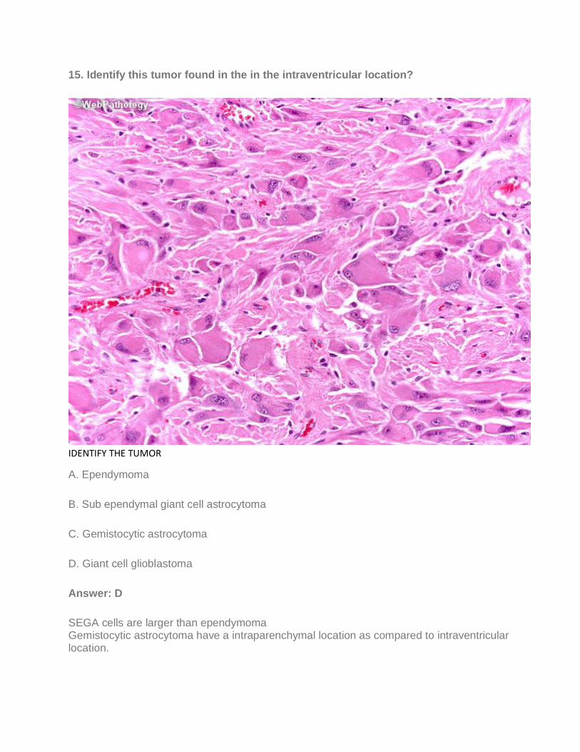
15. Identify this tumor found in the in the intraventricular location?
IDENTIFY THE TUMOR
A. Ependymoma
B. Sub ependymal giant cell astrocytoma
C. Gemistocytic astrocytoma
D. Giant cell glioblastoma
Answer: D
SEGA cells are larger than ependymoma Gemistocytic astrocytoma have a intraparenchymal location as compared to intraventricular location.

16. What is the WHO grade of this tumor commonly arising from cauda equina, conus medullaris or filum terminale?
Find the tumor
A. Grade I
B. Grade II
C. Grade III
D. Grade IV

Answer: A
Explanation: Myxopapillary ependymoma is grade I
17. All of the following are subtypes of ependymoma except?
A. Clear cell
B. Papillary
C. Tancytic
D. Giant cell
Answer: D
18. All of the following are good prognostic markers of glioblastoma multiforme except?
A. Age<50 years
B. Secondary glioblastoma
C. MGMT Promoter methylation
D. Presence of necrosis
Answer: D
19. The picture is from a tumor in a 10-year old male in the lateral ventricle-the tumor cells are CK7 positive and transthyretin positive. What is the diagnosis?
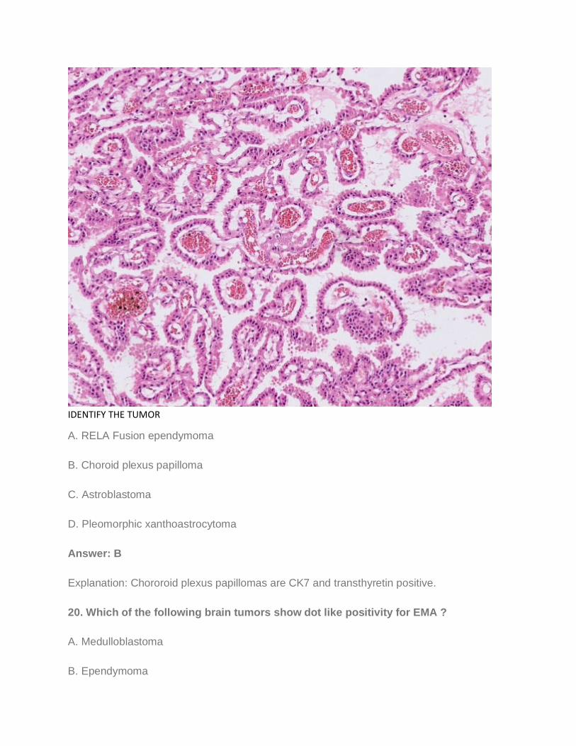
IDENTIFY THE TUMOR
A. RELA Fusion ependymoma
B. Choroid plexus papilloma
C. Astroblastoma
D. Pleomorphic xanthoastrocytoma
Answer: B
Explanation: Chororoid plexus papillomas are CK7 and transthyretin positive.
20. Which of the following brain tumors show dot like positivity for EMA ?
A. Medulloblastoma
B. Ependymoma

C. Diffuse astrocytoma
D. Meningioma
Answer: B
You may join telegram channel- Pathology mcqs
For pathology mcqs, quizzes, interesting facts and updates - Visit HOME - Pathology for all
Join the Facebook page for daily questions- Pathology mcq
Useful for NEET-SS Oncopathology, DM Histopathology, FRCPath, and Hematopathology
fellowships.