Embed Size (px)
Citation preview

THE JOURNAL OF COMPARATIVE NEUROLOGY 295530-547 (1990)
Central Projections of Cochlear Nerve Fibers in the Alligator Lizard
M.R. SZPIR, S. SENTO, AND DX. RYUGO Department of Anatomy and Cellular Biology, Harvard Medical School, Boston, Massachusetts
02115 (M.R.S., S.S., D.K.R.); Eaton-Peabody Laboratory, Massachusetts Eye and Ear Infirmary, Boston, Massachusetts 02114 (M.R.S., S.S., D.K.R.); Department of
Otolaryngology-Head and Neck Surgery, Johns Hopkins University School of Medicine, Baltimore, Maryland 21205 (D.K.R.)
ABSTRACT The auditory (cochlear) ganglion cells of the alligator lizard (Gerrhonotus rnulticarinatus)
give rise to two types of peripheral fibers: tectorial fibers, which contact hair cells covered by a tectorial membrane, and free-standing fibers, which contact hair cells without a tectorial membrane. To determine the central projections of these fibers, we applied intracellular and extracellular injections of horseradish peroxidase (HRP) to the peripheral component of the cochlear nerve. After histological processing with diaminobenzidine, individual cochlear nerve fibers could be traced through serial sections with the aid of a light microscope and drawing tube. The projection patterns formed two morphologically distinct groups. Neurons whose peripheral processes contacted tectorial hair cells in the cochlea projected to three divisions of the cochlear nucleus: nucleus magnocellularis lateralis (NML), nucleus magnocellularis medialis (NMM), and nucleus angularis lateralis (NAL). Neurons whose peripheral processes contacted free- standing hair cells projected primarily to the nucleus angularis medialis (NAM), although some also sent a single, thin branch to the NML; these neurons never projected to NAL or NMM. Morphometric comparisons of tectorial and free-standing fibers demonstrate that tectorial fibers have a larger axonal diameter, form a greater number of terminal swellings, and make proportionally more somatic contacts. By correlating the morphologically defined groups with previously reported physiologically defined groups, we conclude that different divisions of the cochlear nucleus are associated with separate frequency ranges and that stimuli in the different frequency ranges may be processed separately in the brain.
Key words: axon, cochlear nucleus, comparative anatomy, hearing, horseradish peroxidase, primary afferent, reptile, synaptic endings
The cochlear nerve in lizards conveys auditory informa- tion from the hair cells of the basilar papilla to the ipsilat- era1 cochlear nucleus. Although the anatomy and physiology of the peripheral auditory system of lizards have been relatively well described (Mulroy, '74, '83, '86; Weiss et al., '74, '76, '78; Bagger-Sjoback, '76, Manley, '77; Holton and Weiss, '78, '83; Wever, '78; Miller, '80, '85; Eatock et al., %l), little is known about the central auditory system. Knowl- edge of the central projections of reptilian cochlear nerve fibers has been based on anterograde degeneration tech- niques and whole nerve stains (Beccari, '12; Hamilton, '63; Leake, '74; DeFina and Webster, '74; Foster and Hall, '78; Barbas-Henry and Lohman, '88). These studies describe the distribution of nerve fibers and terminals within the dorsal medulla for the entire nerve fiber population but do not provide information on arborization patterns or morphology of terminals.
In many species of lizards, the basilar papilla has more
than one population of hair cells (Mulroy, '68, '74; Baird and Marovitz, '71; Bagger-Sjoback, '76; Miller, '80). A common situation is that one population has hair cells all having kinocilia oriented in the same direction and a second population with some hair cells having kinocilia oriented in one direction and some in the opposite direction. In the alligator lizard, the hair cells in the unidirectional region are covered by an overlying tectorial membrane and can be called tectorial hair cells. The bidirectional hair cells in this species are not covered by a tectorial membrane and can be
Accepted December 1,1989. Address reprint requests to Dr. D.K. Ryugo, Department of Otolaryngol-
ogy-Head and Neck Surgery, Johns Hopkins University School of Medicine, 417 Traylor Research Building, 720 Rutland Avenue, Baltimore, MD 21205. S. Sento's present address is Science Research Laboratory, You-I Clinic,
1-366-5 Sakuragicho, Oomiya, Saitama, 330 Japan.
D 1990 WILEY-LISS, INC.

LIZARD COCHLEAR NERVE FIBERS 531
called free-standing hair cells. Intracellular recordings show that tectorial and free-standing hair cells respond differ- ently to acoustic stimulation (Weiss et al., '74). Morphologi- cally, the two types of hair cells make different types of contacts with the peripheral processes of cochlear nerve fibers (Miller, '85; Mulroy and Oblak, '85; Mulroy, '86). These differences may be reflected in the physiology of single cochlear nerve fibers with respect to tuning-curve shapes, spontaneous discharge rates, characteristic frequen- cies (CF; frequency to which the fiber is most sensitive), and other electrophysiological characteristics (Frezza, '76; Weiss et al., '76, '78; Manley, '77; Holton and Weiss, '78; Turner et al., '81; Turner, '87). In the alligator lizard, tectorial fibers (which contact tectorial hair cells) have low CFs (100-800 Hz), low spontaneous discharge rates ( t 2 5 s/s), and sharp, asymmetric tuning curves. In contrast, free-standing fibers (which contact free-standing hair cells) have high CFs (9004,000 Hz), high spontaneous discharge rates (>25 s/s), and broad symmetric tuning curves. These data demon- strate a clear dichotomy between the two populations of hair cells and primary fibers in the auditory periphery and suggest that the two populations of fibers may differ in their central projections. In the present study, horseradish perox- idase (HRP) techniques were applied to stain individual cochlear nerve fibers in order to address this issue. In these preparations, fibers could be traced with the light micro- scope and subjected to morphometric analyses.
MATERIALS AND METHODS A total of 18 adult alligator lizards (Gerrhonotus rnulticar-
inatus), each weighing between 10 and 40 g, were used in this study. The lizards were anesthetized by intraperitoneal injections of a 25% urethane solution dissolved in distilled water at a dosage of 3.13 mg/g body weight. In the 16 HRP experiments, the middle ear was exposed by a ventral pharyngotomy and the inner ear was exposed by removing the round window membrane and some of the surrounding bone (described in detail by Weiss et al., '74). This exposure made it possible to view the cochlear nerve and the basilar papilla through the scala tympani.
Protargol staining One lizard was perfused through the heart with a lizard
Ringer's solution containing 1% sodium nitrite (30 ml, pH 7.2), followed by 120 ml of a slightly modified Bouin's fixative (0.9% picric acid, 25 % formalin, 5 76 glacial acetic acid) and postfixed overnight in the same fixative. The next day the brain was dissected out of the head, and the Bouin's fixative was leached out of the brain in a 10% formalin solution over a period of 1 week. The brain was dehydrated, embedded in paraffin, cut on a rotary microtome into 15-pm-thick horizontal sections, and mounted onto gelatin- coated slides. The brain was stained by using a protargol method (Bodian, '36).
Cres yl violet staining One lizard was perfused through the heart with a 20 ml
wash of 0.15 M phosphate buffer, pH 7.4, and then with 100 ml of a fixative containing 4% glutaraldehyde, 1 % paraform- aldehyde, 0.2% picric acid, and 4% sucrose, in a 0.15 M phosphate buffer, pH 7.4, at room temperature (Barbas- Henry and Wouterlood, '88). The head was postfixed over- night in the same fixative. The next day the brain was dissected out of the head, embedded in a gelatin-albumin
mixture (Frank et al., '80), and sectioned in the horizontal plane into 50-pm-thick sections on a vibratome. The sec- tions were mounted onto gelatin-coated slides, air dried overnight, and stained with a 0.570 cresyl violet solution.
Physiological recording and HRP injection The technical details of the acoustic stimulus generation
and recording have been previously described (Weiss et al., '76; Holton and Weiss, '83). The intracellular injection of HRP into individual cochlear nerve fibers is similar to the method used in studies of the cat cochlear nerve (Liberman, '82). Briefly, the micropipette electrodes for the intracellu- lar labeling experiments were backfilled with a filtered solution containing 10% HRP (Sigma type VI), 0.15 M KC1, and 0.05 M Tris buffer, pH 7.3. In the one successful case, the initial impedance was 60 MQ and the pipette was bevelled (30" off horizontal) to a final resistance of 38 Ma.
The extracellular labeling experiments involved proce- dures similar to those used for the intracellular recording and injection experiments. The micropipettes had tips that were broken to 9-13 pm I.D. and were backfilled with a solution containing 30% HRP and 0.05 M Tris buffer, pH 7.3. The injection of HRP was made (>40 nA positive current, 50% duty cycle, 15 min duration) into the cochlear nerve peripheral to the intraotic ganglion. To place the micropipette in the nerve, the compound action potential in response to an acoustic click was monitored while the electrode was advanced. When the compound action poten- tial was at its maximum amplitude, the HRP was ionto- phoretically injected into the nerve.
HRP histochemistry Approximately 15-30 hours after the last injection of
HRP, each animal was perfused through the heart with a lizard Ringer's solution containing 1 O/o sodium nitrite (30 ml, pH 7.2), followed by 60-100 ml of fixative (2.5% glutaraldehyde, 0.5% paraformaldehyde, in 0.1 M phos- phate buffer, pH 7.2). The same fixative was then perfused through the scalae of the inner ear. The animals were decapitated, and the heads were postfixed in the same fixative overnight at 5°C. The next day, the entire hindbrain and most of the eighth nerve were removed from the skull and isolated in a single tissue block. The tissue was embed- ded in a gelatin-albumin mixture and sectioned with a vibratome in the horizontal plane a t a thickness of 50 pm. This orientation plane minimizes the number of histological sections required to span the cochlear nucleus and nerve.
Sections were maintained in serial order in a 0.1 M solution of Tris buffer, pH 7.6, washed several times in fresh changes of buffer, and then incubated for 1 hour in a solution of 0.1 M Tris buffer, pH 7.6, containing 0.5% CoC1, and 1% dimethylsulfoxide (DMSO). The tissue was then washed twice for 5 min in Tris buffer and washed twice more for 5 rnin in 0.1 M phosphate buffer, pH 7.3. The sections were immersed for 15 min in a solution containing 0.05% 3,3'-diaminobenzidine (DAB) and 1% DMSO and then immersed for 60 rnin in a solution containing 0.05% DAB, 1 % DMSO, and 0.01 % H,O,. The processed sections were washed in phosphate buffer, mounted onto gelatin-coated slides, air dried overnight, and counterstained with 0.5% cresyl violet. The basilar papillae were histologically pro- cessed en bloc according to the same procedures as for the brain. The papillae were embedded in JB-4 and either whole mounted or sectioned at 20 pm on a rotary microtome perpendicular to the long axis of the papilla.
532 M.R. SZPIR ET AL.
Identification of HRP-labeled fibers One fiber, recorded from intracellularly, was character-
ized as a tectorial unit on the basis of the shape of its tuning curve, characteristic frequency (325 Hz), and spontaneous discharge rate (2.5 s h ) . The fiber was then labeled by inject- ing HRP through the same recording micropipette. A contin- uously negative DC potential plus the similarity of pre- and postinjection tuning curves argues that the labelled fiber is the same fiber that was intracellularly characterized and injected. The identification of labeled fibers following extra- cellular injections of HRP depended on the presence of labeled peripheral processes under either the free-standing or tectorial hair cells. Labeled peripheral processes were located throughout the tectorial and free-standing hair cell regions. The assumption was that labeled central fibers would be correlated with their labeled peripheral exten- sions. Typically only one population of fibers was labeled in any nerve; this result is consistent with anatomical and physiological studies that show that the tectorial and free- standing fibers are segregated in the peripheral component of the cochlear nerve (Weiss et al., ’76; Mulroy and Oblak, ’85).
Fiber reconstruction and terminology Labeled fibers were reconstructed from serial sections
with the aid of a light microscope and drawing tube (total magnification x 800). The continuity of individual fibers was established by matching the cut ends of labeled fiber segments on adjacent section surfaces. Fibers that could not be unambiguously reconstructed were not included in this study. A fiber was considered to be completely labeled when all segments were uniformly and darkly stained; further- more, the tips of all terminal branches were usually marked by a distinct swelling. Swellings that occurred at the tips of branches were called terminal swellings and those that occurred along the branches were called en passant swell- ings. Thus, for each ganglion cell, the entire axonal arboriza- tion and all e n passant and terminal swellings were avail- able for analysis. All fiber reconstructions are shown as if they were obtained from a left cochlear nucleus; fibers from the right side have been deliberately reversed.
Swellings were operationally defined according to the criteria applied to cochlear nerve fibers in cats (Rouiller et al., ’86). Terminal swellings were variable in size and shape, ranging from a simple, continuously convex structure (bou- ton ending) to a multiply-branched and lobulated formation (resembling the mammalian endbulb of Held). Small end- ings appeared as simple boutons, as structures having three or fewer lobulations or (rarely) as blunt endings without a distinct swelling; these endings could be preceded by a short string of en passant swellings. An e n passant swelling was identified when the axon increased abruptly (over a distance of 2 pm) to at least three times its diameter and just as abruptly returned to its original caliber. Blunt endings were included in the counts of terminal number, but they were not used for measurements of terminal area. Small endings were found throughout the nucleus, mostly in the neuropil but sometimes apposed to cell bodies. Large endings were defined by the presence of four or more lobulations from the same branch that appeared to be apposed to a single cell body. Such large axosomatic endings were found only in the NMM. These structures were called endbulbs because of their resemblance to mammalian endbulbs of Held. In general, however, the structure of lizard endbulbs is simpler than that of mammalian endbulbs.
All swellings were drawn with the aid of a drawing tube, and their silhouette areas were calculated by using an electronic planimeter. Endings were also analyzed with respect to fiber type (tectorial or free-standing), location in the cochlear nucleus, and termination target (somata or neuropil). Swelling size was represented by the silhouette area of the drawn ending. The sum of the areas of terminal swellings (i.e., excluding e n passant areas) is defined as the “total terminal area.” Because terminals could be found that appeared to appose cell bodies or that appeared to terminate in the neuropil, there are values defined as “total area of somatic terminals” and “total area of neuropil terminals,” indicating the relationship between the terminal and its postsynaptic target. It is important to note that in some cases only a portion of a “somatic terminal” appeared to contact a cell body; thus the value “total area of somatic terminals” may be greater than that actually actually contacting cell bodies.
The diameter of the central axon in the cochlear nerve root was determined for each of the reconstructed fibers. A 50 pm segment of the fiber was drawn (total magnification x800) from the initial branch point of free-standing fibers or from the bifurcation of tectorial fibers in the cochlear nucleus back towards the cell body. The diameter was calculated by dividing the silhouette area of the drawn fiber segment by its length.
Data analysis Tectorial and free-standing fibers were compared with
respect to central projection pattern; central axon diameter; and distribution, type, size, and number of terminal and e n passant swellings. The underlying hypothesis is that a constellation of structural differences among separate sets of neurons will establish cellular circuits with definably different functions. The mean, standard deviation, and P values (ANOVA model for repeated measures) are provided where appropriate. This ANOVA model was chosen because it takes into consideration the inherent biases of a statistical comparison where many samples are taken from some animals and not from others. Consequently, it is a more rigorous test of statistical significance (Kleinbaum et al., ’88). The cytoarchitectonic criteria and nomenclature of the lizard cochlear nucleus are based on previously reported observations (Miller, ’75).
RESULTS The present results are based on an analysis of 19 cochlear
nuclei from 18 lizards. Sixteen cochlear nuclei involved extracellular injections of HRP into the cochlear nerve; in addition, one cochlear nucleus contained a single fiber intracellularly labeled with HRP, one was prepared with a protargol stain, and one was prepared with a cresyl violet stain.
General description Fibers from the basilar papilla enter the dorsolateral
medulla from the caudal and dorsal portion of the eighth cranial nerve. A t the central end of the cochlear nerve lies an accumulation of cell bodies of second-order neurons upon which primary fibers terminate (Fig. 1). This juncture lies at the dorsal surface of the medulla, forming a slight bulge on the medial edge of the alar elevation, and is called the acoustic tubercle (Holmes, ’03) or the cochlear nucleus (Miller, ’75) . The cochlear nucleus is bordered laterally and

LIZARD COCHLEAR NERVE FIBERS
I 1
I I I
I 1 I
L
Fig. 1. Diagrammatic horizontal section of the lizard hindbrain. This drawing illustrates the cochlear nerve (shaded area on the left), the posterior division of the eighth nerve ganglion (shaded area on the right), and the cochlear nuclei, site of nerve termination. The inset shows a parasagittal section of the lizard brain and the level of the horizontal section (line AB); the shaded region represents the cochlear
ventrally by vestibular nuclei and medially by the fourth ventricle.
The cochlear nucleus of the alligator lizard can be cytoar- chitecturally parcelled into four subdivisions on the basis of Nissl stains (Fig. 2). Nucleus angularis (NA) occupies the
, I $ I
533
BP
nucleus. Abbreviations: BP, basilar papilla; Cb, cerebellum; CNe, co- chlear nerve; Cr, cerebrum; D, dorsal; G, ganglion; IV, fourth ventricle; LN, lagenar nerve; NAL, nucleus angularis lateralis; NAM, nucleus angularis medialis; NML, nucleus magnocellularis lateralis; NMM, nucleus magnocellularis medialis; OT, optic tectum; PAN, posterior ampullary nerve; R, rostral.
rostral position in the cochlear nucleus and is partially segregated from the nucleus magnocellularis lateralis (NML) by cochlear nerve fibers. The NA is composed of two regions: a lateral region (NAL), containing small (10-15 pm), spindle- shaped cells, found only within the most dorsolateral part of

Fig. 2. Photomicrograph of a cresyl violet-stained horizontal section (50 wm) of the left cochlear nucleus. Dashed lines show the outline of the cochlear nucleus and boundaries between cytoarchitectonic subdivi- sions. Abbreviations: IV, fourth ventricle; L, lateral; NAL, nucleus
angularis lateralis; NAM, nucleus angularis medialis; NML, nucleus magnocellularis lateralis; NMM, nucleus magnocellularis medialis; R, rostral; IXn, ninth cranial nerve; VIIIn, eighth cranial nerve. Bar = 250 pm.

LIZARD COCHLEAR NERVE FIBERS
A
535
B
L i
Dorsal-caudal fiber bundle
Ventral-rostra1 fiber bundle
Fig. 3. Fibroarchitecture of the alligator lizard's cochlear nucleus. A: Drawing of protargol-stained horizontal section made with the aid of a drawing tube and light microscope. B Corresponding schematic inter- pretation. This section is through the dorsal part of the nucleus. The dorsal-caudal and ventral-rostra1 fiber bundles represent two groups of
the NA, and a medial region (NAM), containing cell bodies of various diameters (10-20 Wm). Nucleus magnocellularis medialis (NMM) occupies the caudal position in the tuber- cle. NMM is an elongated structure, roughly 500 pm in length but less than 100 pm in width and depth. NMM contains a single type of cell, 15-20 pm in somatic diameter, which exhibits a crescent-shaped, eccentrically placed nu- cleus. Lying between these rostral and caudal extremes of the cochlear nucleus is the nucleus magnocellularis lateralis (NML). The NML contains small (<15 pm) and large (-2.0 gm) cell types. The flanking concavities formed by the bulge of NML correspond to changes in cyto- and fibroarchitec- ture, and help to define the borders of this subdivision.
Central projections of the cochlear nerve A protargol stain of the lizard's hindbrain reveals two
distinct bundles of fibers entering the cochlear nucleus from the eighth cranial nerve. One fiber bundle passes dorsally in the medulla and bifurcates upon entering the middle of the cochlear nucleus (Fig. 3). In more ventral sections, a second fiber bundle enters the rostral one-third of the nucleus without bifurcating; this latter bundle partially separates the NA from the NML (Fig. 4). The HRP experiments, as
i
NAL NAM
NML
NMM
cochlear nerve fibers. The subdivisions of the cochlear nuclei (NAL, NAM, NML, NMM) may be distinguished on the basis of fibroarchitec- ture. Abbreviations: L, lateral; NAL, nucleus angularis lateralis; NAM, nucleus angularis medialis; NML, nucleus magnocellularis medialis; NMM, nucleus magnocellularis medialis; R, rostral. Bar = 250 pm.
described below, allowed us to identify the origin of these two fiber bundles.
Population patterns Tectorial fibers. Extracellular injections of HRP into
the cochlear nerve typically labeled axons that were re- stricted to one or the other of the fiber bundles described above. Labeled fibers streamed away from the injection site, but individual fibers could not be traced through the dark reaction product at this site. We therefore relied on the relationship between the locations of central and peripheral HRP labeling to infer fiber identification. In all cases the location of labeled peripheral processes was correlated with the location of labeled fibers found in the dorsal-caudal or ventral-rostra1 fiber bundles in the medulla. Labeled fibers of the dorsal-caudal bundle (n = 8) were associated exclu- sively with peripheral processes that contacted tectorial hair cells of the basilar papilla, whereas fibers of the ventral- rostral bundle (n = 4) were associated only with peripheral processes that contacted free-standing hair cells (Table 1). In four cases, no peripheral labeling was found despite the presence of labeled fibers in the dorsal bundle. Since the
536
A B M.R. SZPIR ET AL.
7- Dorsal-caudal fiber bundle
Ventral-rostra1 fiber bundle
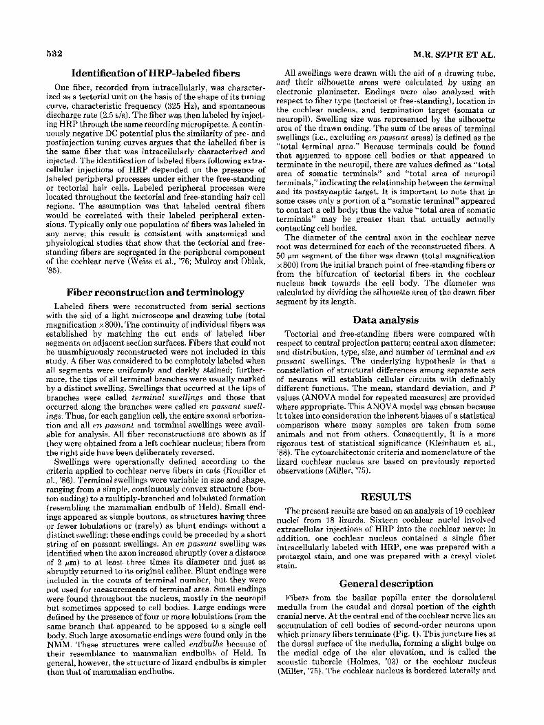
Fig. 4. Fibroarchitecture of the alligator lizard’s cochlear nucleus. A Drawing of a protargol-stained horizontal section made with the aid of a drawing tube and light microscope. B: Corresponding schematic inter- pretation. The top of this section is 15 pm ventral to the bottom of the section shown in Figure 3. The dorsal-caudal and ventral-rostra1 fiber bundles represent two groups of cochlear nerve fibers. The subdivisions
basilar papilla is oriented such that the tectorial hair cell population is located rostral to the free-standing hair cell population, our results imply that the two primary fiber bundles must cross en route to the cochlear nucleus.
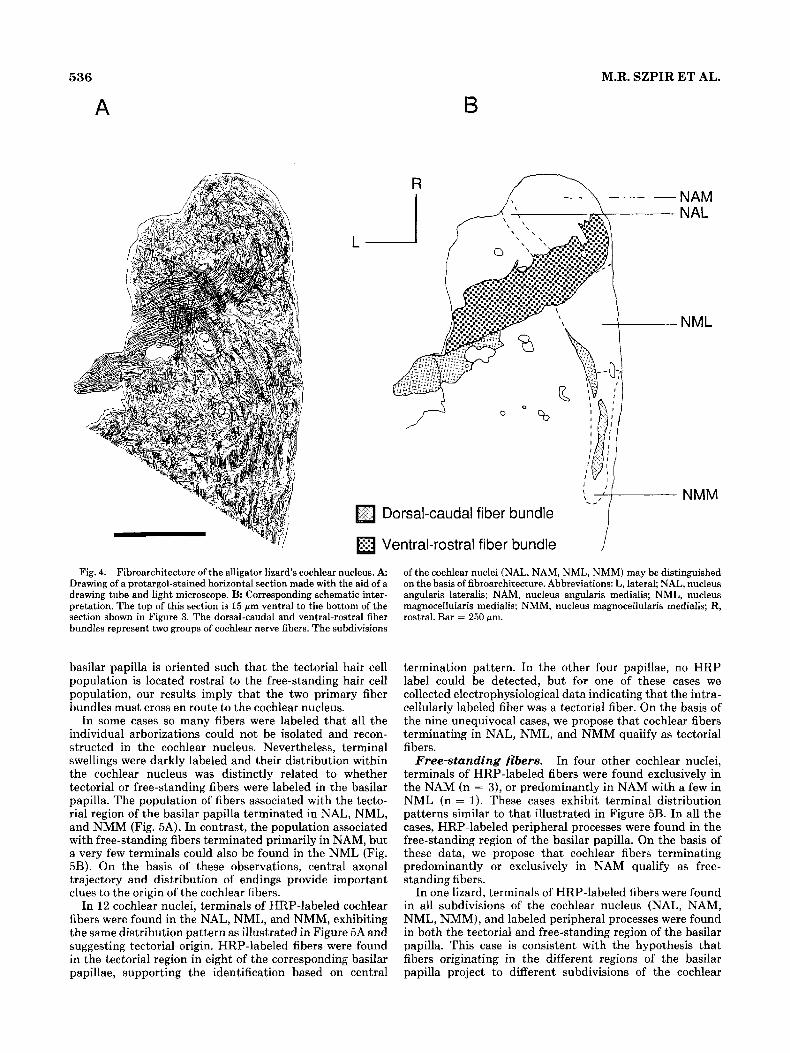
In same cases so many fibers were labeled that all the individual arborizations could not be isolated and recon- structed in the cochlear nucleus. Nevertheless, terminal swellings were darkly labeled and their distribution within the cochlear nucleus was distinctly related to whether tectorial or free-standing fibers were labeled in the basilar papilla. The population of fibers associated with the tecto- rial region of the basilar papilla terminated in NAL, NML, and NMM (Fig. 5A). In contrast, the population associated with free-standing fibers terminated primarily in NAM, but a very few terminals could also be found in the NML (Fig. 5B). On the basis of these observations, central axonal trajectory and distribution of endings provide important clues to the origin of the cochlear fibers.
In 12 cochlear nuclei, terminals of HRP-labeled cochlear fibers were found in the NAL, NML, and NMM, exhibiting the same distribution pattern as illustrated in Figure 5A and suggesting tectorial origin. HRP-labeled fibers were found in the tectorial region in eight of the corresponding basilar papillae, supporting the identification based on central
I
NAM NAL
NML
NMM
of the cochlear nuclei (NAL, NAM, NML, NMM) may be distinguished on the basis of fibroarchitecture. Abbreviations: L, lateral; NAL, nucleus angularis lateralis; NAM, nucleus angularis medialis; NML, nucleus magnocellularis medialis; NMM, nucleus magnocellularis medialis; R, rostral. Bar = 250 pm.
termination pattern. In the other four papillae, no HRP label could be detected, but for one of these cases we collected electrophysiological data indicating that the intra- cellularly labeled fiber was a tectorial fiber. On the basis of the nine unequivocal cases, we propose that cochlear fibers terminating in NAL, NML, and NMM qualify as tectorial fibers.
In four other cochlear nuclei, terminals of HRP-labeled fibers were found exclusively in the NAM (n = 3), or predominantly in NAM with a few in NML (n = 1). These cases exhibit terminal distribution patterns similar to that illustrated in Figure 5B. In all the cases, HRP-labeled peripheral processes were found in the free-standing region of the basilar papilla. On the basis of these data, we propose that cochlear fibers terminating predominantly or exclusively in NAM qualify as free- standing fibers.
In one lizard, terminals of HRP-labeled fibers were found in all subdivisions of the cochlear nucleus (NAL, NAM, NML, NMM), and labeled peripheral processes were found in both the tectorial and free-standing region of the basilar papilla. This case is consistent with the hypothesis that fibers originating in the different regivns of the basilar papilla project to different subdivisions of the cochlear
Free-standing fibers.
LIZARD COCHLEAR NERVE FIBERS 537
TABLE 1. Distribution of HRP-Labeled Term& and Location of Label in the Basilar Papilla'
Location of Loeation of Physiologically labeled fiben labeled fibers characterized in the in the Single fibers
Case fibers b a s h papilla cochlear nucleus reconstructed
37L 41L 55R 56R 67R ESL 46L 49L 39L 41L 53L 1L Tectorial 68R 69L 75L 44R 481,
THC THC THC THC THC THC THC THC - - -
unit - FHC FHC FHC FHC THC. FHC
NAL, NML, NMM NAL, NML, NMM NAL, NML, NMM NAL, NML, NMM NAL, NML, NMM NAL, NML, NMM NAL, NML, NMM NAL, NML, NMM NAL, NML, NMM NAL, NML, NMM NAL, NML, NMM NAL, NML, NMM NAM NAM NAM NAM, (NML) NAL,NAM.NML.NMM 1
- 1T 4T 1T 1T 3T 1T - - ~
1OF T, 3F
'Abbreviations: F, free-atandmg fiber; FHC, free-standing hair cell region; L, left; (NML) small projection tu NML; R, right; T, htorial fiber; THC, tectorial hair cell region
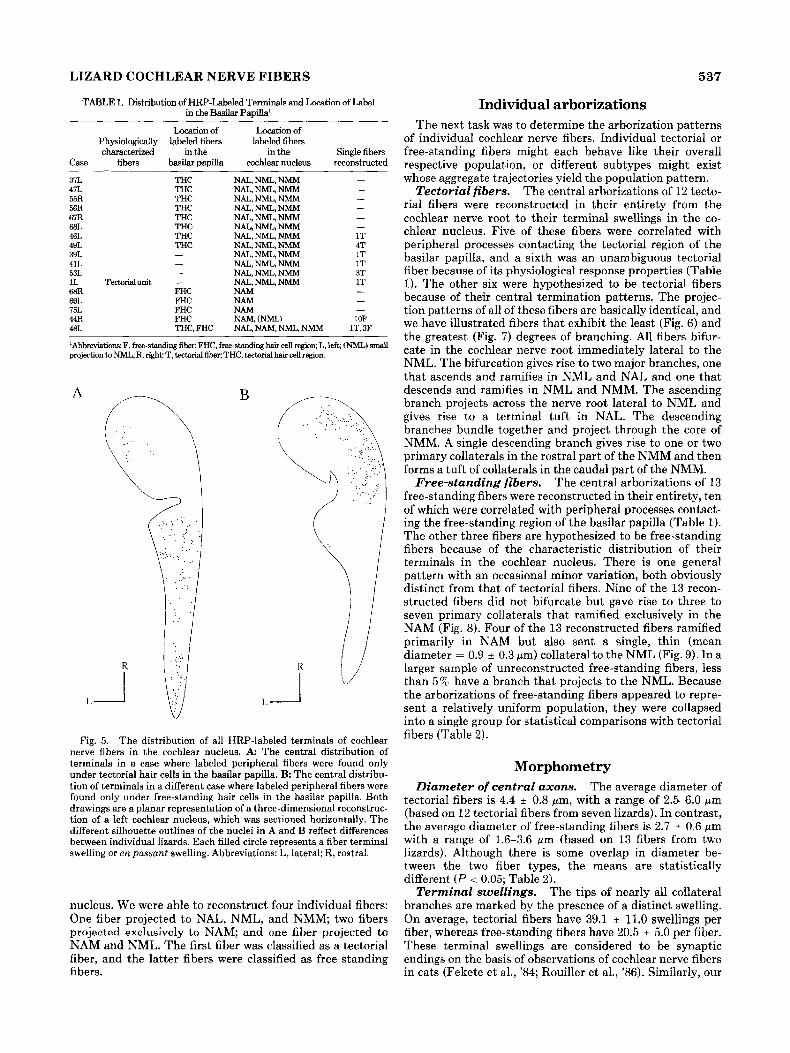
Fig. 5. The distribution of all HRP-labeled terminals of cochlear nerve fibers in the cochlear nucleus. A: The central distribution of terminals in a case where labeled peripheral fibers were found only under tectorial hair cells in the basilar papilla. B The central distribu- tion of terminals in a different case where labeled peripheral fibers were found only under free-standing hair cells in the basilar papilla. Both drawings are a planar representation of a three-dimensional reconstruc- tion of a left cochlear nucleus, which was sectioned horizontally. The different. silhouette outlines of the nuclei in A and B reflect differences between individual lizards. Each filled circle represents a fiber terminal swelling or en passant swelling. Abbreviations: L, lateral; R, rostral.
nucleus. We were able to reconstruct four individual fibers: One fiber projected to NAL, NML, and NMM; two fibers projected exclusively to NAM; and one fiber projected to NAM and NML. The first fiber was classified as a tectorial fiber, and the latter fibers were classified as free-standing fibers.
Individual arborizations The next task was to determine the arborization patterns
of individual cochlear nerve fibers. Individual tectorial or free-standing fibers might each behave like their overall respective population, or different subtypes might exist whose aggregate trajectories yield the population pattern.
The central arborizations of 12 tecto- rial fibers were reconstructed in their entirety from the cochlear nerve root to their terminal swellings in the co- chlear nucleus. Five of these fibers were correlated with peripheral processes contacting the tectorial region of the basilar papilla, and a sixth was an unambiguous tectorial fiber because of its physiological response properties (Table 1). The other six were hypothesized to be tectorial fibers because of their central termination patterns. The projec- tion patterns of all of these fibers are basically identical, and we have illustrated fibers that exhibit the least (Fig. 6) and the greatest (Fig. 7 ) degrees of branching. All fibers bifur- cate in the cochlear nerve root immediately lateral to the NML. The bifurcation gives rise to two major branches, one that ascends and ramifies in NML and NAL and one that descends and ramifies in NML and NMM. The ascending branch projects across the nerve root lateral to NML and gives rise to a terminal tuft in NAL. The descending branches bundle together and project through the core of NMM. A single descending branch gives rise to one or two primary collaterals in the rostral part of the NMM and then forms a tuft of collaterals in the caudal part of the NMM.
The central arborizations of 13 free-standing fibers were reconstructed in their entirety, ten of which were correlated with peripheral processes contact- ing the free-standing region of the basilar papilla (Table 1). The other three fibers are hypothesized to be free-standing fibers because of the characteristic distribution of their terminals in the cochlear nucleus. There is one general pattern with an occasional minor variation, both obviously distinct from that of tectorial fibers. Nine of the 13 recon- structed fibers did not bifurcate but gave rise to three to seven primary collaterals that ramified exclusively in the NAM (Fig. 8). Four of the 13 reconstructed fibers ramified primarily in NAM but also sent a single, thin (mean diameter = 0.9 0.3 pm) collateral to the NML (Fig. 9). In a larger sample of unreconstructed free-standing fibers, less than 5% have a branch that projects to the NML. Because the arborizations of free-standing fibers appeared to repre- sent a relatively uniform population, they were collapsed into a single group for statistical comparisons with tectorial fibers (Table 2).
Tectorial fibers.
Free-standing fibers.
Morphometry Diameter of central axons. The average diameter of
tectorial fibers is 4.4 f 0.8 ym, with a range of 2.5-6.0 pm (based on 12 tectorial fibers from seven lizards). In contrast, the average diameter of free-standing fibers is 2.7 0.6 pm with a range of 1.6-3.6 wm (based on 13 fibers from two lizards). Although there is some overlap in diameter be- tween the two fiber types, the means are statistically different ( P < 0.05; Table 2).
Terminal swellings. The tips of nearly all collateral branches are marked by the presence of a distinct swelling. On average, tectorial fibers have 39.1 11.0 swellings per fiber, whereas free-standing fibers have 20.5 * 5.0 per fiber. These terminal swellings are considered to be synaptic endings on the basis of observations of cochlear nerve fibers in cats (Fekete et al., '84; Rouiller et al., '86). Similarly, our

538
R
L i Fig. 6. Drawing tube reconstruction of the central arborization of a
tectorial fiber. This fiber was labeled with an extracellular injection of HRP into the auditory nerve. It has 26 terminals, a central axon diameter of 4.6 pm, and a total terminal area of 309 pm' and forms
preliminary electron microscopic observations of HRP- labeled swellings from tectorial and free-standing fibers in the lizard reveal the presence of active zones, characterized by clear round vesicles associated with a regular expansion of the intercellular cleft, and a postsynaptic density.
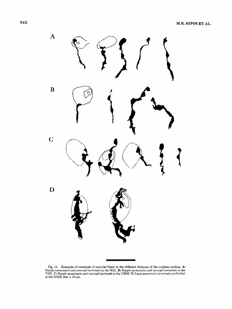
Generally, the terminals of lizard cochlear nerve fibers fall into two separate classes, much like that reported for mammals (e.g., Held, 1896; Ramon y Cajal, '09; Lorente de N6, '81). There are small, relatively simple endings and large, relatively complex endings. The small endings are bouton-like and are often preceded by a short string of en passant swellings having various sizes and shapes (Figs. 10 A-C, 12 A,B). Small endings are found in all subdivisions of the cochlear nucleus and are usually, but not always, found
M.R. SZPIR ET AL.
A
Descending Branch
terminals in the NAL, NML, and NMM. The inset (A) illustrates the location of this relatively simple arborization within the cochlear nucleus. Abbreviations: L, lateral; R, rostral. Bar = 100 pm.
in neuropil. The large endings appear as prominent swell- ings having lobulations, branchlets, and filopodia with swell- ings. These large endings were obviously distinct from the others, and each characteristically formed an extensive axosomatic association with a second-order neuron in the NMM (Figs. 10D, 11). We refer to such endings as endbulbs because of their resemblance to the endbulbs of Held so typical of primary auditory fibers of birds and mammals. Free-standing fibers never exhibit endbulbs, whereas four of the 12 tectorial fibers gave rise to one or two endbulbs in the NMM.
The distribution of termi- nals within the subdivisions of the cochlear nucleus is qualitatively and quantitatively distinct for the two types of
Distribution of terminals.

LIZARD COCHLEAR NERVE FIBERS 539
8; /Ascending Branch
Descending Branch
R
L _I
! 4 A
Fig. 7. Drawing tube reconstruction of the central arborization of a tectorial fiber. This fiber was electrophysiologically characterized by using a pipette inserted into the axon and then labeled with an injection of HRP through the same pipette. This fiber has 65 terminals, a central axon diameter of 5.0 pm, and a total terminal area of 694 pm'. Its basic morphology is consistent with that of tectorial fibers labelled with extracellular injections of HRP (see Fig. 6). The upper right inset (A) illustrates the location of this relatively complex arborization within the cochlear nucleus. The lower inset (B) shows tuning curves measured from this fiber. The HRP was iontophoretically injected with a positive current of 5.0 nA for 2.5 minutes through a microelectrode. A tuning curve was taken before and after the iontophoresis of HRP. The
fibers. The terminals of free-standing fibers are restricted mostly to the NAM. Although four of the 13 fibers also sent a small branch that delivered terminals to the NML, for the entire free-standing sample population, 97 76 of the termi- nals are located in the NAM. In contrast, every tectorial
A
I B
kf -i - 1 1 .o
FREQUENCY RHz) I
preinjection characteristic frequency (CF; 325 Hz) and spontaneous rate (SR; 2.7 spikes/second) and postinjection CF (350 Hz) and SR (2.5 spikesisecond) identify the fiber as a tectorial unit. The DC resting potential was monitored throughout the recording session. Continuity of the intracellular DC potential and similarities in the pre- and postinjec- tion tuning curve indicated that the electrode remained within the same fiber throughout the recording and injection period. The preinjection tuning curve is incomp1et.e below 200 Hz, and the postinjection tuning curve is incomplete below 275 Hz. Abbreviations: dB, decibel; kHz, kiloHertz; L, lateral; R, rostral; SPL, sound pressure level. Bar = 100 urn.
fiber gave rise to terminals in the NAL, NML, and NMM. For the population of 12 tectorial fibers, 22.2% of the terminals were located in the NAL, 45.7 70 were in the NML, and 32.1 % were in the NMM.
In the NAM, NAL, and NML, approximately 90% of the

540 M.R. SZPIR ET AL.
Parent Fiber
- Fig. 8. Drawing-tube reconstruction of the central arborization of a
free-standing fiber that was labeled with an extracellular injection of HRP into the cochlear nerve. It has 22 terminals, a central axon
diameter of 2.8 gm, and a total terminal area of 178 pm' and forms terminals exclusively in the NAM. The inset (A) shows the location of the arborization within the cochlear nucleus. Bar = 100 wm.
terminals of either fiber type are in the neuropil, whereas the remaining terminals were apposed to cell bodies. In the NML, terminals of free-standing fibers tend to make so- matic contacts, but these results should not be emphasized because the sample size is small (eight terminals). In the NMM, 64% of the terminals of tectorial fibers are found in the neuropil, and the remainder are apposed to cell bodies.
We use the silhouette area of individ- ual terminal swellings as an approximation of terminal size. Because synaptic active zones are contained within termi- nals, we assume that larger terminals contain more active zones and so propose that the summed area will reflect the amount of synaptic input to a region. In this context, the total area of the terminals for individual tectorial fibers is 312 -t 159.2 pm2, compared to 77.0 2 40.6 ym2 for free- standing fibers. The total area of terminals that are apposed to cell bodies is about 20 times greater for tectorial fibers compared to free-standing fibers, and the total area of terminals in the neuropil is almost three times greater (Table 2). In addition to the greater number of terminals per fiber, tectorial fibers tend to have larger terminals than do free-standing fibers (Table 2). If we use the average total terminal area per fiber as a point of reference, then tectorial fibers have 38% of their terminal area apposed to cell bodies, whereas free-standing fibers have less than 10 % apposed to cell bodies. Not only do tectorial and free- standing fibers distribute their terminals to different re- gions, they also appear to distribute them to different parts of the target neurons in those regions.
E n passant swellings are found only on the terminal branches of cochlear nerve fibers. All the e n passant swellings of the 13 free-standing fibers are located in the NAM. In contrast, the e n passant swellings of the 1 2 tectorial fibers are distributed evenly among the other subdivisions of the cochlear nucleus (20 in NAL, 17 in
Terminal area.
En passant swellings.
NML, 16 in NMM). Although the tectorial fibers are occasionally seen to pass through the NAM, they were never observed to form en passant swellings there. On average, the total number, the total area, and the mean area of individual e n passant swellings were similar for the two types of fibers (Table 2).
DISCUSSION The present study used HRP staining and light micro-
scopic reconstruction of single neurons to analyze the two known types of cochlear nerve fibers in the alligator lizard. These two types of fibers, called tectorial and free-standing, have been shown to differ across a wide variety of peripheral anatomical features (Mulroy, '74, '83, '86) and electrophysi- ological response properties (e.g., Weiss et al., '74, '76, '78), and we have demonstrated that they have separate projec- tion patterns into the brain. A summary of the two patterns of central projections is schematically illustrated (Fig. 13).
In addition to the qualitative differences in the central projection patterns, the tectorial and free-standing fibers were also markedly different in a number of anatomic parameters, including fiber diameter, number of terminals, total terminal area, and average terminal size. These distinc- tions between fiber types are consistent with the idea that neural signals conveyed by the two fiber populations are processed in separate ways.
One finds that a greater number of terminals is correlated with a larger axon diameter when comparing tectorial and free-standing fibers. Because the number of terminals equals the number of branch points plus one, the observation is consistent with the notion that large-diameter axons have more branching potential than do small axons (Lasek, '88). The peripheral processes of tectorial fibers are also thicker (2.8 pm) than those of free-standing fibers (2.0 pm; Mulroy

LIZARD COCHLEAR NERVE FIBERS 54 1
A
'\i
Fig. 9. The central arborization of a free-standing fiber that forms terminals in the NAM but also emits a single, small caliber branch to the NML. The fiber is from the same case as that shown in Figure 8 and has 22 terminals, a central axon diameter of 2.3 pm, and a total terminal area
TABLE 2. Characteristics of Individual Tectnrial and Free-Standing Fibem: Means, Standard Deviations, and Comparisons Between the
Two Fiber Populations
Tectorial Free-standing P fibers f i b value
Number of fibers Mean diameter Gm) Nnmber of terminnla
Total number of terminals Number of endbulb
Total terminal area Total area of somatic terminals Total men of neuropil terminals Area of individual simple terminals Area of individual endbnlte Area of individual simple somatic tern Area of individual nempil terminals
En possant swellings Nnmber per fiber Area of individual swelling (MI?) Total swelling area bun')
Aren of terminals ( 0 2 )
12 4.43 i 0.8
39.1 i 11.0 0.4 * 0.7
312.0 + 159.2 111.7 ? 72.6 200.3 * 112.0 11.2 * 3.3 47.8 -f 21.2
linals 23.8+ 9.9 9.5 * 3.4
4.4 * 4.7 10.9 * 4.1 50.0 i 62.0
13 2.71 i 0.6
20.5 + 5.0 0
77.0 2 40.6 5.5 i 8.4
71.5 t 41.3 6.6 + 2.6
7.0 f 4.9 6.2 i 2.4
3.5 i 1.8 7.4 + 3.1
24.3 * 14.5
~
- <0.05
e0.05 ~
~0 .05 <0.05 n.8.
<0.05
<0.05 ns.
n.s. n.8. n.3.
and Oblak, '85). One consequence of these diameter differ- ences is that tectorial fibers may have greater conduction velocity (Hursch, '39; Bullock and Horridge, '65; Mountcas- tle, '74), and the auditory information conveyed by the two fiber populations may be temporally separated in the co- chlear nucleus.
The observation that individual tectorial fibers have on average a greater number of terminals and a greater termi- nal area than do individual free-standing fibers suggests that the former may provide a greater number of synapses to
s of 47 pm2. The inset (A) shows the location of the arborization within the cochlear nucleus. Abbreviation: NML, nucleus magnocellularis lateralis. Bar = 100 um.
the cochlear nucleus. It has been shown (at least in mam- mals) that large endings have multiple synaptic active zones (e.g., Lenn and Reese, '66; Ibata and Pappas, '76; Cant and Morest, '79), and it has been hypothesized that large endings are synaptically more efficacious than small endings (e.g., Kuno, '71; Bourk, '76). Consequently, we predict that individual tectorial fibers are more synaptically efficacious than free-standing fibers in the cochlear nucleus.
These data suggest that the alligator lizard has two separate systems for conveying sensory information from the hearing organ to the brain. The two systems may differ with respect to the kind of information conducted from the periphery, the time of arrival of this information, and the postsynaptic neural targets. The functional significance of these differences remains to be determined.
Tectorial fibers There are many morphological, connectional, and physio-
logical similarities suggesting that tectorial fibers may corre- spond to mammalian type I fibers and avian cochlear nerve fibers. All such fibers contact unidirectionally oriented hair cells covered by a tectorial membrane (Weiss e t al., '76; Mulroy, '87), an arrangement thought to have been present in the stem reptiles from which modern reptiles, birds, and mammals evolved (Baird, '74; Miller, '80; Manley, '81). In the brain of alligator lizards, tectorial fibers occasionally contact cells in the NMM by way of large, axosomatic endings, which resemble mammalian and avian endbulbs of
542 M.R. SZPIR ET AL.
B
C
D
I'
Fig. 10. Examples of terminals of tectorial fibers in the different divisions of the cochlear nucleus. A: Simple axosomatic and neuropil terminals in the NAL. B: Simple axosomatic and neuropil terminals in the NML. C: Simple axosomatic and neuropil terminals in the NMM. D: Large axosomatic terminals (endbulbs) in the NMM. Bar = 20 pm.

LIZARD COCHLEAR NERVE FIBERS 543
Fig. 11. Photomicrograph of a large axosomatic terminal (endbulb) in the NMM. This photomicrograph corresponds to the second endbulb shown in Figure 10D. Abbreviations: L, lateral; R, rostral. Bar = 10 fim.
Held (Ram6n y Cajal, '08; Parks and Rubel, '78). This has been substantiated by electron microscopic examinations of the NMM, which reveal the presence of large axosomatic terminals that appose as much as one-quarter of the target cell circumference (unpublished observations). These target neurons make up a homogeneous population, which may correspond to the bushy/spherical cells in the anteroventral cochlear nucleus (AVCN) of mammals (Brawer et al., '74), the medial NM of birds (Jhaveri and Morest, '82)' and the NM of turtles (Browner and Pierz, '86). Consequently, it may be that the relationship between endbulb-like endings and spherical neurons in the auditory pathway is homolo- gous across amniotic vertebrates (Roord and Rasmussen, '63; Miller, '75; Rubel and Parks, '75; Miller and Kasahara, '79; Marbey and Browner, '87; Browner and Marbey, '88). In the NA, terminals of tectorial fibers consist of small bouton endings in the neuropil, and so are reminiscent of terminals of myelinated cochlear nerve fibers distributed to the NA of
birds (Takahashi, reference in Sullivan, '85) and the poster- oventral cochlear nucleus (PVCN) of mammals (Harrison and Irving, '66; Ryugo and Rouiller, '88). Indeed, certain cells of the avian and reptilian NA may correspond to cells of the mammalian PVCN (Foster and Hall, '78; Sullivan, '85).
Electrophysiological data from the cochlear nerve and nucleus are also consistent with the idea that tectorial fibers, avian cochlear nerve fibers, and mammalian type I fibers may be fulfilling comparable functions. Both tectorial and type I fibers exhibit sharp tuning, asymmetric tuning curves, two-tone rate suppression, and phase-locked responses to low frequency sounds at low intensity levels (Weiss et al., '76; Holton and Weiss, '78; Rose and Weiss, '88). The phenomenon of phase locking to the stimulus waveform is of particular interest in that it allows cochlear nerve fibers to convey faithfully information on the timing of the stimulus. In birds, notably the barn owl, phase-locking neurons of the NM are thought to be key to the timing pathway used for localizing sounds in azimuthal space (Sullivan and Konishi, '84; Takahashi et al., '84; Sullivan, '85). This capacity is presumably ensured by the highly efficacious synapses, the endbulbs of Held, that connect cochlear fibers to the second-order bushy cells (Parks and Rubel, '78 Parks, '81; Jhaveri and Morest, '82) and preserves the primary-like PSTH response patterns across the synapse (Pfeiffer, '66; Manley, '76; Sullivan, '85).
The pathway that permits the use of binaural phase comparisons to localize sound sources may proceed through the NMM of the alligator lizard by way of tectorial fibers, since it is here that we found the largest axosomatic contacts. In the periphery, individual tectorial fibers make a greater number of synapses on their presynaptic hair cells (Mulroy, '86). This increase in active sites per fiber may enhance the ability of tectorial fibers to synchronize (phase lock) to the acoustic stimulus. The ascending projections of the NMM are not known, but in the turtle bushy cells of the NM project to the nucleus laminaris (NL; R. Browner, personal communication). In reptiles, the size of the NL and NMM is correlated with the size of the unidirectional hair cell region [Miller, '75; Miller and Kasahara, '79). Further- more, the NL of birds, which corresponds to the medial superior olive of mammals, is the first site of binaural convergence from the NM in the time-coding pathway (Young and Rubel, '83; Takahashi and Konishi, '85; Sullivan and Konishi, '86). A similar pathway may be present in the alligator lizard. However, since interaural phase difference is dependent on the distance between the two ears and the wavelength of the incident sound, the small dimension of the alligator lizard's head (1 cm) limits its ability to use binaural time cues. The extent of this limitation can be understood from the following "best case" scenario. For a sound source that is maximally displaced to one side of the head (90' from the midline), the time for the sound to travel between the two ears of the alligator lizard is approximately 30 psec. Thus a 0.5 kHz' sound (a period of approximately 2,000 psec) will have a maximal interaural phase difference of 1/67 of a wavelength (5" in phase). Since this value approximates the best discriminative performance of humans (Zwislocki and Feldman, '56), the alligator lizard
'This is the corner frequency of the synchronization filter-function for tectorial fibers (Rose and Weiss, '88). It was chosen to represent the highest frequency at which the tectorial fibers can phase lock withnut a significant loss in fidelity.

544 M.R. SZPIR ET AL.
-------
Fig. 12. Examples of terminals of free-standing fibers in the NAM and NML. A: Simple axosomatic and neuropil terminals in the NAM. B: Simple axosomatic and neuropil terminals in the NML. Bar = 20 pm.
would require a comparative mechanism for interaural phase differences that was a t least as sensitive as that of humans merely to determine whether the source was located to the left or to the right of the midline. Presently, there is no evidence that the alligator lizard has such a capacity.
Free-standing fibers The relationship of free-standing fibers to cochlear nerve
fibers of other species lacking free-standing, bidirectional
hair cells (e.g., birds and mammals) is not clear. Free- standing fibers, mammalian type I fibers, and avian cochlear nerve fibers have myelinated and relatively thick central axons and give rise to small synaptic endings. On the other hand, free-standing fibers differ from avian and mammalian cochlear nerve fibers in many ways: Their tuning curves are broader; their peripheral processes contact hair cells having long stereocilia (9-31 pm) that are free of an overlying tectorial membrane; they do not phase lock to low-

LIZARD COCHLEAR NERVE FIBERS
NAL 545
a Tectorial 100-800 HZ
NMM
Tectorial 100-800 HZ
NMM
NAM
a 3 NML Free-standing 900-4000 HZ '
Fig. 13. Summary figure of the central projections of tectorial and free-standing fibers in the alligator lizard. Tectorial fibers project to NAL, NML, and NMM. Free-standing fibers project primarily to NAM, but a small fraction of free-standing fibers also project a fine branch to
frequency, low-intensity sounds; they do not exhibit two- tone suppression, neither they nor their presynaptic hair cells are associated with efferent terminals; and their sponta- neous discharge rates are all above 25 s/s (Mulroy, '68; Weiss et al., '74; '76; Frezza, '76; Holton and Weiss, '78; Rose and Weiss, '88). Furthermore, we have shown that the central projections of free-standing fibers avoid certain major subdi- visions of the cochlear nucleus entirely. This system that uses bidirectional hair cells and free-standing fibers may be a uniquely derived (autapomorphic) feature of lizards; it has not been found in any other vertebrate group.
CONCLUDING REMARKS In general, we have confirmed and extended the observa-
tions provided by population studies of eighth-nerve projec- tions in the brainstem of lizards (Beccari, '12; Hamilton, '63; DeFina and Webster, '74; Foster and Hall, '78; Barbas- Henry and Lohman, '88). The greatest concentration of primary afferent fibers is found in the NA and the NM. For the tegu and monitor lizard, however, it has been suggested that the nucleus laminaris (which is a second-order nucleus in birds, crocodiles, and turtles) receives primary input (DeFina and Webster, '74; Barbas-Henry and Lohman, '88), whereas for the iguana, it has been explicitly reported that the nucleus laminaris does not receive primary input (Foster and Hall, '78). In the present study, we were unable to identify a distinct nucleus laminaris, a situation common to other lizard species (Beccari, '12; Weston, '36; Miller, '75). The variance in observations among studies of the lizard may be attributable to differences in the following: species examined, methods used, or schemes for partitioning the cochlear nucleus. Regardless, our contribution is in the reconstruction of single cochlear nerve fibers and the demon- stration that such fibers may be reliably classified into one of two categories.
Although it was not possible with our material to trace individual fibers continuously from their peripheral termina- tion in the basilar papilla to their central termination in the cochlear nucleus, the labeling patterns were sufficiently
NML. Abbreviations: NAL, nucleus angularis lateralis; NAM, nucleus angularis medialis; NML, nucleus magnocellularis lateralis; NMM, nucleus magnocellularis medialis.
consistent for us to be secure in our identification of central fibers as being either tectorial or free-standing. That is, whenever it was possible to recover labeled fibers in the periphery, labeling under tectorial hair cells was always correlated with projections to the NAL, NML, and NMM, and labeling under free-standing hair cells was always correlated with projections to NAM. The poorer recovery of label in the papilla compared to the nerve resembled what has been reported in cats, where labeling in the organ of Corti is always less than that in the auditory nerve (Liber- man and Oliver, '84).
In our light microscopic studies of the cochlear nerve, we found no evidence for a population of very thin axons. Our observations are in basic agreement with those of Mulroy ('83). This concern is relevant to comparative issues in auditory neurobiology because of the presence of thin (<0.5 Fm in diameter), unmyelinated axons of type I1 spiral ganglion neurons in mammals (Ryugo et al., '86; Brown et al., '88). The diameter of tectorial and free-standing fibers is more in accord with measures of the mammalian type I axons (2.5-4.0 pm in cat, Arnesen and Osen, '78; 2 pm in guinea pig, Brown, '87) or avian cochlear nerve fibers (1-7 Fm in pigeon; Boord and Rasmussen, '63). The thinnest of the free-standing fibers in our sample (1.6 wm) is well outside the diameter range of type I1 axons. An additional line of circumstantial evidence is that mammalian type I1 fibers are associated with granule cell regions in the cochlear nucleus (Brown et al., '88), whereas we have never seen granule cells in the lizard cochlear nucleus.
The structural organization of the basilar papilla is remark- ably variable across lizard families (Miller, '80, '85). There are, however, some features found in all lizard auditory systems described thus far. For example, no lizard species has a papilla with entirely abneural, unidirectional hair cells such as is found in mammals; all lizards have bidirectional hair cells (Miller, '80; Miller and Beck, '88; Wever, '78). Low-frequency reception (tl kHz) is confined to fibers that innervate unidirectional (tectorial) hair cells (Turner, '80; Manley, '81). Furthermore, those fibers innervating unidirec-

546 M.R. SZPIR ET AL.
tional hair cells always have greater axon diameters than do fibers that innervate bidirectional hair cells (Miller, '85). The structural variation of the basilar papilla and its representation in the cochlear nucleus by central projections of cochlear nerve fibers raises a number of questions from both evolutionary and functional perspectives. For example, is the diversity in the periphery systematically related to variations in the structure and organization of the central pathways or can a particular set of central neurons accommo- date extensive peripheral variation? Are particular neuronal morphologies in the cochlear nucleus consistently associ- ated with certain frequency ranges? We mentioned that the size of the NMM is related to the size of the unidirectionally oriented hair cell region (Miller, '75; Miller and Kasahara, '79); this observation may be explained by our results, which demonstrate that the only primary afferent supply to the NMM is via tectorial fibers arising from unidirectional hair cells. In turn, cells of the NMM may be sensitive exclusively to low-frequency (tl kHz) sounds. Ultimately, more data on different species are needed before we can understand how form and function are related.
ACKNOWLEDGMENTS We are grateful to C.M. Gabriel, B. Martindale, and S.
Zagorski for technical assistance, Dr. J. Orav for statistical consultation, and Drs. N.Y.S. Kiang, J.B. Kobler, S.L. Palay, and J.J. Rosowski for helpful discussions of the data. We also thank the following colleagues for critically reading the manuscript: A.M. Berglund, M.C. Brown, R.A. Eatock, M.C. Liberman, S.R. Vacher, and T.F. Weiss. This work was supported by NIH grants NS13126 and DC00232, and an Albert J. Ryan Foundation Fellowship to M.R.S. Some of these results were presented earlier in abstract form (Szpir et al., '88).
LITERATURE CITED Arnesen, A.R., and K.K. Osen (1978) The cochlear nerve in the cat:
Topography, cochleotopy, and fiber spectrum. J. Comp. Neurol. 178.661- 678.
Bagger-Sjobkk, D. (1976) The cellular organization and nervous supply of the papilla basilaris in the lizard, Calotes uersicolor. Cell Tissue Res. 165:141-156.
Baird, I.L. (1974) Anatomical features of the inner ear in submammalian vertebrates. In W.D. Keidel and W.D. Neff (eds): Handbook of Sensory Physiology, Vol. V, Part 1. New York Springer-Verlag, pp. 159-212.
Baird, I.L., and W.F. Manovitz (1971) Some findings of scanning and transmission electron microscopy of the basilar papilla of the lizard Iguana iguana. Anat. Rec. 169:270.
Barbas-Henry, H.A., and A.H.M. Lohman (1988) Primary projections and efferent cells of the VIIIth cranial nerve in the monitor lizard, Varanus exanthematicus. J. Comp. Neurol. 277:234-249.
Barbas-Henry, H.A., and F.G. Wouterlood (1988) Synaptic connections between primary trigeminal afferent8 and accessory abducens motoneu- rons in the monitor lizard, Varanw exanthematicus. J. Comp. Neurol. 267:387-397.
Beccari, N. (1912) La constituzione, i nuclei terminali e le vie di connessione del nervo acustico nella Lacerta muralis. Archiv. Ital. Anat. Embriol. ZOt646-698.
Bodian, D. (1936) A new method for staining nerve fibers and nerve endings in mounted paraffin sections. Anat. Rec. 65:89-95.
Boord, R.L., and G.L. Rasmussen (1963) Projection of the cochlear and lagenar nerves on the cochlear nuclei of the pigeon. J. Comp. Neurol. 120r463-475.
Bourk, T.R. (1976) Electrical responses of neural units in the anteroventral cochlear nucleus of the cat. Doctoral dissertation, Dept. Electrical Engineering and Computer Science, M.I.T.
Brawer, J.R., D.K. Morest, and E.C. Kane (1974) The neuronal architecture of the cochlear nucleus of the cat. J. Comp. Neurol. 155t251-300.
Brown, M.C. (1987) Morphology of labeled afferent fibers in the guinea pig cochlea. J. Comp. Neurol. 260:591404.
Brown, M.C., A.M. Berglund, N.Y.S. Kiang, and D.K. Ryugo (1988) Central trajectories of type I1 spiral ganglion neurons. J. Comp. Neurol. 278:581- 590.
Browner, R.H., and D. Marbey (1988) The nucleus rnagnocellularis in the red-eared turtle, Chrysemys scripta elegans: Eighth nerve endings and neuronal types. Hearing Res. 33t257-272.
Browner, R.H., and D.M. Pierz (1986) Endbulbs of Held in a cochlear nucleus, nucleus magnocellularis in the red-eared turtle, Chrysemys scripta elegans. SOC. Neurosci. Abstr. 12:1265.
Bullock, T.H., and G.A. Horridge (1965) Structure and Function in the Nervous Systems of Invertebrates. San Francisco: W.H. Freeman.
Cant, N.B., and D.K. Morest (1979) The bushy cells in the anteroventral cochlear nucleus of the cat. A study with the electron microscope. Neuroscience 4:1925-1945.
DeFina, A.V., and D.B. Webster (1974) Projections of intraotic ganglion to the medullary nuclei in the Tegu lizard, Tupinambis nigropunctatus. Brain Behav. Evol. 10:197-211.
Eatock, R.A., G.A. Manley, and L. Pawson (1981) Auditory nerve fibre activity in the Tokay gecko. I. Implications for cochlear nerve processing. J. Comp. Physiol. 242,203-218.
Fekete, D.M., E.M. Rouiller, M.C. Liberman, and D.K. Ryugo (1984) The central projections of intracellularly labelled auditory nerve fibers in cats. J. Comp. Neurol. 229:432-450.
Foster, R.E., and W.C. Hall (1978) The organization of central auditory pathways in a reptile, Iguana iguana. J. Comp. Neurol. 178r783-832.
Frank, E., W.A. Harris, and M.B. Kennedy (1980) Lysopbosphatidyl choline facilitates labelling of CNS projections with horseradish peroxidase. J. Neurosci. Methods 2:183-189.
Frezza, W.A. (1976) Spontaneous activity in the auditory nerve of the alligator lizard. B.S. Thesis, Dept. Electrical Engineering, M.I.T.
Hamilton, D.W. (1963) Posterior division of the eighth cranial nerve in Lacerta uiuipara. Nature 200:705-706.
Harrison, J.M., and R. Irving (1966) The organization of the posterior ventral cochlear nucleus in the rat. J. Comp. Neurol. 126r391-402.
Held, H. (1893) Die centrale Gehorleitung. Arch. Anat. Physiol. Anat. Abteil. 17:201-248.
Holmes, G. (1903) On the comparative anatomy of the nervus acusticus. Trans. R. Irish Acad. 32(B)r101-144.
Holton, T., and T.F. Weiss (1978) Two-tone rate suppression in lizard cochlear nerve fibers, relation to receptor organ morphology. Brain Res. 159:219-222.
Holton, T., and T.F. Weiss (1983) Receptor potentials of lizard cochlear hair cells with free-standing stereocilia in response to tones. J. Physiol. 345:205-240.
Hursch, J.B. (1939) Conduction velocity and diameter of nerve fibers. Am. J. Physiol. 127t131-139.
Ibata, Y., and G.D. Pappas (1976) The fine structure of synapses in relation to the large spherical neurons in the anterior ventral cochlear [sic] of the cat. J. Neurocytol. 5:395406.
Jbaveri, S.R., and Morest, D.K. (1982) Neuronal architecture in nucleus magnocellularis of the chicken auditory system with observations on nucleus Iaminaris: A light and electron microscope study. Neuroscience 7:809-836.
Kleinbaum, D.G., L.L. Kupper, and K.E. Muller (1988) Applied Regression Analysis and Other Multivariable Methods. Boston: PWS-Kent.
Kuno, M. (1971) Quantum aspects of central and ganglionic synaptic transmission in vertebrates. Physiol. Rev. 51t647-678.
Lasek, R.J. (1988) Studying the intrinsic determinants of neuronal form and function. In R..J. Lasek and M.M. Black (eds): Intrinsic Determinants of Neuronal Form and Function. New York Alan R. Liss, Inc., pp. 3-58.
Leake, P.A. (1974) Central projections of the statoacoustic nerve in Caiman crocodilus. Brain Rebnv. Evol. 10:170-196.
Lenn, N.Y., and T.S. Reese (1966) The fine structure of nerve endings in the cochlear nucleus of the trapezoid body and the ventral cochlear nucleus. Am. J. Anat. 118:375-389.
Liberman, M.C. (1982) The cochlear frequency map for the c a t Labelling auditory-nerve fibers of known characteristic frequency. J. Acoust. Soc. Am. 72:1441-1449.

LIZARD COCHLEAR NERVE FIBERS 547
Liberman, M.C., and M.E. Oliver (1984) Morphometry of intracellularly labelled neurons of the auditory-nerve: Correlations with functional properties. J. Comp. Neurol. 223:163-176.
Lorente de Nb, R. (1981) The Primary Acoustic Nuclei. New York: Raven Press.
Manley, G.A. (1976) Auditory responses from the medulla of the monitor lizard Varanw bengalensis. Brain Res. 102329334,
Manley, G.A. (1977) Response patterns and peripheral origin of auditory nerve fibers in the monitor lizard, Varanus bengalensis. J. Comp. Physiol. 118t249-260.
Manley, G.A. (1981) A review of the auditory physiology of reptiles. In H. Autrum, D. Ottoson, E. Perl, and R.F. Schmidt (eds): Progress in Sensory Physiology. New York Springer, pp. 45134.
Marbey, D., and R.H. Browner (1987) Regeneration of the eighth nerve fibers after transection in the. red-eared turtle, Chrysemys scripta elegans. J. Morphol. 193:197-216.
Miller, M.R. (1975) The cochlear nuclei of lizards. J. Comp. Neurol. 159:375- 406.
Miller, M.R. (1980) The reptilian cochlear duct. In A.N. Popper and R.R. Fay (eds): Comparative Studies of Hearing in Vertebra-. Berlin: Springer, pp. 169-204.
Miller, M.R. (1985) Quantitative studies of auditory hair cells and nerves in lizards. J. Comp. Neurol. 2321-24.
Miller, M.R., and J. Beck (1988) Auditory hair cell innervational patterns in lizards. J. Comp. Neurol. 271:604-628.
Miller, M.R., and M. Kasahara (1979) The cochlear nuclei of some turtles. J. Comp. Neurol. 285221-236.
Mountcastle, V.B. (1974) Medical Physiology. Saint Louis: C.V. Mosby. Mulroy, M.J. (1968) Ultrastructure of the basilar papillae of reptiles. Ph.D.
Dissertation, Dept. Anatomy, U.C.S.F. (Univ. Microfilms Int., Ann Arbor, Michigan).
Mulroy, M.J. (1974) Cochlear anatomy of the alligator lizard. Brain Behav. Evol. 10.69-87.
Mulroy, M.J. (1983) Cochlear ganglion cells in the alligator lizard. Hearing Res. 12:121-137.
Mulroy, M.J. (1986) Patterns of afferent synaptic contacts in the digator lizard’s cochlea. J. Comp. Neurol. 248r263-271.
Mulroy, M.J. (1987) Auditory stereocilia in the alligator lizard. Hearing Res. 25:11-21.
Mulroy, M.J., and T.G. Oblak (1985) Cochlear nerve of the alligator lizard. J. Comp. Neurol. 233:463472.
Parks, T.N. (1981) Morphology of axosomatic endings in the avian cochlear nucleus: Nucleus magnocellularis of the chicken. J. Comp. Neurol. 203t425-440.
Parks, T.N., and E.W. Rubel (1978) Organization and development of brain stem auditory nuclei of the chicken: Primary afferent projections. J. Comp. Neurol. 180:439448.
Pfeiffer, R.R. (1966) Classification of response patterns of spike discharges for units in the cochlear nucleus: tone burst stimulation. Exp. Brain Res. 1:220-235.
Rambn y Cajal S. (1909) Hktologie du Systeme Nerveux de I’Homme et des VertBbrBs. Paris: Maloine.
Rose, C., and T.F. Weiss (1988) Frequency dependence of synchronization of cochlear nerve fibers in the alligator lizard Evidence for a cochlear origin of timing and non-timing neural pathways. Hearing Res. 33:151-166
Rouiller, E.M., R. Cronin-Schreiber, D.M. Fekete, and D.K. Ryugo (1986) The central projections of intracellularly labeled auditory nerve fibers in cats: An analysis of terminal morphology. J. Comp. Neurol. 249261-2’78.
Rubel, E.W., and T.N. Parks (1975) Organization and development of brainstem auditory nuclei of the chicken: tonotopic organization of N. magnocellularis and N. laminaris. J. Comp. Neurol. 164:411-434.
Ryugo, D.K., L.W. Dodds, and N.Y.S. Kiang (1986) Axon morphology of type I1 spiral ganglion cells in cats. Soc. Neurosci. Abstr. 12.779.
Ryugo, D.K., and E.M. Rouiller (1988) Central projections of intracellularly labeled auditory nerve fibers in cats: morphometric correlations with physiological properties. J. Comp. Neurol. 27lt130-142.
Sullivan, W.E. (1985) Classification of response patterns in cochlear nucleus of barn owl: correlation with functional response properties. J. Neurosci. 53:201-216.
Sullivan, W.E., and M. Konishi (1984) Segregation of stimulus phase and intensity coding in the cochlear nucleus of the barn owl. J. Neurosci. 4t1787-1799.
Sullivan, W.E., and M. Konishi (1986) A map of interaural phase difference in the owl’s brainstem. Proc. Natl. Acad. Sci. USA83:8400-8404.
Szpir, M.R., S. Sento, and D.K. Ryugo (1988) The central projections of individual auditory nerve fibers in the alligator lizard (Gerrhonotus multicarinatus). SOC. Neurosci. Abstr. 14:491.
Takahashi, T., and M. Konishi (1985) Parallel pathways in the owl’s brainstem auditory system. Anat. Rec. 211:191A.
Takahashi, T., A. Moiseff, and M. Konishi (1984) Time and intensity cues are processed independently in the auditory system of the owl. J. Neurosci. 4:1781-1786.
Turner, R.G. (1980) Physiology and bioacoustics in reptiles. In A.N. Popper and R.R. Fay (eds): Comparative Studies of Hearing in Vertebrates. Berlin: Springer, pp. 205-237.
Turner, R.G. (1987) Neural tuning in the granite spiny lizard. Hearing Res. 26:287-299.
Turner, R.G., A.A. Muraski, and D.W. Nielsen (1981) Cilium length Influ- ence on neural tonotopic organization. Science 213t1519-1521.
Weiss, T.F., M.J. Mulroy, and D.W. Altman (1974) Intracellular responses to acoustic clicks in the inner ear of the alligator lizard. J. Acoust. SOC. Am. 5 5 . m 6 1 9 .
Weiss, T.F., M.J. Mulroy, R.G. Turner, and C.L. Pike (1976) Tuning of single fibers in the cochlear nerve of the alligator lizard: Relation to receptor organ morphology. Brain Res. 115t71-90.
Weiss, T.F., W.T. Peake, A. Ling, Jr.. and T. Holton (1978) Which structures determine frequency selectivity and tonotopic organization of vertebrate nerve fibers? Evidence from the alligator lizard. In R.F. Naunton and C. Fernhdez (eds): Evoked Electrical Activity in the Auditory Nervous System. New York Academic Press, pp. 91-112.
Weston, J.K. (1936) The reptilian vestibular and cerebellar gray with fiber connections. J. Comp. Neurol. 65t93-199.
Wever, E.G. (1978) The Reptile Ear. Princeton, N J Princeton University Press.
Young, S.R., and E.W. Ruhel (1983) Frequency-specific projections of individual neurons in chick brainstem auditory nuclei. J. Neurosci. 3:1373-1378.
Zwislocki, J., and R.S. Feldman (1956) Just noticeable differences in dichotic phase. J. Acoust. Soc. Am. 28:860-864.


![Untitled-9 [pages.jh.edu]pages.jh.edu/~ryugolab/pdfs/2009_ryugo_limb.pdf · is defined as any change in behavior as a result of experi- ence. Behavior is shaped by the interactions](https://img.pdfslide.net/doc/110x75/5b449e8c7f8b9ae0668bd4a6/untitled-9-pagesjhedupagesjheduryugolabpdfs2009ryugolimbpdf-is.jpg)