Embed Size (px)
Citation preview
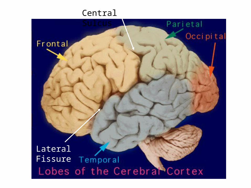
Central Sulcus
Lateral Fissure
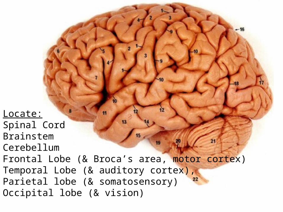
Locate:Spinal CordBrainstemCerebellumFrontal Lobe (& Broca’s area, motor cortex)Temporal Lobe (& auditory cortex),Parietal lobe (& somatosensory)Occipital lobe (& vision)
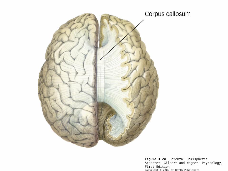
Figure 3.20 Cerebral HemispheresSchacter, Gilbert and Wegner: Psychology, First EditionCopyright © 2009 by Worth Publishers
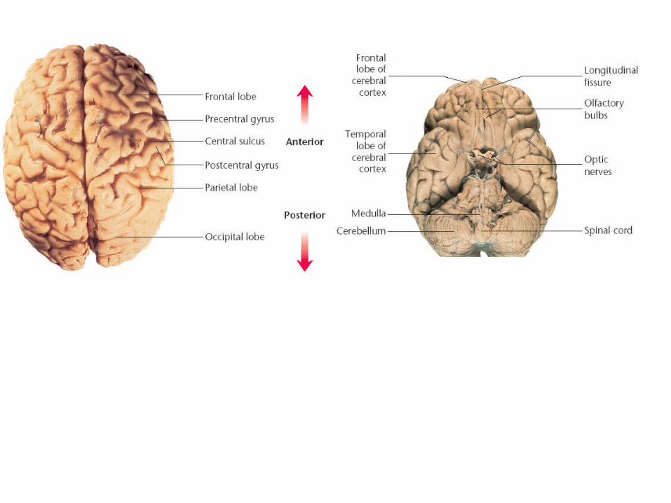
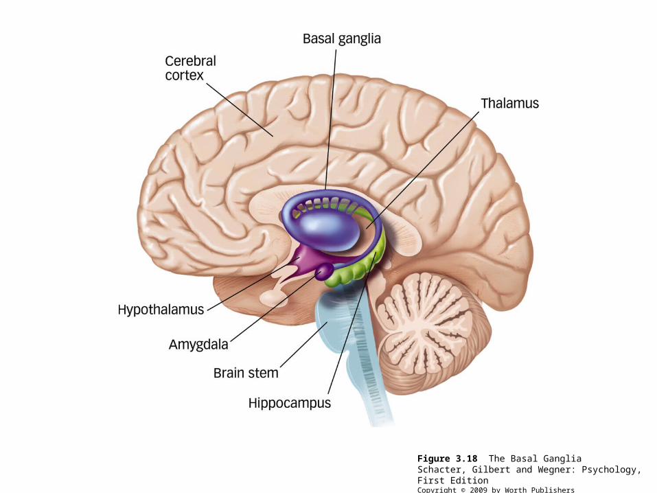
Figure 3.18 The Basal GangliaSchacter, Gilbert and Wegner: Psychology, First EditionCopyright © 2009 by Worth Publishers
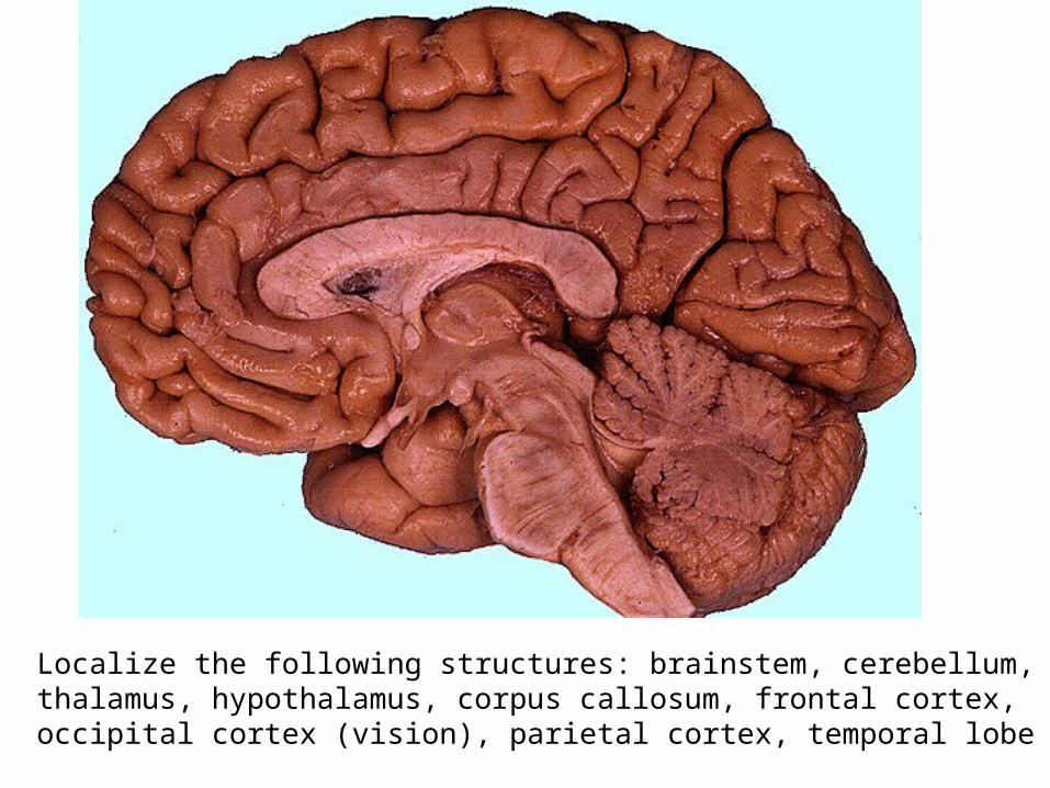
Localize the following structures: brainstem, cerebellum, thalamus, hypothalamus, corpus callosum, frontal cortex, occipital cortex (vision), parietal cortex, temporal lobe
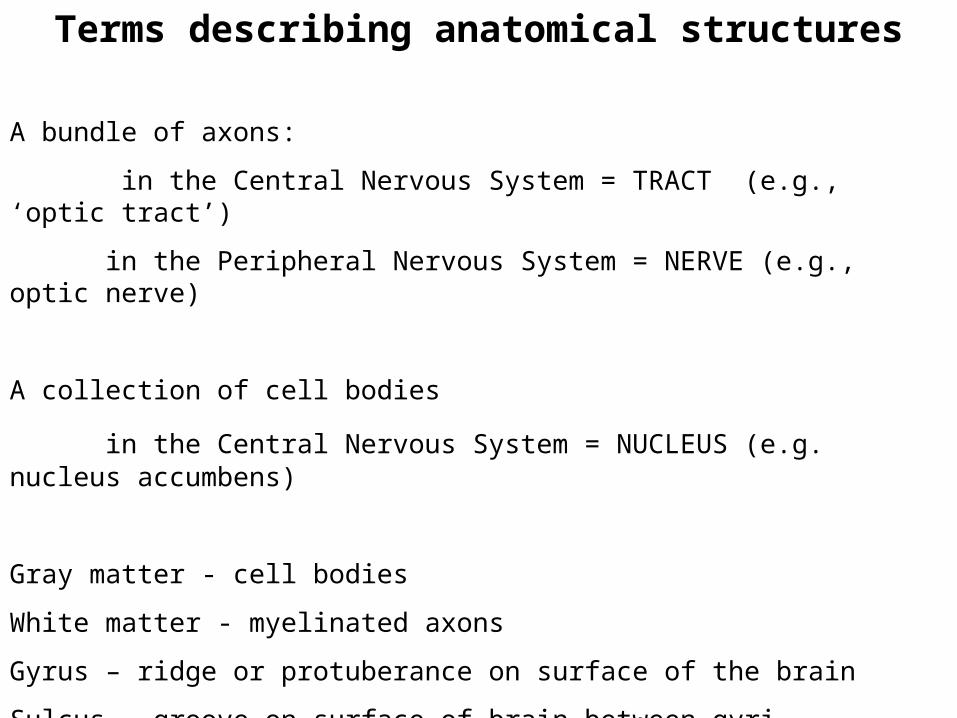
Terms describing anatomical structures
A bundle of axons:
in the Central Nervous System = TRACT (e.g., ‘optic tract’)
in the Peripheral Nervous System = NERVE (e.g., optic nerve)
A collection of cell bodies
in the Central Nervous System = NUCLEUS (e.g. nucleus accumbens)
Gray matter - cell bodies
White matter - myelinated axons
Gyrus – ridge or protuberance on surface of the brain
Sulcus – groove on surface of brain between gyri
Fissure – long, deep sulcus
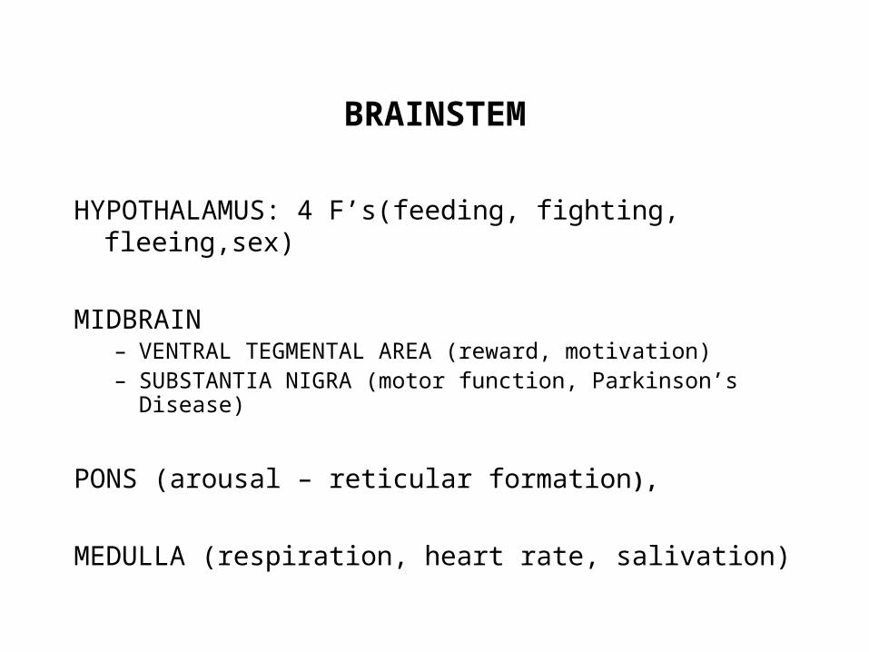
BRAINSTEM
HYPOTHALAMUS: 4 F’s(feeding, fighting, fleeing,sex)
MIDBRAIN– VENTRAL TEGMENTAL AREA (reward, motivation)– SUBSTANTIA NIGRA (motor function, Parkinson’s Disease)
PONS (arousal – reticular formation),
MEDULLA (respiration, heart rate, salivation)
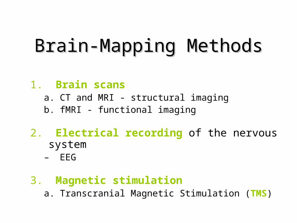
Brain-Mapping MethodsBrain-Mapping Methods
1. Brain scans a. CT and MRI - structural imagingb. fMRI - functional imaging
2. Electrical recording of the nervous system– EEG
3. Magnetic stimulation a. Transcranial Magnetic Stimulation (TMS)
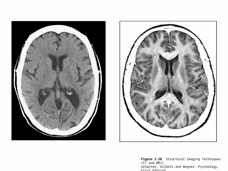
Figure 3.28 Structural Imaging Techniques (CT and MRI)Schacter, Gilbert and Wegner: Psychology, First EditionCopyright © 2009 by Worth Publishers
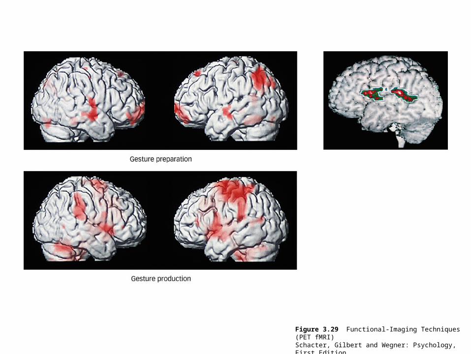
Figure 3.29 Functional-Imaging Techniques (PET fMRI)Schacter, Gilbert and Wegner: Psychology, First EditionCopyright © 2009 by Worth Publishers
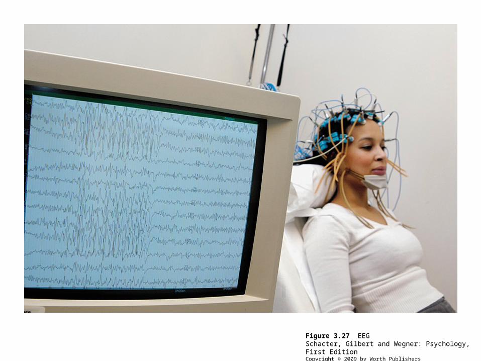
Figure 3.27 EEGSchacter, Gilbert and Wegner: Psychology, First EditionCopyright © 2009 by Worth Publishers

Spare slides
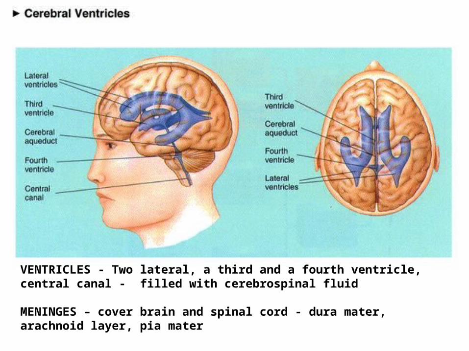
VENTRICLES - Two lateral, a third and a fourth ventricle, central canal - filled with cerebrospinal fluid
MENINGES – cover brain and spinal cord - dura mater, arachnoid layer, pia mater
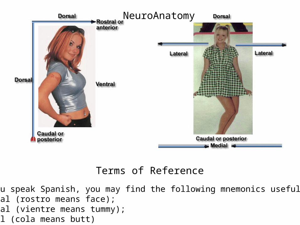
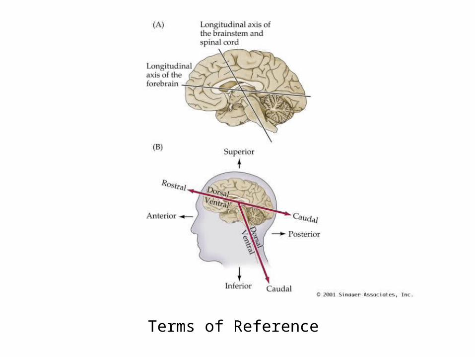
Terms of Reference
If you speak Spanish, you may find the following mnemonics useful:Rostral (rostro means face); Ventral (vientre means tummy);Caudal (cola means butt)
NeuroAnatomy
Terms of Reference
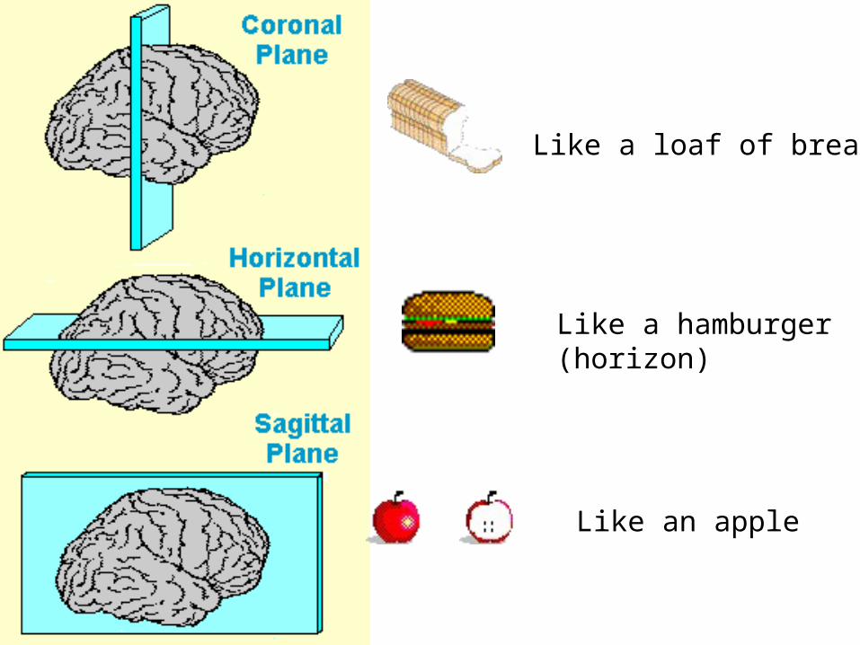
Like an apple
Like a loaf of bread
Like a hamburger(horizon)
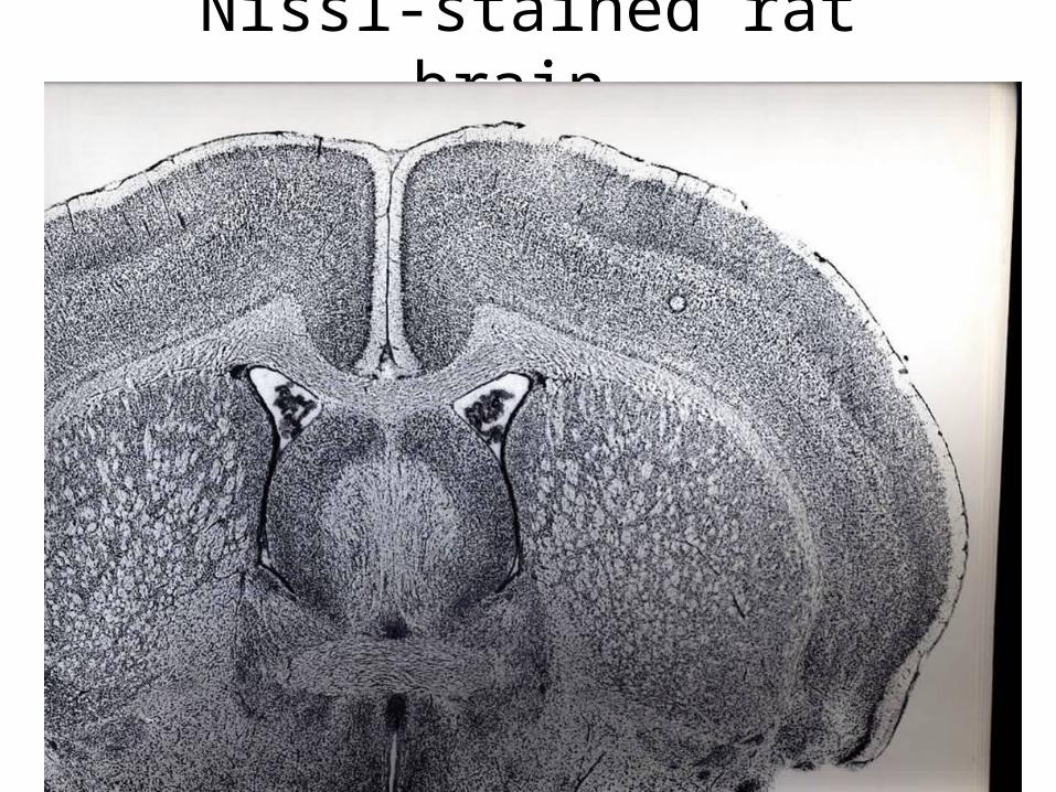
Nissl-stained rat brain