Embed Size (px)
Citation preview
ARTICLE IN PRESS
1350-9462/$ - se
doi:10.1016/j.pr
�CorrespondE-mail addr
1Senior autho
Please cite thi
doi:10.1016/j.
Progress in Retinal and Eye Research ] (]]]]) ]]]–]]]
www.elsevier.com/locate/prer
Centrins in retinal photoreceptor cells: Regulators in theconnecting cilium
Philipp Trojana, Norbert Kraussb, Hui-Woog Choec,d, Andreas GieXla,Alexander Pulvermullerc,1, Uwe Wolfruma,�,1
aInstitut fur Zoologie, Johannes Gutenberg-Universitat Mainz, 55099 Mainz, GermanybSchool of Biological and Chemical Sciences, Queen Mary, University of London, Mile End Road, London E1 4NS, UK
cInstitut fur Medizinische Physik und Biophysik, Humboldt-Universitat zu Berlin, Universitatsklinikum Charite, 10098 Berlin, GermanydDepartment of Chemistry, College of Natural Science, Chonbuk National University, 561-756 Chonju, South Korea
Abstract
Changes in the intracellular Ca2+ concentration regulate the visual signal transduction cascade directly or more often indirectly
through Ca2+-binding proteins. Here we focus on centrins, which are members of a highly conserved subgroup of the EF-hand
superfamily of Ca2+-binding proteins in photoreceptor cells of the vertebrate retina. Centrins are commonly associated with centrosome-
related structures. In mammalian retinal photoreceptor cells, four centrin isoforms are expressed as prominent components in the
connecting cilium linking the light-sensitive outer segment compartment with the metabolically active inner segment compartment. Our
data indicate that Ca2+-activated centrin isoforms assemble into protein complexes with the visual heterotrimeric G-protein transducin.
This interaction of centrins with transducin is mediated by binding to the bg-dimer of the heterotrimeric G-protein. More recent findings
show that these interactions of centrins with transducin are reciprocally regulated via site-specific phosphorylations mediated by the
protein kinase CK2. The assembly of centrin/G-protein complexes is a novel aspect of translocation regulation of signalling proteins in
sensory cells, and represents a potential link between molecular trafficking and signal transduction in general.
r 2008 Elsevier Ltd. All rights reserved.
Keywords: Ca2+-binding proteins; Centrins; Retina; Photoreceptor cells; Transducin; Light-dependent translocation
Contents
1. Introduction . . . . . . . . . . . . . . . . . . . . . . . . . . . . . . . . . . . . . . . . . . . . . . . . . . . . . . . . . . . . . . . . . . . . . . . . . . . . . . . . . 2
2. Centrin genes and protein structures . . . . . . . . . . . . . . . . . . . . . . . . . . . . . . . . . . . . . . . . . . . . . . . . . . . . . . . . . . . . . . . . 3
2.1. Centrin genes and proteins in their phylogenic context . . . . . . . . . . . . . . . . . . . . . . . . . . . . . . . . . . . . . . . . . . . . . . . 3
2.2. Primary domain structure of centrins . . . . . . . . . . . . . . . . . . . . . . . . . . . . . . . . . . . . . . . . . . . . . . . . . . . . . . . . . . . 4
2.2.1. Ca2+-binding at EF-hand motifs of centrins . . . . . . . . . . . . . . . . . . . . . . . . . . . . . . . . . . . . . . . . . . . . . . . . 6
2.2.2. Site-specific phosphorylation of centrins . . . . . . . . . . . . . . . . . . . . . . . . . . . . . . . . . . . . . . . . . . . . . . . . . . . 6
2.3. High-resolution molecular structure of centrins and their complexes . . . . . . . . . . . . . . . . . . . . . . . . . . . . . . . . . . . . . 7
3. Subcellular localization and cellular function of centrins . . . . . . . . . . . . . . . . . . . . . . . . . . . . . . . . . . . . . . . . . . . . . . . . . . 8
3.1. Centrins as ubiquitous components of centrosomes, spindle poles and basal bodies. . . . . . . . . . . . . . . . . . . . . . . . . . . 8
4. Centrins in the vertebrate retina . . . . . . . . . . . . . . . . . . . . . . . . . . . . . . . . . . . . . . . . . . . . . . . . . . . . . . . . . . . . . . . . . . 10
4.1. Centrin expression in the vertebrate retina. . . . . . . . . . . . . . . . . . . . . . . . . . . . . . . . . . . . . . . . . . . . . . . . . . . . . . . 10
4.2. Subcellular localization of centrins in retinal cells — in particular in photoreceptor cells . . . . . . . . . . . . . . . . . . . . . . 10
e front matter r 2008 Elsevier Ltd. All rights reserved.
eteyeres.2008.01.003
ing author. Tel.: +496131 3925148; fax: +49 6131 3923815.
ess: [email protected] (U. Wolfrum).
rs contributed equally to the present work.
s article as: Trojan, P., et al., Centrins in retinal photoreceptor cells: Regulators in the connecting cilium. Progress Retinal Eye Res (2008),
preteyeres.2008.01.003
ARTICLE IN PRESSP. Trojan et al. / Progress in Retinal and Eye Research ] (]]]]) ]]]–]]]2
5. Centrin-binding proteins in vertebrates . . . . . . . . . . . . . . . . . . . . . . . . . . . . . . . . . . . . . . . . . . . . . . . . . . . . . . . . . . . . . 12
5.1. Centrin-binding proteins in mammalian photoreceptor cells . . . . . . . . . . . . . . . . . . . . . . . . . . . . . . . . . . . . . . . . . . 14
5.2. Molecular characteristics of centrin/transducin complexes. . . . . . . . . . . . . . . . . . . . . . . . . . . . . . . . . . . . . . . . . . . . 14
N
CC
OS
IS
RPE
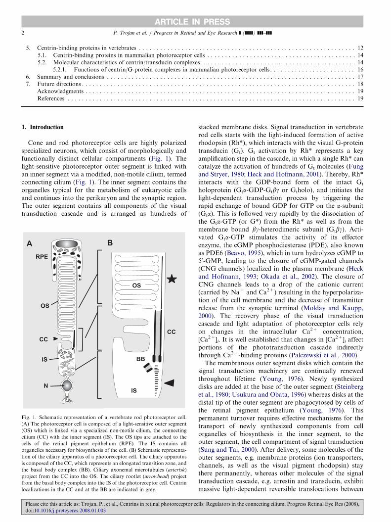
Fig. 1. Schem
(A) The phot
(OS) which is
cilium (CC) w
cells of the
organelles nec
tion of the cil
is composed o
the basal bod
project from
from the basa
localizations i
Please cite th
doi:10.1016/
5.2.1. Functions of centrin/G-protein complexes in mammalian photoreceptor cells . . . . . . . . . . . . . . . . . . . . . . . . 16
6. Summary and conclusions . . . . . . . . . . . . . . . . . . . . . . . . . . . . . . . . . . . . . . . . . . . . . . . . . . . . . . . . . . . . . . . . . . . . . . 17
7. Future directions . . . . . . . . . . . . . . . . . . . . . . . . . . . . . . . . . . . . . . . . . . . . . . . . . . . . . . . . . . . . . . . . . . . . . . . . . . . . . 18
Acknowledgments . . . . . . . . . . . . . . . . . . . . . . . . . . . . . . . . . . . . . . . . . . . . . . . . . . . . . . . . . . . . . . . . . . . . . . . . . . . . 19
References . . . . . . . . . . . . . . . . . . . . . . . . . . . . . . . . . . . . . . . . . . . . . . . . . . . . . . . . . . . . . . . . . . . . . . . . . . . . . . . . . 19
1. Introduction
Cone and rod photoreceptor cells are highly polarizedspecialized neurons, which consist of morphologically andfunctionally distinct cellular compartments (Fig. 1). Thelight-sensitive photoreceptor outer segment is linked withan inner segment via a modified, non-motile cilium, termedconnecting cilium (Fig. 1). The inner segment contains theorganelles typical for the metabolism of eukaryotic cellsand continues into the perikaryon and the synaptic region.The outer segment contains all components of the visualtransduction cascade and is arranged as hundreds of
OS
IS
BB
CC
atic representation of a vertebrate rod photoreceptor cell.
oreceptor cell is composed of a light-sensitive outer segment
linked via a specialized non-motile cilium, the connecting
ith the inner segment (IS). The OS tips are attached to the
retinal pigment epithelium (RPE). The IS contains all
essary for biosynthesis of the cell. (B) Schematic representa-
iary apparatus of a photoreceptor cell. The ciliary apparatus
f the CC, which represents an elongated transition zone, and
y complex (BB). Ciliary axonemal microtubules (asterisk)
the CC into the OS. The ciliary rootlet (arrowhead) project
l body complex into the IS of the photoreceptor cell. Centrin
n the CC and at the BB are indicated in grey.
is article as: Trojan, P., et al., Centrins in retinal photoreceptor ce
j.preteyeres.2008.01.003
stacked membrane disks. Signal transduction in vertebraterod cells starts with the light-induced formation of activerhodopsin (Rh*), which interacts with the visual G-proteintransducin (Gt). Gt activation by Rh* represents a keyamplification step in the cascade, in which a single Rh* cancatalyze the activation of hundreds of Gt molecules (Fungand Stryer, 1980; Heck and Hofmann, 2001). Thereby, Rh*interacts with the GDP-bound form of the intact Gt
holoprotein (Gta-GDP-Gtbg or Gtholo), and initiates thelight-dependent transduction process by triggering therapid exchange of bound GDP for GTP on the a-subunit(Gta). This is followed very rapidly by the dissociation ofthe Gta-GTP (or G*) from the Rh* as well as from themembrane bound bg-heterodimeric subunit (Gtbg). Acti-vated Gta-GTP stimulates the activity of its effectorenzyme, the cGMP phosphodiesterase (PDE), also knownas PDE6 (Beavo, 1995), which in turn hydrolyzes cGMP to50-GMP, leading to the closure of cGMP-gated channels(CNG channels) localized in the plasma membrane (Heckand Hofmann, 1993; Okada et al., 2002). The closure ofCNG channels leads to a drop of the cationic current(carried by Na+ and Ca2+) resulting in the hyperpolariza-tion of the cell membrane and the decrease of transmitterrelease from the synaptic terminal (Molday and Kaupp,2000). The recovery phase of the visual transductioncascade and light adaptation of photoreceptor cells relyon changes in the intracellular Ca2+ concentration,[Ca2+]i. It is well established that changes in [Ca2+]i affectportions of the phototransduction cascade indirectlythrough Ca2+-binding proteins (Palczewski et al., 2000).The membranous outer segment disks which contain the
signal transduction machinery are continually renewedthroughout lifetime (Young, 1976). Newly synthesizeddisks are added at the base of the outer segment (Steinberget al., 1980; Usukura and Obata, 1996) whereas disks at thedistal tip of the outer segment are phagocytosed by cells ofthe retinal pigment epithelium (Young, 1976). Thispermanent turnover requires effective mechanisms for thetransport of newly synthesized components from cellorganelles of biosynthesis in the inner segment, to theouter segment, the cell compartment of signal transduction(Sung and Tai, 2000). After delivery, some molecules of theouter segments, e.g. membrane proteins (ion transporters,channels, as well as the visual pigment rhodopsin) staythere permanently, whereas other molecules of the signaltransduction cascade, e.g. arrestin and transducin, exhibitmassive light-dependent reversible translocations between
lls: Regulators in the connecting cilium. Progress Retinal Eye Res (2008),
ARTICLE IN PRESSP. Trojan et al. / Progress in Retinal and Eye Research ] (]]]]) ]]]–]]] 3
photoreceptor compartments (Brann and Cohen, 1987;Philp et al., 1987; Whelan and McGinnis, 1988; Organis-ciak et al., 1991; McGinnis et al., 2002; Pulvermuller et al.,2002; Sokolov et al., 2002, 2004; Wolfrum et al., 2002;Mendez et al., 2003; Peterson et al., 2003). Thesebidirectional translocations of components of the trans-duction cascade are thought to contribute to slow but longlasting adaptation of photoreceptor cells (Sokolov et al.,2002, 2004; Hardie, 2003; Frechter and Minke, 2006).
All intracellular exchanges between these two functionalcompartments of vertebrate photoreceptor cells occurthrough the slender connecting cilium, which is the onlycytoplasmic bridge between the inner segment and theouter segment (Fig. 1). During recent years, an increasingnumber of proteins and protein complexes have beenidentified at the ciliary apparatus of vertebrate photo-receptor cells, which is composed of the connecting ciliumand the basal body complex (Liu et al., 2007; Roepmanand Wolfrum, 2007). The identified molecules of the ciliumwere very often suggested to play a role in the ciliarytransport (Schmitt and Wolfrum, 2001; Stohr et al., 2003;Liu et al., 2007; Roepman and Wolfrum, 2007). The list ofmolecules identified in the photoreceptor cilium containsproteins from different classes and families, but alsoincludes molecular motors associated with microtubulesand actin filaments and represent good candidates for theactive molecular transport through the connecting cilium(e.g. myosin VIIa and kinesin II) (Liu et al., 1997, 1999;Marszalek et al., 2000; Wolfrum and Schmitt, 2000;Williams, 2002; Luby-Phelps et al., 2007). In addition, allfour known centrin isoforms are also found in the ciliaryapparatus of rodent photoreceptor cells (GieXl et al.,2004a, b, 2006).
The present review deals with the current view ofstructure, expression, subcellular localization and functionof centrins. In particular, we focus on these aspects of thesmall Ca2+-binding centrins in the vertebrate retina. Theprominent localization of centrin isoforms is described inthe connecting cilium of the photoreceptor cell and theputative role of centrin/transducin protein complexes in theregulation of transducin movements through the cilium isdiscussed.
2. Centrin genes and protein structures
2.1. Centrin genes and proteins in their phylogenic context
Centrins, also termed ‘‘caltractins’’, are highly conservedlow molecular weight phospho-proteins (Salisbury, 1995;Schiebel and Bornens, 1995; GieXl et al., 2004a). Theybelong to the large EF-hand superfamily of Ca2+-bindingproteins which includes parvalbumin, troponin C, the S100protein and the well-known Ca2+-sensor calmodulin(Kretsinger and Nockolds, 1973; Kretsinger, 1976; Persechiniet al., 1989; Nakayama and Kretsinger, 1994). Centrinswere first described in unicellular green algae, where theyare associated with the basal apparatus of flagella (Salisbury
Please cite this article as: Trojan, P., et al., Centrins in retinal photoreceptor ce
doi:10.1016/j.preteyeres.2008.01.003
et al., 1984). In these protists, centrins are thought toparticipate in Ca2+-dependent contractions of striatedflagellar rootlets (Salisbury et al., 1984). More recently,centrins have been found to be ubiquitously associatedwith centrioles of basal bodies and centrosomes, as well asmitotic spindle poles in cells from diverse eukaryoticorganisms, from yeast to man (Salisbury, 1995; Schiebeland Bornens, 1995). The centrin protein family is one classof about 350 ‘‘eukaryotic signature proteins’’ (ESPs) thatoccur in all eukaryotic cells but have no significanthomology to proteins in archaea and bacteria (Hartmanand Fedorov, 2002; Salisbury, 2007). These ESPs define anancient class of proteins that might be uniquely critical forthe structure and function of the eukaryotic cell in general(Hartman and Fedorov, 2002).Over the last two decades, centrin genes were described in
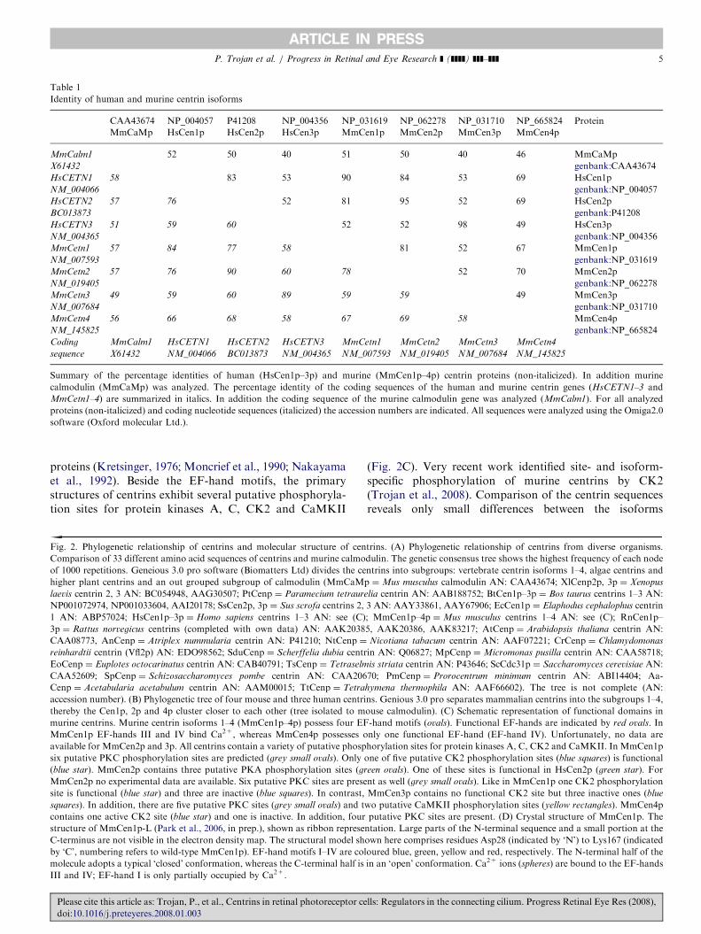
a large variety of species from all kingdoms of eukaryoticorganisms, protists, fungi, plants and animals (Huang et al.,1988a; Baum et al., 1986, 1988; Lee and Huang, 1993;Errabolu et al., 1994; Zhu et al., 1995; Levy et al., 1996;Madeddu et al., 1996; Meng et al., 1996; Middendorp et al.,1997; Wottrich, 1998; Daunderer et al., 2001; Gavet et al.,2003; Guerra et al., 2003; Correa et al., 2004; Lemullois et al.,2004; Ribichich and Gomes, 2005; Nagamune and Sibley,2006; Boutet et al., 2008). Comparisons of amino acidsequences deduced from cDNA clones certainly show thatcentrins are highly conserved, and yet distinct members of asubfamily of the EF-hand superfamily of Ca2+-bindingproteins, also termed the parvalbumin or troponin Csuperfamily (Fig. 2) (Kretsinger and Nockolds, 1973;Kretsinger, 1976; Persechini et al., 1989; Nakayama andKretsinger, 1994). Centrins are small acidic proteins (�170amino acids in length; apparent molecular mass �20kDa)(Salisbury, 1995; Schiebel and Bornens, 1995).To date, in lower eukaryotes like yeast, only one centrin
gene (e.g. Saccharomyces cerevisiae: ScCDC31) has beenidentified (Baum et al., 1986, 1988). In the unicellular algaeChlamydomonas reinhardtii, database searches also provideone centrin gene listed as CrCEN or VFL2. However, in theproteomic analysis of isolated Chlamydomonas centrioles,Keller et al. (2005) identified two other centrins related tomammalian Cen2p and 3p in addition to the previouslyfound centrin (CrCenp/Vfl2p) (Keller et al., 2005).Whereas for lower vertebrate species, an incomplete (mostprobably) set of one to two centrin genes are deposited indatabases, in mammals up to four centrin genes have beendescribed (Fig. 2A) (Friedberg, 2006). In the rodents Mus
musculus and Rattus norvegicus four centrin genes(MmCetn1–4; RnCetn1–4) were identified (Lee and Huang,1993; Middendorp et al., 1997; Gavet et al., 2003; Trojan,2003). In the human genome, three centrin genes arepresent (HsCETN1–3). A predicted fourth centrin gene isfound on chromosome 4 (accession number: XR_015512),but the potential gene transcript encodes a very short, 98amino acids long peptide (GieXl, 2004). It is doubtfulwhether this transcript will exist as a functional polypep-tide in the cell (GieXl, 2004).
lls: Regulators in the connecting cilium. Progress Retinal Eye Res (2008),

ARTICLE IN PRESSP. Trojan et al. / Progress in Retinal and Eye Research ] (]]]]) ]]]–]]]4
Cluster analyses of the deduced amino acid sequences ofthe diverse centrins of different organisms reveal severalphylogenetic groups within the centrin protein family(Fig. 2). While some centrins of protists cannot be classifiedto homogeneous groups, most centrins of higher plants,green algae and all vertebrate centrin isoforms formphylogenetic groups. In mammals, Cen1p, 2p and 4pisoforms are very closely related exhibiting high amino acididentities (Table 1). In contrast, sequences of vertebrateCen3p isoforms, related to the yeast centrin (ScCdc31p),only have high amino acid identities among each other(Table 1, Fig. 2). In mammals, Cen1p, 2p and 4p isoformsare closer related to algal centrin (e.g. Chlamydomonas
CrCenp/Vfl2p) than to the mammalian Cen3p isoform.This strongly suggests two divergent centrin subfamilies
Please cite this article as: Trojan, P., et al., Centrins in retinal photoreceptor ce
doi:10.1016/j.preteyeres.2008.01.003
(Middendorp et al., 1997): one centrin subfamily groupedaround the mammalian Cen1p, 2p and 4p and anothercentrin subfamily related to yeast centrin Cdc31p. Since theseparation of both centrin subfamilies is already imple-mented in the unicellular green algae (see above; Kelleret al. 2005), this division was a very early event in themolecular evolution of eukaryotes.
2.2. Primary domain structure of centrins
Analyses of the primary structures of centrins demon-strate that the most characteristic domains are the fourhelix–loop–helix EF-hand consensus motifs (Fig. 2C).These potential Ca2+-binding sites define centrins asmembers of the superfamily of EF-hand Ca2+-binding
lls: Regulators in the connecting cilium. Progress Retinal Eye Res (2008),
ARTICLE IN PRESS
Table 1
Identity of human and murine centrin isoforms
CAA43674
MmCaMp
NP_004057
HsCen1p
P41208
HsCen2p
NP_004356
HsCen3p
NP_031619
MmCen1p
NP_062278
MmCen2p
NP_031710
MmCen3p
NP_665824
MmCen4p
Protein
MmCalm1
X61432
52 50 40 51 50 40 46 MmCaMp
genbank:CAA43674
HsCETN1
NM_004066
58 83 53 90 84 53 69 HsCen1p
genbank:NP_004057
HsCETN2
BC013873
57 76 52 81 95 52 69 HsCen2p
genbank:P41208
HsCETN3
NM_004365
51 59 60 52 52 98 49 HsCen3p
genbank:NP_004356
MmCetn1
NM_007593
57 84 77 58 81 52 67 MmCen1p
genbank:NP_031619
MmCetn2
NM_019405
57 76 90 60 78 52 70 MmCen2p
genbank:NP_062278
MmCetn3
NM_007684
49 59 60 89 59 59 49 MmCen3p
genbank:NP_031710
MmCetn4
NM_145825
56 66 68 58 67 69 58 MmCen4p
genbank:NP_665824
Coding
sequence
MmCalm1
X61432
HsCETN1
NM_004066
HsCETN2
BC013873
HsCETN3
NM_004365
MmCetn1
NM_007593
MmCetn2
NM_019405
MmCetn3
NM_007684
MmCetn4
NM_145825
Summary of the percentage identities of human (HsCen1p–3p) and murine (MmCen1p–4p) centrin proteins (non-italicized). In addition murine
calmodulin (MmCaMp) was analyzed. The percentage identity of the coding sequences of the human and murine centrin genes (HsCETN1–3 and
MmCetn1–4) are summarized in italics. In addition the coding sequence of the murine calmodulin gene was analyzed (MmCalm1). For all analyzed
proteins (non-italicized) and coding nucleotide sequences (italicized) the accession numbers are indicated. All sequences were analyzed using the Omiga2.0
software (Oxford molecular Ltd.).
P. Trojan et al. / Progress in Retinal and Eye Research ] (]]]]) ]]]–]]] 5
proteins (Kretsinger, 1976; Moncrief et al., 1990; Nakayamaet al., 1992). Beside the EF-hand motifs, the primarystructures of centrins exhibit several putative phosphoryla-tion sites for protein kinases A, C, CK2 and CaMKII
Fig. 2. Phylogenetic relationship of centrins and molecular structure of cen
Comparison of 33 different amino acid sequences of centrins and murine calmo
of 1000 repetitions. Geneious 3.0 pro software (Biomatters Ltd) divides the ce
higher plant centrins and an out grouped subgroup of calmodulin (MmCaMp
laevis centrin 2, 3 AN: BC054948, AAG30507; PtCenp ¼ Paramecium tetraure
NP001072974, NP001033604, AAI20178; SsCen2p, 3p ¼ Sus scrofa centrins 2,
1 AN: ABP57024; HsCen1p–3p ¼ Homo sapiens centrins 1–3 AN: see (C)
3p ¼ Rattus norvegicus centrins (completed with own data) AN: AAK2038
CAA08773, AnCenp ¼ Atriplex nummularia centrin AN: P41210; NtCenp ¼
reinhardtii centrin (Vfl2p) AN: EDO98562; SduCenp ¼ Scherffelia dubia centr
EoCenp ¼ Euplotes octocarinatus centrin AN: CAB40791; TsCenp ¼ Tetraselm
CAA52609; SpCenp ¼ Schizosaccharomyces pombe centrin AN: CAA206
Cenp ¼ Acetabularia acetabulum centrin AN: AAM00015; TtCenp ¼ Tetra
accession number). (B) Phylogenetic tree of four mouse and three human centri
thereby the Cen1p, 2p and 4p cluster closer to each other (tree isolated to m
murine centrins. Murine centrin isoforms 1–4 (MmCen1p–4p) possess four EF
MmCen1p EF-hands III and IV bind Ca2+, whereas MmCen4p possesses o
available for MmCen2p and 3p. All centrins contain a variety of putative phosp
six putative PKC phosphorylation sites are predicted (grey small ovals). Only o
(blue star). MmCen2p contains three putative PKA phosphorylation sites (gr
MmCen2p no experimental data are available. Six putative PKC sites are prese
site is functional (blue star) and three are inactive (blue squares). In contrast,
squares). In addition, there are five putative PKC sites (grey small ovals) and t
contains one active CK2 site (blue star) and one is inactive. In addition, four
structure of MmCen1p-L (Park et al., 2006, in prep.), shown as ribbon represe
C-terminus are not visible in the electron density map. The structural model sh
by ‘C’, numbering refers to wild-type MmCen1p). EF-hand motifs I–IV are col
molecule adopts a typical ‘closed’ conformation, whereas the C-terminal half is
III and IV; EF-hand I is only partially occupied by Ca2+.
Please cite this article as: Trojan, P., et al., Centrins in retinal photoreceptor ce
doi:10.1016/j.preteyeres.2008.01.003
(Fig. 2C). Very recent work identified site- and isoform-specific phosphorylation of murine centrins by CK2(Trojan et al., 2008). Comparison of the centrin sequencesreveals only small differences between the isoforms
trins. (A) Phylogenetic relationship of centrins from diverse organisms.
dulin. The genetic consensus tree shows the highest frequency of each node
ntrins into subgroups: vertebrate centrin isoforms 1–4, algae centrins and
¼Mus musculus calmodulin AN: CAA43674; XlCenp2p, 3p ¼ Xenopus
lia centrin AN: AAB188752; BtCen1p–3p ¼ Bos taurus centrins 1–3 AN:
3 AN: AAY33861, AAY67906; EcCen1p ¼ Elaphodus cephalophus centrin
; MmCen1p–4p ¼Mus musculus centrins 1–4 AN: see (C); RnCen1p–
5, AAK20386, AAK83217; AtCenp ¼ Arabidopsis thaliana centrin AN:
Nicotiana tabacum centrin AN: AAF07221; CrCenp ¼ Chlamydomonas
in AN: Q06827; MpCenp ¼Micromonas pusilla centrin AN: CAA58718;
is striata centrin AN: P43646; ScCdc31p ¼ Saccharomyces cerevisiae AN:
70; PmCenp ¼ Prorocentrum minimum centrin AN: ABI14404; Aa-
hymena thermophila AN: AAF66602). The tree is not complete (AN:
ns. Genious 3.0 pro separates mammalian centrins into the subgroups 1–4,
ouse calmodulin). (C) Schematic representation of functional domains in
-hand motifs (ovals). Functional EF-hands are indicated by red ovals. In
nly one functional EF-hand (EF-hand IV). Unfortunately, no data are
horylation sites for protein kinases A, C, CK2 and CaMKII. In MmCen1p
ne of five putative CK2 phosphorylation sites (blue squares) is functional
een ovals). One of these sites is functional in HsCen2p (green star). For
nt as well (grey small ovals). Like in MmCen1p one CK2 phosphorylation
MmCen3p contains no functional CK2 site but three inactive ones (blue
wo putative CaMKII phosphorylation sites (yellow rectangles). MmCen4p
putative PKC sites are present. (D) Crystal structure of MmCen1p. The
ntation. Large parts of the N-terminal sequence and a small portion at the
own here comprises residues Asp28 (indicated by ‘N’) to Lys167 (indicated
oured blue, green, yellow and red, respectively. The N-terminal half of the
in an ‘open’ conformation. Ca2+ ions (spheres) are bound to the EF-hands
lls: Regulators in the connecting cilium. Progress Retinal Eye Res (2008),

ARTICLE IN PRESSP. Trojan et al. / Progress in Retinal and Eye Research ] (]]]]) ]]]–]]]6
(Table 1) (GieXl et al., 2004a; Salisbury, 2007). In additionto the EF-hand motifs, the most conserved region is theC-terminal half of the centrins, in particular the shortC-terminal sequence (-KKTSLY). This sequence could beresponsible for general features of centrins, like thepositioning at centrosomal structures (e.g. centrosomes,basal bodies or transition zones of cilia). In contrast to theconserved C-terminal domain, the N-terminus, especiallythe first 20 amino acids, represent the most variable regionof the centrin sequences (Hart et al., 1999, 2001; Salisbury,2007). Therefore, this region has been suggested to beresponsible for some functional diversity among centrinspecies (Bhattacharya et al., 1993; Salisbury, 1995; Wiechet al., 1996; Wolfrum et al., 2002; GieXl et al., 2004b; Yanget al., 2006b).
2.2.1. Ca2+-binding at EF-hand motifs of centrins
The most prominent characteristic of the centrin proteinfamily are the four EF-hand motifs which posses thepotential capacity for Ca2+-binding (Fig. 2C). Therefore, itis not surprising that the function of centrins is accompaniedby binding of Ca2+ ions. As a prerequisite, centrins need tobe activated by bound Ca2+ for the interaction with most ofits known interacting partners (details see in Sections 3.1and 5) (Schiebel and Bornens, 1995; Wiech et al., 1996;Durussel et al., 2000; Pulvermuller et al., 2002; GieXl et al.,2004b, 2006; Hu et al., 2004; Cox et al., 2005). In green algae,centrins serve as Ca2+ sensors at the contractile flagellarrootlets by recognizing the increase of the intracellular Ca2+
concentration (Sanders and Salisbury, 1989, 1994; Schiebeland Bornens, 1995). This binding of Ca2+ leads to an ATP-independent contraction of Ca affinity centrin-containingfibres of the flagellar rootlets (Sanders and Salisbury, 1994;Schiebel and Bornens, 1995).
Although the four EF-hand motifs of centrins are highlyconserved, the Ca2+ affinities between the different EF-hands in centrins are not identical. In the green algaeChlamydomonas, centrin molecules bind two Ca2+ ionswith high affinity at the N-terminal domain and two withlow affinity at the C-terminal domain (Weber et al., 1994;Durussel et al., 2000). The N-terminus of Chlamydomonas
centrin serves as a Ca2+ sensor and strengthens protein–protein interactions, like the complex formation with Sfi1p(Sheehan et al., 2006). In contrast to Chlamydomonas
centrin where all EF-hands bind Ca2+, in higher eukar-yotic cells some EF-hand motifs lost the ability to bindCa2+. In HsCen2p, for example, Ca2+-binding is onlymediated via the EF-hand motif IV in the C-terminaldomain (Durussel et al., 2000). This Ca2+-binding inducesconformational changes which lead to the exposure ofhydrophobic surfaces and therefore supporting the forma-tion of homodimers (Durussel et al., 2000; Tourbez et al.,2004). This functional EF-hand IV of HsCen2p is strictlyCa2+-specific and does not bind other cations like Mg2+
(Durussel et al., 2000; Cox et al., 2005). Such mixed cation-binding sites were identified in HsCen3p and indicateisoform-specific regulations of the EF-hands. HsCen3p
Please cite this article as: Trojan, P., et al., Centrins in retinal photoreceptor ce
doi:10.1016/j.preteyeres.2008.01.003
belongs to the second subfamily of centrins, related to yeastCdc31p, and contains three functional EF-hands (Coxet al., 2005). However, two of them show only low Ca2+
affinity and one has a rather unspecific, but high affinityto both cations Ca2+ and Mg2+ (Cox et al., 2005).Preliminary data on MmCen1p indicate extraordinary highCa2+ affinity of EF-hands in the C-terminal domain of themolecule (�1000 times higher than human calmodulin)(Black et al., 2006; Park et al., 2006). Although all centrinscontain the same distribution of EF-hand motifs theregulation is highly selective since the binding propertiesdiffer between species and between isoforms in one species.Due to the fact that most known homomeric andheteromeric protein–protein interactions of centrins areCa2+-triggered, the Ca2+-binding represents the mostimportant, but not the only, molecular regulatory mechan-ism of centrins.
2.2.2. Site-specific phosphorylation of centrins
Besides Ca2+-binding, phosphorylation represents asecond principle modification of centrin molecules whichregulates the functions of centrins in yeast, green algae andin mammalian cells (Salisbury et al., 1984; Martindale andSalisbury, 1990; Salisbury, 1995; Lutz et al., 2001; GieXlet al., 2004b; Trojan et al., 2008). In unicellular green algae,centrins are the major components of contractile fibres atthe basal bodies. These fibres contract upon an increase ofintracellular Ca2+. For fibre relaxation, centrins have to bephosphorylated at the C-terminal domain (Salisbury et al.,1984; Martindale and Salisbury, 1990). In the green algaeChlamydomonas, protein kinase A (PKA) phosphorylates acentrin species at Ser167 in vitro (Meyn et al., 2006). Theidentified target sequence is located in the C-terminus, thehighest conserved region of centrins. In human HeLa cells,phosphorylation of this conserved PKA phosphorylationsite occurs in HsCen2p during the cell cycle at the G1/Stransition (Lutz et al., 2001). Interestingly, centrins arehyperphosphorylated during the abnormal cell cycle ofbreast cancer cells obtained from human patients (Lingleet al., 1998).Recently, we have identified CK2 as the protein kinase
which phosphorylates murine centrin isoforms with highspecificity in fully differentiated retinal photoreceptor cells(for details see Section 5.2) (Trojan et al., 2008). CK2phosphorylation is centrin isoform specific. Only Cen1p, 2pand 4p, but not Cen3p are phosphorylated by CK2 at aspecific site. Furthermore, CK2-mediated phosphorylationoccurs in the dark and strongly reduces the bindingaffinities of centrins for target interactor proteins likeG-proteins. The latter effect is probably due to the reductionof centrin Ca2+ affinity induced by phosphorylation.In conclusion, regulatory modifications of centrins
include two major events: Ca2+-binding on the one handand phosphorylation on the other hand. Comparisons ofhigh-resolution structures in centrins under different Ca2+
concentration and phosphorylation states may providemore insights in these mechanisms.
lls: Regulators in the connecting cilium. Progress Retinal Eye Res (2008),
ARTICLE IN PRESS
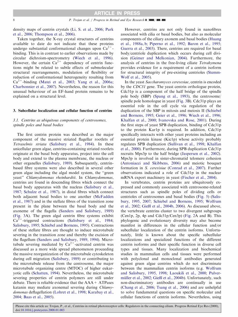
Table 2
Domain and full-length centrin structures from different sources determined by either NMR spectroscopy or X-ray crystallography
Structure Reference Method PDB code Deposition
C-HsCen2pa Matei et al. (2003) NMR 1M39 2003
C-CrCenp/Kar1pb Hu and Chazin (2003) NMR 1OQP 2003
C-HsCen2p/XPCpc Yang et al. (2006a) NMR 2A4J 2005
N-CrCenpd Sheehan et al. (2006) NMR 2AMI 2006
N-HsCen2p Yang et al. (2006b) NMR 1ZMZ 2006
HsCen2p/XPCp Thompson et al. (2006) X-ray 2GGM 2006
Cdc31p/Sfi1pe (Ca2+ bound) Li, S. et al. (2006) X-ray 2DOQ 2006
Cdc31p/Sfi1p (Ca2+ free) Li, S. et al. (2006) X-ray 2GV5 2006
HsCen2p/XPCp Charbonnier et al. (2007) X-ray 2OBH 2007
MmCen1p-Lf Park and Pulvermuller (unpublished) X-ray – –
a‘C-’ indicates the C-terminal half of a centrin containing EF-hand motifs III and IV.bKar1p is a peptide derived from the cell division control protein Kar1p.cXPC is a peptide derived from Xeroderma pigmentosum group C protein.d‘N-’ indicates the N-terminal half of a centrin containing EF-hand motifs I and II.eSfi1p is a protein from the half bridge attached to the spindle pole body.fMmCen1p-L is an N-terminally extended mouse Cen1p (Park et al., 2006, 2005).
2N-terminally extended MmCen1p containing the additional
GSPGISGGGGGIRLRAPLRSQLLWR peptide sequence.
P. Trojan et al. / Progress in Retinal and Eye Research ] (]]]]) ]]]–]]] 7
2.3. High-resolution molecular structure of centrins and
their complexes
Detailed protein structures at high-resolution based onnuclear magnetic resonance (NMR) spectroscopy andX-ray crystallography provide more insights into themolecular composition and function of molecules. High-resolution structural data on centrins from differentorganisms are available from solution studies by NMRspectroscopy and from X-ray crystallography (Table 2).These studies were carried out either on individual C- orN-terminal domains, or on full-length proteins, and theproteins were either in their unbound states or incomplexes with target proteins.
As a result of these analyses, centrins are known to formdumbbell-like structures, which closely resemble those ofthe other proteins belonging to the calmodulin-parvalbu-min superfamily (Fig. 2D). Pairs of the first and secondEF-hands and of the third and the fourth EF-hands formcompact structures in the N- and C-terminal halves,respectively, which are connected by a long central a-helix(Fig. 2D). Typically an ‘open’ conformation is reported forthe C-terminal domains and a ‘closed’ conformation isfound for the N-terminal domains of the centrin molecules.‘Closed’ conformations are usually adopted by the Ca2+-free domains of calmodulin-parvalbumin-type proteins,whereas Ca2+-binding triggers a switch to the ‘open’conformation as was shown in detail for troponin C byX-ray crystallography (Herzberg and James, 1985;Sundaralingam et al., 1985; Satyshur et al., 1994; Houdusseet al., 1997; Strynadka et al., 1997). Correspondingly, in thetwo crystal structures of the HsCen2p–XCPp complex(Table 2; for functional details see Section 5), theN-terminal domains are Ca2+-free and each of theEF-hand motifs III and IV in the C-terminal domain hasa Ca2+ ion bound (Thompson et al., 2006; Charbonnieret al., 2007). In agreement with a relatively low Ca2+
Please cite this article as: Trojan, P., et al., Centrins in retinal photoreceptor ce
doi:10.1016/j.preteyeres.2008.01.003
affinity of EF-hand III this site is occupied by Ca2+ in only70% of the HsCen2p molecules in the crystal (Charbonnieret al., 2007). Surprisingly, a typical ‘open’ conformation isreported for the C-terminal domains of centrin moleculeseven in the Ca2+-free Cdc31p/Sfi1p complex. In contrast,in the Ca2+-bound Cdc31p/Sfi1p complex and inMmCen1p-L2 Ca2+ ions are bound to the EF-hand I andboth centrins adopt a ‘closed’ conformation (Li, S. et al.,2006; Park et al., 2006). Assuming that the Ca2+ affinitiesof the N-terminal domains of HsCen2p (Yang et al., 2006a)and Cdc31p are comparably low, the observed binding ofCa2+ to EF-hand I in one of the Cdc31p/Sfi1p complexstructures is a result of the unphysiologically high Ca2+
concentration (0.1M) used for crystallization (Li, S. et al.,2006). The fact that Ca2+ binding does not change theconformation was also shown by NMR spectroscopy forthe N-terminal domain of HsCen2p (Yang et al., 2006a),but, on a structural level, the reason for this unusualbehaviour of an EF-hand protein remains to be explained.Clearly different from the majority of centrins, which havebeen structurally investigated, the N-terminal domain ofCrCenp adopts an ‘open’ conformation in the presence ofCa2+ and is proposed to act as a Ca2+ sensor (Sheehanet al., 2006).Analyses of polymerization properties of centrins in-
dicate that their Ca2+-induced polymerization is mainlydependent on the N-terminal subdomain (Wiech et al.,1996). Studies on HsCen2p suggest that polymerization isfacilitated by intermolecular interactions of the N- and theC-terminal subdomains (Yang et al., 2006a). NMRspectroscopy revealed the N-terminal subdomains to beof irregular and dynamic structure in solution (Yang et al.,2006a). These regions were also not visible in the electron
lls: Regulators in the connecting cilium. Progress Retinal Eye Res (2008),
ARTICLE IN PRESSP. Trojan et al. / Progress in Retinal and Eye Research ] (]]]]) ]]]–]]]8
density maps of centrin crystals (Li, S. et al., 2006; Parket al., 2006; Thompson et al., 2006).
Taken together, the X-ray crystal structures of centrinsavailable to date do not indicate that these proteinsundergo substantial conformational changes upon Ca2+-binding. This is in contrast to earlier observations made bycircular dichroism-spectrometry (Wiech et al., 1996).However, the certain Ca2+ dependency of centrin func-tions might be related to subtle effects of submolecularstructural rearrangements, modulation of flexibility orreduction of conformational heterogeneity resulting fromCa2+-binding (Matei et al., 2003; Yang et al., 2006a;Charbonnier et al., 2007). Nevertheless, the reason for thisunusual behaviour of an EF-hand protein remains to beexplained on a structural level.
3. Subcellular localization and cellular function of centrins
3.1. Centrins as ubiquitous components of centrosomes,
spindle poles and basal bodies
The first centrin protein was described as the majorcomponent of the massive striated flagellar rootlets ofTetraselmis striata (Salisbury et al., 1984). In theseunicellular green algae, centrins-containing striated rootletsoriginate at the basal body apparatus, project into the cellbody and extend to the plasma membrane, the nucleus orother organelles (Salisbury, 1989). Subsequently, centrin-based fibre systems were also described in several othergreen algae including the algal model system, the ‘‘greenyeast’’ Chlamydomonas rheinhardtii. In Chlamydomonas,centrins are found in descending fibres which connect thebasal body apparatus with the nucleus (Salisbury et al.,1987; Schulze et al., 1987), in distal fibres which connectboth adjacent basal bodies to one another (McFaddenet al., 1987) and in the stellate fibres of the transition zonepresent in the plane between the basal body and theaxoneme of the flagella (Sanders and Salisbury, 1989)(Fig. 3A). The green algal centrin fibre systems exhibitCa2+-triggered contractions (Salisbury et al., 1984;Salisbury, 1995; Schiebel and Bornens, 1995). Contractionsof these stellate fibres are thought to induce microtubulesevering in the transition zone and thereby the excision ofthe flagellum (Sanders and Salisbury, 1989, 1994). Micro-tubule severing mediated by Ca2+-activated centrin wasdiscussed as a more wide spread phenomenon proceedingthe massive reorganization of the microtubule cytoskeletonduring cell migration (Salisbury, 1989) or contributing tothe microtubule release from the centrosome, the majormicrotubule organizing centre (MTOC) of higher eukar-yotic cells (Schatten, 1994). Nevertheless, the microtubulesevering properties of centrin polymers are still underdebate. There is reliable evidence that the AAA+ ATPaseskatanin may mediate axonemal severing during Chlamy-
domonas deflagellation (Lohret et al., 1998; Karabay et al.,2004; Baas et al., 2005).
Please cite this article as: Trojan, P., et al., Centrins in retinal photoreceptor ce
doi:10.1016/j.preteyeres.2008.01.003
However, centrins are not only found in nanofibresassociated with cilia or basal bodies, but also as molecularcomponents of the ciliary axonem and basal bodies (Huanget al., 1988a, b; Piperno et al., 1992; Baron et al., 1995;Guerra et al., 2003). There, centrins are required for basalbody/centriole duplication which occurs during cell divi-sion (Geimer and Melkonian, 2004). Furthermore, theanalysis of centrins in the free-living ciliate Tetrahymena
provides evidence for a requirement of a centrin isoformfor structural integrity of pre-existing centrioles (Stemm-Wolf et al., 2005).In the yeast Saccharomyces cerevesiae, centrin is encoded
by the CDC31 gene. The yeast centrin orthologue protein,Cdc31p is a component of the half bridge of the spindlepole body (SBP) (Spang et al., 1993), the centrosome/spindle pole homologue in yeast (Fig. 3B). Cdc31p plays anessential role in the cell cycle via regulation of theduplication of the SBP in mitosis and meiosis II (Schiebeland Bornens, 1995; Geier et al., 1996; Wiech et al., 1996;Khalfan et al., 2000; Ivanovska and Rose, 2001). Duringthe first steps of yeast SPB duplication, binding of Cdc31pto the protein Kar1p is required. In addition, Cdc31pspecifically interacts with other yeast proteins including anessential protein kinase (Kic1p) whose activity probablyregulates SPB duplication (Sullivan et al., 1998; Khalfanet al., 2000). Furthermore, during SPB duplication Cdc31precruits Mps3p to the half bridge (Jaspersen et al., 2002).Mps3p is involved in sister-chromatid telomere cohesion(Antoniacci and Skibbens, 2006) and meiotic bouquetformation in S. cerevisiae (Conrad et al., 2007). Furtherobservations indicated a role of Cdc31p in the nuclearmRNA export machinery in yeast (Fischer et al., 2004).In vertebrates, centrin proteins are ubiquitously ex-
pressed and commonly associated with centrosome-relatedstructures such as spindle poles of dividing cells orcentrioles of centrosomes and basal bodies (Fig. 5) (Salis-bury, 1995, 2007; Schiebel and Bornens, 1995; Wolfrumet al., 2002; GieXl et al., 2004b, 2006). As discussed above,the vertebrate centrins cluster to two divergent subgroups(Cen1p, 2p, 4p and Cdc31p/Cen3p) (Fig. 2A and B). Thisphylogenic and evolutionary diversity may also becomemanifest in differences in the cellular function and/orsubcellular localization of the centrin isoforms. Unfortu-nately, little is known about the specific subcellularlocalizations and specialized functions of the differentcentrin isoforms and their specific function in diverse celltypes and tissues. Many localization and biochemicalstudies in mammalian cells and tissues were performedwith polyclonal and monoclonal antibodies generatedagainst green algae centrins which do not discriminatebetween the mammalian centrin isoforms (e.g. Wolfrumand Salisbury, 1995, 1998; Laoukili et al., 2000; Pulver-muller et al., 2002; GieXl et al., 2004b). Unfortunately, suchnon-discriminatory antibodies are continually in use(Chang et al., 2006; Tsang et al., 2006) and are unhelpfulto solve fundamental questions on specific molecular andcellular functions of centrin isoforms. Nevertheless, using
lls: Regulators in the connecting cilium. Progress Retinal Eye Res (2008),
ARTICLE IN PRESS
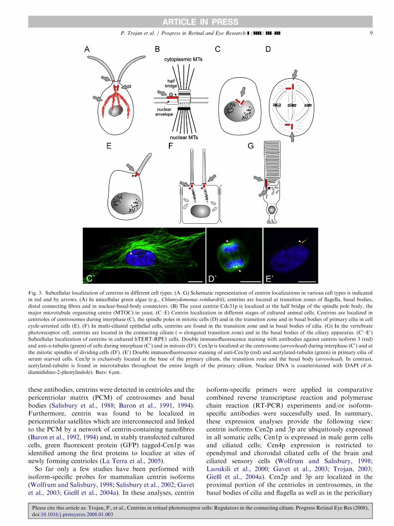
Fig. 3. Subcellular localization of centrins in different cell types. (A–G) Schematic representation of centrin localizations in various cell types is indicated
in red and by arrows. (A) In unicellular green algae (e.g., Chlamydomonas reinhardtii), centrins are located at transition zones of flagella, basal bodies,
distal connecting fibres and in nuclear-basal-body connectors. (B) The yeast centrin Cdc31p is localized at the half bridge of the spindle pole body, the
major microtubule organizing centre (MTOC) in yeast. (C–E) Centrin localization in different stages of cultured animal cells. Centrins are localized in
centrioles of centrosomes during interphase (C), the spindle poles in mitotic cells (D) and in the transition zone and in basal bodies of primary cilia in cell
cycle-arrested cells (E). (F) In multi-ciliated epithelial cells, centrins are found in the transition zone and in basal bodies of cilia. (G) In the vertebrate
photoreceptor cell, centrins are located in the connecting cilium ( ¼ elongated transition zone) and in the basal bodies of the ciliary apparatus. (C0–E0)
Subcellular localization of centrins in cultured hTERT-RPE1 cells. Double immunofluorescence staining with antibodies against centrin isoform 3 (red)
and anti-a-tubulin (green) of cells during interphase (C0) and in mitosis (D0). Cen3p is localized at the centrosome (arrowhead) during interphase (C0) and at
the mitotic spindles of dividing cells (D0). (E0) Double immunofluorescence staining of anti-Cen3p (red) and acetylated-tubulin (green) in primary cilia of
serum starved cells. Cen3p is exclusively located at the base of the primary cilium, the transition zone and the basal body (arrowhead). In contrast,
acetylated-tubulin is found in microtubules throughout the entire length of the primary cilium. Nuclear DNA is counterstained with DAPI (40,6-
diamididino-2-phenylindole). Bars: 6mm.
P. Trojan et al. / Progress in Retinal and Eye Research ] (]]]]) ]]]–]]] 9
these antibodies, centrins were detected in centrioles and thepericentriolar matrix (PCM) of centrosomes and basalbodies (Salisbury et al., 1988; Baron et al., 1991, 1994).Furthermore, centrin was found to be localized inpericentriolar satellites which are interconnected and linkedto the PCM by a network of centrin-containing nanofibres(Baron et al., 1992, 1994) and, in stably transfected culturedcells, green fluorescent protein (GFP) tagged-Cen1p wasidentified among the first proteins to localize at sites ofnewly forming centrioles (La Terra et al., 2005).
So far only a few studies have been performed withisoform-specific probes for mammalian centrin isoforms(Wolfrum and Salisbury, 1998; Salisbury et al., 2002; Gavetet al., 2003; GieXl et al., 2004a). In these analyses, centrin
Please cite this article as: Trojan, P., et al., Centrins in retinal photoreceptor ce
doi:10.1016/j.preteyeres.2008.01.003
isoform-specific primers were applied in comparativecombined reverse transcriptase reaction and polymerasechain reaction (RT-PCR) experiments and/or isoform-specific antibodies were successfully used. In summary,these expression analyses provide the following view:centrin isoforms Cen2p and 3p are ubiquitously expressedin all somatic cells; Cen1p is expressed in male germ cellsand ciliated cells; Cen4p expression is restricted toependymal and choroidal ciliated cells of the brain andciliated sensory cells (Wolfrum and Salisbury, 1998;Laoukili et al., 2000; Gavet et al., 2003; Trojan, 2003;GieXl et al., 2004a). Cen2p and 3p are localized in theproximal portion of the centrioles in centrosomes, in thebasal bodies of cilia and flagella as well as in the periciliary
lls: Regulators in the connecting cilium. Progress Retinal Eye Res (2008),
ARTICLE IN PRESSP. Trojan et al. / Progress in Retinal and Eye Research ] (]]]]) ]]]–]]]10
matrix surrounding the centrioles (Paoletti et al., 1996;Laoukili et al., 2000; GieXl et al., 2004a). In the course ofmitosis, both, Cen2p and 3p, appear and stay in the spindlepoles. In contrast to Cen2p and 3p, Cen1p was mapped tothe transition zone of cilia and Cen4p was found in thebasal body of ciliated neurons and sensory cells (Gavetet al., 2003; GieXl et al., 2004a).
The prominent localization of centrins at the centrosomesand basal bodies gave rise to several hypotheses regardingthe cellular functions of centrins. In animal interphase cellsor in arrested cells of differentiated tissue, the centrosomefunctions as the major MTOC (Fig. 3) (Bettencourt-Diasand Glover, 2007). At the MTOC, microtubules are de novo
synthesized; the number and polarity of cytoplasmicmicrotubules is determined. It has been suggested thatcentrins are involved in the microtubule severing whichshould occur to release de novo synthesized microtubulesfrom the pericentriolar origin (Schatten, 1994). However,more reliable evidence was gathered for important, butprobably distinct roles of centrins at the centrosome duringthe cell cycle. The centrosome is duplicated once during thecell cycle to give rise to two spindle poles that organize themicrotubule array of the mitotic spindle (Fig. 3D and D0).Like its close relative, the yeast Cdc31p, Cen3p mayparticipate in centrosome reproduction and duplicationduring G2 of the interphase of mitosis (Middendorp et al.,2000). Cen2p seems to play a specific role in centrioleseparation preceding centrosome duplication (Lutz et al.,2001). Gene silencing experiments using RNA interferencein human HeLa cells confirmed the requirement of Cen2pfor correct centrosome duplication and for proper cytokin-esis (Salisbury et al., 2002).
Nevertheless, centrins are not only expressed in thecentrioles during the de novo formation of basal bodies orduring the prearrangement and execution of centriolduplication, but also at basal bodies and centrioles andcentrosomes in interphase G1 or in fully differentiated cells(G0) (Fig. 3E and E0). However, little is known about thefunction of centrins in the latter cell stages. In G1 and G0cells, vertebrate centrins are probably required for struc-tural integrity of pre-existing centrioles as shown forcentrins in the ciliate Tetrahymena (Stemm-Wolf et al.,2005; Salisbury, 2007). Furthermore, centrins may con-tribute to membrane-independent G-protein signalling atthe centrosomes and the basal body apparatus of ciliatedcells (GieXl et al., 2004b). In the highly specialized, fullydifferentiated photoreceptor cells of the vertebrate retinabinding of centrins to the visual G-protein may regulate thetranslocation of transducin through the connecting cilium(see below, Section 5.1).
4. Centrins in the vertebrate retina
4.1. Centrin expression in the vertebrate retina
Comparative studies demonstrate expression of centrinsin the retina of species distributed throughout the
Please cite this article as: Trojan, P., et al., Centrins in retinal photoreceptor ce
doi:10.1016/j.preteyeres.2008.01.003
subphylum of vertebrates (Fig. 4) (Wolfrum and Salisbury,1998; Wolfrum et al., 2002). In mammals, RT-PCRanalyses with isoform-specific primers demonstrate expres-sion of all four known mammalian centrin isoforms in theretina (Wolfrum and Salisbury, 1998; Trojan, 2003; GieXlet al., 2004a). The RT-PCR results were confirmed inprotein expression studies by Western blot analyses usingspecific antibody probes for specific centrin isoforms (GieXlet al., 2004a).
4.2. Subcellular localization of centrins in retinal cells —
in particular in photoreceptor cells
As in other cell types of animal tissues, centrins arecommon components of centrioles of the centrosomes andof basal body apparatuses in neurons of the vertebrateretina (Figs. 3 and 6) (Wolfrum and Salisbury, 1998;Wolfrum et al., 2002). Furthermore, centrins were found inthe connecting cilium of rod and cone photoreceptor cellsin all vertebrate species investigated so far (Fig. 4)(Wolfrum, 1992; Wolfrum and Salisbury, 1995, 1998;Schmitt and Wolfrum, 2001; Pulvermuller et al., 2002;Wolfrum et al., 2002; GieXl et al., 2004a, b, 2006). Ourdetailed analysis of the diverse centrins in the mouse retinarevealed differential expression and subcellular distributionof centrin isoforms (Figs. 5 and 6) (GieXl et al., 2004a, b,2006): Cen2p and 3p are expressed in all cell types of theretina and the associated cells of the retinal pigmentepithelium. As in other cell types, Cen2p and 3p arelocalized at the centrosomes of non-photoreceptor retinalneurons. In rod and cone photoreceptor cells, subcellularlocalization of Cen2p and 3p is found in the basal body andthe connecting cilium. In contrast, the expression of Cen1pand 4p in the retina is restricted to photoreceptor cells.Cen1p and 4p are localized in the connecting cilium or inthe basal body of rod and cone photoreceptor cells,respectively (Fig. 6). In conclusion, it is worth noting thatrod and cone photoreceptor cells of the mammalian retinaare the only cell types known so far that express all fourcentrins in parallel; three isoforms (Cen1p–3p) in the ciliumand three in the basal body (Cen2p–4p) (Fig. 6).High-resolution immunofluorescence techniques and
immunoelectron microscopy enabled assignment of cen-trins to organelle substructures in retinal cells. As incentrioles of other cell types, centrins are found in theapical part of the centrioles. In the connecting cilium ofphotoreceptor cells, the ‘‘ciliary centrins’’, Cen1p–3p arelocalized along the entire extension of the connectingcilium (Figs. 5 and 6) (Wolfrum and Salisbury, 1995;Pulvermuller et al., 2002; Wolfrum et al., 2002; GieXl et al.,2004a, b, 2006). Therefore, antibodies raised againstcentrins are frequently used as molecular markers not onlyfor centrioles (e.g. Nagasato and Motomura, 2004; LaTerra et al., 2005; Dahm et al., 2007), but also for theconnecting cilium (e.g. Liu et al., 1997; den Hollanderet al., 2007; Overlack et al., 2008; Maerker et al., 2008).Immunoelectron microscopy data demonstrate that the
lls: Regulators in the connecting cilium. Progress Retinal Eye Res (2008),

ARTICLE IN PRESS
Fig. 4. Localization of centrins in vertebrate photoreceptor cells. (A–J) Immunofluorescence localizations of centrins in photoreceptor cells of species from
different vertebrate classes. (A, C, E, G and I) Differential interference contrast images. (B, D, F, H and J) Indirect immunofluorescence staining with anti
pan-centrin antibodies (green) and DAPI DNA staining (blue). Centrins are predominantly located in the ciliary apparatus, composed of the connecting
cilium and the basal body. (A, B) Cryosection of a light-adapted fish retina (Danio rerio). Centrins are localized at the joint between the inner and outer
segment of rods (arrowhead) and cones (arrow). Note: fish photoreceptor cells and RPE cells exhibit light-dependent retinomotor movement. In light-
adapted retinas, melanin granules in the long microvilli-like extensions of the retinal pigment epithelium cells (asterisk) are located between the layer of rod
and cone photoreceptor cells. (C, D) In an isolated rod photoreceptor cell of the teleost Lepomis cyanellus, centrins are stained in the short connecting
cilium (green upper dot) and the basal body (green lower dot) (arrowhead). (E, F) In amphibian photoreceptor cells (Ambystoma mexicanum) centrin
antibodies label connecting cilia in rods (arrowhead) and cones (arrow). (G, H) In chicken (Gallus gallus), pan-centrin antibodies label the ciliary apparatus
of photoreceptor cells. (I, J) In the pig Sus scrofa (mammal), centrins are localized in the cilary apparatus of rod (arrowhead) and cone (arrow)
photoreceptor cells.
P. Trojan et al. / Progress in Retinal and Eye Research ] (]]]]) ]]]–]]] 11
‘‘ciliary centrins’’ co-localize at the inner surface of themicrotubule doublets of the connecting cilium (Fig. 5H;Wolfrum and Salisbury, 1998; Pulvermuller et al., 2002; U.Wolfrum and A. GieXl, unpublished data). Our recentobservation of a direct binding of Cen1p to microtubulesfurther supports this attachment to the ciliary microtubulepairs (Trojan et al., 2008).
The modified connecting cilium of vertebrate photo-receptor cells is the structural equivalent of an extendedtransition zone present at the base of a common motilecilium (Besharse and Horst, 1990; Liu et al., 2007;Roepman and Wolfrum, 2007). The presence of centrinsalong the entire extension of the connecting cilium is inagreement with the localization of centrins in the transitionzone of motile cilia or the sensory cilia of mammalianolfactory cells (Wolfrum and Salisbury, 1998; Laoukiliet al., 2000). The prominent localization of ‘‘ciliarycentrins’’ in the connecting cilium certainly indicates a
Please cite this article as: Trojan, P., et al., Centrins in retinal photoreceptor ce
doi:10.1016/j.preteyeres.2008.01.003
specific role of centrins in the function of the photoreceptorcilium. At the joint between the outer and the innersegment of the photoreceptor cell ‘‘ciliary centrins’’ mayparticipate in the alignment of the photoreceptor outersegment (Wolfrum and Salisbury, 1995). In mammals,variation of the alignment angle of each outer segment isthought to achieve optimal light infiltration in eachphotoreceptor outer segment (Enoch, 1981). In addition,‘‘ciliary centrins’’ may contribute to the massive moleculartransport through the connecting cilium (Wolfrum andSalisbury, 1995). They may also contribute to the barrierfor soluble proteins which is thought to be established inthe connecting cilium for the regulation of moleculardiffusion between the inner and the outer segment ofphotoreceptor cells (Spencer et al., 1988; Besharse andHorst, 1990; Wolfrum and Salisbury, 1998). In any case,centrin-based processes in the connecting cilium should bedependent on regulatory changes of the free Ca2+
lls: Regulators in the connecting cilium. Progress Retinal Eye Res (2008),
ARTICLE IN PRESS
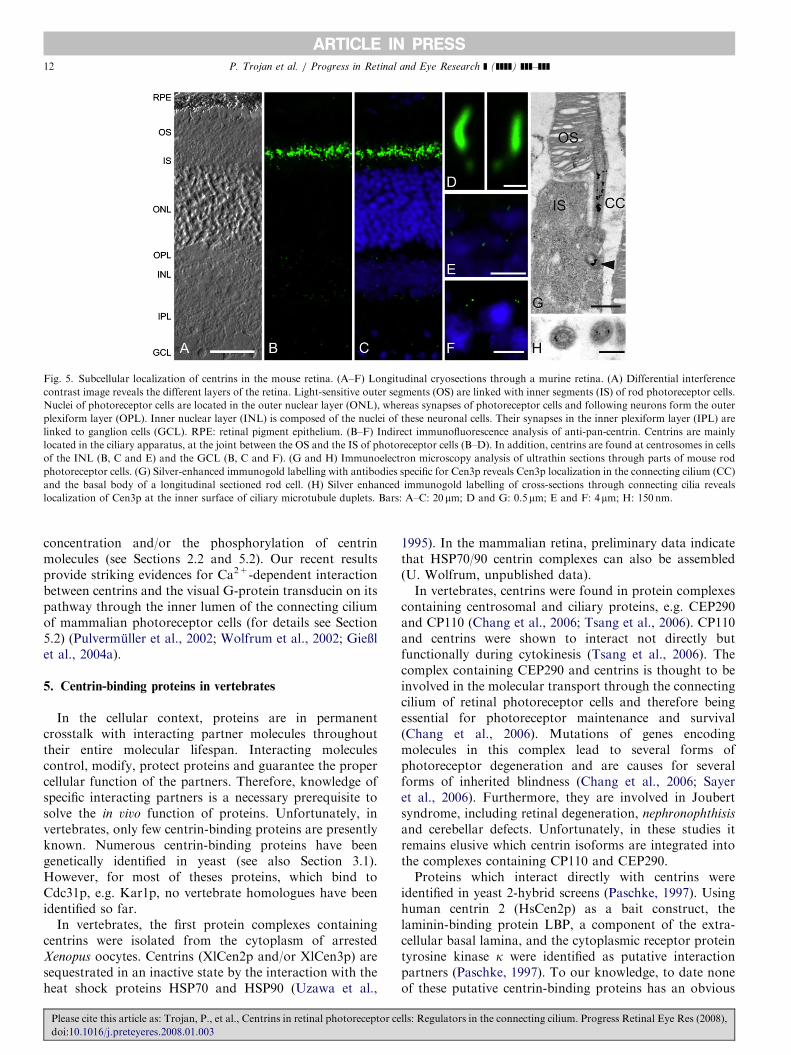
Fig. 5. Subcellular localization of centrins in the mouse retina. (A–F) Longitudinal cryosections through a murine retina. (A) Differential interference
contrast image reveals the different layers of the retina. Light-sensitive outer segments (OS) are linked with inner segments (IS) of rod photoreceptor cells.
Nuclei of photoreceptor cells are located in the outer nuclear layer (ONL), whereas synapses of photoreceptor cells and following neurons form the outer
plexiform layer (OPL). Inner nuclear layer (INL) is composed of the nuclei of these neuronal cells. Their synapses in the inner plexiform layer (IPL) are
linked to ganglion cells (GCL). RPE: retinal pigment epithelium. (B–F) Indirect immunofluorescence analysis of anti-pan-centrin. Centrins are mainly
located in the ciliary apparatus, at the joint between the OS and the IS of photoreceptor cells (B–D). In addition, centrins are found at centrosomes in cells
of the INL (B, C and E) and the GCL (B, C and F). (G and H) Immunoelectron microscopy analysis of ultrathin sections through parts of mouse rod
photoreceptor cells. (G) Silver-enhanced immunogold labelling with antibodies specific for Cen3p reveals Cen3p localization in the connecting cilium (CC)
and the basal body of a longitudinal sectioned rod cell. (H) Silver enhanced immunogold labelling of cross-sections through connecting cilia reveals
localization of Cen3p at the inner surface of ciliary microtubule duplets. Bars: A–C: 20 mm; D and G: 0.5mm; E and F: 4mm; H: 150 nm.
P. Trojan et al. / Progress in Retinal and Eye Research ] (]]]]) ]]]–]]]12
concentration and/or the phosphorylation of centrinmolecules (see Sections 2.2 and 5.2). Our recent resultsprovide striking evidences for Ca2+-dependent interactionbetween centrins and the visual G-protein transducin on itspathway through the inner lumen of the connecting ciliumof mammalian photoreceptor cells (for details see Section5.2) (Pulvermuller et al., 2002; Wolfrum et al., 2002; GieXlet al., 2004a).
5. Centrin-binding proteins in vertebrates
In the cellular context, proteins are in permanentcrosstalk with interacting partner molecules throughouttheir entire molecular lifespan. Interacting moleculescontrol, modify, protect proteins and guarantee the propercellular function of the partners. Therefore, knowledge ofspecific interacting partners is a necessary prerequisite tosolve the in vivo function of proteins. Unfortunately, invertebrates, only few centrin-binding proteins are presentlyknown. Numerous centrin-binding proteins have beengenetically identified in yeast (see also Section 3.1).However, for most of theses proteins, which bind toCdc31p, e.g. Kar1p, no vertebrate homologues have beenidentified so far.
In vertebrates, the first protein complexes containingcentrins were isolated from the cytoplasm of arrestedXenopus oocytes. Centrins (XlCen2p and/or XlCen3p) aresequestrated in an inactive state by the interaction with theheat shock proteins HSP70 and HSP90 (Uzawa et al.,
Please cite this article as: Trojan, P., et al., Centrins in retinal photoreceptor ce
doi:10.1016/j.preteyeres.2008.01.003
1995). In the mammalian retina, preliminary data indicatethat HSP70/90 centrin complexes can also be assembled(U. Wolfrum, unpublished data).In vertebrates, centrins were found in protein complexes
containing centrosomal and ciliary proteins, e.g. CEP290and CP110 (Chang et al., 2006; Tsang et al., 2006). CP110and centrins were shown to interact not directly butfunctionally during cytokinesis (Tsang et al., 2006). Thecomplex containing CEP290 and centrins is thought to beinvolved in the molecular transport through the connectingcilium of retinal photoreceptor cells and therefore beingessential for photoreceptor maintenance and survival(Chang et al., 2006). Mutations of genes encodingmolecules in this complex lead to several forms ofphotoreceptor degeneration and are causes for severalforms of inherited blindness (Chang et al., 2006; Sayeret al., 2006). Furthermore, they are involved in Joubertsyndrome, including retinal degeneration, nephronophthisis
and cerebellar defects. Unfortunately, in these studies itremains elusive which centrin isoforms are integrated intothe complexes containing CP110 and CEP290.Proteins which interact directly with centrins were
identified in yeast 2-hybrid screens (Paschke, 1997). Usinghuman centrin 2 (HsCen2p) as a bait construct, thelaminin-binding protein LBP, a component of the extra-cellular basal lamina, and the cytoplasmic receptor proteintyrosine kinase k were identified as putative interactionpartners (Paschke, 1997). To our knowledge, to date noneof these putative centrin-binding proteins has an obvious
lls: Regulators in the connecting cilium. Progress Retinal Eye Res (2008),
ARTICLE IN PRESS
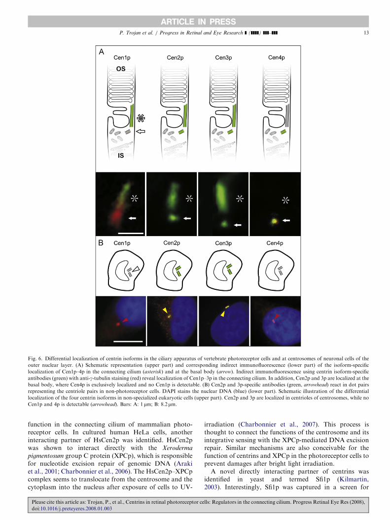
Fig. 6. Differential localization of centrin isoforms in the ciliary apparatus of vertebrate photoreceptor cells and at centrosomes of neuronal cells of the
outer nuclear layer. (A) Schematic representation (upper part) and corresponding indirect immunofluorescence (lower part) of the isoform-specific
localization of Cen1p–4p in the connecting cilium (asterisk) and at the basal body (arrow). Indirect immunofluorescence using centrin isoform-specific
antibodies (green) with anti-g-tubulin staining (red) reveal localization of Cen1p–3p in the connecting cilium. In addition, Cen2p and 3p are localized at the
basal body, where Cen4p is exclusively localized and no Cen1p is detectable. (B) Cen2p and 3p-specific antibodies (green, arrowhead) react in dot pairs
representing the centriole pairs in non-photoreceptor cells. DAPI stains the nuclear DNA (blue) (lower part). Schematic illustration of the differential
localization of the four centrin isoforms in non-specialized eukaryotic cells (upper part). Cen2p and 3p are localized in centrioles of centrosomes, while no
Cen1p and 4p is detectable (arrowhead). Bars: A: 1 mm; B: 8.2 mm.
P. Trojan et al. / Progress in Retinal and Eye Research ] (]]]]) ]]]–]]] 13
function in the connecting cilium of mammalian photo-receptor cells. In cultured human HeLa cells, anotherinteracting partner of HsCen2p was identified. HsCen2pwas shown to interact directly with the Xeroderma
pigmentosum group C protein (XPCp), which is responsiblefor nucleotide excision repair of genomic DNA (Arakiet al., 2001; Charbonnier et al., 2006). The HsCen2p–XPCpcomplex seems to translocate from the centrosome and thecytoplasm into the nucleus after exposure of cells to UV-
Please cite this article as: Trojan, P., et al., Centrins in retinal photoreceptor ce
doi:10.1016/j.preteyeres.2008.01.003
irradiation (Charbonnier et al., 2007). This process isthought to connect the functions of the centrosome and itsintegrative sensing with the XPCp-mediated DNA excisionrepair. Similar mechanisms are also conceivable for thefunction of centrins and XPCp in the photoreceptor cells toprevent damages after bright light irradiation.A novel directly interacting partner of centrins was
identified in yeast and termed Sfi1p (Kilmartin,2003). Interestingly, Sfi1p was captured in a screen for
lls: Regulators in the connecting cilium. Progress Retinal Eye Res (2008),
ARTICLE IN PRESSP. Trojan et al. / Progress in Retinal and Eye Research ] (]]]]) ]]]–]]]14
Ca2+-independent centrin interaction partners. This pro-tein contains multiple conserved centrin-binding repeatsand was discussed as a structural scaffolding proteinforming Ca2+-sensitive contractile fibres (Kilmartin,2003; Li, X. et al., 2006). Mutations in SFI1 gene lead todrastic spindle pole defects in budding yeast indicating arole during the duplication of the MTOC and duringmitotic spindle assembly (Kilmartin, 2003; Li, X. et al.,2006; Anderson et al., 2007). Sfi1p-like proteins containingcentrin-binding repeats are conserved from yeast tohumans (Kilmartin, 2003; Salisbury, 2004; Li, X. et al.,2006; Gogendeau et al., 2007). However, no clear evidencefor an interaction of centrins with Sfi1p-like proteins and afunctional role of such protein complexes has beendescribed in higher eukaryotes so far.
5.1. Centrin-binding proteins in mammalian photoreceptor
cells
Little is known about the expression and function ofcentrin-binding proteins like XPCp-protein or Sfi1p in theretina. Unpublished data obtained by RT-PCR indicatethat Sfi1p is expressed in the murine retina (Ph. Trojan andU. Wolfrum, unpublished). Unfortunately, a furtherevaluation of the Sfi1p function in retinal photoreceptorcells is lacking.
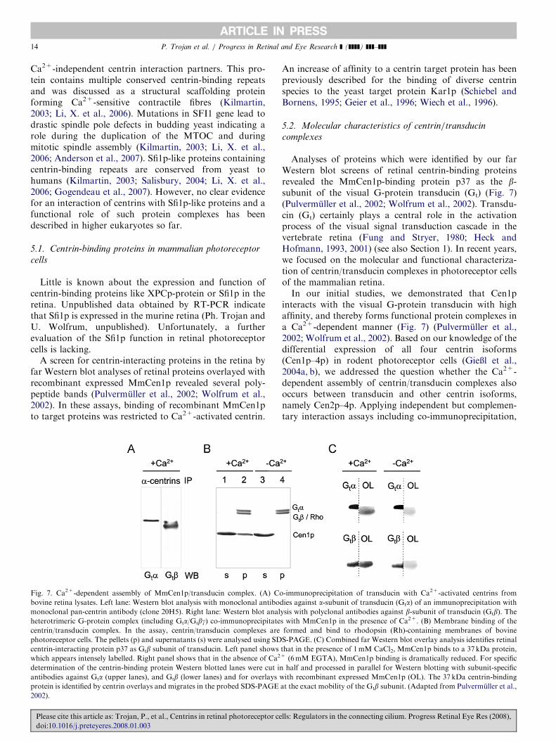
A screen for centrin-interacting proteins in the retina byfar Western blot analyses of retinal proteins overlayed withrecombinant expressed MmCen1p revealed several poly-peptide bands (Pulvermuller et al., 2002; Wolfrum et al.,2002). In these assays, binding of recombinant MmCen1pto target proteins was restricted to Ca2+-activated centrin.
Fig. 7. Ca2+-dependent assembly of MmCen1p/transducin complex. (A) Co
bovine retina lysates. Left lane: Western blot analysis with monoclonal antibo
monoclonal pan-centrin antibody (clone 20H5). Right lane: Western blot anal
heterotrimeric G-protein complex (including Gta/Gtbg) co-immunoprecipitate
centrin/transducin complex. In the assay, centrin/transducin complexes are
photoreceptor cells. The pellets (p) and supernatants (s) were analysed using SD
centrin-interacting protein p37 as Gtb subunit of transducin. Left panel shows
which appears intensely labelled. Right panel shows that in the absence of Ca2
determination of the centrin-binding protein Western blotted lanes were cut in
antibodies against Gta (upper lanes), and Gtb (lower lanes) and for overlays w
protein is identified by centrin overlays and migrates in the probed SDS-PAGE
2002).
Please cite this article as: Trojan, P., et al., Centrins in retinal photoreceptor ce
doi:10.1016/j.preteyeres.2008.01.003
An increase of affinity to a centrin target protein has beenpreviously described for the binding of diverse centrinspecies to the yeast target protein Kar1p (Schiebel andBornens, 1995; Geier et al., 1996; Wiech et al., 1996).
5.2. Molecular characteristics of centrin/transducin
complexes
Analyses of proteins which were identified by our farWestern blot screens of retinal centrin-binding proteinsrevealed the MmCen1p-binding protein p37 as the b-subunit of the visual G-protein transducin (Gt) (Fig. 7)(Pulvermuller et al., 2002; Wolfrum et al., 2002). Transdu-cin (Gt) certainly plays a central role in the activationprocess of the visual signal transduction cascade in thevertebrate retina (Fung and Stryer, 1980; Heck andHofmann, 1993, 2001) (see also Section 1). In recent years,we focused on the molecular and functional characteriza-tion of centrin/transducin complexes in photoreceptor cellsof the mammalian retina.In our initial studies, we demonstrated that Cen1p
interacts with the visual G-protein transducin with highaffinity, and thereby forms functional protein complexes ina Ca2+-dependent manner (Fig. 7) (Pulvermuller et al.,2002; Wolfrum et al., 2002). Based on our knowledge of thedifferential expression of all four centrin isoforms(Cen1p–4p) in rodent photoreceptor cells (GieXl et al.,2004a, b), we addressed the question whether the Ca2+-dependent assembly of centrin/transducin complexes alsooccurs between transducin and other centrin isoforms,namely Cen2p–4p. Applying independent but complemen-tary interaction assays including co-immunoprecipitation,
-immunoprecipitation of transducin with Ca2+-activated centrins from
dies against a-subunit of transducin (Gta) of an immunoprecipitation with
ysis with polyclonal antibodies against b-subunit of transducin (Gtb). Thes with MmCen1p in the presence of Ca2+. (B) Membrane binding of the
formed and bind to rhodopsin (Rh)-containing membranes of bovine
S-PAGE. (C) Combined far Western blot overlay analysis identifies retinal
that in the presence of 1mM CaCl2, MmCen1p binds to a 37 kDa protein,+ (6mM EGTA), MmCen1p binding is dramatically reduced. For specific
half and processed in parallel for Western blotting with subunit-specific
ith recombinant expressed MmCen1p (OL). The 37 kDa centrin-binding
at the exact mobility of the Gtb subunit. (Adapted from Pulvermuller et al.,
lls: Regulators in the connecting cilium. Progress Retinal Eye Res (2008),

ARTICLE IN PRESSP. Trojan et al. / Progress in Retinal and Eye Research ] (]]]]) ]]]–]]] 15
GST-pull down, overlay and co-sedimentation assays aswell as size exclusion chromatography and kinetic light-scattering experiments, we have shown that not onlyCen1p, but also the three other centrin isoforms, Cen2p–4p,bind with high affinity to transducin (Pulvermuller et al.,2002; GieXl et al., 2004a, b, 2006). Further analyses usingkinetic light-scattering experiments (see description inFig. 8D) indicate that the centrin/transducin interactionsare highly specific: centrin-related EF-hand proteins,calmodulin and recoverin, which are highly expressed inphotoreceptor cells do not show any detectable Ca2+-dependent interaction with transducin (Pulvermuller et al.,2002; Wolfrum et al., 2002; GieXl et al., 2004b). Inaddition, centrins do not bind to any other molecule ofthe visual transduction cascade, neither to arrestin,rhodopsin-kinase or rhodopsin, nor do they influence theactivity of the cGMP PDE (Fig. 8) (Pulvermuller et al.,2002; GieXl et al., 2004b).
Fig. 8. Light-scattering setup to analyse the centrin/transducin assembly. (A
signal with unphosphorylated membranes and transducin (Gtholo) in the p
rhodopsin, 0.5 mm Gtholo) in the presence of Ca2+, and 0 (control, black curve
Lower panel represents KLS binding signals under the same conditions as in th
with unphosphorylated or prephosphorylated membranes and arrestin and rh
curves) of MmCen1p. Upper panels represent KLS binding signals (0.5mm ar
MmCen1p and the lower panels KLS binding signals in the absence of Ca2+. E
5mM MgCl2 and either 100mm CaCl2 or 1mM EGTA at 20 1C, sample volume
per flash (500720 nm). (D) Real-time monitoring of rhodopsin-transducin co
scattering intensity originated from rhodopsin-containing disk vesicles. Left
Reactions were triggered by flash photolysis of rhodopsin with a green (5007intensity is quantified photometrically by the amount of rhodopsin bleached and
binding signals (Rh*/Rh ¼ 32%) were corrected by a reference signal (N-
(Pulvermuller et al., 1993). KLS dissociation signals were recorded with a 0.5
presence of 1mM GTP and with catalytic amounts of flash activated rhodopsin
added to the sample. The KLS binding signal is interpreted as a gain of protein
of protein mass from the disk vesicle (Heck et al., 2000). The right panel illus
causing the binding and dissociation signal, respectively.
Please cite this article as: Trojan, P., et al., Centrins in retinal photoreceptor ce
doi:10.1016/j.preteyeres.2008.01.003
Analyses with our set of complementary protein–proteininteraction assays further demonstrate that assembly ofcentrin/transducin complexes is mediated by the bg-heterodimer (Fig. 7) (Pulvermuller et al., 2002; Wolfrumet al., 2002; GieXl et al., 2004a). Later studies also revealeda strict dependence of the assembly of these complexes onthe free Ca2+ concentration. Titrations of the centrinisoforms in kinetic light-scattering experiments in thepresence of Ca2+ showed differences in the affinity of thecentrin isoforms to transducin. Cen3p has a significantlylower affinity to the transducin holoprotein than the othercentrin isoforms (GieXl et al., 2004a). In the case of Cen1pand 2p, at least two Ca2+ ions are required for theactivation of these centrin isoforms and for centrin/transducin complex formation (Pulvermuller et al., 2002;Trojan et al., 2008). Further analyses of these complexesindicate that Cen1p, 2p and 4p bind as homooligomers tothe Gtbg-heterodimer, in contrast to Cen3p which binds as
) Ca2+-dependent enhancement of kinetic light-scattering (KLS) binding
resence of Cen1p. Upper panel represents KLS binding signals (3mm), 0.6, 1.2, 2.5, 3.6, 5, 7.3, and 10 mm MmCen1p (grey curves), respectively.
e upper panel, but with EGTA instead of Ca2+. (B, C) KLS binding signal
odopsin kinase (GRK1) in the presence (grey curves) or absence (black
restin or GRK1 and 3mm rhodopsin) in the presence of Ca2+ plus/minus
xperimental conditions were 50mM BTP, pH 7.5 containing 80mM NaCl,
of 300ml, and cuvette path length of 1 cm; 32% rhodopsin was photolyzed
mplex-formation by KLS. Shown is the time course of normalized light-
panel represents an example of KLS binding and dissociation signals.
20 nm) flash, attenuated by appropriate neutral density filters. The flash
expressed as the mole fraction of photoexcited rhodopsin (Rh*/Rh). KLS
signal) measured on a sample without added protein as described by
–5ms dwell time of the A/D converter (Nicolet 400, Madison, WI) in the
(Rh*/Rh ¼ 0.5%). To suppress base-line activation, 2.5mM NH2OH was
mass bound to the disk membranes and the KLS dissociation signal as loss
trates light-induced mass changes of the scattering membranous particles
lls: Regulators in the connecting cilium. Progress Retinal Eye Res (2008),

ARTICLE IN PRESS
Fig. 9. Immunofluorescence localization of opsin, transducin and centrins
in dark- and light-adapted mouse retinas. Upper panel: triple labelled
dark-adapted (left) and light-adapted (right) mouse retina. Transducin
(green) is localized to the outer segment (OS) of dark-adapted
photoreceptor cells and moves into the inner segment (IS) and cell body
(CB) during light adaptation. Opsin (red) stays in the outer segment
during both conditions. Lower panel: Triple labelled dark-adapted (left)
and light-adapted (right) mouse retina. Transducin (green) is localized as
described for the upper panel. Centrins (red) are stained in the connecting
cilium and basal bodies at the joint between the outer segment and the
inner segment of dark- and light-adapted photoreceptor cells. Transducin
passes centrins during its passage through the connecting cilium. Bars:
13.1mm.
P. Trojan et al. / Progress in Retinal and Eye Research ] (]]]]) ]]]–]]]16
a monomer to each Gtbg (Pulvermuller et al., 2002; GieXlet al., 2004a).
5.2.1. Functions of centrin/G-protein complexes in
mammalian photoreceptor cells
At first view the spatial distribution of centrins (presentin the connecting cilium and the basal bodies) andtransducin (associated with the visual signal transductionmachinery in the outer segment) should exclude anymolecular interaction between centrins and transducin inphotoreceptor cells. However, the visual G-protein trans-ducin (Gt) is not a permanent resident of the outersegment. It undergoes light-dependent reversible bidirec-tional translocation in vertebrate photoreceptor cells(Fig. 9) (Brann and Cohen, 1987; Philp et al., 1987;Whelan and McGinnis, 1988). In the dark, Gt is highlyconcentrated in the rod outer segment, while under brightlight conditions, the majority of Gt is translocated into theinner segment, the cell body, and even to the synapse ofphotoreceptor cells (Fig. 9) (Organisciak et al., 1991;Pulvermuller et al., 2002; Sokolov et al., 2002; Calvertet al., 2006). Interestingly, the clearance of transducin fromthe outer segment is completed in a few minutes afterillumination and is much faster than transducin movementsback into the outer segment (Sokolov et al., 2002; Calvertet al., 2006) indicating that different cellular mechanismsare involved (Peterson et al., 2003). Nevertheless, thebidirectional translocations of transducin are thought tocontribute to slow but long lasting adaptation of rodphotoreceptor cells (Sokolov et al., 2002, 2004; Hardie,2003; Frechter and Minke, 2006).
Since any intracellular exchange between the inner andouter segmental compartments of photoreceptor cellsoccurs through the slender non-motile connecting cilium(Besharse and Horst, 1990; Roepman and Wolfrum, 2007)the bidirectional translocation of transducin should takethis intracellular route (Fig. 9). Indeed, the power ofimmunoelectron microscopy provided us with the insightthat transducin travels through the connecting cilium on itsway between the two photoreceptor cell compartments(Pulvermuller et al., 2002). The slender, closely definedconnecting cilium represents not only a track for themolecular translocation but also a suitable domain forregulation of intersegmental molecular exchange (Spenceret al., 1988; Besharse and Horst, 1990; Wolfrum andSalisbury, 1998), and the subcellular compartmentalizationof the binding of transducin to the ciliary centrins.
As described in Section 4, centrins are present in theciliary apparatus (connecting cilium plus basal bodycomplex) of photoreceptor cells of all vertebrate retinasinvestigated. Our extensive analyses have shown that thefour mammalian centrin isoforms (Cen1p–4p) are differ-entially localized in the ciliary apparatus of retinalphotoreceptor cells (see Fig. 6) (GieXl et al., 2004a, 2006).These studies revealed the localization of the centrinisoforms Cen1p–3p in the connecting cilium of photo-receptor cells. Double-immunofluorescence analyses using
Please cite this article as: Trojan, P., et al., Centrins in retinal photoreceptor ce
doi:10.1016/j.preteyeres.2008.01.003
antibodies against centrins and transducin subunits showco-localization of the ciliary centrins in the photoreceptorconnecting cilium (Pulvermuller et al., 2002; Wolfrumet al., 2002; GieXl et al., 2004a, b, 2006). Quantitativeanalyses of silver-enhanced immunogold labelling revealedthat ciliary centrins and transducin share the same
lls: Regulators in the connecting cilium. Progress Retinal Eye Res (2008),
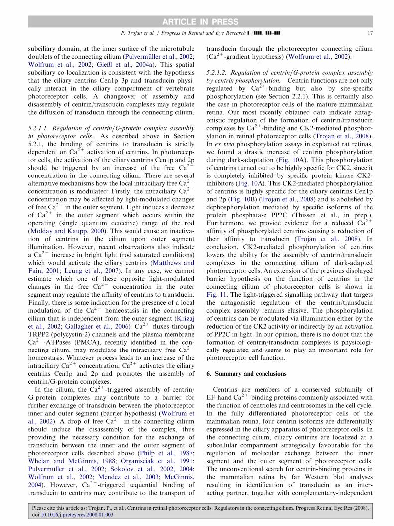
ARTICLE IN PRESSP. Trojan et al. / Progress in Retinal and Eye Research ] (]]]]) ]]]–]]] 17
subciliary domain, at the inner surface of the microtubuledoublets of the connecting cilium (Pulvermuller et al., 2002;Wolfrum et al., 2002; GieXl et al., 2004a). This spatialsubciliary co-localization is consistent with the hypothesisthat the ciliary centrins Cen1p–3p and transducin physi-cally interact in the ciliary compartment of vertebratephotoreceptor cells. A changeover of assembly anddisassembly of centrin/transducin complexes may regulatethe diffusion of transducin through the connecting cilium.
5.2.1.1. Regulation of centrin/G-protein complex assembly
in photoreceptor cells. As described above in Section5.2.1, the binding of centrins to transducin is strictlydependent on Ca2+ activation of centrins. In photorecep-tor cells, the activation of the ciliary centrins Cen1p and 2pshould be triggered by an increase of the free Ca2+
concentration in the connecting cilium. There are severalalternative mechanisms how the local intraciliary free Ca2+
concentration is modulated: Firstly, the intraciliary Ca2+
concentration may be affected by light-modulated changesof free Ca2+ in the outer segment. Light induces a decreaseof Ca2+ in the outer segment which occurs within theoperating (single quantum detective) range of the rod(Molday and Kaupp, 2000). This would cause an inactiva-tion of centrins in the cilium upon outer segmentillumination. However, recent observations also indicatea Ca2+ increase in bright light (rod saturated conditions)which would activate the ciliary centrins (Matthews andFain, 2001; Leung et al., 2007). In any case, we cannotestimate which one of these opposite light-modulatedchanges in the free Ca2+ concentration in the outersegment may regulate the affinity of centrins to transducin.Finally, there is some indication for the presence of a localmodulation of the Ca2+ homeostasis in the connectingcilium that is independent from the outer segment (Krizajet al., 2002; Gallagher et al., 2006): Ca2+ fluxes throughTRPP2 (polycystin-2) channels and the plasma membraneCa2+-ATPases (PMCA), recently identified in the con-necting cilium, may modulate the intraciliary free Ca2+
homeostasis. Whatever process leads to an increase of theintraciliary Ca2+ concentration, Ca2+ activates the ciliarycentrins Cen1p and 2p and promotes the assembly ofcentrin/G-protein complexes.
In the cilium, the Ca2+-triggered assembly of centrin/G-protein complexes may contribute to a barrier forfurther exchange of transducin between the photoreceptorinner and outer segment (barrier hypothesis) (Wolfrum etal., 2002). A drop of free Ca2+ in the connecting ciliumshould induce the disassembly of the complex, thusproviding the necessary condition for the exchange oftransducin between the inner and the outer segment ofphotoreceptor cells described above (Philp et al., 1987;Whelan and McGinnis, 1988; Organisciak et al., 1991;Pulvermuller et al., 2002; Sokolov et al., 2002, 2004;Wolfrum et al., 2002; Mendez et al., 2003; McGinnis,2004). However, Ca2+-triggered sequential binding oftransducin to centrins may contribute to the transport of
Please cite this article as: Trojan, P., et al., Centrins in retinal photoreceptor ce
doi:10.1016/j.preteyeres.2008.01.003
transducin through the photoreceptor connecting cilium(Ca2+-gradient hypothesis) (Wolfrum et al., 2002).
5.2.1.2. Regulation of centrin/G-protein complex assembly
by centrin phosphorylation. Centrin functions are not onlyregulated by Ca2+-binding but also by site-specificphosphorylation (see Section 2.2.1). This is certainly alsothe case in photoreceptor cells of the mature mammalianretina. Our most recently obtained data indicate antag-onistic regulation of the formation of centrin/transducincomplexes by Ca2+-binding and CK2-mediated phosphor-ylation in retinal photoreceptor cells (Trojan et al., 2008).In ex vivo phosphorylation assays in explanted rat retinas,we found a drastic increase of centrin phosphorylationduring dark-adaptation (Fig. 10A). This phosphorylationof centrins turned out to be highly specific for CK2, since itis completely inhibited by specific protein kinase CK2-inhibitors (Fig. 10A). This CK2-mediated phosphorylationof centrins is highly specific for the ciliary centrins Cen1pand 2p (Fig. 10B) (Trojan et al., 2008) and is abolished bydephosphorylation mediated by specific isoforms of theprotein phosphatase PP2C (Thissen et al., in prep.).Furthermore, we provide evidence for a reduced Ca2+
affinity of phosphorylated centrins causing a reduction oftheir affinity to transducin (Trojan et al., 2008). Inconclusion, CK2-mediated phosphorylation of centrinslowers the ability for the assembly of centrin/transducincomplexes in the connecting cilium of dark-adaptedphotoreceptor cells. An extension of the previous displayedbarrier hypothesis on the function of centrins in theconnecting cilium of photoreceptor cells is shown inFig. 11. The light-triggered signalling pathway that targetsthe antagonistic regulation of the centrin/transducincomplex assembly remains elusive. The phosphorylationof centrins can be modulated via illumination either by thereduction of the CK2 activity or indirectly by an activationof PP2C in light. In our opinion, there is no doubt that theformation of centrin/transducin complexes is physiologi-cally regulated and seems to play an important role forphotoreceptor cell function.
6. Summary and conclusions
Centrins are members of a conserved subfamily ofEF-hand Ca2+-binding proteins commonly associated withthe function of centrioles and centrosomes in the cell cycle.In the fully differentiated photoreceptor cells of themammalian retina, four centrin isoforms are differentiallyexpressed in the ciliary apparatus of photoreceptor cells. Inthe connecting cilium, ciliary centrins are localized at asubcellular compartment strategically favourable for theregulation of molecular exchange between the innersegment and the outer segment of photoreceptor cells.The unconventional search for centrin-binding proteins inthe mammalian retina by far Western blot analysesresulting in identification of transducin as an inter-acting partner, together with complementary-independent
lls: Regulators in the connecting cilium. Progress Retinal Eye Res (2008),
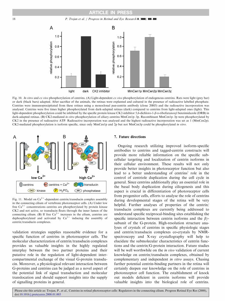
ARTICLE IN PRESS
Fig. 10. In vitro and ex vivo phosphorylation of centrins. (A) Light-dependent ex vivo phosphorylation of endogenous centrins. Rats were light (grey bar)
or dark (black bars) adapted. After sacrifice of the animals, the retinas were explanted and cultured in the presence of radioactive labelled phosphate.
Centrins were immunoprecipitated from these retinas using a monoclonal pan-centrin antibody (clone 20H5) and the radioactive incorporation was
analysed. Centrins were five times higher phosphorylated from dark-adapted retinas (dark) compared to centrins from light-adapted ones (light). This
light-dependent phosphorylation could be inhibited by the specific protein kinase CK2-inhibitor 5,6-dichloro-1-b-D-ribofuranosyl benzimidazole (DRB) in
dark-adapted retinas. (B) CK2-mediated in vitro phosphorylation of ciliary centrins MmCen1p–3p. Recombinant MmCen1p–3p were phosphorylated by
CK2 in the presence of radioactive ATP. Radioactive incorporation was analysed and the highest radioactive incorporation was set as 1 (MmCen2p).
CK2-mediated phosphorylation is isoform specific, since only MmCen1p and 2p but not MmCen3p could be phosphorylated in vitro.
low Ca2+
G t
G t
GDP
P P
2+centrins centrins + Ca
kinase phosphataseH PO4
transducin
P
high Ca2+
G t
G t
CK2
Ca
PP
Ca
CaCa
PP
PPP
CK2
CK2 CK2
Ca
3
Fig. 11. Model on Ca2+-dependent centrin/transducin complex assembly
in the connecting cilium of vertebrate photoreceptor cells. (A) Under low
free Ca2+ concentrations centrins are phosphorylated by protein kinase
CK2 and not active, so transducin floats through the inner lumen of the
connecting cilium. (B) If free Ca2+ increases in the cilium, centrins are
dephosphorylated and activated by Ca2+ inducing the assembly of
centrin/transducin complexes.
P. Trojan et al. / Progress in Retinal and Eye Research ] (]]]]) ]]]–]]]18
validation strategies supplies reasonable evidence for aspecific function of centrins in photoreceptor cells. Themolecular characterization of centrin/transducin complexesprovides us valuable insights in the highly regulatedinterplay between the two partner proteins and theirputative role in the regulation of light-dependent inter-compartmental exchange of the visual G-protein transdu-cin. Moreover, a physiological relevant interaction betweenG-proteins and centrins can be judged as a novel aspect ofthe potential link of signal transduction and moleculartranslocation and should support insights into the supplyof signalling proteins in general.
Please cite this article as: Trojan, P., et al., Centrins in retinal photoreceptor ce
doi:10.1016/j.preteyeres.2008.01.003
7. Future directions
Ongoing research utilizing improved isoform-specificantibodies to centrins and tagged-centrin constructs willprovide more reliable information on the specific sub-cellular targeting and localization of centrin isoforms intheir cellular environment. These results will not onlyprovide better insights in photoreceptor function but alsolead to a better understanding of centrins’ role in thecontrol of centriole duplication during the cell cycle ingeneral. Since centrins additionally play an essential role inthe basal body duplication during ciliogenesis and thisaspect is crucial in differentiation of photoreceptor cellsfrom progenitor cells, efforts to analyse the role of centrinsduring developmental stages of the retina will be veryhelpful. Further analyses of properties of the centrin/transducin complexes are currently being addressed tounderstand specific reciprocal-binding sites establishing thespecific interaction between centrin isoforms and the bg-subunit of the G-protein. High-resolution structure ana-lyses of crystals of centrins in specific physiologic stagesand centrin/transducin complexes co-crystals by NMR-spectroscopy and X-ray crystallography will help toelucidate the submolecular characteristics of centrin func-tions and the centrin/G-protein interaction. Future studieswill be well worthwhile on the in vivo validation of currentknowledge on centrin/transducin complexes, obtained bycomplementary and independent in vitro assays. Chasingfurther potential centrin-binding partners in the retina willcertainly deepen our knowledge on the role of centrins inphotoreceptor cell function. The establishment of knockout models deficient in centrin isoforms will providevaluable insights into the biological role of centrins.
lls: Regulators in the connecting cilium. Progress Retinal Eye Res (2008),
ARTICLE IN PRESSP. Trojan et al. / Progress in Retinal and Eye Research ] (]]]]) ]]]–]]] 19
However, because of the fundamental roles of most centrinisoforms in the cell cycle, where they are essential for theproper embryonic development all known attempts of thegeneration of centrin knock out mice have failed due tolethal defects in early developmental stages. Ongoingresearch utilizing conditional knock out mice will over-come the latter problems. An alternative strategy thatsubstitutes the time and resource-consuming knock outapproaches would be the future application of RNAitechnologies in vivo or ex vivo in organotypic retinacultures. For this purpose, improved gene transfer tech-nologies for the application of centrin probes in the livinganimal or in organotypic retina cultures are currently beingtested. Functional analysis of centrins in retinal photo-receptor cells will provide further insights in the cellularand molecular function of centrins in general and mayalso elucidate the role of transducin and heterotrimericG-proteins at the centrosome of the eukaryotic cell.
Acknowledgments
This work was partly supported by grants by FAUN-Stiftung, Nurnberg, Germany to U.W. and by theDeutsche Forschungsgemeinschaft (DFG) to U.W.(Wo548/6-2), to A.P. (Pu186/3-1). H.W.C. gratefullyacknowledges his support by CBNU funds for overseasresearch 2006–2007. We are grateful to our colleagues whoparticipated in the original studies. We thank Dr. KerstinNagel-Wolfrum, Nora Overlack, Sebastian Rausch, TinaSedmak, Dr. Martha Sommer for critical reading of themanuscript and their helpful comments. We thank JungHee Park, Ulli Maas, Elisabeth Sehn, and Gabi Stern-Schneider for their assistance in experiments.
References
Anderson, V.E., Prudden, J., Prochnik, S., Giddings Jr., T.H., Hardwick,
K.G., 2007. Novel sfi1 alleles uncover additional functions for Sfi1p in
bi-polar spindle assembly and function. Mol. Biol. Cell. 18, 2047–2056.
Antoniacci, L.M., Skibbens, R.V., 2006. Sister-chromatid telomere
cohesion is nonredundant and resists both spindle forces and telomere
motility. Curr. Biol. 16, 902–906.
Araki, M., Masutani, C., Takemura, M., Uchida, A., Sugasawa, K.,
Kondoh, J., Ohkuma, Y., Hanaoka, F., 2001. Centrosome protein
centrin 2/caltractin 1 is part of the Xeroderma pigmentosum group C
complex that initiates global genome nucleotide excision repair. J. Biol.
Chem. 276, 18665–18672.
Baas, P.W., Karabay, A., Qiang, L., 2005. Microtubules cut and run.
Trends Cell Biol. 15, 518–524.
Baron, A.T., Greenwood, T.M., Salisbury, J.L., 1991. Localization of the
centrin-related 165,000-Mr protein of PtK2 cells during the cell cycle.
Cell Motil. Cytoskeleton 18, 1–14.
Baron, A.T., Greenwood, T.M., Bazinet, C.W., Salisbury, J.L., 1992.
Centrin is a component of the pericentriolar lattice. Biol. Cell 76,
383–388.
Baron, A.T., Suman, V.J., Nemeth, E., Salisbury, J.L., 1994. The
pericentriolar lattice of PtK2 cells exhibits temperature and calcium-
modulated behavior. J. Cell Sci. 107, 2993–3003.
Baron, A.T., Errabolu, R., Dinusson, J., Salisbury, J.L., 1995. Centrin-
based contractile fibers: chromatographic purification of centrin. In:
Please cite this article as: Trojan, P., et al., Centrins in retinal photoreceptor ce
doi:10.1016/j.preteyeres.2008.01.003
Dentler, W.L., Witman, G.B. (Eds.), Methods in Cell Biology, Vol. 47:
Cilia and Flagella. Academic Press, San Diego, CA, pp. 341–351.
Baum, P., Furlong, C., Byers, B.E., 1986. Yeast gene required for spindle
pole body duplication: homology of its product with Ca2+-binding
proteins. Proc. Natl. Acad. Sci. USA 83, 5512–5516.
Baum, P., Yip, C., Goetsch, L., Byers, B., 1988. A yeast gene essential for
regulation of spindle pole dulication. Mol. Cell Biol. 8, 5386–5397.
Beavo, J.A., 1995. Cyclic nucleotide phosphodiesterases: functional
implications of multiple isoforms. Physiol. Rev. 75, 725–748.
Besharse, J.C., Horst, C.J., 1990. The photoreceptor connecting cilium—
a model for the transition tone. In: Bloodgood, R.A. (Ed.), Ciliary and
Flagellar Membranes. Plenum, New York, pp. 389–417.
Bettencourt-Dias, M., Glover, D.M., 2007. Centrosome biogenesis and
function: centrosomics brings new understanding. Nat. Rev. Mol. Cell
Biol. 8, 451–463.
Bhattacharya, D., Steinkotter, J., Melkonian, M., 1993. Molecular cloning
and evolutionary analysis of the calcium-modulated contractile
protein, centrin, in green algae and land plants. Plant Mol. Biol. 23
(6), 1243–1254.
Black, D.J., Leonard, J., Persechini, A., 2006. Biphasic Ca2+-dependent
switching in a calmodulin-IQ domain complex. Biochemistry 45,
6987–6995.
Boutet, I., Moraga, D., Marinovic, L., Obreque, J., Chavez-Crooker, P.,
2008. Characterization of reproduction-specific genes in a marine
bivalve mollusc: influence of maturation stage and sex on mRNA
expression. Gene 407, 130–138.
Brann, M.R., Cohen, L.V., 1987. Diurnal expression of transducin mRNA
and translocation of transducin in rods of rat retina. Science 235,
585–587.
Calvert, P.D., Strissel, K.J., Schiesser, W.E., Pugh Jr., E.N., Arshavsky,
V.Y., 2006. Light-driven translocation of signaling proteins in
vertebrate photoreceptors. Trends Cell Biol. 16, 560–568.
Chang, B., Khanna, H., Hawes, N., Jimeno, D., He, S., Lillo, C.,
Parapuram, S.K., Cheng, H., Scott, A., Hurd, R.E., Sayer, J.A., Otto,
E.A., Attanasio, M., O’Toole, J.F., Jin, G., Shou, C., Hildebrandt, F.,
Williams, D.S., Heckenlively, J.R., Swaroop, A., 2006. In-frame
deletion in a novel centrosomal/ciliary protein CEP290/NPHP6
perturbs its interaction with RPGR and results in early-onset retinal
degeneration in the rd16 mouse. Hum. Mol. Genet. 15, 1847–1857.
Charbonnier, J.B., Christova, P., Shosheva, A., Stura, E., Le Du, M.H.,
Blouquit, Y., Duchambon, P., Miron, S., Craescu, C.T., 2006.
Crystallization and preliminary X-ray diffraction data of the complex
between human centrin 2 and a peptide from the protein XPC. Acta
Crystallogr. Sect. F. Struct. Biol. Cryst. Commun. 62, 649–651.
Charbonnier, J.B., Renaud, E., Miron, S., Le Du, M.H., Blouquit, Y.,
Duchambon, P., Christova, P., Shosheva, A., Rose, T., Angulo, J.F.,
Craescu, C.T., 2007. Structural, thermodynamic, and cellular char-
acterization of human centrin 2 interaction with Xeroderma pigmento-
sum group C protein. J. Mol. Biol. 373, 1032–1046.
Conrad, M.N., Lee, C.Y., Wilkerson, J.L., Dresser, M.E., 2007. MPS3
mediates meiotic bouquet formation in Saccharomyces cerevisiae. Proc.
Natl. Acad. Sci. USA 104, 8863–8868.
Correa, G., Morgado-Diaz, J.A., Benchimol, M., 2004. Centrin in Giardia
lamblia—ultrastructural localization. FEMSMicrobiol. Lett. 233, 91–96.
Cox, J.A., Tirone, F., Durussel, I., Firanescu, C., Blouquit, Y.,
Duchambon, P., Craescu, C.T., 2005. Calcium and magnesium binding
to human centrin 3 and interaction with target peptides. Biochemistry
44, 840–850.
Dahm, R., Procter, J.E., Ireland, M.E., Lo, W.K., Mogensen, M.M.,
Quinlan, R.A., Prescott, A.R., 2007. Reorganization of centrosomal
marker proteins coincides with epithelial cell differentiation in the
vertebrate lens. Exp. Eye Res. 85, 696–713.
Daunderer, C., Schliwa, M., Graf, R., 2001. Dictyostelium centrin-related
protein (DdCrp), the most divergent member of the centrin family,
possesses only two EF hands and dissociates from the centrosome
during mitosis. Eur. J. Cell Biol. 80, 621–630.
den Hollander, A.I., Koenekoop, R.K., Mohamed, M.D., Arts, H.H.,
Boldt, K., Towns, K.V., Sedmak, T., Beer, M., Nagel-Wolfrum, K.,
lls: Regulators in the connecting cilium. Progress Retinal Eye Res (2008),
ARTICLE IN PRESSP. Trojan et al. / Progress in Retinal and Eye Research ] (]]]]) ]]]–]]]20
McKibbin, M., Dharmaraj, S., Lopez, I., Ivings, L., Williams, G.A.,
Springell, K., Woods, C.G., Jafri, H., Rashid, Y., Strom, T.M., van
der, Z.B., Gosens, I., Kersten, F.F., van Wijk, E., Veltman, J.A.,
Zonneveld, M.N., van Beersum, S.E., Maumenee, I.H., Wolfrum, U.,
Cheetham, M.E., Ueffing, M., Cremers, F.P., Inglehearn, C.F.,
Roepman, R., 2007. Mutations in LCA5, encoding the ciliary protein
lebercilin, cause Leber congenital amaurosis. Nat. Genet. 39, 889–895.
Durussel, I., Blouquit, Y., Middendorp, S., Craescu, C.T., Cox, J.A.,
2000. Cation- and peptide-binding properties of human centrin 2.
FEBS Lett. 472, 208–212.
Enoch, J.M., 1981. Retinal receptor orientation and photoreceptor optics.
In: Enoch, J.M., Tobey, F.L. (Eds.), Vertebrate Photoreceptor Optics.
Springer, Berlin, Heidelberg, New York, pp. 125–168.
Errabolu, R., Sanders, M.A., Salisbury, J.L., 1994. Cloning of a cDNA
encoding human centrin, an EF-hand protein of centrosomes and
mitotic spindle poles. J. Cell Sci. 107, 9–16.
Fischer, T., Rodriguez-Navarro, S., Pereira, G., Racz, A., Schiebel, E.,
Hurt, E., 2004. Yeast centrin Cdc31 is linked to the nuclear mRNA
export machinery. Nat. Cell Biol. 6, 840–848.
Frechter, S., Minke, B., 2006. Light-regulated translocation of signaling
proteins in Drosophila photoreceptors. J. Physiol. Paris 99, 133–139.
Friedberg, F., 2006. Centrin isoforms in mammals. Relation to
calmodulin. Mol. Biol. Rep. 33, 243–252.
Fung, B.K., Stryer, L., 1980. Photolyzed rhodopsin catalyzes the exchange
of GTP for bound GDP in retinal rod outer segments. Proc. Natl.
Acad. Sci. USA 77, 2500–2504.
Gallagher, A.R., Hoffmann, S., Brown, N., Cedzich, A., Meruvu, S.,
Podlich, D., Feng, Y., Konecke, V., de Vries, U., Hammes, H.P.,
Gretz, N., Witzgall, R., 2006. A truncated polycystin-2 protein causes
polycystic kidney disease and retinal degeneration in transgenic rats.
J. Am. Soc. Nephrol. 17, 2719–2730.
Gavet, O., Alvarez, C., Gaspar, P., Bornens, M., 2003. Centrin4p, a novel
mammalian centrin specifically expressed in ciliated cells. Mol. Biol.
Cell 14, 1818–1834.
Geier, B.M., Wiech, H., Schiebel, E., 1996. Binding of centrin and yeast
calmodulin to synthetic peptides corresponding to binding sites in the
spindle pole body components Kar1p and Spc110p. J. Biol. Chem. 271,
28366–28374.
Geimer, S., Melkonian, M., 2004. The ultrastructure of the Chlamydo-
monas reinhardtii basal apparatus: identification of an early marker of
radial asymmetry inherent in the basal body. J. Cell Sci. 117,
2663–2674.
GieXl, A., 2004. Molekulare charakterisierung der centrin-isoformen in der
retina von saugetieren. Dissertation, Johannes Gutenberg Universitat,
Mainz.
GieXl, A., Pulvermuller, A., Trojan, P., Park, J.H., Choe, H.W., Ernst,
O.P., Hofmann, K.P., Wolfrum, U., 2004a. Differential expression and
interaction with the visual G-protein transducin of centrin isoforms in
mammalian photoreceptor cells. J. Biol. Chem. 279, 51472–51481.
GieXl, A., Trojan, P., Pulvermuller, A., Wolfrum, U., 2004b. Centrins,
potential regulators of transducin translokation in photoreceptor cells.
In: Williams, D.S. (Ed.), Cell Biology and Related Disease of the Outer
Retina. World Scientific Publishing Company Pte. Ltd., Singapore,
pp. 122–195.
GieXl, A., Trojan, P., Rausch, S., Pulvermuller, A., Wolfrum, U., 2006.
Centrins, gatekeepers for the light-dependent translocation of trans-
ducin through the photoreceptor cell connecting cilium. Vision Res.
46, 4502–4509.
Gogendeau, D., Beisson, J., de Loubresse, N.G., Le Caer, J.P., Ruiz, F.,
Cohen, J., Sperling, L., Koll, F., Klotz, C., 2007. An Sfi1p-like centrin-
binding protein mediates centrin-based Ca2+-dependent contractility
in Paramecium tetraurelia. Eukaryot. Cell 6, 1992–2000.
Guerra, C., Wada, Y., Leick, V., Bell, A., Satir, P., 2003. Cloning,
localization, and axonemal function of Tetrahymena centrin. Mol.
Biol. Cell 14, 251–261.
Hardie, R.C., 2003. Phototransduction: shedding light on translocation.
Curr. Biol. 13, R775–R777.
Please cite this article as: Trojan, P., et al., Centrins in retinal photoreceptor ce
doi:10.1016/j.preteyeres.2008.01.003
Hart, P.E., Glantz, J.N., Orth, J.D., Poynter, G.M., Salisbury, J.L., 1999.
Testis-specific murine centrin, Cetn1: genomic characterization and
evidence for retroposition of a gene encoding a centrosome protein.
Genomics 60, 111–120.
Hart, P.E., Poynter, G.M., Whitehead, C.M., Orth, J.D., Glantz, J.N.,
Busby, R.C., Barrett, S.L., Salisbury, J.L., 2001. Characterization of
the X-linked murine centrin Cetn2 gene. Gene 264, 205–213.
Hartman, H., Fedorov, A., 2002. The origin of the eukaryotic cell: a
genomic investigation. Proc. Natl. Acad. Sci. USA 99, 1420–1425.
Heck, M., Hofmann, K.P., 1993. G-protein–effector coupling: a real-time
light-scattering assay for transducin–phosphodiesterase interaction.
Biochemistry 32, 8220–8227.
Heck, M., Hofmann, K.P., 2001. Maximal rate and nucleotide dependence
of rhodopsin-catalyzed transducin activation: initial rate analysis based
on a double displacement mechanism. J. Biol. Chem. 276, 10000–10009.
Heck, M., Pulvermuller, A., Hofmann, K.P., 2000. Light scattering
methods to monitor interactions between rhodopsin-containing
membranes and soluble proteins. Methods Enzymol. 315, 329–347.
Herzberg, O., James, M.N., 1985. Structure of the calcium regulatory
muscle protein troponin-C at 2.8 A resolution. Nature 313, 653–659.
Houdusse, A., Love, M.L., Dominguez, R., Grabarek, Z., Cohen, C.,
1997. Structures of four Ca2+-bound troponin C at 2.0 A resolution:
further insights into the Ca2+-switch in the calmodulin superfamily.
Structure 5, 1695–1711.
Hu, H., Chazin, W.J., 2003. Unique features in the C-terminal domain
provide caltractin with target specificity. J. Mol. Biol. 330, 473–484.
Hu, H., Sheehan, J.H., Chazin, W.J., 2004. The mode of action of centrin.
Binding of Ca2+ and a peptide fragment of Kar1p to the C-terminal
domain. J. Biol. Chem. 279, 50895–50903.
Huang, B., Mengerson, A., Lee, V.D., 1988a. Molecular cloning of cDNA
for caltractin, a basal body-associated Ca2+-binding protein: homo-
logy in its protein sequence with calmodulin and the yeast CDC31 gene
product. J. Cell Biol. 107, 133–140.
Huang, B., Watterson, D.M., Lee, V.D., Schibler, M.J., 1988b. Purifica-
tion and characterization of a basal body-associated Ca2+-binding
protein. J. Cell Biol. 107, 121–131.
Ivanovska, I., Rose, M.D., 2001. Fine structure analysis of the yeast
centrin, Cdc31p, identifies residues specific for cell morphology and
spindle pole body duplication. Genetics 157, 503–518.
Jaspersen, S.L., Giddings Jr., T.H., Winey, M., 2002. Mps3p is a novel
component of the yeast spindle pole body that interacts with the yeast
centrin homologue Cdc31p. J. Cell Biol. 159, 945–956.
Karabay, A., Yu, W., Solowska, J.M., Baird, D.H., Baas, P.W., 2004.
Axonal growth is sensitive to the levels of katanin, a protein that severs
microtubules. J. Neurosci. 24, 5778–5788.
Keller, L.C., Romijn, E.P., Zamora, I., Yates III, J.R., Marshall, W.F.,
2005. Proteomic analysis of isolated chlamydomonas centrioles reveals
orthologs of ciliary-disease genes. Curr. Biol. 15, 1090–1098.
Khalfan, W., Ivanovska, I., Rose, M.D., 2000. Functional interaction
between the PKC1 pathway and CDC31 network of SPB duplication
genes. Genetics 155, 1543–1559.
Kilmartin, J.V., 2003. Sfi1p has conserved centrin-binding sites and an
essential function in budding yeast spindle pole body duplication.
J. Cell Biol. 162, 1211–1221.
Kretsinger, R.H., 1976. Calcium-binding proteins. Annu. Rev. Biochem.
45, 239–266.
Kretsinger, R.H., Nockolds, C.E., 1973. Carp muscle calcium-binding
protein. II. Structure determination and general description. J. Biol.
Chem. 248, 3313–3326.
Krizaj, D., Demarco, S.J., Johnson, J., Strehler, E.E., Copenhagen, D.R.,
2002. Cell-specific expression of plasma membrane calcium ATPase
isoforms in retinal neurons. J. Comp. Neurol. 451, 1–21.
La Terra, S., English, C.N., Hergert, P., McEwen, B.F., Sluder, G.,
Khodjakov, A., 2005. The de novo centriole assembly pathway in
HeLa cells: cell cycle progression and centriole assembly/maturation. J.
Cell Biol. 168, 713–722.
Laoukili, J., Perret, E., Middendorp, S., Houcine, O., Guennou, C.,
Marano, F., Bornens, M., Tournier, F., 2000. Differential expression
lls: Regulators in the connecting cilium. Progress Retinal Eye Res (2008),

ARTICLE IN PRESSP. Trojan et al. / Progress in Retinal and Eye Research ] (]]]]) ]]]–]]] 21
and cellular distribution of centrin isoforms during human ciliated cell
differentiation in vitro. J. Cell Sci. 11, 1355–1364.
Lee, V.D., Huang, B., 1993. Molecular cloning and centrosomal localiza-
tion of human caltractin. Proc. Natl. Acad. Sci. USA 90, 11039–11043.
Lemullois, M., Fryd-Versavel, G., Fleury-Aubusson, A., 2004. Localiza-
tion of centrins in the hypotrich ciliate Paraurostyla weissei.
Protistologica 155, 331–346.
Leung, Y.T., Fain, G.L., Matthews, H.R., 2007. Simultaneous measure-
ment of current and calcium in the ultraviolet-sensitive cones of
zebrafish. J. Physiol. 579, 15–27.
Levy, Y.Y., Lai, E.Y., Remillard, S.P., Heintzelman, M.B., Fulton, C.,
1996. Centrin is a conserved protein that forms diverse associations
with centrioles and MTOCs in Naegleria and other organisms. Cell
Motil. Cytoskeleton 33, 298–323.
Li, S., Sandercock, A.M., Conduit, P., Robinson, C.V., Williams, R.L.,
Kilmartin, J.V., 2006. Structural role of Sfi1p-centrin filaments in
budding yeast spindle pole body duplication. J. Cell Biol. 173,
867–877.
Li, X., Guan, B., Maghami, S., Bieberich, C.J., 2006. NKX3.1 is regulated
by protein kinase CK2 in prostate tumor cells. Mol. Cell Biol. 26,
3008–3017.
Lingle, W.L., Lutz, W.H., Ingle, J.N., Maihle, N.J., Salisbury, J.L., 1998.
Centrosome hypertrophy in human breast tumors: implications for
genomic stability and cell polarity. Proc. Natl. Acad. Sci. USA 95,
2950–2955.
Liu, Q., Tan, G., Levenkova, N., Li, T., Pugh Jr., E.N., Rux, J., Speicher,
D.W., Pierce, E.A., 2007. The proteome of the mouse photoreceptor
sensory cilium complex. Mol. Cell Proteomics 6, 1299–1317.
Liu, X., Vansant, G., Udovichenko, I.P., Wolfrum, U., Williams, D.S.,
1997. Myosin VIIa, the product of the Usher 1B syndrome gene, is
concentrated in the connecting cilia of photoreceptor cells. Cell Motil.
Cytoskeleton 37, 240–252.
Liu, X., Udovichenko, I.P., Brown, S.D., Steel, K.P., Williams, D.S.,
1999. Myosin VIIa participates in opsin transport through the
photoreceptor cilium. J. Neurosci. 19, 6267–6274.
Lohret, T.A., McNally, F.J., Quarmby, L.M., 1998. A role for katanin-
mediated axonemal severing during Chlamydomonas deflagellation.
Mol. Biol. Cell 9, 1195–1207.
Luby-Phelps, K., Fogerty, J., Baker, S.A., Pazour, G.J., Besharse, J.C., 2007.
Spatial distribution of intraflagellar transport proteins in vertebrate
photoreceptors. Vision Res., 2007 October 9 (Epub ahead of print).
Lutz, W., Lingle, W.L., McCormick, D., Greenwood, T.M., Salisbury,
J.L., 2001. Phosphorylation of centrin during the cell cycle and its role
in centriole separation preceding centrosome duplication. J. Biol.
Chem. 276 (23), 20774–20780.
Madeddu, L., Klotz, C., Lecaer, J.P., Beisson, J., 1996. Characterization
of centrin genes in Paramecium. Eur. J. Biochem. 238, 121–128.
Maerker, T., Wijk, E.V., Overlack, N., Kersten, F.F., McGee, J.,
Goldmann, T., Sehn, E., Roepman, R., Walsh, E.J., Kremer, H.,
Wolfrum, U., 2008. A novel Usher protein network at the periciliary
reloading point between molecular transport machineries in vertebrate
photoreceptor cells. Hum. Mol. Genet. 17, 71–86.
Marszalek, J.R., Liu, X., Roberts, E.A., Chui, D., Marth, J.D., Williams,
D.S., Goldstein, L.S.B., 2000. Genetic evidence for selective transport
of opsin and arrestin by kinesin-II in mammalian photoreceptors. Cell
102, 175–187.
Martindale, V.E., Salisbury, J.L., 1990. Phosphorylation of algal centrin is
rapidly responsive to changes in the external milieu. J. Cell Sci. 96,
395–402.
Matei, E., Miron, S., Blouquit, Y., Duchambon, P., Durussel, I., Cox,
J.A., Craescu, C.T., 2003. C-terminal half of human centrin 2 behaves
like a regulatory EF-hand domain. Biochemistry 42, 1439–1450.
Matthews, H.R., Fain, G.L., 2001. A light-dependent increase in free
Ca2+ concentration in the salamander rod outer segment. J. Physiol.
532, 305–321.
McFadden, G.I., Schulze, D., Surek, B., Salisbury, J.L., Melkonian, M.,
1987. Basal body reorientation mediated by a Ca2+-modulated
contractile protein. J. Cell Biol. 105, 903–912.
Please cite this article as: Trojan, P., et al., Centrins in retinal photoreceptor ce
doi:10.1016/j.preteyeres.2008.01.003
McGinnis, J., 2004. Light-dependent protein trafficking in photoreceptor
cells. In: Williams, D.S. (Ed.), Recent Advances in Human Biololgy:
Cell Biology and Disease of the Outer Retina: Problems of Protein
Trafficking. World Scientific, Singapore.
McGinnis, J.F., Matsumoto, B., Whelan, J.P., Cao, W., 2002. Cytoske-
leton participation in subcellular trafficking of signal transduction
proteins in rod photoreceptor cells. J. Neurosci. Res. 67, 290–297.
Mendez, A., Lem, J., Simon, M., Chen, J., 2003. Light-dependent
translocation of arrestin in the absence of rhodopsin phosphorylation
and transducin signaling. J. Neurosci. 23, 3124–3129.
Meng, T.C., Aley, S.B., Svard, S.G., Smith, M.W., Huang, B., Kim, J.,
Gillin, F.D., 1996. Immunolocalization and sequence of caltractin/
centrin from the early branching eukaryote Giardia lamblia. Mol.
Biochem. Parasitol. 79, 103–108.
Meyn, S.M., Seda, C., Campbell, M., Weiss, K.L., Hu, H., Pastrana-Rios,
B., Chazin, W.J., 2006. The biochemical effect of Ser167 phosphoryla-
tion on Chlamydomonas reinhardtii centrin. Biochem. Biophys. Res.
Commun. 342, 342–348.
Middendorp, S., Paoletti, A., Schiebel, E., Bornens, M., 1997. Identifica-
tion of a new mammalian centrin gene, more closely related to
Saccharomyces cerevisiae CDC31 gene. Proc. Natl. Acad. Sci. USA 94,
9141–9146.
Middendorp, S., Kuntziger, T., Abraham, Y., Holmes, S., Bordes, N.,
Paintrand, M., Paoletti, A., Bornens, M., 2000. A role for centrin 3 in
centrosome reproduction. J. Cell Biol. 148 (3), 405–415.
Molday, R.S., Kaupp, U.B., 2000. Ion channels of vertebrate photo-
receptors. In: Stavenga, D.G., DeGrip, W.J., Pugh, Jr., E.N. (Eds.),
Molecular Mechanism in Visual Transduction. Elsevier Science
Publishers B.V., Amsterdam, pp. 143–182.
Moncrief, N.D., Kretsinger, R.H., Goodman, M., 1990. Evolution of EF-
hand calcium-modulated proteins. I. Relationships based on amino
acid sequences. J. Mol. Evol. 30, 522–562.
Nagamune, K., Sibley, L.D., 2006. Comparative genomic and phyloge-
netic analyses of calcium ATPases and calcium-regulated proteins in
the apicomplexa. Mol. Biol. Evol. 23, 1613–1627.
Nagasato, C., Motomura, T., 2004. Destruction of maternal centrioles
during fertilization of the brown alga, Scytosiphon lomentaria
(Scytosiphonales, Phaeophyceae). Cell Motil. Cytoskeleton 59,
109–118.
Nakayama, S., Kretsinger, R.H., 1994. Evolution of the EF-hand family
of proteins. Annu. Rev. Biophys. Biomol. Struct. 23, 473–507.
Nakayama, S., Moncrief, N.D., Kretsinger, R.H., 1992. Evolution of EF-
hand calcium-modulated proteins. II. Domains of several subfamilies
have diverse evolutionary histories. J. Mol. Evol. 34, 416–448.
Okada, T., Fujiyoshi, Y., Silow, M., Navarro, J., Landau, E.M., Shichida,
Y., 2002. Functional role of internal water molecules in rhodopsin
revealed by X-ray crystallography. Proc. Natl. Acad. Sci. USA 99,
5982–5987.
Organisciak, D.T., Xie, A., Wang, H.M., Jiang, Y.L., Darrow, R.M.,
Donoso, L.A., 1991. Adaptive changes in visual cell transduction
protein levels: effect of light. Exp. Eye Res. 53, 773–779.
Overlack, N., Maerker, T., Latz, M., Nagel-Wolfrum, K., Wolfrum, U.,
2008. SANS (USH1G) expression in developing and mature mamma-
lian retina. Vision Res 48, 400–412.
Palczewski, K., Polans, A.S., Baehr, W., Ames, J.B., 2000. Ca(2+)-
binding proteins in the retina: structure, function, and the etiology of
human visual diseases. Bioessays 22, 337–350.
Paoletti, A., Moudjou, M., Paintrand, M., Salisbury, J.L., Bornens, M.,
1996. Most of centrin in animal cells is not centrosome-associated and
centrosomal centrin is confined to the distal lumen of centrioles. J. Cell
Sci. 109, 3089–3102.
Park, J.H., Krauss, N., Pulvermuller, A., Scheerer, P., Hohne, W.,
GieXl, A., Wolfrum, U., Hofmann, K.P., Ernst, O.P., Choe, H.W.,
2005. Crystallization and preliminary X-ray studies of mouse
centrin1. Acta Crystallogr. Sect. F Struct. Biol. Cryst. Commun. 61,
510–513.
Park, J.H., Pulvermuller, A., Scheerer, P., Rausch, S., GieXl, A.,
Hohne, W., Wolfrum, U., Hofmann, K.P., Ernst, O.P., Choe, H.W.,
lls: Regulators in the connecting cilium. Progress Retinal Eye Res (2008),
ARTICLE IN PRESSP. Trojan et al. / Progress in Retinal and Eye Research ] (]]]]) ]]]–]]]22
Krauss, N., 2006. Insights into functional aspects of centrins from the
structure of N-terminally extended mouse centrin 1. Vision Res. 46,
4568–4574.
Paschke, T., 1997. Untersuchungen zur familie der Ca2+-bindenden
centrine: biochemische charakterisierung und identifikation von
interagierenden proteinen. Ph.D. Thesis, University of Cologne,
Germany.
Persechini, A., Moncrief, N.D., Kretsinger, R.H., 1989. The EF-hand
family of calcium-modulated proteins. Trends Neurosci. 12, 462–467.
Peterson, J.J., Tam, B.M., Moritz, O.L., Shelamer, C.L., Dugger, D.R.,
McDowell, J.H., Hargrave, P.A., Papermaster, D.S., Smith, W.C.,
2003. Arrestin migrates in photoreceptors in response to light: a study
of arrestin localization using an arrestin-GFP fusion protein in
transgenic frogs. Exp. Eye Res. 76, 553–563.
Philp, N.J., Chang, W., Long, K., 1987. Light-stimulated protein
movement in rod photoreceptor cells of the rat retina. FEBS Lett.
225, 127–132.
Piperno, G., Mead, K., Shestak, W., 1992. The inner dynein arms I2
interact with a ‘‘dynein regulatory complex’’ in Chlamydomonas
flagella. J. Cell Biol. 118, 1455–1463.
Pulvermuller, A., Palczewski, K., Hofmann, K.P., 1993. Interaction
between photoactivated rhodopsin and its kinase: stability and kinetics
of complex formation. Biochemistry 32, 14082–14088.
Pulvermuller, A., GieXl, A., Heck, M., Wottrich, R., Schmitt, A., Ernst,
O.P., Choe, H.W., Hofmann, K.P., Wolfrum, U., 2002. Calcium-
dependent assembly of centrin-G-protein complex in photoreceptor
cells. Mol. Cell Biol. 22, 2194–2203.
Ribichich, K.F., Gomes, S.L., 2005. Blastocladiella emersonii expresses a
centrin similar to Chlamydomonas reinhardtii isoform not found in
late-diverging fungi. FEBS Lett. 579, 4355–4360.
Roepman, R., Wolfrum, U., 2007. Protein networks and complexes in
photoreceptor cilia. In: Bertrand, E., Faupel, M. (Eds.), Subcellular
Proteomics from Cell Deconstruction to System Reconstruction,
vol. 43, pp. 209–235.
Salisbury, J.L., 1989. Centrin and the algal flagellar apparatus. J. Phycol.
25, 201–206.
Salisbury, J.L., 1995. Centrin, centrosomes, and mitotic spindle poles.
Curr. Opin. Cell Biol. 7, 39–45.
Salisbury, J.L., 2004. Centrosomes: Sfi1p and centrin unravel a structural
riddle. Curr. Biol. 14, R27–R29.
Salisbury, J.L., 2007. A mechanistic view on the evolutionary origin for
centrin-based control of centriole duplication. J. Cell Physiol. 213,
420–428.
Salisbury, J.L., Baron, A., Surek, B., Melkonian, M., 1984. Striated
flagellar roots: isolation and characterization of a calcium-modulated
contractile organelle. J. Cell Biol. 99, 962–970.
Salisbury, J.L., Sanders, M.A., Harpst, L., 1987. Flagellar root contrac-
tion and nuclear movement during flagellar regeneration in Chlamy-
domonas reinhardtii. J. Cell Biol. 105, 1799–1805.
Salisbury, J.L., Baron, A.T., Sanders, M.A., 1988. The centrin-based
cytoskeleton of Chlamydomonas reinhardtii: distribution in interphase
and mitotic cells. J. Cell Biol. 107, 635–641.
Salisbury, J.L., Suino, K.M., Busby, R., Springett, M., 2002. Centrin-2 is
required for centriole duplication in mammalian cells. Curr. Biol. 12,
1287–1292.
Sanders, M.A., Salisbury, J.L., 1989. Centrin-mediated microtubule
serving during flagellar excision in Chlamydomonas reinhardtii. J. Cell
Biol. 108, 1751–1760.
Sanders, M.A., Salisbury, J.L., 1994. Centrin plays an essential role in
microtubule severing during flagellar excision in Chlamydomonas
reinhardtii. J. Cell Biol. 124, 795–805.
Satyshur, K.A., Pyzalska, D., Greaser, M., Rao, S.T., Sundaralingam, M.,
1994. Structure of chicken skeletal muscle troponin C at 1.78 A
resolution. Acta Crystallogr. D. Biol. Crystallogr. 50, 40–49.
Sayer, J.A., Otto, E.A., O’Toole, J.F., Nurnberg, G., Kennedy, M.A.,
Becker, C., Hennies, H.C., Helou, J., Attanasio, M., Fausett, B.V.,
Utsch, B., Khanna, H., Liu, Y., Drummond, I., Kawakami, I.,
Kusakabe, T., Tsuda, M., Ma, L., Lee, H., Larson, R.G., Allen, S.J.,
Please cite this article as: Trojan, P., et al., Centrins in retinal photoreceptor ce
doi:10.1016/j.preteyeres.2008.01.003
Wilkinson, C.J., Nigg, E.A., Shou, C., Lillo, C., Williams, D.S.,
Hoppe, B., Kemper, M.J., Neuhaus, T., Parisi, M.A., Glass, I.A.,
Petry, M., Kispert, A., Gloy, J., Ganner, A., Walz, G., Zhu, X.,
Goldman, D., Nurnberg, P., Swaroop, A., Leroux, M.R., Hildebrandt,
F., 2006. The centrosomal protein nephrocystin-6 is mutated in
Joubert syndrome and activates transcription factor ATF4. Nat.
Genet. 38, 674–681.
Schatten, G., 1994. The centrosome and its mode of inheritance: the
reduction of the centrosome during gametogenesis and its restoration
during fertilization. Dev. Biol. 165, 299–335.
Schiebel, E., Bornens, M., 1995. In search of a function for centrins.
Trends Cell Biol. 5, 197–201.
Schmitt, A., Wolfrum, U., 2001. Identification of novel molecular
components of the photoreceptor connecting cilium by immunosc-
reens. Exp. Eye Res. 73, 837–849.
Schulze, D., Robenek, H., McFadden, G.I., Melkonian, M., 1987.
Immunolocalization of a Ca2+-modulated contractile protein in the
flagellar apparatus of green algae: the nucleus-basal body connector.
Eur. J. Cell Biol. 45, 51–61.
Sheehan, J.H., Bunick, C.G., Hu, H., Fagan, P.A., Meyn, S.M., Chazin,
W.J., 2006. Structure of the N-terminal calcium sensor domain of
centrin reveals the biochemical basis for domain-specific function.
J. Biol. Chem. 281, 2876–2881.
Sokolov, M., Lyubarsky, A.L., Strissel, K.J., Savchenko, A.B., Govar-
dovskii, V.I., Pugh Jr., E.N., Arshavsky, V.Y., 2002. Massive light-
driven translocation of transducin between the two major compart-
ments of rod cells: a novel mechanism of light adaptation. Neuron 33,
95–106.
Sokolov, M., Strissel, K.J., Leskov, I.B., Michaud, N.A., Govardovskii,
V.I., Arshavsky, V.Y., 2004. Phosducin facilitates light-driven
transducin translocation in rod photoreceptors. Evidence from the
phosducin knockout mouse. J. Biol. Chem. 279, 19149–19156.
Spang, A., Courtney, I., Fackler, U., Matzner, M., Schiebel, E., 1993. The
calcium-binding protein cell division cycle 31 of Saccharomyces
cerevisiae is a component of the half bridge of the spindle pole body.
J. Cell Biol. 123, 405–416.
Spencer, M., Detwiler, P.B., Bunt-Milam, A.H., 1988. Distribution of
membrane proteins in mechanically dissociated retinal rods. Invest
Ophthalmol. Vis. Sci. 29, 1012–1020.
Steinberg, R.H., Fisher, S.K., Anderson, D.H., 1980. Disc morphogenesis
in vertebrate photoreceptors. J. Comp. Neurol. 190, 501–508.
Stemm-Wolf, A.J., Morgan, G., Giddings Jr., T.H., White, E.A.,
Marchione, R., McDonald, H.B., Winey, M., 2005. Basal body
duplication and maintenance require one member of the Tetrahymena
thermophila centrin gene family. Mol. Biol. Cell 16, 3606–3619.
Stohr, H., Stojic, J., Weber, B.H., 2003. Cellular localization of the MPP4
protein in the mammalian retina. Invest Ophthalmol. Vis. Sci. 44,
5067–5074.
Strynadka, N.C., Cherney, M., Sielecki, A.R., Li, M.X., Smillie, L.B.,
James, M.N., 1997. Structural details of a calcium-induced molecular
switch: X-ray crystallographic analysis of the calcium-saturated
N-terminal domain of troponin C at 1.75 A resolution. J. Mol. Biol.
273, 238–255.
Sullivan, D.S., Biggins, S., Rose, M.D., 1998. The yeast centrin, cdc31p,
and the interacting protein kinase, Kic1p, are required for cell
integrity. J. Cell Biol. 143, 751–765.
Sundaralingam, M., Drendel, W., Greaser, M., 1985. Stabilization of the
long central helix of troponin C by intrahelical salt bridges between
charged amino acid side chains. Proc. Natl. Acad. Sci. USA 82,
7944–7947.
Sung, C.H., Tai, A.W., 2000. Rhodopsin trafficking and its role in retinal
dystrophies. Int. Rev. Cytol. 195, 215–267.
Thompson, J.R., Ryan, Z.C., Salisbury, J.L., Kumar, R., 2006. The
structure of the human centrin 2-Xeroderma pigmentosum group C
protein complex. J. Biol. Chem. 281 (27), 18746–18752.
Tourbez, M., Firanescu, C., Yang, A., Unipan, L., Duchambon, P.,
Blouquit, Y., Craescu, C.T., 2004. Calcium-dependent self-assembly of
human centrin 2. J. Biol. Chem. 279, 47672–47680.
lls: Regulators in the connecting cilium. Progress Retinal Eye Res (2008),

ARTICLE IN PRESSP. Trojan et al. / Progress in Retinal and Eye Research ] (]]]]) ]]]–]]] 23
Trojan, P., 2003. Charakterisierung funktioneller domanen von centrin-
isoformen. Diplomarbeit, Johannes Gutenberg Universitat Mainz.
Trojan, P., Rausch, S., Giebl, A., Klemm, C., Krause, E., Pulvermuller.
A., Wolfrum U., 2008. Light-dependent CK2-mediated phosphoryla-
tion of centrins regulates complex formation with visual G-protein.
Biochim. Biophys. Acta. 2008. January 17 (Epub ahead of print).
Tsang, W.Y., Spektor, A., Luciano, D.J., Indjeian, V.B., Chen, Z.,
Salisbury, J.L., Sanchez, I., Dynlacht, B.D., 2006. CP110 cooperates
with two calcium-binding proteins to regulate cytokinesis and genome
stability. Mol. Biol. Cell 17, 3423–3434.
Usukura, J., Obata, S., 1996. Morphogenesis of photoreceptor outer
segments in retinal development. Prog. Retin. Eye Res. 15, 113–125.
Uzawa, M., Grams, J., Madden, B., Toft, D., Salisbury, J.L., 1995.
Identification of a complex between centrin and heat shock proteins in
CSF-arrested Xenopus oocytes and dissociation of the complex
following oocyte activation. Dev. Biol. 171, 51–59.
Weber, C., Lee, V.D., Chazin, W.J., Huang, B., 1994. High level
expression in Escherichia coli and characterization of the EF-hand
calcium-binding protein caltractin. J. Biol. Chem. 269, 15795–15802.
Whelan, J.P., McGinnis, J.F., 1988. Light-dependent subcellular move-
ment of photoreceptor proteins. J. Neurosci. Res. 20, 263–270.
Wiech, H., Geier, B.M., Paschke, T., Spang, A., Grein, K., Steinkotter, J.,
Melkonian, M., Schiebel, E., 1996. Characterization of green alga,
yeast, and human centrins. J. Biol. Chem. 271, 22453–22461.
Williams, D.S., 2002. Transport to the photoreceptor outer segment by
myosin VIIa and kinesin II. Vision Res. 42, 455–462.
Wolfrum, U., 1992. Cytoskeletal elements in ciliary receptor systems.
Verh. Dtsch. Zool. Ges. 85, 218.
Please cite this article as: Trojan, P., et al., Centrins in retinal photoreceptor ce
doi:10.1016/j.preteyeres.2008.01.003
Wolfrum, U., Salisbury, J.L., 1995. Centrin: a new Ca-binding protein and
a noval component of the connecting cilium of photoreceptors in
mammals and man. Invest. Ophthalmol. Vis. Sci. 36, 2379 (Abstract
Book).
Wolfrum, U., Salisbury, J.L., 1998. Expression of centrin isoforms in the
mammalian retina. Exp. Cell Res. 242, 10–17.
Wolfrum, U., Schmitt, A., 2000. Rhodopsin transport in the membrane of
the connecting cilium of mammalian photoreceptor cells. Cell Motil.
Cytoskeleton 46, 95–107.
Wolfrum, U., GieXl, A., Pulvermuller, A., 2002. Centrins, a novel group of
Ca2+-binding proteins in vertebrate photoreceptor cells. Adv. Exp.
Med. Biol. 514, 155–178.
Wottrich, R., 1998. Klonierung und computergestutzte strukturanalyse
von centrinisoformen der ratte (Rattus norvegicus). Diplomarbeit,
Universitat Karlsruhe?
Yang, A., Miron, S., Duchambon, P., Assairi, L., Blouquit, Y., Craescu,
C.T., 2006a. The N-terminal domain of human centrin 2 has a closed
structure, binds calcium with a very low affinity, and plays a role in the
protein self-assembly. Biochemistry 45, 880–889.
Yang, A., Miron, S., Mouawad, L., Duchambon, P., Blouquit, Y.,
Craescu, C.T., 2006b. Flexibility and plasticity of human centrin 2
binding to the Xeroderma pigmentosum group C protein (XPC) from
nuclear excision repair. Biochemistry 45, 3653–3663.
Young, R.W., 1976. Visual cells and the concept of renewal. Invest
Ophthalmol. Vis. Sci. 15, 700–725.
Zhu, J.A., Bloom, S.E., Lazarides, E., Woods, C., 1995. Identification of a
novel Ca2+-regulated protein that is associated with the marginal band
and centrosomes of chicken erythrocytes. J. Cell Sci. 108, 685–698.
lls: Regulators in the connecting cilium. Progress Retinal Eye Res (2008),