Embed Size (px)
Citation preview
1
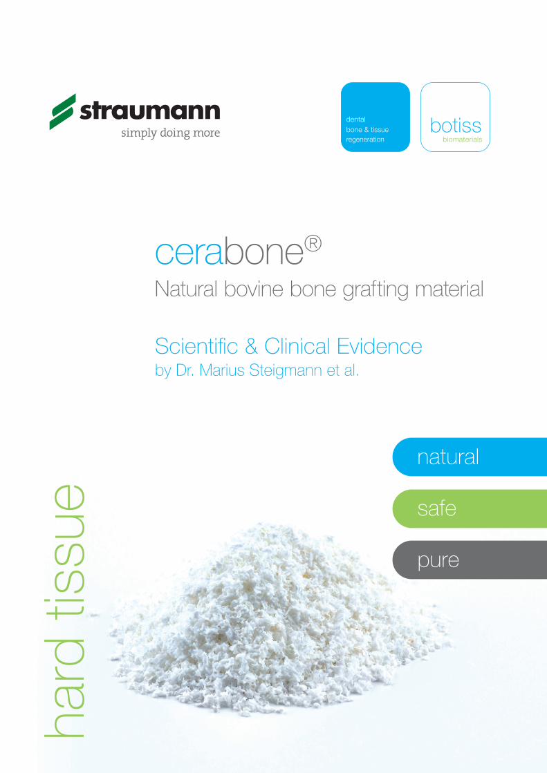
cerabone®Natural bovine bone grafting material
Scientific & Clinical Evidenceby Dr. Marius Steigmann et al.
hard tissue
natural
safe
pure
botissbiomaterials
dental
bone & tissue regeneration
2
cerabone®
Natural bovine bone graft
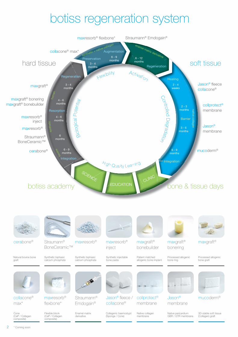
maxresorb®
inject
maxgraft®
bonering
maxgraft®
bonebuilder
Patient matched allogenic bone implant
maxgraft®
Processed allogenicbone graft
Synthetic injectable bone paste
maxresorb®
Synthetic biphasiccalcium phosphate
Processed allogenic bone ring
maxresorb®
flexbone*
Flexible block (CaP / Collagen composite)
Straumann®
Emdogain®
Enamel matrix derivative
collacone®
max*
Cone(CaP / Collagen composite)
mucoderm®
3D-stable soft tissue (Collagen) graft
Jason®
membrane
Native pericardium GBR / GTR membrane
collprotect®
membrane
Native collagen membrane
Jason® fleece /
collacone®
Collagenic haemostypt (Sponge / Cone)
* Coming soon
High Quality Learning
Activation
Flexib
ility
High Quality Learning
Activation
AcFlexibility
Flexibility
Controlled Degradation
Biological Potential
mucoderm®
collprotect® membrane
Jason® membrane
Jason® fleececollacone®......
hard tissue
cerabone®
Straumann® BoneCeramic™
maxresorb®
inject
maxgraft® boneringmaxgraft® bonebuilder
maxgraft®
EDUCATION
SCIENCE CLINIC
6 - 9
months6 - 9
months
6
months
4 - 6
months
4 - 6
months
3 - 4
months
6 - 9
months3 - 4
months
2 - 4
weeks
Regeneration
Augmentation
Preservation
Healing
IntegrationIntegration
Barrier
Resorption
2 - 3
months
3 - 6
months
bovi
ne
synth
etic
hum
an
nativ
e c
olla
gen
collacone®..max*
soft tissue
botiss regeneration system
synthetic + native collagen
botiss academy bone & tissue days
6 - 12
months
Regeneration
enamel matrix derivative
Straumann® Emdogain®maxresorb® flexbone*
Straumann®
BoneCeramic™
Synthetic biphasiccalcium phosphate
maxresorb®
3
The Steigmann Implant Institute is a private teaching institution
founded in 2006. The mission is to teach dentists all aspects of
dental implants and biologics. However the main focus is on aest-
hetics with soft tissue management and bone regeneration.
Dr. Steigmann is considered a specialist and pioneer of modern
dental implantology.
Global clinical and scientific network include: Prof. Dr. Hom-Lay
Wang, Prof. Dr. Anton Sculean, Dr. Maurice Salama, Dr. Philippe
Russe, Dr. Tiziano Testori, Dr. Scott Ganz, Dr. Olaf Daum, PD Dr. Dr.
Daniel Rothamel, Dr. Damir Jelušic , Dr. Ophir Fromovich.
The Steigmann Implant Institute
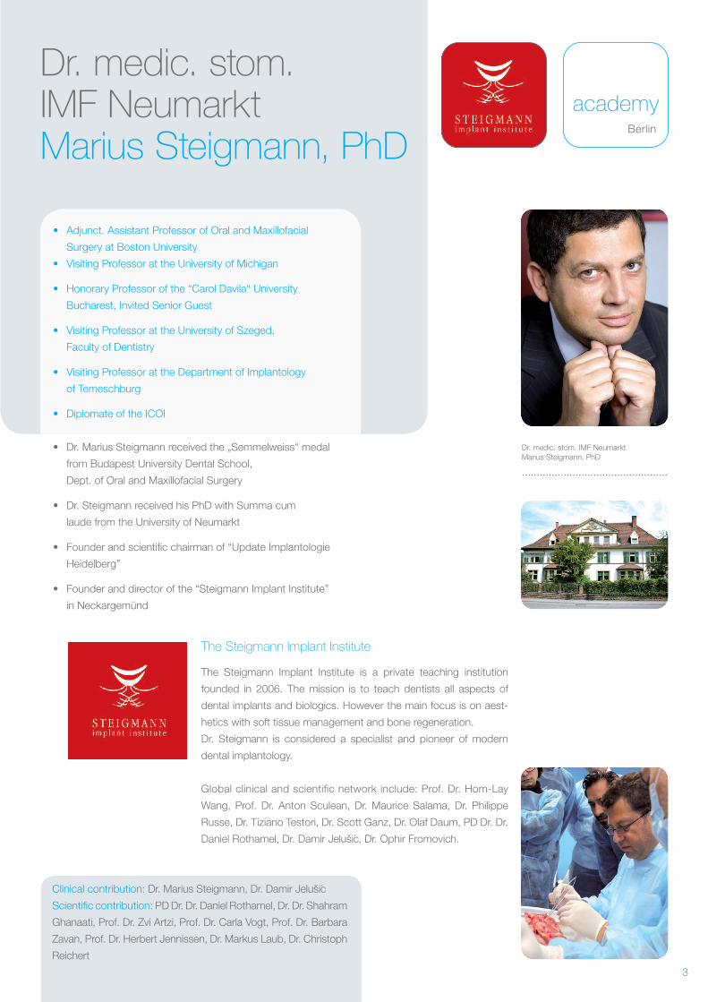
Dr. medic. stom. IMF Neumarkt Marius Steigmann, PhD
Dr. medic. stom. IMF Neumarkt Marius Steigmann, PhD
• Adjunct. Assistant Professor of Oral and Maxillofacial
Surgery at Boston University
• Visiting Professor at the University of Michigan
• Honorary Professor of the “Carol Davila“ University
Bucharest, Invited Senior Guest
• Visiting Professor at the University of Szeged,
Faculty of Dentistry
• Visiting Professor at the Department of Implantology
of Temeschburg
• Diplomate of the ICOI
• Dr. Marius Steigmann received the „Semmelweiss“ medal
from Budapest University Dental School,
Dept. of Oral and Maxillofacial Surgery
• Dr. Steigmann received his PhD with Summa cum
laude from the University of Neumarkt
• Founder and scientific chairman of “Update Implantologie
Heidelberg”
• Founder and director of the “Steigmann Implant Institute”
in Neckargemünd
academyBerlin
.................................................
Clinical contribution: Dr. Marius Steigmann, Dr. Damir Jelušic
Scientific contribution: PD Dr. Dr. Daniel Rothamel, Dr. Dr. Shahram
Ghanaati, Prof. Dr. Zvi Artzi, Prof. Dr. Carla Vogt, Prof. Dr. Barbara
Zavan, Prof. Dr. Herbert Jennissen, Dr. Markus Laub, Dr. Christoph
Reichert
4
Bone and Regeneration Techniques
Classification
The use of bone graft materials
Bone graft materials are applied to replace and regenerate bone
matrix lost by various reasons such as tooth extraction, cystectomy
or bone atrophy following loss of teeth or inflammatory processes.
For the filling of bone defects, the patients own (autologous) bone
is considered the „gold standard“, because of its biological activity
due to vital cells and growth factors. Nevertheless, the harvesting of
autologous bone requires a second surgical site associated with an
additional bony defect and potential donor site morbidity.
In addition, the quantity of autologous bone is limited. Today, due to
a constant development, bone graft materials provide a reliable and
safe alternative to autologous bone grafts.
Clinicians can choose between a variety of different bone graft
materials and augmentation techniques. Bone graft materials are
classified by their origin into four groups.
The principle of Guided Bone Regeneration (GBR)
or Guided Tissue Regeneration (GTR) is based
on the separation of the grafted site from the sur-
rounding soft tissue by application of a barrier.
Collagen membranes act as a resorbable matrix
to avoid the ingrowth of the faster proliferating fi-
broblasts and/or epithelium into the defect, and
The GBR/GTR technique
Guided Tissue Regeneration (GTR) Guided Bone Regeneration (GBR)
.................................................
Autologous:
- patients own bone, mostly
harvested intraorally or from
the iliac crest
- intrinsic biological activity
Allogenic:
- bone from human donors
(cadaver bone or femoral
heads of living donors)
- natural bone composition and
structure
Xenogenic:
- from other organisms, mainly
bovine origin
- Long term volume stability
Alloplastic:
- synthetically produced, pre-
ferably calcium phosphate
ceramics
- no risk of disease transmission
to maintain the space for controlled regeneration
of bone.
The application of a bone graft material into the
defect prevents a collapse of the collagen memb-
rane, acting as a place holder for the regenerating
bone and as an osteoconductive scaffold for the
ingrowth of blood vessels and bone forming cells.
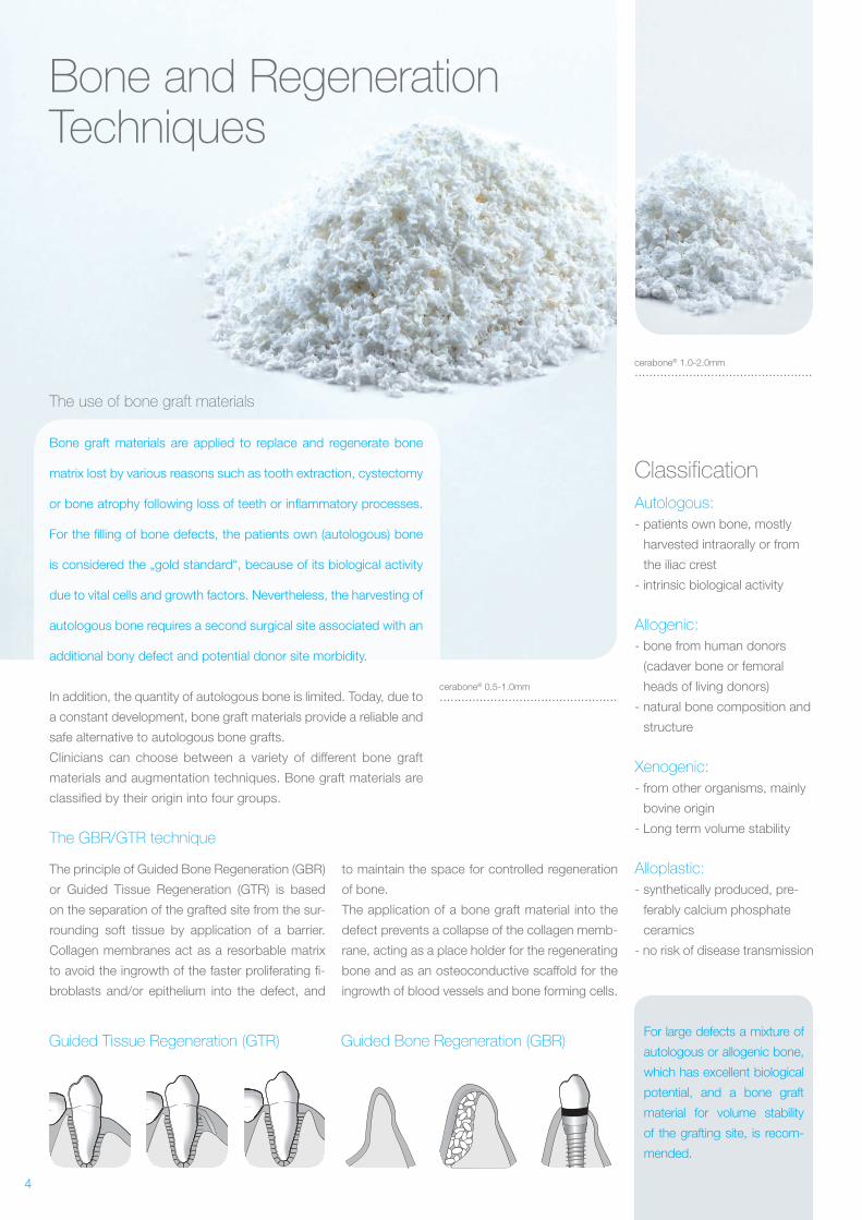
cerabone® 1.0-2.0mm
.................................................cerabone® 0.5-1.0mm
For large defects a mixture of
autologous or allogenic bone,
which has excellent biological
potential, and a bone graft
material for volume stability
of the grafting site, is recom-
mended.
5
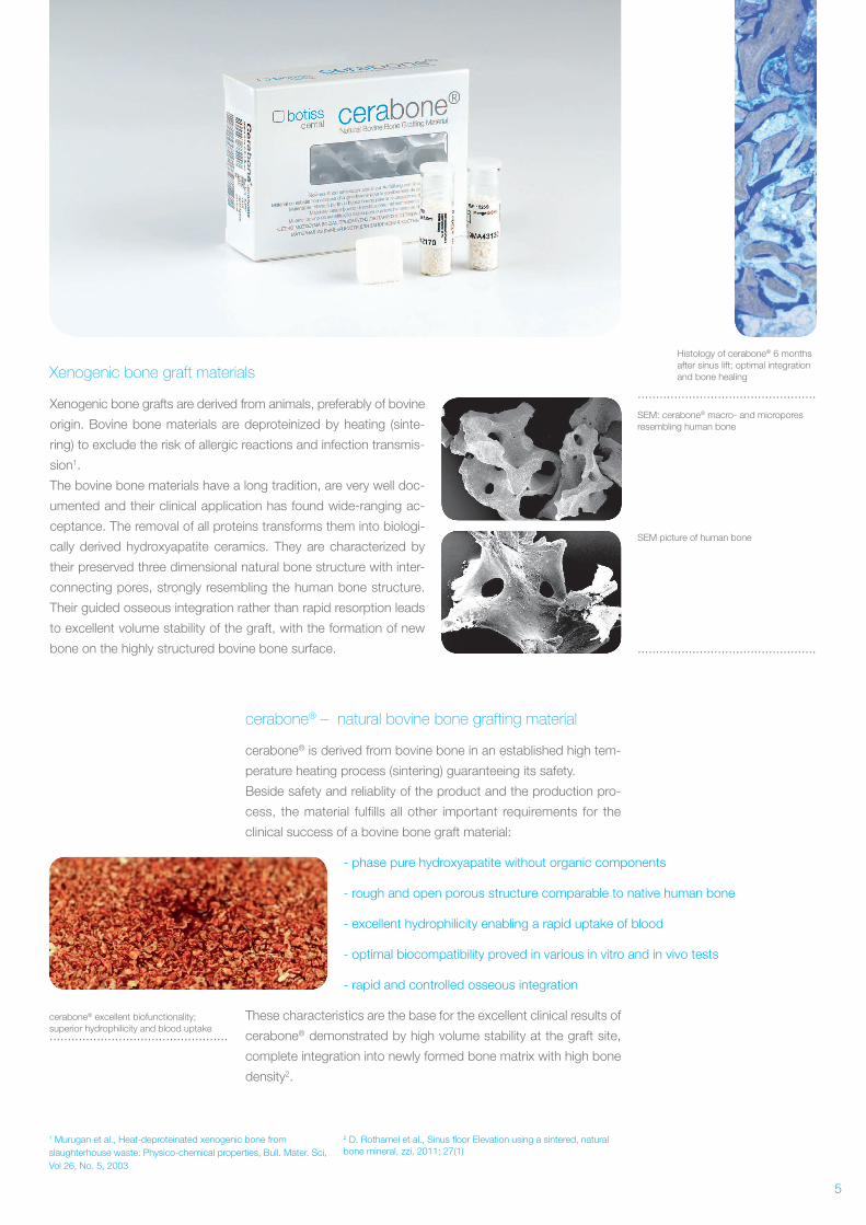
Xenogenic bone grafts are derived from animals, preferably of bovine
origin. Bovine bone materials are deproteinized by heating (sinte-
ring) to exclude the risk of allergic reactions and infection transmis-
sion1.
The bovine bone materials have a long tradition, are very well doc-
umented and their clinical application has found wide-ranging ac-
ceptance. The removal of all proteins transforms them into biologi-
cally derived hydroxyapatite ceramics. They are characterized by
their preserved three dimensional natural bone structure with inter-
connecting pores, strongly resembling the human bone structure.
Their guided osseous integration rather than rapid resorption leads
to excellent volume stability of the graft, with the formation of new
bone on the highly structured bovine bone surface.
Xenogenic bone graft materials
cerabone® is derived from bovine bone in an established high tem-
perature heating process (sintering) guaranteeing its safety.
Beside safety and reliablity of the product and the production pro-
cess, the material fulfills all other important requirements for the
clinical success of a bovine bone graft material:
These characteristics are the base for the excellent clinical results of
cerabone® demonstrated by high volume stability at the graft site,
complete integration into newly formed bone matrix with high bone
density2.
cerabone® – natural bovine bone grafting material
Histology of cerabone® 6 months after sinus lift; optimal integration and bone healing
SEM: cerabone® macro- and micropores resembling human bone
SEM picture of human bone
cerabone® excellent biofunctionality; superior hydrophilicity and blood uptake
- phase pure hydroxyapatite without organic components
- rough and open porous structure comparable to native human bone
- excellent hydrophilicity enabling a rapid uptake of blood
- optimal biocompatibility proved in various in vitro and in vivo tests
- rapid and controlled osseous integration
1 Murugan et al., Heat-deproteinated xenogenic bone from slaughterhouse waste: Physico-chemical properties, Bull. Mater. Sci, Vol 26, No. 5, 2003
2 D. Rothamel et al., Sinus floor Elevation using a sintered, natural bone mineral, zzi, 2011; 27(1)
.................................................
.................................................
.................................................
6
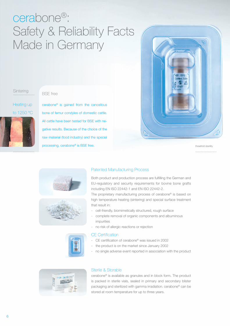
cerabone®: Safety & Reliability FactsMade in Germany
cerabone® is gained from the cancellous
bone of femur condyles of domestic cattle.
All cattle have been tested for BSE with ne-
gative results. Because of the choice of the
raw material (food industry) and the special
processing, cerabone® is BSE free.
Both product and production process are fulfilling the German and
EU-regulatory and security requirements for bovine bone grafts
including EN ISO 22442-1 and EN ISO 22442-2.
The proprietary manufacturing process of cerabone® is based on
high temperature heating (sintering) and special surface treatment
that result in:
- cell-friendly, biomimetically structured, rough surface
- complete removal of organic components and albuminous
impurities
- no risk of allergic reactions or rejection
BSE free
Patented Manufacturing Process
Heating up
to 1250 °C
Sintering
- CE certification of cerabone® was issued in 2002
- the product is on the market since January 2002
- no single adverse event reported in association with the product
CE Certification
...................... ......................
cerabone® is available as granules and in block form. The product
is packed in sterile vials, sealed in primary and secondary blister
packaging and sterilized with gamma irradiation. cerabone® can be
stored at room temperature for up to three years.
Sterile & Storable
threefold sterility
......................
7
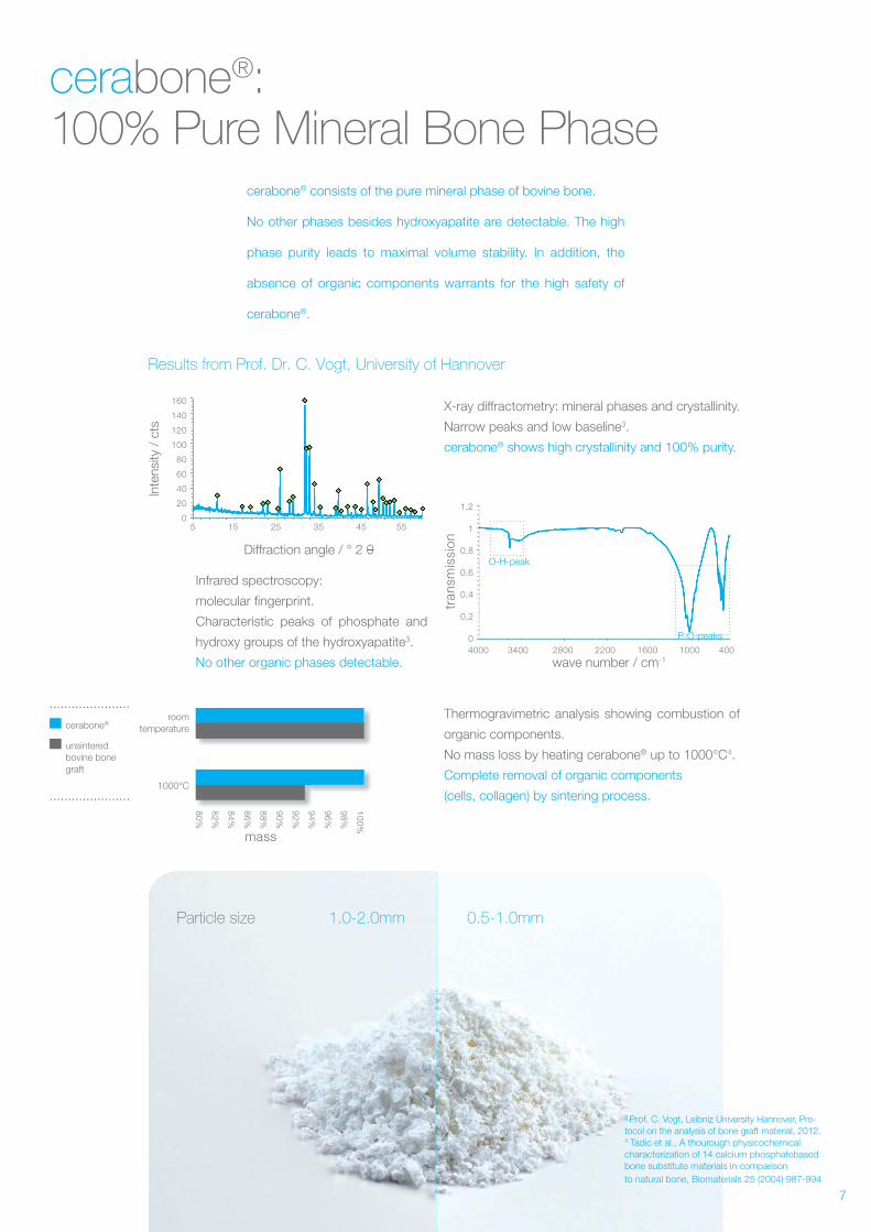
Results from Prof. Dr. C. Vogt, University of Hannover
Particle size
cerabone®: 100% Pure Mineral Bone Phase
cerabone® consists of the pure mineral phase of bovine bone.
No other phases besides hydroxyapatite are detectable. The high
phase purity leads to maximal volume stability. In addition, the
absence of organic components warrants for the high safety of
cerabone®.
X-ray diffractometry: mineral phases and crystallinity.
Narrow peaks and low baseline3.
cerabone® shows high crystallinity and 100% purity.
Infrared spectroscopy:
molecular fingerprint.
Characteristic peaks of phosphate and
hydroxy groups of the hydroxyapatite3.
No other organic phases detectable.
Thermogravimetric analysis showing combustion of
organic components.
No mass loss by heating cerabone® up to 1000°C4.
Complete removal of organic components
(cells, collagen) by sintering process.
......................
cerabone®
unsintered bovine bone graft
0.5-1.0mm1.0-2.0mm
room temperature
1000°C
10
0%
98
%
96
%
94
%
92
%
90
%
88
%
86
%
84
%
82
%
80
%
mass
160
140
120
100
80
60
40
20
01,2
1
0,8
0,6
0,4
0,2
0
Inte
nsity
/ c
ts
tran
smis
sio
n
Diffraction angle / ° 2 O
5 15 25 35 45 55
4000 3400 2800 2200 1600 1000 400
wave number / cm-1
O-H-peak
P-O-peaks
......................
3 Prof. C. Vogt, Leibniz University Hannover, Pro-tocol on the analysis of bone graft material, 2012. 4 Tadic et al., A thourough physicochemicalcharacterization of 14 calcium phosphatebasedbone substitute materials in comparisonto natural bone, Biomaterials 25 (2004) 987-994
8
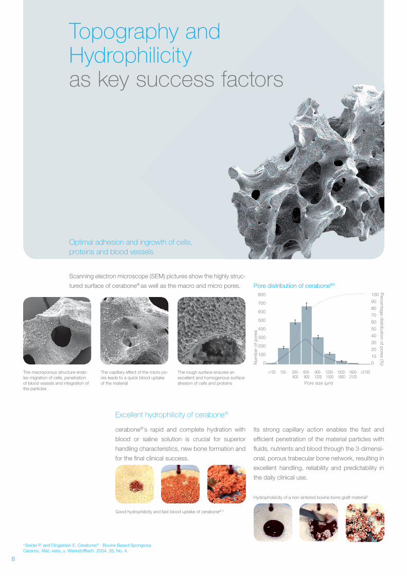
Topography and Hydrophilicity as key success factors
Optimal adhesion and ingrowth of cells,
proteins and blood vessels
Excellent hydrophilicity of cerabone®
cerabone®´s rapid and complete hydration with
blood or saline solution is crucial for superior
handling characteristics, new bone formation and
for the final clinical success.
Its strong capillary action enables the fast and
efficient penetration of the material particles with
fluids, nutrients and blood through the 3-dimensi-
onal, porous trabecular bone network, resulting in
excellent handling, reliability and predictability in
the daily clinical use.
Scanning electron microscope (SEM) pictures show the highly struc-
tured surface of cerabone® as well as the macro and micro pores.
Hydrophobicity of a non sintered bovine bone graft material3
Good hydrophilicity and fast blood uptake of cerabone® 3
The macroporous structure enab-les migration of cells, penetration of blood vessels and integration of the particles
The capillary effect of the micro po-res leads to a quick blood uptake of the material
The rough surface ensures an excellent and homogenous surface ahesion of cells and proteins
5 Seidel P. and Dingeldein E. Cerabone® - Bovine Based Spongiosa Ceramic. Mat.-wiss. u. Werkstofftech. 2004, 35, No. 4.
Nu
mb
er o
f p
ore
s
Pore size (µm)
Percen
tage d
istribu
tion
of p
ores (%
)
800
700
600
500
400
300
200
100
0
100
90
80
70
60
50
40
30
20
10
0
<150 150- 300-600
600-900
900-1200
1200-1500
1500-1800
1800-2100
>2100
Pore distribution of cerabone®5
9
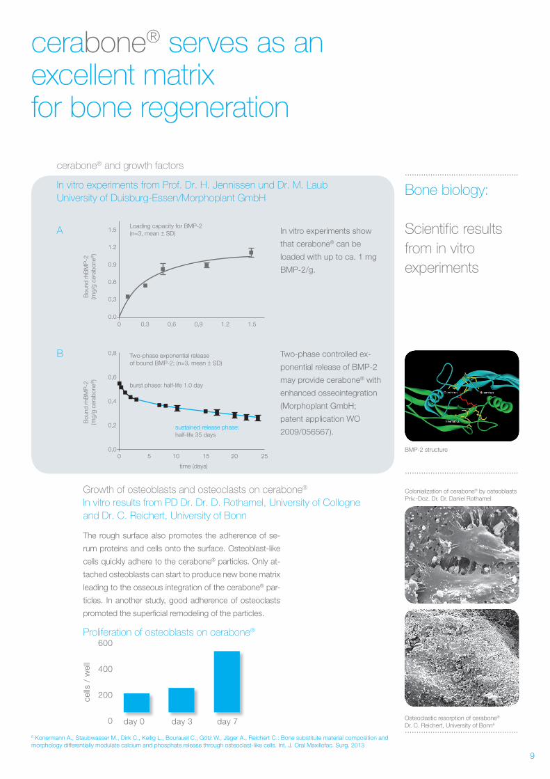
cerabone® serves as an excellent matrix for bone regeneration
Growth of osteoblasts and osteoclasts on cerabone®
In vitro results from PD Dr. Dr. D. Rothamel, University of Collogne
and Dr. C. Reichert, University of Bonn
.................................................
The rough surface also promotes the adherence of se-
rum proteins and cells onto the surface. Osteoblast-like
cells quickly adhere to the cerabone® particles. Only at-
tached osteoblasts can start to produce new bone matrix
leading to the osseous integration of the cerabone® par-
ticles. In another study, good adherence of osteoclasts
promoted the superficial remodeling of the particles.
Proliferation of osteoblasts on cerabone®
day 0 day 3 day 7
600
400
200
0
cells
/ w
ell
Bone biology:
Scientific results
from in vitro
experiments
.................................................
.................................................
In vitro experiments show
that cerabone® can be
loaded with up to ca. 1 mg
BMP-2/g.
Two-phase controlled ex-
ponential release of BMP-2
may provide cerabone® with
enhanced osseointegration
(Morphoplant GmbH;
patent application WO
2009/056567).
Colonialization of cerabone® by osteoblastsPriv.-Doz. Dr. Dr. Daniel Rothamel
Osteoclastic resorption of cerabone®
Dr. C. Reichert, University of Bonn6
cerabone® and growth factors
In vitro experiments from Prof. Dr. H. Jennissen und Dr. M. Laub
University of Duisburg-Essen/Morphoplant GmbH
A
B
0 5 10 15 20 25
0 0,3 0,6 0,9 1.2 1.5
time (days)
0,8
0,6
0,4
0,2
0,0
1.5
1.2
0.9
0.6
0,3
0.0
Bou
nd r
hBM
P-2
(mg/
g ce
rab
one®
)B
ound
rhB
MP
-2(m
g/g
cera
bon
e®)
sustained release phase:half-life 35 days
Two-phase exponential releaseof bound BMP-2; (n=3, mean + SD)
burst phase: half-life 1.0 day
Loading capacity for BMP-2(n=3, mean + SD)
_
_
BMP-2 structure
6 Konermann A., Staubwasser M., Dirk C., Keilig L., Bourauel C., Götz W., Jäger A., Reichert C.: Bone substitute material composition and morphology differentially modulate calcium and phosphate release through osteoclast-like cells. Int. J. Oral Maxillofac. Surg. 2013
10
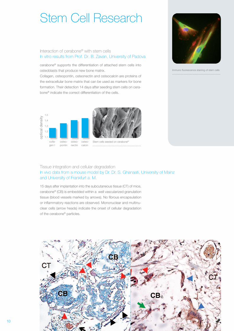
Stem Cell Research
cerabone® supports the differentiation of attached stem cells into
osteoblasts that produce new bone matrix.
Collagen, osteopontin, osteonectin and osteocalcin are proteins of
the extracellular bone matrix that can be used as markers for bone
formation. Their detection 14 days after seeding stem cells on cera-
bone® indicate the correct differentiation of the cells.
Interaction of cerabone® with stem cells
In vitro results from Prof. Dr. B. Zavan, University of Padova
colla-gen I
osteo-pontin
osteo-nectin
osteo-calcin
1,5
1,4
1,3
1,2
1,1op
tical
den
sity
15 days after implantation into the subcutaneous tissue (CT) of mice,
cerabone® (CB) is embedded within a well vascularized granulation
tissue (blood vessels marked by arrows). No fibrous encapsulation
or inflammatory reactions are observed. Mononuclear and multinu-
clear cells (arrow heads) indicate the onset of cellular degradation
of the cerabone® particles.
Tissue integration and cellular degradation
In vivo data from a mouse model by Dr. Dr. S. Ghanaati, University of Mainz
and University of Frankfurt a. M.
.................................................Stem cells seeded on cerabone®
................................................. .................................................
Immuno fluorescence staining of stem cells.................................................
11
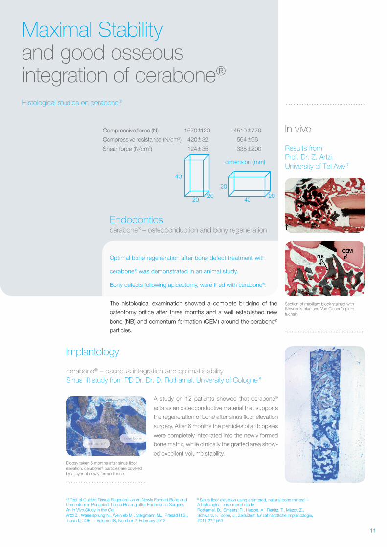
Maximal Stabilityand good osseous integration of cerabone®
cerabone® – osteoconduction and bony regeneration
Endodontics
Implantology
Histological studies on cerabone®
Optimal bone regeneration after bone defect treatment with
cerabone® was demonstrated in an animal study.
Bony defects following apicectomy, were filled with cerabone®.
The histological examination showed a complete bridging of the
osteotomy orifice after three months and a well established new
bone (NB) and cementum formation (CEM) around the cerabone®
particles.
A study on 12 patients showed that cerabone®
acts as an osteoconductive material that supports
the regeneration of bone after sinus floor elevation
surgery. After 6 months the particles of all biopsies
were completely integrated into the newly formed
bone matrix, while clinically the grafted area show-
ed excellent volume stability.
cerabone® – osseous integration and optimal stability
Sinus lift study from PD Dr. Dr. D. Rothamel, University of Cologne 8
.................................................
Results from
Prof. Dr. Z. Artzi,
University of Tel Aviv 7
Section of maxillary block stained with Stevenels blue and Van Gieson’s picro fuchsin
.................................................
Biopsy taken 6 months after sinus floor elevation. cerabone® particles are covered by a layer of newly formed bone.
7Effect of Guided Tissue Regeneration on Newly Formed Bone and Cementum in Periapical Tissue Healing after Endodontic Surgery: An In Vivo Study in the CatArtzi Z., Wasersprung N., Weinreb M., Steigmann M., Prasad H.S., Tsesis I.; JOE — Volume 38, Number 2, February 2012
cerabone®new bone
8 Sinus floor elevation using a sintered, natural bone mineral – A histological case report studyRothamel, D., Smeets, R., Happe, A., Fienitz, T., Mazor, Z., Schwarz, F., Zöller, J., Zeitschrift für zahnärztliche Implantologie, 2011;27(1):60
In vivo
.................................................
Compressive force (N) 1670 120 4510 770
Compressive resistance (N/cm2) 420 32 564 96
Shear force (N/cm2) 124 35 338 200
2020
40
4020
20
+– +– +– +–
+–+–
dimension (mm)
12
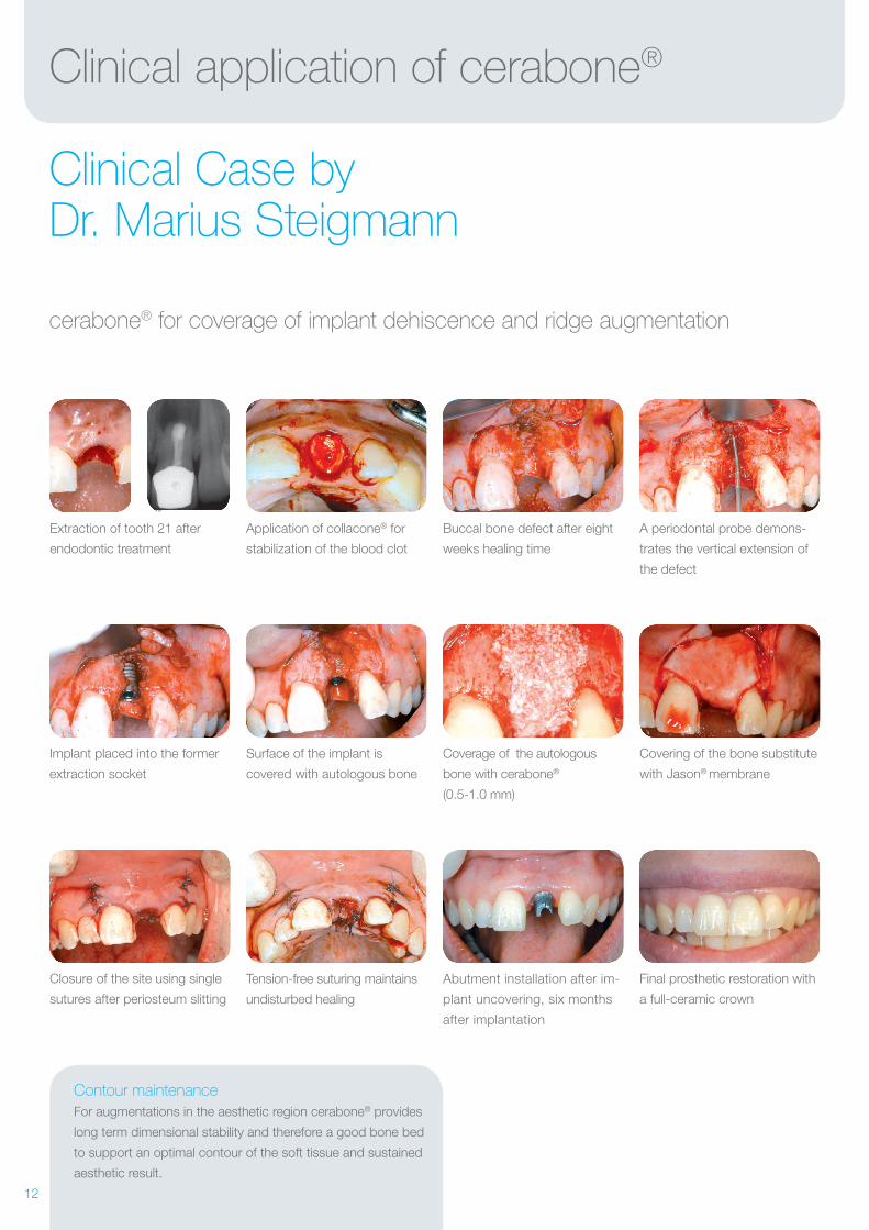
Clinical application of cerabone®
Clinical Case by Dr. Marius Steigmann
cerabone® for coverage of implant dehiscence and ridge augmentation
Implant placed into the former
extraction socket
Surface of the implant is
covered with autologous bone
Coverage of the autologous
bone with cerabone®
(0.5-1.0 mm)
Extraction of tooth 21 after
endodontic treatment
Application of collacone® for
stabilization of the blood clot
Buccal bone defect after eight
weeks healing time
A periodontal probe demons-
trates the vertical extension of
the defect
Covering of the bone substitute
with Jason® membrane
Closure of the site using single
sutures after periosteum slitting
Tension-free suturing maintains
undisturbed healing
Abutment installation after im-
plant uncovering, six months
after implantation
Final prosthetic restoration with
a full-ceramic crown
Contour maintenance
For augmentations in the aesthetic region cerabone® provides
long term dimensional stability and therefore a good bone bed
to support an optimal contour of the soft tissue and sustained
aesthetic result.
13
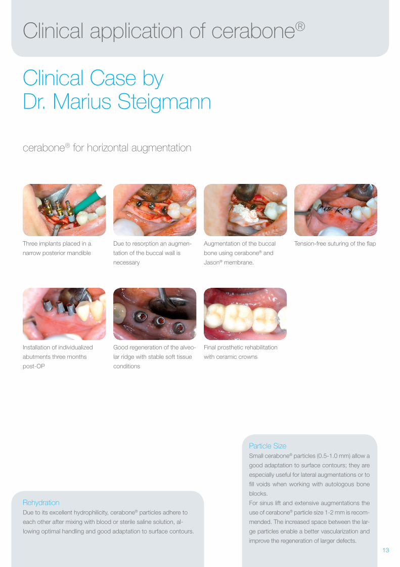
Clinical application of cerabone®
Clinical Case by Dr. Marius Steigmann
cerabone® for horizontal augmentation
Installation of individualized
abutments three months
post-OP
Good regeneration of the alveo-
lar ridge with stable soft tissue
conditions
Final prosthetic rehabilitation
with ceramic crowns
Three implants placed in a
narrow posterior mandible
Due to resorption an augmen-
tation of the buccal wall is
necessary
Augmentation of the buccal
bone using cerabone® and
Jason® membrane.
Tension-free suturing of the flap
Rehydration
Due to its excellent hydrophilicity, cerabone® particles adhere to
each other after mixing with blood or sterile saline solution, al-
lowing optimal handling and good adaptation to surface contours.
Particle Size
Small cerabone® particles (0.5-1.0 mm) allow a
good adaptation to surface contours; they are
especially useful for lateral augmentations or to
fill voids when working with autologous bone
blocks.
For sinus lift and extensive augmentations the
use of cerabone® particle size 1-2 mm is recom-
mended. The increased space between the lar-
ge particles enable a better vascularization and
improve the regeneration of larger defects.
14
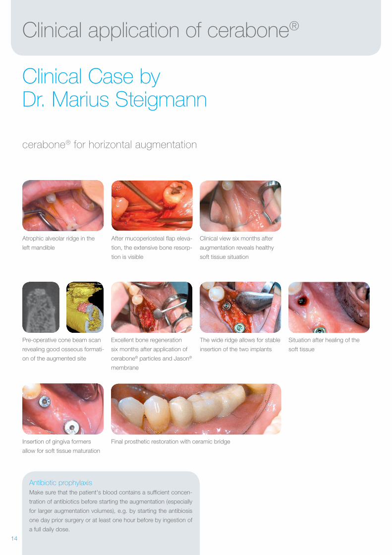
Clinical application of cerabone®
Clinical Case by Dr. Marius Steigmann
cerabone® for horizontal augmentation
Pre-operative cone beam scan
revealing good osseous formati-
on of the augmented site
The wide ridge allows for stable
insertion of the two implants
Situation after healing of the
soft tissue
Atrophic alveolar ridge in the
left mandible
After mucoperiosteal flap eleva-
tion, the extensive bone resorp-
tion is visible
Clinical view six months after
augmentation reveals healthy
soft tissue situation
Excellent bone regeneration
six months after application of
cerabone® particles and Jason®
membrane
Insertion of gingiva formers
allow for soft tissue maturation
Final prosthetic restoration with ceramic bridge
Antibiotic prophylaxis
Make sure that the patient's blood contains a sufficient concen-
tration of antibiotics before starting the augmentation (especially
for larger augmentation volumes), e.g. by starting the antibiosis
one day prior surgery or at least one hour before by ingestion of
a full daily dose.
15
Clinical application of cerabone®
Clinical Case by Dr. Marius Steigmann
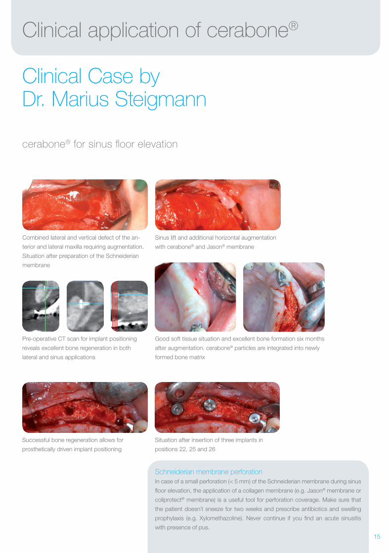
cerabone® for sinus floor elevation
Good soft tissue situation and excellent bone formation six months
after augmentation. cerabone® particles are integrated into newly
formed bone matrix
Pre-operative CT scan for implant positioning
reveals excellent bone regeneration in both
lateral and sinus applications
Combined lateral and vertical defect of the an-
terior and lateral maxilla requiring augmentation.
Situation after preparation of the Schneiderian
membrane
Sinus lift and additional horizontal augmentation
with cerabone® and Jason® membrane
Successful bone regeneration allows for
prosthetically driven implant positioning
Situation after insertion of three implants in
positions 22, 25 and 26
Schneiderian membrane perforation
In case of a small perforation (< 5 mm) of the Schneiderian membrane during sinus
floor elevation, the application of a collagen membrane (e.g. Jason® membrane or
collprotect® membrane) is a useful tool for perforation coverage. Make sure that
the patient doesn´t sneeze for two weeks and prescribe antibiotics and swelling
prophylaxis (e.g. Xylomethazoline). Never continue if you find an acute sinusitis
with presence of pus.
16
Clinical application of cerabone®
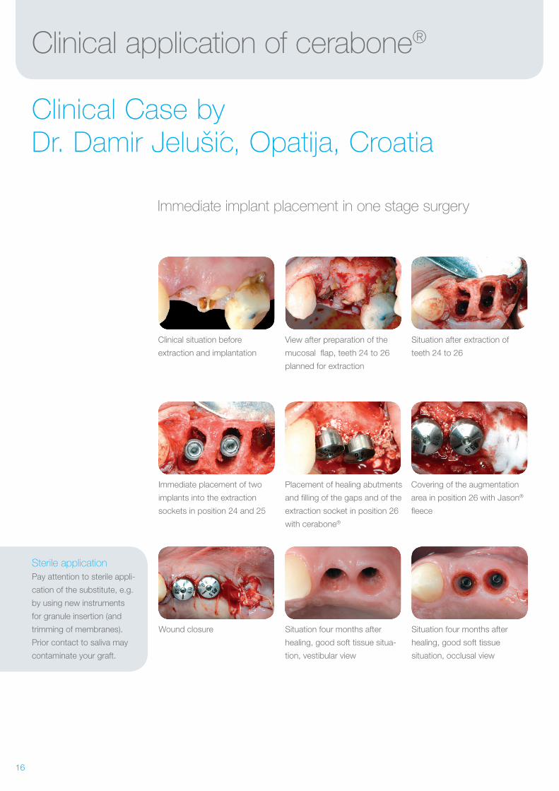
Clinical Case by Dr. Damir Jelušic, Opatija, Croatia
Immediate implant placement in one stage surgery
Immediate placement of two
implants into the extraction
sockets in position 24 and 25
Placement of healing abutments
and filling of the gaps and of the
extraction socket in position 26
with cerabone®
Clinical situation before
extraction and implantation
View after preparation of the
mucosal flap, teeth 24 to 26
planned for extraction
Situation after extraction of
teeth 24 to 26
Covering of the augmentation
area in position 26 with Jason®
fleece
Wound closure Situation four months after
healing, good soft tissue situa-
tion, vestibular view
Situation four months after
healing, good soft tissue
situation, occlusal view
Sterile application
Pay attention to sterile appli-
cation of the substitute, e.g.
by using new instruments
for granule insertion (and
trimming of membranes).
Prior contact to saliva may
contaminate your graft.
17
Clinical application of cerabone®
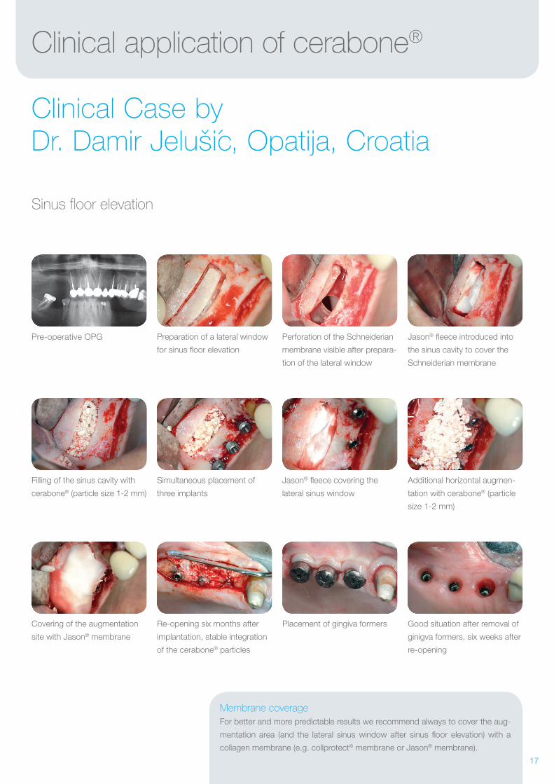
Clinical Case by Dr. Damir Jelušic, Opatija, Croatia
Sinus floor elevation
Filling of the sinus cavity with
cerabone® (particle size 1-2 mm)
Simultaneous placement of
three implants
Jason® fleece covering the
lateral sinus window
Pre-operative OPG Preparation of a lateral window
for sinus floor elevation
Perforation of the Schneiderian
membrane visible after prepara-
tion of the lateral window
Jason® fleece introduced into
the sinus cavity to cover the
Schneiderian membrane
Additional horizontal augmen-
tation with cerabone® (particle
size 1-2 mm)
Covering of the augmentation
site with Jason® membrane
Re-opening six months after
implantation, stable integration
of the cerabone® particles
Placement of gingiva formers Good situation after removal of
ginigva formers, six weeks after
re-opening
Membrane coverage
For better and more predictable results we recommend always to cover the aug-
mentation area (and the lateral sinus window after sinus floor elevation) with a
collagen membrane (e.g. collprotect® membrane or Jason® membrane).
18
Clinical application of cerabone®
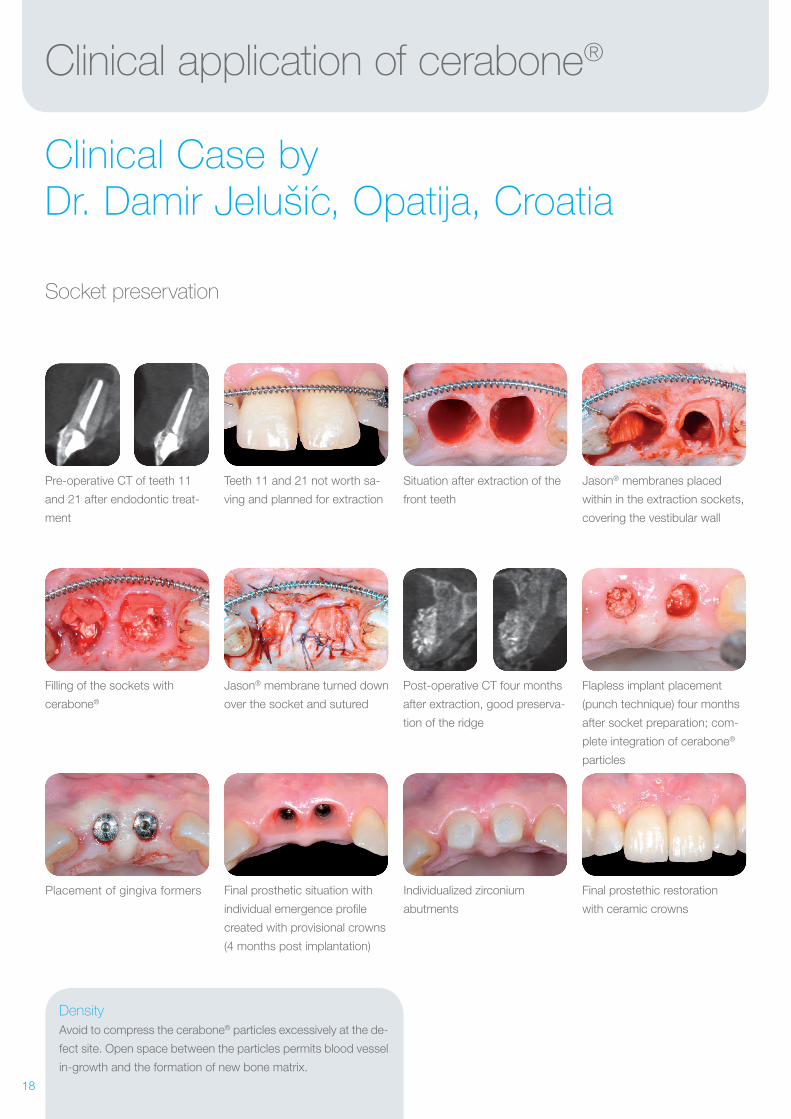
Clinical Case by Dr. Damir Jelušic, Opatija, Croatia
Socket preservation
Filling of the sockets with
cerabone®
Jason® membrane turned down
over the socket and sutured
Post-operative CT four months
after extraction, good preserva-
tion of the ridge
Pre-operative CT of teeth 11
and 21 after endodontic treat-
ment
Teeth 11 and 21 not worth sa-
ving and planned for extraction
Situation after extraction of the
front teeth
Jason® membranes placed
within in the extraction sockets,
covering the vestibular wall
Flapless implant placement
(punch technique) four months
after socket preparation; com-
plete integration of cerabone®
particles
Placement of gingiva formers Final prosthetic situation with
individual emergence profile
created with provisional crowns
(4 months post implantation)
Individualized zirconium
abutments
Final prostethic restoration
with ceramic crowns
Density
Avoid to compress the cerabone® particles excessively at the de-
fect site. Open space between the particles permits blood vessel
in-growth and the formation of new bone matrix.
19
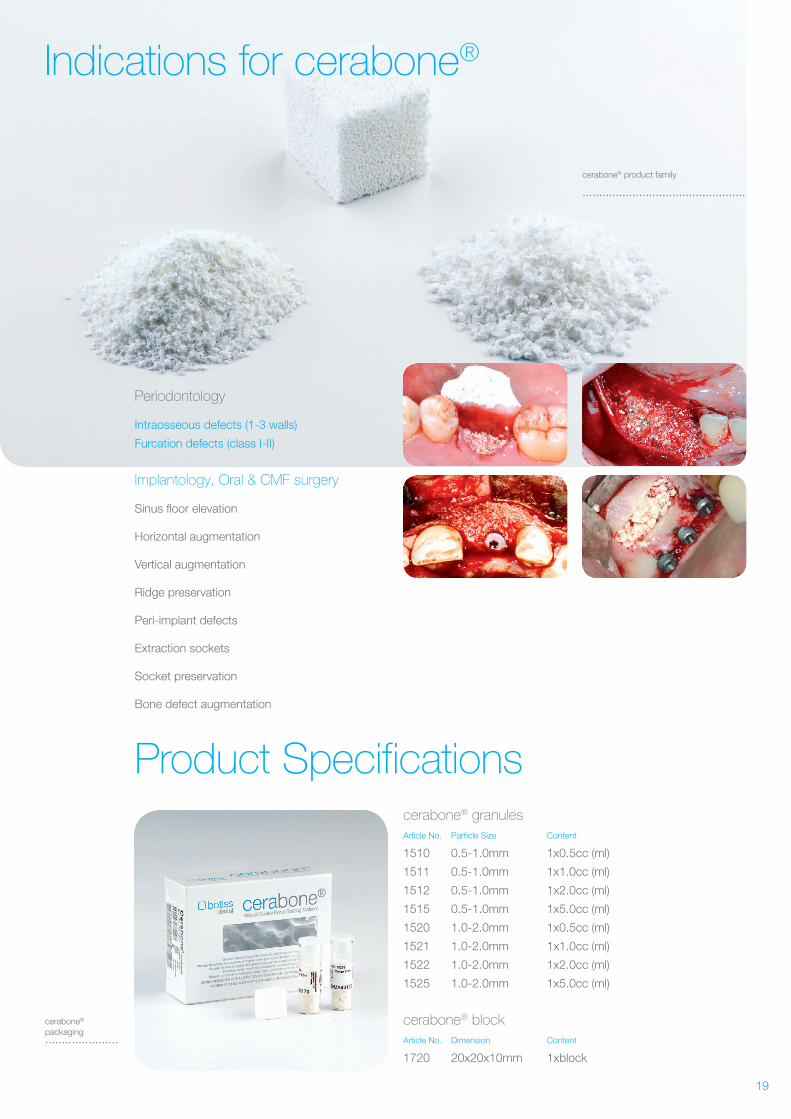
Indications for cerabone®
cerabone® product family
Periodontology
Intraosseous defects (1-3 walls)
Furcation defects (class I-II)
Implantology, Oral & CMF surgery
Sinus floor elevation
Horizontal augmentation
Vertical augmentation
Ridge preservation
Peri-implant defects
Extraction sockets
Socket preservation
Bone defect augmentation
Product Specificationscerabone® granules
Article No. Particle Size Content
1510 0.5-1.0mm 1x0.5cc (ml)
1511 0.5-1.0mm 1x1.0cc (ml)
1512 0.5-1.0mm 1x2.0cc (ml)
1515 0.5-1.0mm 1x5.0cc (ml)
1520 1.0-2.0mm 1x0.5cc (ml)
1521 1.0-2.0mm 1x1.0cc (ml)
1522 1.0-2.0mm 1x2.0cc (ml)
1525 1.0-2.0mm 1x5.0cc (ml)
cerabone® block
Article No. Dimension Content
1720 20x20x10mm 1xblock
......................
cerabone® packaging
.................................................
20Rev.: CBSen-01/2014-08
soft tissue
education
hard tissue
botiss dental GmbH
Uhlandstraße 20-25
10623 Berlin / Germany
Fon +49 30 20 60 73 98 30
Fax +49 30 20 60 73 98 20
www.botiss.com
facebook: botiss biomaterials
Innovation.
Regeneration.
Aesthetics.
Straumann UK
3 Pegasus Place
Gatwick Road
West Sussex
Crawley RH10 9AY
Tel: +44 1293 65 12 30
Fax +44 1293 65 12 39
www.straumann.co.uk
Straumann SA/NV
Belgicastraat 3 Box 3
1930 Zaventem
Tel: +32 2 790 10 00
Fax +32 2 790 10 20
www.straumann.be
...................................
Straumann s.r.o.
Na Žertvách 2196
180 00 Prague 8
Czech Republic
Tel.: +420 284 094 650
Fax: +420 284 094 659
www.straumann.cz
Straumann B.V.
Einsteinweg 15
Postbus 338
3400 AH Ijsselstein
Tel: +31 30 604 66 11
Fax +31 30 604 67 28
www.straumann.nl
...................................
Straumann A/S
Post boks 1751, Vika
0122 Oslo
Tel: +47 23 35 44 88
Fax +47 23 35 44 80
www.straumann.no
...................................
Distributed by:
Straumann AB
Krokslätts Fabriker 45
431 37 Mölndal
Tel: +46 31 708 75 00
Fax +46 31 708 75 19
www.straumann.se
...................................
Some products may not be available in all countries. Please check with your local Straumann sales representative for more information.
botissbiomaterials
dental
bone & tissue regeneration