Embed Size (px)
Citation preview
Andreas Charidimou, MD PhD FESO
Hemorrhagic Stroke Research ProgramJPK Stroke Research Center
Cerebral amyloid angiopathy and
intracerebral haemorrhage:
An update

Boston, Jan 2015 Sept 2017
Objectives
• Definition of CAA as a disease
• Key clinical/MRI signatures – ongoing work
• Implications for treatments, BP, OAC etc.
• No RCTs, latest developments, current trends
We will not cover…
• Pathophysiology of the disease
• Acute CAA-ICH, haematoma expansion-BP etc.
• Role of CAA in dementia
• Advanced neuroimaging aspects, e.g. fMRI, DTI etc.
Keytake-homemessages
• MRI is crucial when CAA is suspected– For diagnosis– For prognosis
• Cortical superficial siderosis – driver of bleeding
• CAA is not only about haemorrhage
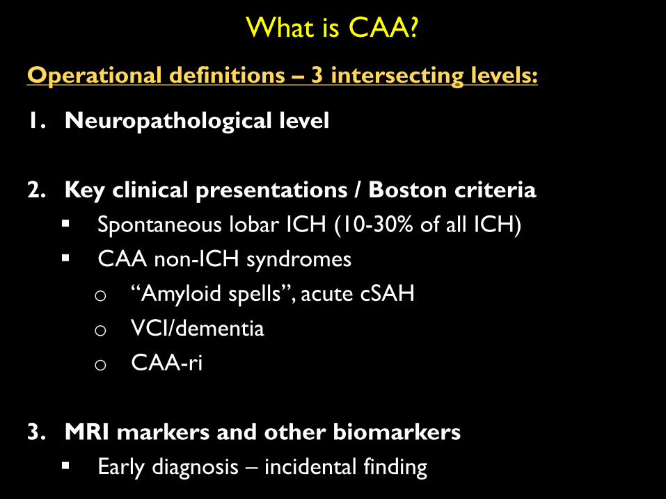
What is CAA?
Operational definitions – 3 intersecting levels:
1. Neuropathological level
2. Key clinical presentations / Boston criteria§ Spontaneous lobar ICH (10-30% of all ICH)§ CAA non-ICH syndromes
o “Amyloid spells”, acute cSAHo VCI/dementiao CAA-ri
3. MRI markers and other biomarkers§ Early diagnosis – incidental finding
What is CAA? Pathologically common, clinically relevant
for developing CAA but may increase the risk of CAA relatedICH. Vinters2din a clinicopathological series of 107 pathologi-cally proven CAA casesdfound the prevalence of hypertensionto be around 32%, similar to community dwelling elderlypopulations,33 while another pathological study reported thatCAA patients with ICH were more frequently hypertensive(50%) than those without ICH (23%), suggesting that hyper-tension may contribute to CAA related cerebral bleeding.34 Ina recent multicentre cohort of patients with spontaneous ICH,we found that the prevalence of hypertension in CAA relatedICH was 62%dsignificantly less than in non-CAA related ICH(85%).35 Whether hypertension in association with CAA confersa greater risk for ICH compared with CAA alone is an importantclinical question.36e38 Evidence from the PROGRESS trial ofblood pressure lowering after stroke showed that a mean bloodpressure reduction of 9/4 mm Hg reduced the risk of futureCAA related ICH by about 77%, supporting an important causalrole for hypertension.38
Apolipoprotein E (ApoE) alleles are the only known geneticrisk factors for sporadic CAA.39 ApoE is a protein with crucialroles in lipoprotein complexes, which regulate lipid metabolismby binding to cell surface receptors and proteins associated withlipid transfer and lipolysis.39 There are three major poly-morphisms in the ApoE genednamely, 34, 32 and 33dresultingin a single amino acid change40 which dramatically alters the
functional properties of ApoE isoforms.41 These alleles havea strong dose dependent effect on the risk of developing CAAand its clinical severity. Thus ApoE 34 in both postmortem andclinical series increases the risk of sporadic CAA related lobarICH; moreover, the number of 34 alleles relates to clinicalseverity.39 42e44 Individuals carrying the ApoE 32 allele also havean increased risk of CAA related lobar ICH.44 45 Both of theserisk alleles are also associated with a younger age of first ICH,46
greater likelihood of haematoma expansion, poorer clinicaloutcome47 48 and a higher risk of recurrence.49 Furthermore, thetwo allelic variants interact: patients with both ApoE 32 and 34alleles have the earliest disease onset and highest risk ofearly ICH recurrence.49 50 The 32 and 34 alleles might promoteCAA related haemorrhage through distinct mechanisms: 34by promoting Ab deposition and 32 by inducing structuralchanges in amyloid laden vessels, making them prone torupture.47 48 50e52 Other as yet unidentified genetic poly-morphisms relating to amyloid metabolic pathways (figure 2A)may also play a role in sporadic CAA, (eg, presenilin-1, neprilysinand transforming growth factor b-1),57e59 and are a topic ofongoing investigation.
NEUROPATHOLOGYMorphological characteristics, natural history and severitygradingCAA primarily involves neocortical and leptomeningeal arteri-oles, to a lesser extent capillaries and, very rarely, venules.3 Incontrast with amyloid plaques found in ADdwhich arepredominantly composed of the 42 amino acid residue fragment(Ab42)dthe vascular amyloid in CAA is mostly composed of themore soluble, 40 amino acid fragment (Ab40), suggestingdifferent pathophysiological mechanisms for pathologicaldeposition (see below).60e63 Cerebral vessels with moderate tosevere CAA show an acellular wall thickening with a stronglyeosinophilic smudgy appearance on haematoxylineeosin stainedsections.64 Congo red staining, under polarised light, revealsamyloid deposits as ‘apple green’ birefringence (hence the termcongophilic angiopathy)2 65 although immunological stains forAb are highly specific and now widely used (figure 3). Thedevelopment of CAA is progressive, with Ab first appearing inthe abluminal aspect of the tunica media, surrounding smoothmuscle cells, and in the adventitia (figure 3).2 At the initial stage,the vessel wall structure is intact, but as the disease progresses,there is pan-mural amyloid accumulation and loss of smoothmuscle cells.3 In severe CAA, detachment and delamination ofthe outer part of the tunica media result in the so-called ‘doublebarrel’ appearance (figure 3)3; fibrinoid necrosis and micro-aneurysm formation also occur in advanced disease. There mayalso be microbleeding with perivascular deposition of erythro-cytes and blood breakdown products.64 Endothelial cells areusually preserved even in vessels severely affected by CAA.66
Occasionally Ab is deposited in the surrounding brain paren-chyma immediately adjacent to an affected vessel (sometimescalled ‘dyshoric CAA’).CAA is also associated with cerebral ischaemic damage,17 26 67 68
including cortical microinfarcts,69 and white matter pathology(demyelination and gliosis).8 17 62 Microinfarcts are predominantlylobar (corticalesubcortical), usually in patients with severe CAA.One possible mechanism for these ischaemic lesions is occlusion orreduced perfusion in amyloid laden cortical vessels affected by CAA.The changes described above provide the basis of neuropath-
ological scoring systems for CAA,34 67 70 each with strengths andlimitations.71 No standardised consensus neuropathologicalcriteria for rating CAA are available72 but are desirable to allow
Box 1 Search strategy and selection criteria
References were identified through PubMed with the searchterms: ‘cerebral amyloid angiopathy’; ‘microbleed(s) or microh(a)emorrhage(s) and cerebral amyloid angiopathy’; ‘intracerebralh(a)emorrhage’; and ‘vascular cognitive impairment’ betweenJanuary 1970 and August 2011. The references from identifiedarticles and the authors’ own files were also searched for relevantpublications. Only papers published in English were reviewed.The final reference list was chosen on the basis of relevance tothe topics covered in this article.
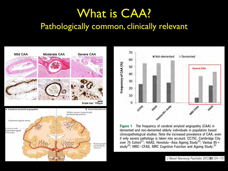
Figure 1 The frequency of cerebral amyloid angiopathy (CAA) indemented and non-demented elderly individuals in population basedclinicopathological studies. Note the increased prevalence of CAA, evenif only severe pathology is taken into account. CC75C, Cambridge Cityover 75 Cohort21; HAAS, HonolulueAsia Ageing Study23; Vantaa 85+study24; MRCeCFAS, MRC Cognitive Function and Ageing Study.22
J Neurol Neurosurg Psychiatry 2012;83:124e137. doi:10.1136/jnnp-2011-301308 125
Cerebrovascular disease
group.bmj.com on January 5, 2015 - Published by http://jnnp.bmj.com/Downloaded from
REVIEW
Sporadic cerebral amyloid angiopathy revisited:recent insights into pathophysiology andclinical spectrumAndreas Charidimou, Qiang Gang, David J Werring
ABSTRACTSporadic cerebral amyloid angiopathy (CAA) is a commonage related cerebral small vessel disease, characterised byprogressive deposition of amyloid-b (Ab) in the wall ofsmall to medium sized arteries, arterioles and capillaries ofthe cerebral cortex and overlying leptomeninges. Previouslyconsidered to be a rare neurological curiosity, CAA is nowrecognised as an important cause of spontaneousintracerebral haemorrhage and cognitive impairment in theelderly, two fundamental challenges in the field ofcerebrovascular disease. Our understanding of thepathophysiology and clinical manifestations of CAAcontinues to evolve rapidly, with the use of transgenicmouse models and advanced structural and/or molecularneuroimaging techniques. Yet, despite remarkable recentinterest, CAA remains under-recognised by neurologistsand stroke physicians. In this review, a fresh look at keydevelopments in understanding the complexpathophysiology, important clinical and radiological features,diagnostic approaches and prospects for rational therapiesfor this enigmatic small vessel disorder is provided.
INTRODUCTIONSporadic cerebral amyloid angiopathy (CAA) isa common small vessel disease of the brain, char-acterised by the progressive deposition of amyloid-b (Ab) protein in the walls of small to mediumsized arteries (up to about 2 mm in diameter1),arterioles and capillaries in the cerebral cortex andoverlying leptomeninges.2 3 CAA can also affectcerebellar vessels but only rarely those in thebrainstem or basal ganglia. Although known topathologists for over a century,4 5 CAA was notlinked to clinical disease until as late as the 1960swhen it was suggested to be a rare cause of intra-cerebral haemorrhage (ICH).6e8 In recent years,CAA has been ‘rediscovered’ as a common andimportant cause of spontaneous ICH, whichremains the most devastating form of stroke, witha death rate approaching 50% in contrast withimproved outcomes from ischaemic stroke.9 10 Anincreased understanding of CAA thus holds promisefor improved prevention and treatment of ICH.The growing interest in CAA is at least partly
thanks to two fields of research, which have beenimportant in defining the expanding clinicaleradiological phenotype and the underlying patho-physiology of the disease: (1) neuroimaging, whichnow allows an unprecedented ability to investigateCAA dynamics in vivo using MRI to reveal complex
patterns of cerebral bleeding (including lobarmicrobleeds11) and ischaemia, and an increasingrepertoire of specific amyloid binding ligands3 12e16;and (2) transgenic mouse studies, which haveallowed the experimental alteration of amyloidpeptide expression and molecular structure,providing significant mechanistic insights. Despitethese advances, CAA remains under-recognised byneurologists and stroke physicians, making a freshlook especially timely. In this review (see box 1 forsearch strategy), we provide a comprehensiveupdate, emphasising the widening spectrum ofCAA clinical presentations and neuroimagingfeatures, including diagnostic approaches to reliablyidentify the disease in vivo. Finally, we discussimproved prospects for rational preventive ordisease modifying therapies for this common anddevastating microangiopathic disorder.
EPIDEMIOLOGY AND RISK FACTORSPathologically defined CAA is common in theelderly.17e20 Population based autopsy studiesindicate a CAA prevalence of 20e40% in non-demented and 50e60% in demented elderlypopulations (figure 1).19 21e24 Furthermore, CAApathology may be severe in older individuals(figure 1): in the HonolulueAsia Ageing AutopsyStudy, severe CAA was found in 43% of dementedand 24% of non-demented elderly individuals(mean age at death 85 years).23 In Alzheimer ’sdisease (AD), CAA is almost invariable being foundat autopsy in more than 90% of cases.17 25
However, most of these patients have mild CAA;severe CAA is found in about 25% of AD brains.26
Advancing age is the strongest known clinical riskfactor for developing CAA.2 In a community basedsample of 100 individuals, the prevalence of corticalvascular Ab deposition progressively increased fromthe seventh to the ninth decades,27 a pattern alsoobserved in 784 consecutive autopsies, corrected forover-representation of AD.28 Moreover, patientswith CAA related ICH (suggesting advanceddisease) in large autopsy series were all older than60 years (and most over 70 years of age).7 29 30
Sporadic CAA is seldom reported before the sixthdecade of life; occasional patients presenting in their50s have been described.31
In contrast with hypertensive arteriopathydtheother main form of small vessel disease and cause ofICH32dthe risk of CAA is not accounted for byconventional cardiovascular risk factors other thanage.2 Hypertension is not considered a risk factor
Stroke Research Group,Department of Brain Repair andRehabilitation, UCL Institute ofNeurology and the NationalHospital for Neurology andNeurosurgery, Queen Square,London, UK
Correspondence toDr D J Werring, NationalHospital for Neurology andNeurosurgery, Box 6, QueenSquare, London WC1N 3BG,UK; [email protected]
Received 30 August 2011Accepted 2 October 2011Published Online First5 November 2011
124 J Neurol Neurosurg Psychiatry 2012;83:124e137. doi:10.1136/jnnp-2011-301308
Cerebrovascular disease
group.bmj.com on January 5, 2015 - Published by http://jnnp.bmj.com/Downloaded from
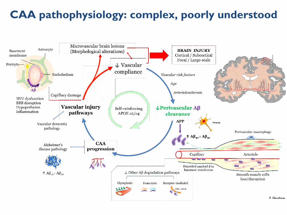
CAA pathophysiology: complex, poorly understood
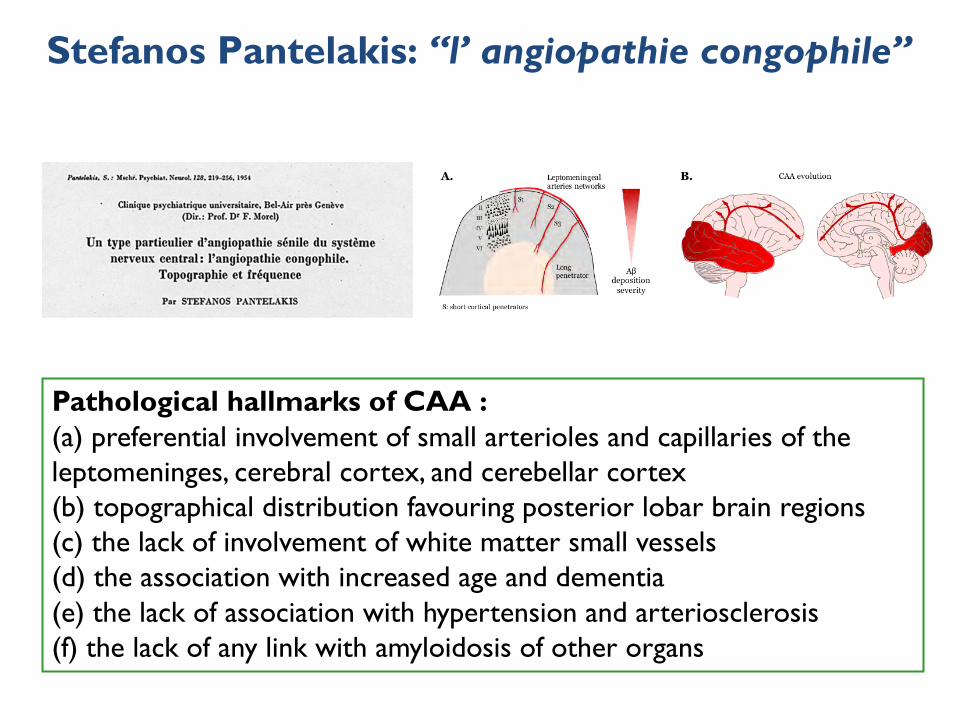
Pathological hallmarks of CAA :(a) preferential involvement of small arterioles and capillaries of the leptomeninges, cerebral cortex, and cerebellar cortex(b) topographical distribution favouring posterior lobar brain regions (c) the lack of involvement of white matter small vessels(d) the association with increased age and dementia(e) the lack of association with hypertension and arteriosclerosis (f) the lack of any link with amyloidosis of other organs
Stefanos Pantelakis: “l’ angiopathie congophile”
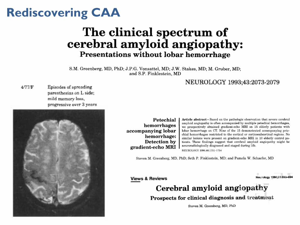
Rediscovering CAA
*
*
1 2a 2b
3a 3b
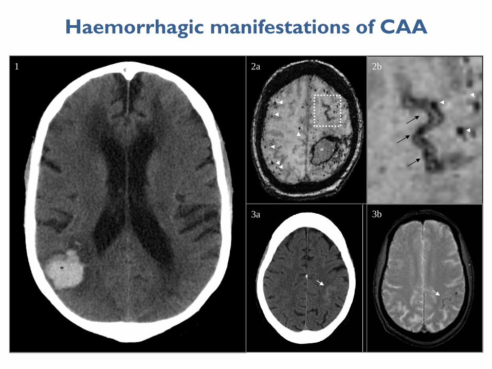
Haemorrhagic manifestations of CAA
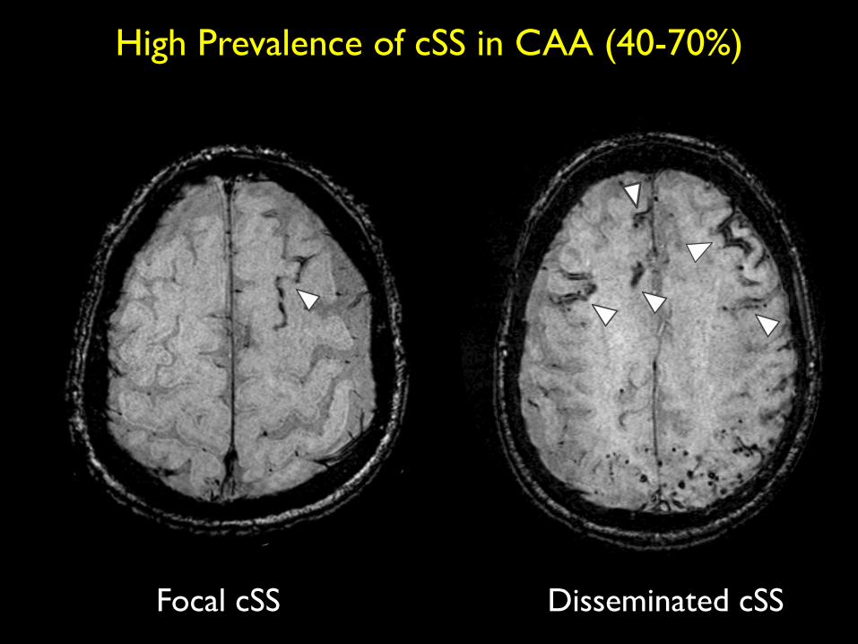
Focal cSS Disseminated cSS
High Prevalence of cSS in CAA (40-70%)
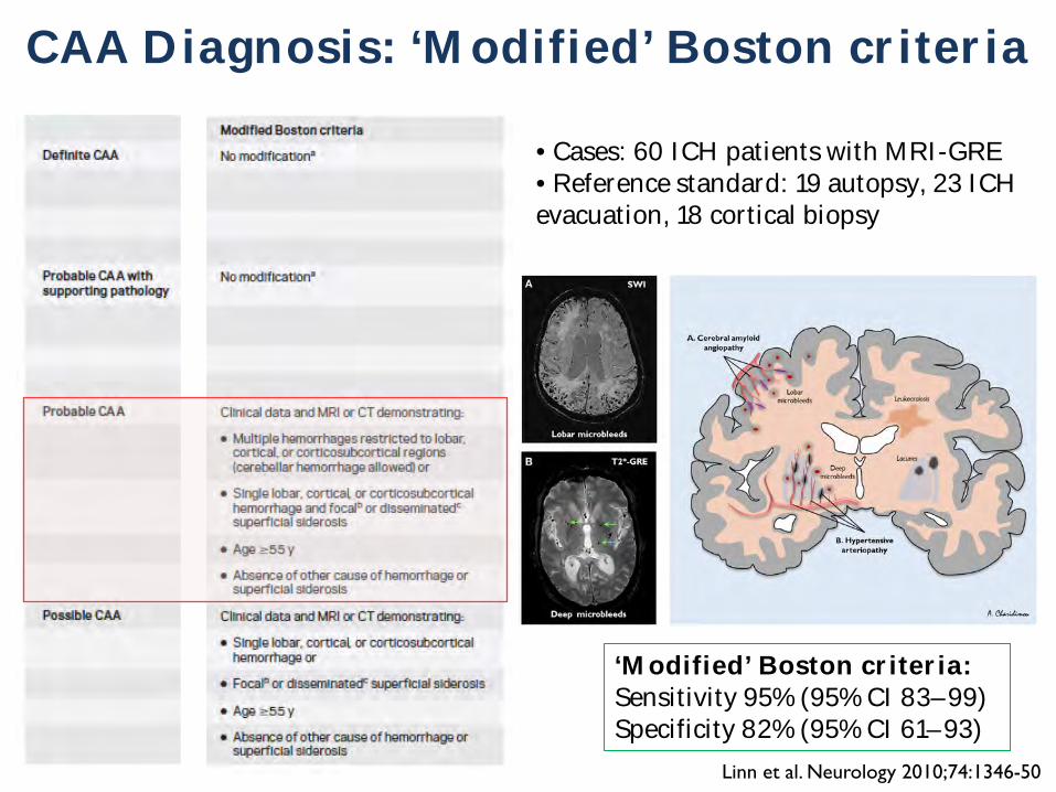
Linn et al. Neurology 2010;74:1346-50
CAA Diagnosis: ‘Modified’ Boston criteria
‘Modified’ Boston criteria:Sensitivity 95% (95% CI 83–99)Specificity 82% (95% CI 61–93)
• Cases: 60 ICH patients with MRI-GRE• Reference standard: 19 autopsy, 23 ICH evacuation, 18 cortical biopsy
Charidimou et al. Cerebral microbleeds and anticoagulant-associated intracerebral hemorrhage
that the risk of ICH is increased by an age-related disorder of smallbrain blood vessels. Anticoagulant use per se should not cause ICHif cerebral vessels are intact, but the presence of microangiopathy,rendering small vessels brittle and fragile, is a plausible causal oraggravating factor for such hemorrhage. Indeed, some risk stratifi-cation scores (e.g., HAS-BLED; Pisters et al., 2010) includes clinicalelements that may correlate with small vessel disease (e.g., age andhypertension).
Cerebral small vessel disease is one of the most prevalent brainconditions described, especially as people live longer (Greenberg,2006; Pantoni, 2010). The common sporadic forms are: (a) hyper-tensive arteriopathy (including lipohyalinosis and arterioloscle-rosis), which typically affects the small perforating end-arteriesof the deep gray nuclei and deep white matter, and as the nameimplies is related to hypertension and other traditional cardio-vascular risk factors (Pantoni, 2010); and (b) CAA, a commonage-related condition characterized by the progressive deposi-tion of amyloid-b in the media and adventitia of small arteries,arterioles, and capillaries in the cerebral cortex, overlying lep-tomeninges, and gray–white matter junction (Charidimou et al.,2012; Figure 3). The rupture of small arteries affected by thesetwo disease processes underlies the majority of ICHs (>75%) inthe elderly, classified as spontaneous ICH (sometimes also termedprimary or non-traumatic).
Cerebral amyloid angiopathy is most often recognized in life bysymptomatic, spontaneous, lobar ICH in elderly patients (Charidi-mou et al., 2012). Evidence supporting a link between CAA andanticoagulation-associated ICH includes the demonstration ofCAA in 7 of 11 lobar ICHs occurring on warfarin in the largestconsecutive pathological series reported (Rosand et al., 2000). Inaddition, the apolipoprotein E e2 allele, a known genetic riskfactor of CAA-related lobar ICH (Nicoll et al., 1997; Biffi et al.,2010b), is more common in warfarin-associated ICH than in con-trol patients on warfarin without ICH, supporting a role for CAA(Rosand et al., 2000). There are also individual cases of ICH fol-lowing anticoagulation or coronary thrombolysis, which demon-strated advanced CAA on autopsy (Melo et al., 1993; McCarronand Nicoll, 2004). However, the mechanisms of spontaneousand anticoagulation-associated ICH are complex and involve adynamic interplay between underlying bleeding-prone small ves-sel diseases, genetic factors, cardiovascular risk factors, and the useof oral anticoagulation treatments (Figure 4).
Modern MRI allows an unprecedented ability to identify cere-bral small vessel disease in vivo. Leukoaraiosis has been recognizedfor many years as a characteristic MRI manifestation of small ves-sel disease. Some studies suggest that the presence of leukoaraiosisis associated with an increased the risk of oral anticoagulant-associated ICH (Gorter, 1999; Smith et al., 2002). However,
FIGURE 3 |The distribution of sporadic small vessel disease in thebrain and the topography of cerebral microbleeds (CMBs). (A) Cerebralamyloid angiopathy (CAA) preferentially affects the small arteries andarterioles of the cerebral cortex and gray–white matter junction by thedeposition of amyloid-b in the vessel walls (purple); (B) hypertensivearteriopathy typically affects small deep arterial perforators (black). CMBsare a marker for the severity and type of small vessel disease; their
anatomic distribution is meant to reflect the underlying pathological vesseldamage. Hence, CMBs (dark, rounded lesions) located incortical-subcortical regions are presumably caused by CAA (A), whereasCMBs located in deep brain regions mainly result from hypertensivearteriopathy (B). (A) is an axial susceptibility-weighted imaging (SWI)which is currently the most sensitive means to image CMBs. (B) is an axialT2*-weighted gradient-recalled echo (T2*-GRE) MRI.
www.frontiersin.org September 2012 | Volume 3 | Article 133 | 5
3a 3b
2a1 2b
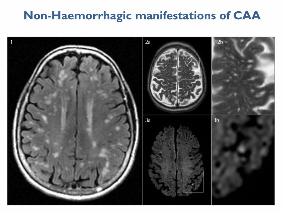
Non-Haemorrhagic manifestations of CAA
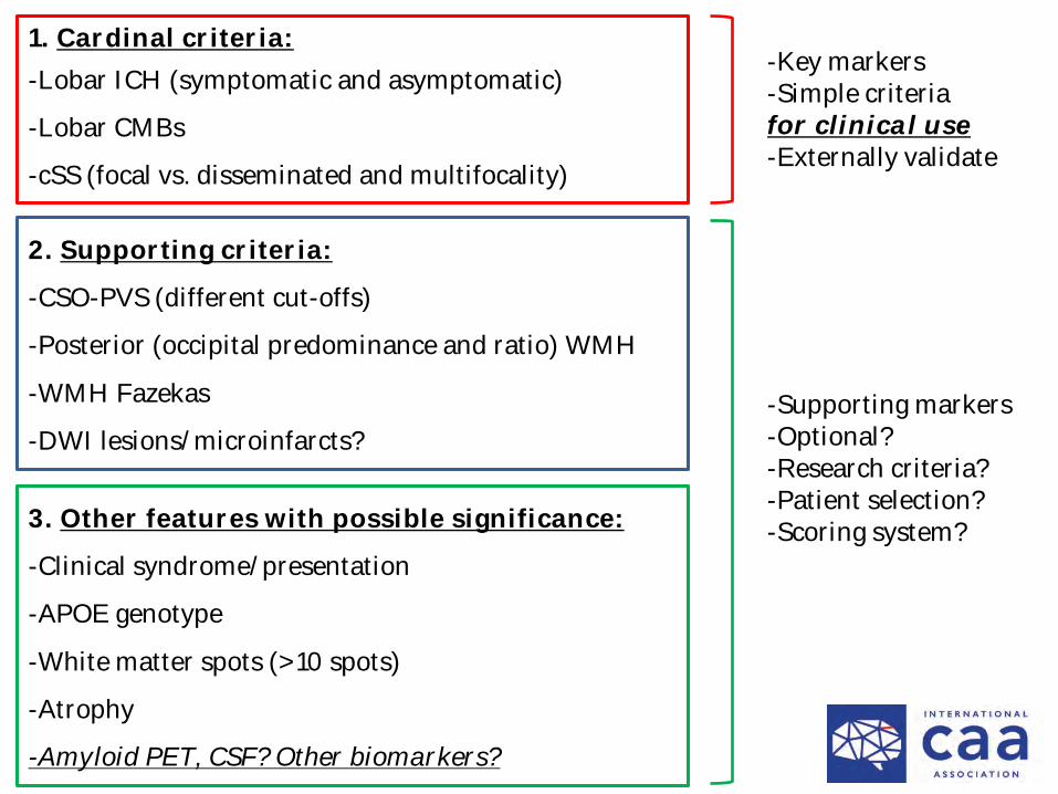
1. Cardinal criteria:-Lobar ICH (symptomatic and asymptomatic)
-Lobar CMBs
-cSS (focal vs. disseminated and multifocality)
2. Supporting criteria:
-CSO-PVS (different cut-offs)
-Posterior (occipital predominance and ratio) WMH
-WMH Fazekas
-DWI lesions/microinfarcts?
3. Other features with possible significance:
-Clinical syndrome/presentation
-APOE genotype
-White matter spots (>10 spots)
-Atrophy
-Amyloid PET, CSF? Other biomarkers?
-Key markers-Simple criteriafor clinical use-Externally validate
-Supporting markers-Optional?-Research criteria?-Patient selection?-Scoring system?
0.2
.4.6
.81
Sens
itivi
ty
0.2.4.6.81
Specificity
Study estimate Summary point
HSROC curve 95% confidence region
95% prediction region
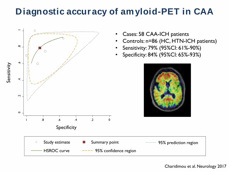
Diagnostic accuracy of amyloid-PET in CAA
• Cases: 58 CAA-ICH patients • Controls: n=86 (HC, HTN-ICH patients)• Sensitivity: 79% (95%CI: 61%-90%)• Specificity: 84% (95%CI: 65%-93%)
Vinci Lite Vinci Lite Vinci Lite Vinci Lite Vinci Lite Vinci Lite Vinci Lite Vinci Lite Vinci Lite Vinci Lite Vinci Lite Vinci Lite Vinci Lite Vinci Lite Vinci Lite
Vinci Lite Vinci Lite Vinci Lite Vinci Lite Vinci Lite Vinci Lite Vinci Lite Vinci Lite Vinci Lite Vinci Lite Vinci Lite Vinci Lite Vinci Lite Vinci Lite Vinci Lite
Vinci Lite Vinci Lite Vinci Lite Vinci Lite Vinci Lite Vinci Lite Vinci Lite Vinci Lite Vinci Lite Vinci Lite Vinci Lite Vinci Lite Vinci Lite Vinci Lite Vinci Lite
Charidimou et al. Neurology 2017
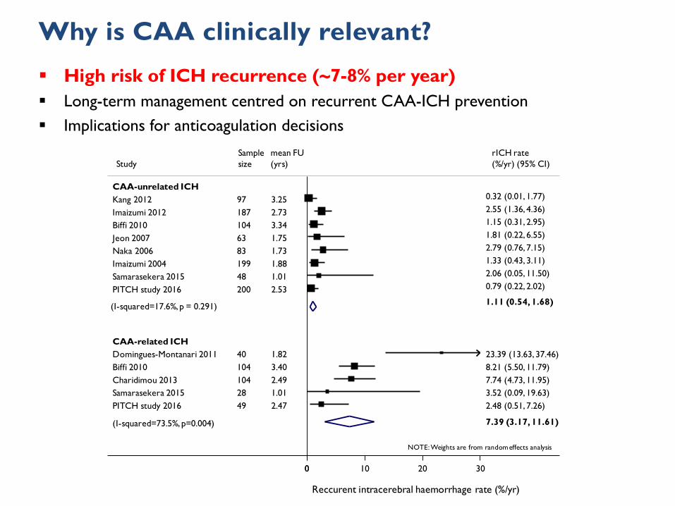
Why is CAA clinically relevant?
Studymean FU(yrs)
rICH rate(%/yr) (95% CI)
Reccurent intracerebral haemorrhage rate (%/yr)
Sample size
NOTE: Weights are from random effects analysis
(I-squared=73.5%, p=0.004)
Samarasekera 2015
CAA-related ICH
Biffi 2010
Domingues-Montanari 2011Biffi 2010
Naka 2006
CAA-unrelated ICH
Jeon 2007
Imaizumi 2004
PITCH study 2016
Samarasekera 2015
Kang 2012
PITCH study 2016
Imaizumi 2012
Charidimou 201328
104
40104
8363
199
49
48
97
200
187
1041.01
3.34
1.823.40
1.731.75
1.88
2.47
1.01
3.25
2.53
2.73
2.493.52 (0.09, 19.63)
7.39 (3.17, 11.61)
1.15 (0.31, 2.95)
23.39 (13.63, 37.46)8.21 (5.50, 11.79)
2.79 (0.76, 7.15)1.81 (0.22, 6.55)
1.11 (0.54, 1.68)
1.33 (0.43, 3.11)
2.48 (0.51, 7.26)
2.06 (0.05, 11.50)
0.32 (0.01, 1.77)
0.79 (0.22, 2.02)
2.55 (1.36, 4.36)
7.74 (4.73, 11.95)
00 10 20 30
(I-squared=17.6%, p = 0.291)
§ High risk of ICH recurrence (~7-8% per year)§ Long-term management centred on recurrent CAA-ICH prevention§ Implications for anticoagulation decisions
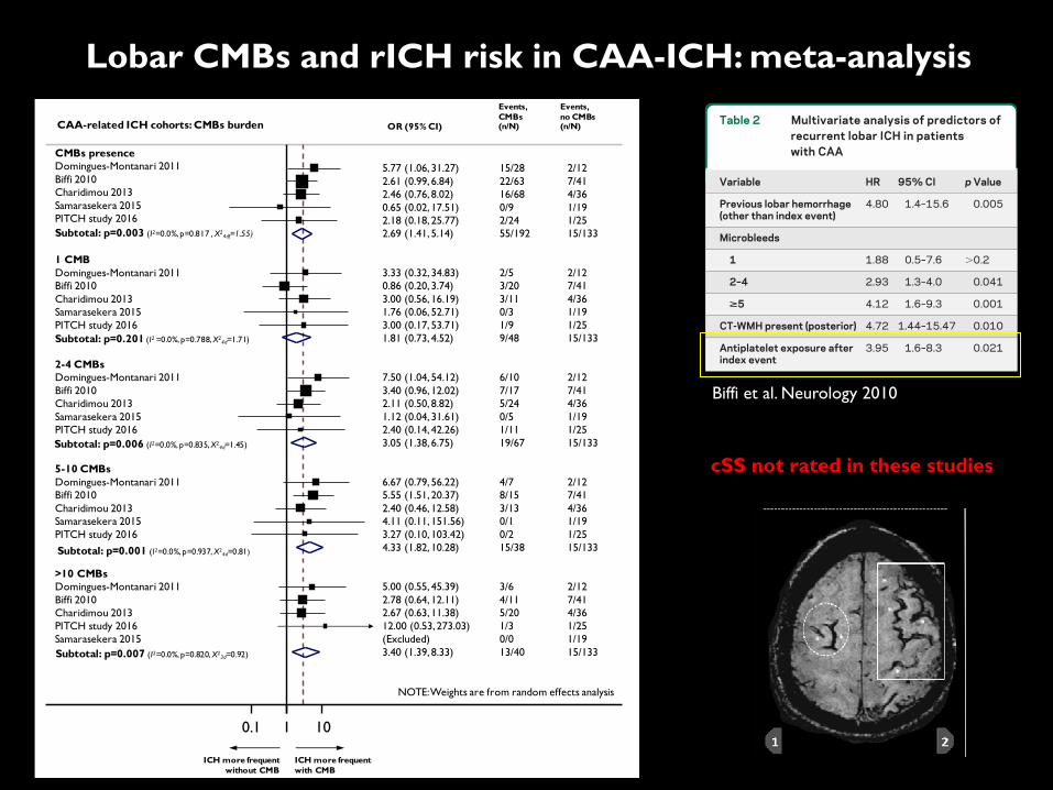
Lobar CMBs and rICH risk in CAA-ICH: meta-analysis
cSS not rated in these studies
farin exposure, statin exposure). The model demon-strated that previous ICH, posterior CT-WMH, thepresence of !2 microbleeds, and antiplatelet use areall independently associated with increased risk ofrecurrent lobar ICH (figure). No effect was evidentfor warfarin or statin exposure. The final multivariatemodel, including only significant predictors of lobarICH recurrence, is detailed in table 2.
To explore whether the risk of aspirin varied ac-cording to the number of baseline microbleeds, theantithrombotic drug-associated HR for recurrence ineach microbleed category was determined (table 3).There was an aspirin-associated increase in HR for
lobar ICH recurrence in those with 0, 1, 2–4, or !5microbleeds (table 3).
DISCUSSION The major finding from this prospec-tive cohort study is that lobar ICH is associated witha high rate of recurrent hemorrhage and that severityof white matter disease and cerebral microbleeds in-dependently increase this risk. The use of antithrom-botic agents in CAA after lobar ICH may alsoincrease this risk when the analysis is controlled forbaseline microbleed count.
Although the association between aspirin expo-sure, microbleeds, and symptomatic ICH was previ-ously reported in case-control studies,21,22 prospectivedata on the influence of aspirin on lobar ICH recur-rence have been lacking. This analysis of prospec-tively followed patients with CAA associates aspirinuse with recurrent hemorrhage when adjusting forbaseline predictors of recurrent ICH.
Several mechanisms might be implicated in theeffect of aspirin exposure in patients with CAA. Re-cently published data on a cross-sectional populationstudy of microbleeds prevalence suggest that expo-sure to aspirin may be associated with increased prev-alence of lobar microbleeds, one of the hallmarks ofCAA.23 Antiplatelet agents might therefore increasethe risk of recurrent ICH by substantially increasingthe number of microbleeds at risk for conversion intoclinically manifest macrobleeds. Antiplatelet therapymight also act on preexisting and new spontaneousmicrobleeds by increasing the risk of evolution intoclinically symptomatic ICH.
Although our analysis was not adequately pow-ered to explore this mechanism in regard to warfarin,anticoagulation therapy might also influence ICH re-currence based on microbleed burden. Limited evi-dence supporting this hypothesis has been recentlyprovided in a small case-control series.24 Additional
Figure Aspirin and recurrent lobar intracerebral hemorrhage
Modified Kaplan-Meier plot of the effect of aspirin use on recurrent lobar intracerebralhemorrhage in patients with lobar intracerebral hemorrhage, adjusting for baseline clinicaland imaging characteristics. Because antiplatelet use varied over time, the graphic displayof the antiplatelet stratum does not include follow-up time during which the individual wasnot exposed to antiplatelet. AP ! acetylsalicylic acid/antiplatelet intercurrent use.
Table 2 Multivariate analysis of predictors ofrecurrent lobar ICH in patientswith CAA
Variable HR 95% CI p Value
Previous lobar hemorrhage(other than index event)
4.80 1.4–15.6 0.005
Microbleeds
1 1.88 0.5–7.6 "0.2
2–4 2.93 1.3–4.0 0.041
>5 4.12 1.6–9.3 0.001
CT-WMH present (posterior) 4.72 1.44–15.47 0.010
Antiplatelet exposure afterindex event
3.95 1.6–8.3 0.021
Abbreviations: CAA ! cerebral amyloid angiopathy; CI ! con-fidence interval; CT-WMH ! CT-defined white matter hypo-density; HR ! hazard ratio; ICH ! intracerebral hemorrhage.
Table 3 Univariate analysis of the effectof antiplatelet exposure on lobarICH recurrence within eachmicrobleed category
Microbleeds
Effect of antiplatelet exposurea
HR p Value
0 1.9 0.66
1 3.2 0.004
2–4 4.8 0.037
>5 5.3 0.048
Abbreviations: HR ! hazard ratio; ICH ! intracerebralhemorrhage.a Univariate analysis (log-rank test) was stratified accord-ing to previous lobar ICH before index event and APOE ge-notypes. Hazard ratio point estimates were obtained viaCox regression.
696 Neurology 75 August 24, 2010
Biffi et al. Neurology 2010
CAA-related ICH cohorts: CMBs burden OR (95% CI)
Events, CMBs (n/N)
Events, no CMBs (n/N)
ICH more frequentwith CMB
ICH more frequent without CMB
NOTE: Weights are from random effects analysis
Domingues-Montanari 2011
Samarasekera 2015
Biffi 2010
CMBs presence
Charidimou 2013
Biffi 2010
PITCH study 2016
PITCH study 2016
Charidimou 2013
Domingues-Montanari 2011
2-4 CMBs
Domingues-Montanari 20115-10 CMBs
PITCH study 2016
Samarasekera 2015Charidimou 2013
PITCH study 2016
Biffi 2010
Samarasekera 2015
>10 CMBs
Charidimou 2013
Samarasekera 2015
Domingues-Montanari 2011
Biffi 2010
1 CMB
Domingues-Montanari 2011
Charidimou 2013
Biffi 2010
PITCH study 2016Samarasekera 2015
7.50 (1.04, 54.12)
0.65 (0.02, 17.51)
3.40 (0.96, 12.02)
2.46 (0.76, 8.02)
0.86 (0.20, 3.74)
3.27 (0.10, 103.42)
2.18 (0.18, 25.77)
2.40 (0.46, 12.58)
4.33 (1.82, 10.28)
5.00 (0.55, 45.39)
2.69 (1.41, 5.14)
3.05 (1.38, 6.75)
6.67 (0.79, 56.22)
3.00 (0.17, 53.71)
1.12 (0.04, 31.61)2.11 (0.50, 8.82)
2.40 (0.14, 42.26)
2.61 (0.99, 6.84)
4.11 (0.11, 151.56)
2.67 (0.63, 11.38)
1.76 (0.06, 52.71)
3.33 (0.32, 34.83)
1.81 (0.73, 4.52)
5.55 (1.51, 20.37)
5.77 (1.06, 31.27)
3.40 (1.39, 8.33)
3.00 (0.56, 16.19)
2.78 (0.64, 12.11)
12.00 (0.53, 273.03)(Excluded)
6/10
0/9
7/17
16/68
3/20
0/2
2/24
3/13
15/38
3/6
55/192
19/67
4/7
1/9
0/55/24
1/11
22/63
0/1
5/20
0/3
2/5
9/48
8/15
15/28
13/40
3/11
4/11
1/30/0
2/12
1/19
7/41
4/36
7/41
1/25
1/25
4/36
15/133
2/12
15/133
15/133
2/12
1/25
1/194/36
1/25
7/41
1/19
4/36
1/19
2/12
15/133
7/41
2/12
15/133
4/36
7/41
1/251/19
0.1 1 10
Subtotal: p=0.003 (I2=0.0%, p=0.817 , X24df=1.55)
Subtotal: p=0.201 (I2 =0.0%, p=0.788, X24d=1.71)
Subtotal: p=0.006 (I2=0.0%, p=0.835, X24d=1.45)
Subtotal: p=0.001 (I2=0.0%, p=0.937, X24d=0.81)
Subtotal: p=0.007 (I2=0.0%, p=0.820, X23d=0.92)
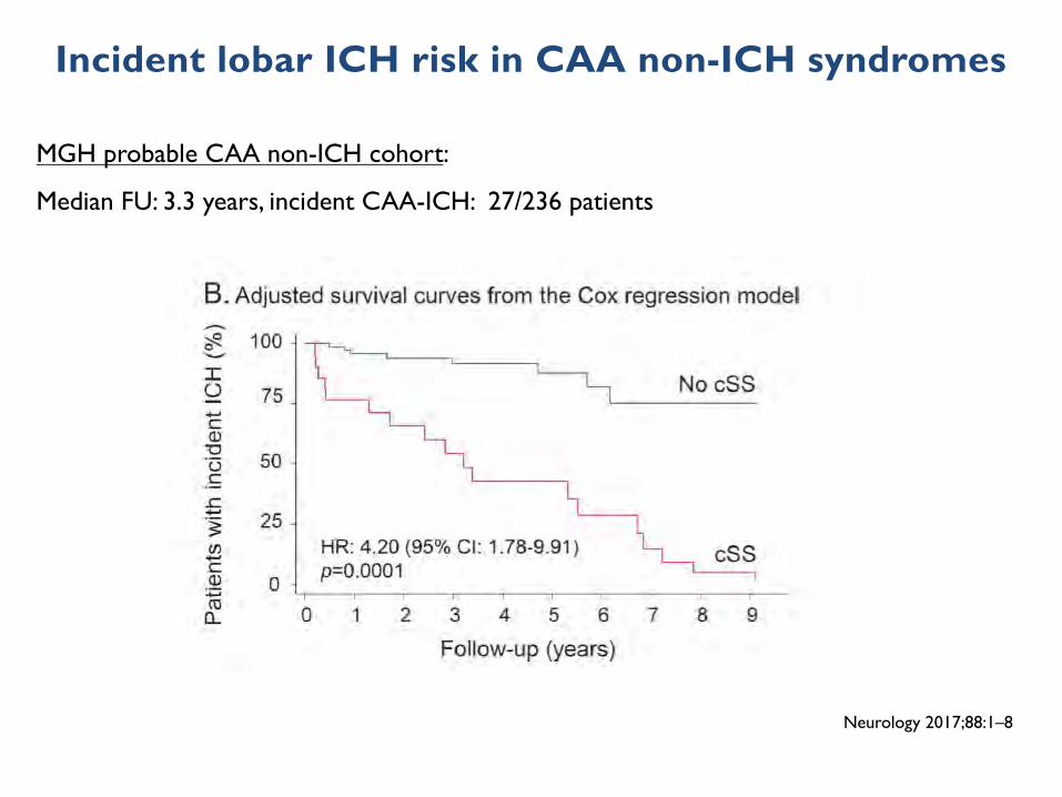
Incident lobar ICH risk in CAA non-ICH syndromes
MGH probable CAA non-ICH cohort:
Median FU: 3.3 years, incident CAA-ICH: 27/236 patients
Neurology 2017;88:1–8
Cumulative rates of new-onset dementia
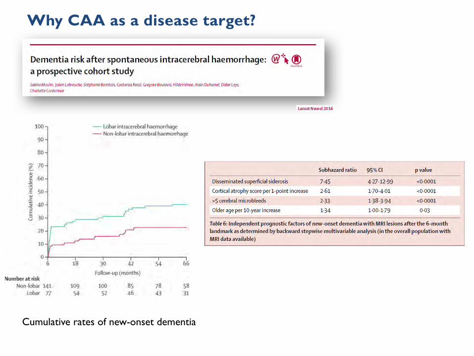
Why CAA as a disease target?
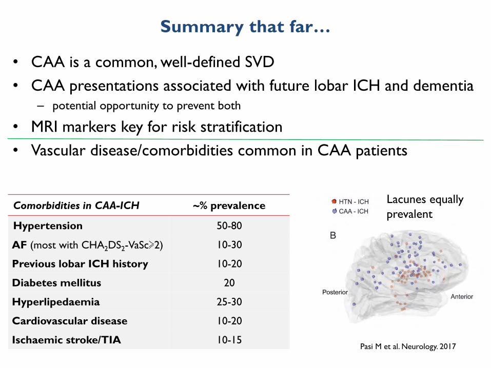
Summary that far…
Comorbidities in CAA-ICH ~% prevalence
Hypertension 50-80
AF (most with CHA2DS2-VaSc≽2) 10-30
Previous lobar ICH history 10-20
Diabetes mellitus 20
Hyperlipedaemia 25-30
Cardiovascular disease 10-20
Ischaemic stroke/TIA 10-15
• CAA is a common, well-defined SVD• CAA presentations associated with future lobar ICH and dementia
– potential opportunity to prevent both
• MRI markers key for risk stratification• Vascular disease/comorbidities common in CAA patients
Pasi M et al. Neurology. 2017
Lacunes equally prevalent
Copyright 2015 American Medical Association. All rights reserved.
bar ICH. Given the limitations of our BP capture strategies, thesefindings are likely to reflect associations between educa-tional achievements and unmeasured BP control via factorssuch as health literacy and lack of access to, or affordabilityof, health care because of socioeconomic status. Although clini-cal trials of aggressive vs conservative BP management in ICHsurvivors should be planned, more proactive management ofBP for ICH survivors according to existing guidelines wouldsubstantially reduce the risk of ICH recurrence (and its asso-ciated toll in terms of mortality and disability).
Our study has several limitations. Because of its single-center observational nature, selection and severity bias maybe reflected in the characteristics of our study population; our
findings will therefore require replication in future studies, aswell as extension to different health care settings. The non-standardized data capture procedures in this study (ie, rely-ing primarily on BP measurements obtained during routine de-livery of care) also represent an important limitation. However,lack of standardization likely introduced additional impreci-sion in the BP exposure data, thus biasing findings toward thenull hypothesis rather than risking generation of false-positive findings. Owing to the observational design of thisstudy, BP management was determined by each patient’s in-dividual physician and did not follow prespecified or stan-dardized protocols. We are therefore limited to describing as-sociations between observed BP control and recurrent ICH,
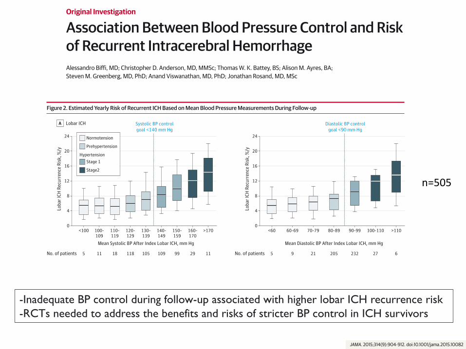
Figure 2. Estimated Yearly Risk of Recurrent ICH Based on Mean Blood Pressure Measurements During Follow-up
24
4
8
12
16
20
0
Loba
r ICH
Rec
urre
nce
Risk
, %/y
Mean Systolic BP After Index Lobar ICH, mm Hg
No. of patients
<100
5
100-109
11
110-119
18
120-129
118
130-139
105
140-149
109
150-159
99
160-170
29
>170
11
Lobar ICHA
Normotension
Prehypertension
HypertensionStage 1
Stage2
Systolic BP controlgoal <140 mm Hg
24
8
12
20
16
4
0
Loba
r ICH
Rec
urre
nce
Risk
, %/y
Mean Diastolic BP After Index Lobar ICH, mm Hg
No. of patients
<60
5
60-69
9
70-79
21
80-89
205
90-99
232
100-110
27
>110
6
Diastolic BP controlgoal <90 mm Hg
15
3
6
9
12
0
Nonl
obar
ICH
Recu
rren
ce R
isk,
%/y
Mean Systolic BP After Index Nonlobar ICH, mm Hg
No. of patients
<100
7
100-109
21
110-119
35
120-129
109
130-139
121
140-149
151
150-159
178
160-170
11
>170
7
Nonlobar ICHB Systolic BP controlgoal <140 mm Hg
15
6
9
12
3
0
Nonl
obar
ICH
Recu
rren
ce R
isk,
%/y
Mean Diastolic BP After Index Nonlobar ICH, mm Hg
No. of patients
<60
13
60-69
22
70-79
33
80-89
225
90-99
330
100-110
12
>110
5
Diastolic BP controlgoal <90 mm Hg
Box upper and lower margins indicate 25th and 75th percentiles of riskdistributions, respectively; heavy horizontal lines in boxes indicate median riskvalues; error bars indicate maximum and minimum estimated risk values in eachdistribution. Vertical lines in blue indicate currently recommended bloodpressure (BP) control goals among survivors of intracerebral hemorrhage (ICH)without diabetes, based on American Heart Association/American StrokeAssociation guidelines for post-ICH secondary prevention (lines are added forillustrative purposes only and have no direct impact on risk estimation results).A, Estimated yearly risk of recurrent lobar ICH based on systolic and diastolic BP
measurements during follow-up. Estimated risk calculated adjusting for otherfactors associated with recurrence of lobar ICH (see main text and eMethods inthe Supplement). B, Estimated yearly risk of recurrent nonlobar ICH based onsystolic and diastolic BP measurements during follow-up. Risk is calculatedassuming mean systolic and diastolic BP measurements as indicated on thehorizontal axes and is expressed as % recurrent rate/y among survivors ofnonlobar ICH. Estimated risk calculated adjusting for other factors associatedwith recurrence of nonlobar ICH (see main text and eTable 2 in theSupplement).
Blood Pressure Control and Recurrent Intracerebral Hemorrhage Original Investigation Research
jama.com (Reprinted) JAMA September 1, 2015 Volume 314, Number 9 911
Copyright 2015 American Medical Association. All rights reserved.
Downloaded From: http://jama.jamanetwork.com/ by a McMaster University User on 09/04/2015
Copyright 2015 American Medical Association. All rights reserved.
Association Between Blood Pressure Control and Riskof Recurrent Intracerebral HemorrhageAlessandro Biffi, MD; Christopher D. Anderson, MD, MMSc; Thomas W. K. Battey, BS; Alison M. Ayres, BA;Steven M. Greenberg, MD, PhD; Anand Viswanathan, MD, PhD; Jonathan Rosand, MD, MSc
IMPORTANCE Intracerebral hemorrhage (ICH) is the most severe form of stroke. Survivors areat high risk of recurrence, death, and worsening functional disability.
OBJECTIVE To investigate the association between blood pressure (BP) after index ICH andrisk of recurrent ICH.
DESIGN, SETTING, AND PARTICIPANTS Single-site, tertiary care referral center observationalstudy of 1145 of 2197 consecutive patients with ICH presenting from July 1994 to December2013. A total of 1145 patients with ICH survived at least 90 days and were followed upthrough December 2013 (median follow-up of 36.8 months [minimum, 9.8 months]).
EXPOSURES Blood pressure measurements at 3, 6, 9, and 12 months, and every 6 monthsthereafter, obtained from medical personnel (inpatient hospital or outpatient clinic medical ornursing staff) or via patient self-report. Exposure was characterized in 3 ways: (1) recordedsystolic and diastolic measurements; (2) classification as adequate or inadequate BP controlbased on American Heart Association/American Stroke Association recommendations; and(3) stage of hypertension based on Joint National Committee on Prevention, Detection,Evaluation, and Treatment of High Blood Pressure 7 criteria.
MAIN OUTCOMES AND MEASURES Recurrent ICH and its location within the brain(lobar vs nonlobar).
RESULTS There were 102 recurrent ICH events among 505 survivors of lobar ICH and 44recurrent ICH events among 640 survivors of nonlobar ICH. During follow-up adequate BPcontrol was achieved on at least 1 measurement by 625 patients (54.6% of total [range,49.2%-58.7%]) and consistently (ie, at all available time points) by 495 patients (43.2% oftotal [range, 34.5%-51.0%]). The event rate for lobar ICH was 84 per 1000 person-yearsamong patients with inadequate BP control compared with 49 per 1000 person-years amongpatients with adequate BP control. For nonlobar ICH the event rate was 52 per 1000person-years with inadequate BP control compared with 27 per 1000 person-years forpatients with adequate BP control. In analyses modeling BP control as a time-varying variable,inadequate BP control was associated with higher risk of recurrence of both lobar ICH (hazardratio [HR], 3.53 [95% CI, 1.65-7.54]) and nonlobar ICH (HR, 4.23 [95% CI, 1.02-17.52]). SystolicBP during follow-up was associated with increased risk of both lobar ICH recurrence (HR, 1.33per 10-mm Hg increase [95% CI, 1.02-1.76]) and nonlobar ICH recurrence (HR, 1.54 [95% CI,1.03-2.30]). Diastolic BP was associated with increased risk of nonlobar ICH recurrence(HR, 1.21 per 10-mm Hg increase [95% CI, 1.01-1.47]) but not with lobar ICH recurrence(HR, 1.36 [95% CI, 0.90-2.10]).
CONCLUSIONS AND RELEVANCE In this observational single-center cohort study of ICHsurvivors, reported BP measurements suggesting inadequate BP control during follow-upwere associated with higher risk of both lobar and nonlobar ICH recurrence. These datasuggest that randomized clinical trials are needed to address the benefits and risks of stricterBP control in ICH survivors.
JAMA. 2015;314(9):904-912. doi:10.1001/jama.2015.10082
Supplemental content atjama.com
CME Quiz atjamanetworkcme.com andCME Questions page 945
Author Affiliations: Center forHuman Genetic Research,Massachusetts General Hospital,Boston (Biffi, Anderson, Battey,Rosand); J. Philip Kistler StrokeResearch Center, MassachusettsGeneral Hospital, Boston (Biffi,Anderson, Battey, Ayres, Greenberg,Viswanathan, Rosand); Division ofNeurocritical Care and EmergencyNeurology, Department of Neurology,Massachusetts General Hospital,Boston (Biffi, Anderson, Battey,Greenberg, Viswanathan, Rosand);Program in Medical and PopulationGenetics, Broad Institute, Cambridge,Massachusetts (Biffi, Anderson,Battey, Rosand).
Corresponding Author: JonathanRosand, MD, MSc, Center for HumanGenetic Research, MassachusettsGeneral Hospital, 185 Cambridge St,CPZN-6818, Boston, MA 02114([email protected]).
Research
Original Investigation
904 (Reprinted) jama.com
Copyright 2015 American Medical Association. All rights reserved.
Downloaded From: http://jama.jamanetwork.com/ by a McMaster University User on 09/04/2015
n=505
Copyright 2015 American Medical Association. All rights reserved.
Association Between Blood Pressure Control and Riskof Recurrent Intracerebral HemorrhageAlessandro Biffi, MD; Christopher D. Anderson, MD, MMSc; Thomas W. K. Battey, BS; Alison M. Ayres, BA;Steven M. Greenberg, MD, PhD; Anand Viswanathan, MD, PhD; Jonathan Rosand, MD, MSc
IMPORTANCE Intracerebral hemorrhage (ICH) is the most severe form of stroke. Survivors areat high risk of recurrence, death, and worsening functional disability.
OBJECTIVE To investigate the association between blood pressure (BP) after index ICH andrisk of recurrent ICH.
DESIGN, SETTING, AND PARTICIPANTS Single-site, tertiary care referral center observationalstudy of 1145 of 2197 consecutive patients with ICH presenting from July 1994 to December2013. A total of 1145 patients with ICH survived at least 90 days and were followed upthrough December 2013 (median follow-up of 36.8 months [minimum, 9.8 months]).
EXPOSURES Blood pressure measurements at 3, 6, 9, and 12 months, and every 6 monthsthereafter, obtained from medical personnel (inpatient hospital or outpatient clinic medical ornursing staff) or via patient self-report. Exposure was characterized in 3 ways: (1) recordedsystolic and diastolic measurements; (2) classification as adequate or inadequate BP controlbased on American Heart Association/American Stroke Association recommendations; and(3) stage of hypertension based on Joint National Committee on Prevention, Detection,Evaluation, and Treatment of High Blood Pressure 7 criteria.
MAIN OUTCOMES AND MEASURES Recurrent ICH and its location within the brain(lobar vs nonlobar).
RESULTS There were 102 recurrent ICH events among 505 survivors of lobar ICH and 44recurrent ICH events among 640 survivors of nonlobar ICH. During follow-up adequate BPcontrol was achieved on at least 1 measurement by 625 patients (54.6% of total [range,49.2%-58.7%]) and consistently (ie, at all available time points) by 495 patients (43.2% oftotal [range, 34.5%-51.0%]). The event rate for lobar ICH was 84 per 1000 person-yearsamong patients with inadequate BP control compared with 49 per 1000 person-years amongpatients with adequate BP control. For nonlobar ICH the event rate was 52 per 1000person-years with inadequate BP control compared with 27 per 1000 person-years forpatients with adequate BP control. In analyses modeling BP control as a time-varying variable,inadequate BP control was associated with higher risk of recurrence of both lobar ICH (hazardratio [HR], 3.53 [95% CI, 1.65-7.54]) and nonlobar ICH (HR, 4.23 [95% CI, 1.02-17.52]). SystolicBP during follow-up was associated with increased risk of both lobar ICH recurrence (HR, 1.33per 10-mm Hg increase [95% CI, 1.02-1.76]) and nonlobar ICH recurrence (HR, 1.54 [95% CI,1.03-2.30]). Diastolic BP was associated with increased risk of nonlobar ICH recurrence(HR, 1.21 per 10-mm Hg increase [95% CI, 1.01-1.47]) but not with lobar ICH recurrence(HR, 1.36 [95% CI, 0.90-2.10]).
CONCLUSIONS AND RELEVANCE In this observational single-center cohort study of ICHsurvivors, reported BP measurements suggesting inadequate BP control during follow-upwere associated with higher risk of both lobar and nonlobar ICH recurrence. These datasuggest that randomized clinical trials are needed to address the benefits and risks of stricterBP control in ICH survivors.
JAMA. 2015;314(9):904-912. doi:10.1001/jama.2015.10082
Supplemental content atjama.com
CME Quiz atjamanetworkcme.com andCME Questions page 945
Author Affiliations: Center forHuman Genetic Research,Massachusetts General Hospital,Boston (Biffi, Anderson, Battey,Rosand); J. Philip Kistler StrokeResearch Center, MassachusettsGeneral Hospital, Boston (Biffi,Anderson, Battey, Ayres, Greenberg,Viswanathan, Rosand); Division ofNeurocritical Care and EmergencyNeurology, Department of Neurology,Massachusetts General Hospital,Boston (Biffi, Anderson, Battey,Greenberg, Viswanathan, Rosand);Program in Medical and PopulationGenetics, Broad Institute, Cambridge,Massachusetts (Biffi, Anderson,Battey, Rosand).
Corresponding Author: JonathanRosand, MD, MSc, Center for HumanGenetic Research, MassachusettsGeneral Hospital, 185 Cambridge St,CPZN-6818, Boston, MA 02114([email protected]).
Research
Original Investigation
904 (Reprinted) jama.com
Copyright 2015 American Medical Association. All rights reserved.
Downloaded From: http://jama.jamanetwork.com/ by a McMaster University User on 09/04/2015
-Inadequate BP control during follow-up associated with higher lobar ICH recurrence risk-RCTs needed to address the benefits and risks of stricter BP control in ICH survivors
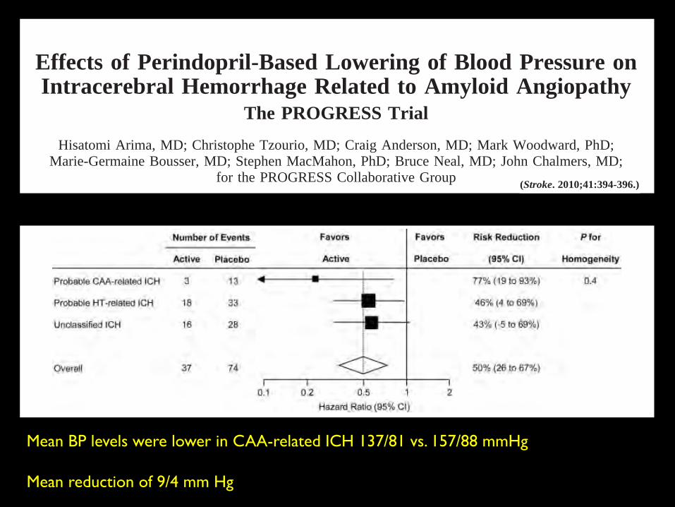
Effects of Perindopril-Based Lowering of Blood Pressure onIntracerebral Hemorrhage Related to Amyloid Angiopathy
The PROGRESS Trial
Hisatomi Arima, MD; Christophe Tzourio, MD; Craig Anderson, MD; Mark Woodward, PhD;Marie-Germaine Bousser, MD; Stephen MacMahon, PhD; Bruce Neal, MD; John Chalmers, MD;
for the PROGRESS Collaborative Group
Background and Purpose—Patients with cerebral amyloid angiopathy (CAA) are at high risk for intracerebral hemorrhage(ICH), but no effective prevention strategies have been established. The objective is to determine whether lowering ofblood pressure (BP) provides protection for this high-risk patient group.
Methods—This study is a subsidiary analysis of the PROGRESS trial—a randomized, placebo-controlled trial thatestablished the beneficial effects of BP lowering in patients with cerebrovascular disease; 6105 patients wererandomly assigned to either active treatment (perindopril for all participants plus indapamide for those with neitheran indication for nor a contraindication to a diuretic) or matching placebo. Outcomes were probable CAA-relatedICH as defined by the Boston criteria, probable hypertension-related ICH, and unclassified ICH.
Results—Over a mean follow-up of 3.9 years, 16 probable CAA-related ICH, 51 probable hypertension-related ICH, and44 unclassified ICH occurred. Active treatment reduced the risk of CAA-related ICH by 77% (95% CI, 19%–93%), thatof hypertension-related ICH by 46% (95% CI, 4%–69%), and that of unclassified ICH by 43% (95% CI, !5%–69%).There was no evidence of differences in the magnitude of the effects of treatment among different types of ICH (Phomogeneity"0.4).
Conclusions—BP-lowering treatment is likely to provide protection against all types of ICH. (Stroke. 2010;41:394-396.)
Key Words: blood pressure ! cerebral amyloid angiopathy ! intracerebral hemorrhage! randomized controlled trials
Intracerebral hemorrhage (ICH) is estimated to affect #1million people worldwide each year, most of whom
either die or are left seriously disabled.1 The most commontype of ICH is hypertension (HT)-related ICH, which isrelated to degenerative changes in the small penetratingarteries of the deep part of the brain.2 The other type ofICH is associated with cerebral amyloid angiopathy(CAA), which is defined by the deposition of congophilicmaterial, preferentially in vessels of the cortex and lepto-meninges.3 CAA-related ICH is characterized by multipleoccurrence of ICH over time, a cortical localization of thehematoma, and an increasing incidence with age.4 Despitethis high rate of ICH, no effective prevention strategieshave been established. The objective of the present anal-ysis is to determine whether blood pressure (BP) loweringprovides protection against probable CAA-related ICH.
Materials and Methods
Study DesignThe PROGRESS trial was a randomized, placebo-controlled trialthat investigated the effects of BP lowering among patients withcerebrovascular disease.5 The design of PROGRESS has beendescribed in detail elsewhere.5 Briefly, 6105 participants withcerebrovascular disease who had no clear indication for, or contra-indication to, an angiotensin-converting enzyme inhibitor wererandomly assigned to active treatment (2–4 mg perindopril for allparticipants plus 2–2.5 mg indapamide for those with neither anindication for nor a contraindication to a diuretic) or matchingplacebo. The institutional ethics committee of each collaboratingcenter approved the trial, and all participants provided writteninformed consent.
OutcomesFor patients with possible stroke, a detailed history was taken andneurological and morphological (CT/MRI) examinations were con-
Received July 28, 2009; final revision received October 13, 2009; accepted November 6, 2009.From The George Institute for International Health, the University of Sydney and the Royal Prince Alfred Hospital (H.A., C.A., S.M., B.N., J.C.),
Sydney, Australia; INSERM U708 (C.T.) and Department of Neurology, Hopital Lariboisiere (C.T., M.-G.B.), Paris, France; Mount Sinai School ofMedicine (M.W.), New York, NY.
For a full list of investigators, see reference 5.Correspondence to Professor John Chalmers, c/o PROGRESS Collaborative Group, The George Institute for International Health, The University
of Sydney and the Royal Prince Alfred Hospital, PO Box M201, Missenden Road, NSW 2050 Australia. E-mail [email protected]© 2010 American Heart Association, Inc.
Stroke is available at http://stroke.ahajournals.org DOI: 10.1161/STROKEAHA.109.563932
394 by guest on December 9, 2014http://stroke.ahajournals.org/Downloaded from
ducted. Stroke was defined according to standard criteria6 andsubclassified into ICH (ICD-9 code 431) or ischemic stroke (ICD-9codes 433, 434).5 The diagnosis of ICH and exclusion of secondarycauses were confirmed using CT/MRI. ICH was classified into lobarhemorrhage or nonlobar hemorrhage(basal ganglia, thalamus, brainstem, or cerebellum) according to the originated location based oninvestigation reports and supporting documentation (medical chartsand CT/MRI reports) independently by 2 investigators (! coeffi-cient!0.84), as described previously.7 Probable CAA-related ICHwas defined according to the Boston Criteria8 as follows: lobarhemorrhage with evidence of multiple ICH (recurrence of ICHamong patients with preexisting ICH or incidence of multiple ICHduring follow-up) and age at onset 55 years or older. ProbableHT-related ICH was defined as follows: ICH with no evidence ofmultiple ICH and presence of HT at baseline (BP "140/90 mm Hgor use of antihypertensive agents). Only the first ICH event duringfollow-up was included in the analysis.
Statistical AnalysisThe effects of randomized treatment on events were calculated usingunivariate Cox proportional hazards models, according to the prin-ciple of intention-to-treat. The constancy of treatment effects wastested using a #2 test of homogeneity.
ResultsThere were no important differences in characteristics be-tween randomized groups (Table).5 Over a mean follow-up of3.9 years, 16 probable CAA-related ICH, 51 probable HT-related ICH, and 44 unclassified ICH occurred. Subjects withICH were more likely to be Asian (65%) than those withoutICH (38%). Mean BP levels at baseline were slightly higheramong patient with ICH (150/88 mm Hg) than among thosewithout (147/86 mm Hg). Frequency of preexisting ICH washigher among patients with ICH (46%) than among thosewithout (10%). Mean BP levels at baseline were lower amongpatients with CAA-related ICH (137/81 mm Hg) than amongthose with HT-related ICH (157/88 mm Hg). Fifty-six per-cent of patients with CAA-related ICH had HT at baselineand 13% of those had HT newly diagnosed during follow-up.Whereas 88% of patients with CAA-related ICH had preex-isting ICH, 84% of those with HT-related ICH had preexist-ing ischemic stroke.
During follow-up, mean BP difference between random-ized groups was 9/4 mm Hg. Active treatment reduced therisk of CAA-related ICH by 77% (95% CI, 19%–93%), thatof HT-related ICH by 46% (95% CI, 4%–69%), and that ofunclassified ICH by 43% (95% CI, "5%–69%; Figure).There was no evidence of differences in the magnitude of theeffects of treatment among different types of ICH (P homo-geneity!0.4). There were also comparable benefits fromactive treatment on CAA-related ICH with and withoutbaseline HT (P homogeneity!0.4) or baseline and newlydiagnosed HT (P homogeneity!0.2).
DiscussionThe main results from the PROGRESS trial showed thatroutine BP-lowering treatment reduced the risk of ICH by50% among patients with cerebrovascular disease.5 Theanalyses reported here expand on this earlier report andsuggest that BP lowering is likely to reduce the risks ofCAA-related ICH and other forms of ICH.
Few studies have investigated the effects of BP on the risksof CAA-related ICH. A study of autopsy cases has demon-strated that definite CAA patients with ICH were morefrequently hypertensive (50%) than those without ICH(23%).9 This finding suggests that HT is likely to have animportant role in development of ICH among patients withCAA and is consistent with our hypothesis that BP loweringhas potential to reduce ICH among patients with CAA.
Table. Baseline Characteristics of Randomized Participants
Active, n!3051 Placebo, n!3054
Demographic
Mean age, years (SD) 64 (10) 64 (10)
Women, % 30 30
Asian, % 39 39
Cerebrovascular disease history, %
Ischemic stroke 71 71
ICH 11 11
Stroke of unknown type 4 5
Transient ischemic attack 22 22
Other medical history, %
Current smoker 20 20
Diabetes 13 12
Coronary heart disease 16 16
BP
Mean systolic BP, mm Hg (SD) 147 (19) 147 (19)
Mean diastolic BP, mm Hg (SD) 86 (11) 86 (11)
Reprinted from Randomised trial of a perindopril-based blood pressure loweringregimen among 6105 individuals with previous stroke or transient ischaemicattack. Lancet. 2001;358:1033–1041, with permission from Elsevier.
Figure. Effects of randomized treatment on the risks of different types of ICH. Solid boxes indicate estimates of treatment effect on therisks of ICH types; horizontal lines, 95% CI; diamond, the estimate and 95% CI for overall effect. Areas of the boxes are proportional tothe event number.
Arima et al BP Lowering and ICH Related to Amyloid Angiopathy 395
by guest on December 9, 2014http://stroke.ahajournals.org/Downloaded from
Mean BP levels were lower in CAA-related ICH 137/81 vs. 157/88 mmHg
Mean reduction of 9/4 mm Hg
AF after CAA-ICH: A hot dilemma
§ Start oral anticoagulation (OAC)?o warfarin vs. DOACs?o vs. no antithrombotico vs. antiplateleto vs. left atrial appendage occlusion
§ Stratification according to CAA MRI signatures?o cSS/CMBs
§ Stratification according to CHA2DS2-VaSc score?
§ No data from RCTs of OAC after ICH
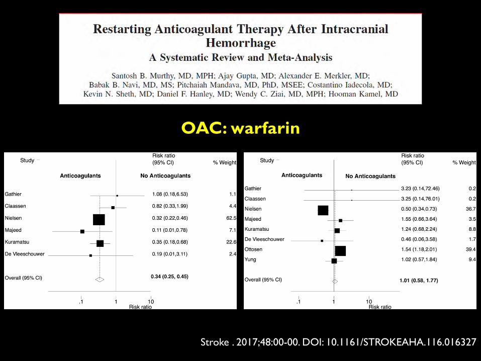
Stroke . 2017;48:00-00. DOI: 10.1161/STROKEAHA.116.016327
OAC: warfarin
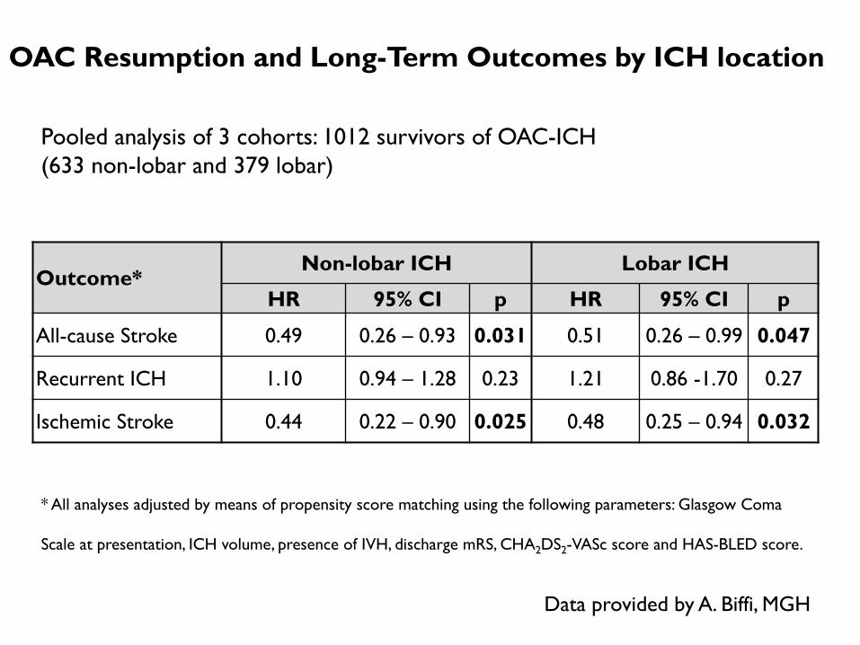
OAC Resumption and Long-Term Outcomes by ICH location
Outcome*Non-lobar ICH Lobar ICH
HR 95% CI p HR 95% CI p
All-cause Stroke 0.49 0.26 – 0.93 0.031 0.51 0.26 – 0.99 0.047
Recurrent ICH 1.10 0.94 – 1.28 0.23 1.21 0.86 -1.70 0.27
Ischemic Stroke 0.44 0.22 – 0.90 0.025 0.48 0.25 – 0.94 0.032
* All analyses adjusted by means of propensity score matching using the following parameters: Glasgow Coma
Scale at presentation, ICH volume, presence of IVH, discharge mRS, CHA2DS2-VASc score and HAS-BLED score.
Pooled analysis of 3 cohorts: 1012 survivors of OAC-ICH (633 non-lobar and 379 lobar)
Data provided by A. Biffi, MGH
Articles
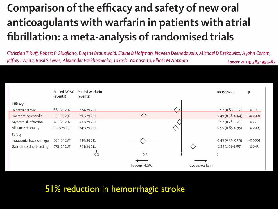
www.thelancet.com Vol 383 March 15, 2014 955
Comparison of the effi cacy and safety of new oral anticoagulants with warfarin in patients with atrial fi brillation: a meta-analysis of randomised trialsChristian T Ruff , Robert P Giugliano, Eugene Braunwald, Elaine B Hoff man, Naveen Deenadayalu, Michael D Ezekowitz, A John Camm, Jeff rey I Weitz, Basil S Lewis, Alexander Parkhomenko, Takeshi Yamashita, Elliott M Antman
SummaryBackground Four new oral anticoagulants compare favourably with warfarin for stroke prevention in patients with atrial fi brillation; however, the balance between effi cacy and safety in subgroups needs better defi nition. We aimed to assess the relative benefi t of new oral anticoagulants in key subgroups, and the eff ects on important secondary outcomes.
Methods We searched Medline from Jan 1, 2009, to Nov 19, 2013, limiting searches to phase 3, randomised trials of patients with atrial fi brillation who were randomised to receive new oral anticoagulants or warfarin, and trials in which both effi cacy and safety outcomes were reported. We did a prespecifi ed meta-analysis of all 71 683 participants included in the RE-LY, ROCKET AF, ARISTOTLE, and ENGAGE AF–TIMI 48 trials. The main outcomes were stroke and systemic embolic events, ischaemic stroke, haemorrhagic stroke, all-cause mortality, myocardial infarction, major bleeding, intracranial haemorrhage, and gastrointestinal bleeding. We calculated relative risks (RRs) and 95% CIs for each outcome. We did subgroup analyses to assess whether diff erences in patient and trial characteristics aff ected outcomes. We used a random-eff ects model to compare pooled outcomes and tested for heterogeneity.
Findings 42 411 participants received a new oral anticoagulant and 29 272 participants received warfarin. New oral anticoagulants signifi cantly reduced stroke or systemic embolic events by 19% compared with warfarin (RR 0·81, 95% CI 0·73–0·91; p<0·0001), mainly driven by a reduction in haemorrhagic stroke (0·49, 0·38–0·64; p<0·0001). New oral anticoagulants also signifi cantly reduced all-cause mortality (0·90, 0·85–0·95; p=0·0003) and intracranial haemorrhage (0·48, 0·39–0·59; p<0·0001), but increased gastrointestinal bleeding (1·25, 1·01–1·55; p=0·04). We noted no heterogeneity for stroke or systemic embolic events in important subgroups, but there was a greater relative reduction in major bleeding with new oral anticoagulants when the centre-based time in therapeutic range was less than 66% than when it was 66% or more (0·69, 0·59–0·81 vs 0·93, 0·76–1·13; p for interaction 0·022). Low-dose new oral anticoagulant regimens showed similar overall reductions in stroke or systemic embolic events to warfarin (1·03, 0·84–1·27; p=0·74), and a more favourable bleeding profi le (0·65, 0·43–1·00; p=0·05), but signifi cantly more ischaemic strokes (1·28, 1·02–1·60; p=0·045).
Interpretation This meta-analysis is the fi rst to include data for all four new oral anticoagulants studied in the pivotal phase 3 clinical trials for stroke prevention or systemic embolic events in patients with atrial fi brillation. New oral anticoagulants had a favourable risk–benefi t profi le, with signifi cant reductions in stroke, intracranial haemorrhage, and mortality, and with similar major bleeding as for warfarin, but increased gastrointestinal bleeding. The relative effi cacy and safety of new oral anticoagulants was consistent across a wide range of patients. Our fi ndings off er clinicians a more comprehensive picture of the new oral anticoagulants as a therapeutic option to reduce the risk of stroke in this patient population.
Funding None.
IntroductionAtrial fi brillation, the most common sustained cardiac arrhythmia, predisposes patients to an increased risk of embolic stroke and has a higher mortality than sinus rhythm.1,2 Until 2009, warfarin and other vitamin K antagonists were the only class of oral anticoagulants available. Although these drugs are highly eff ective in prevention of thromboembolism, their use is limited by a narrow therapeutic index that necessitates frequent monitoring and dose adjustments resulting in substantial
risk and inconvenience. This limitation has translated into poor patient adherence and probably contributes to the systematic underuse of vitamin K antagonists for stroke prevention.3,4
Several new oral anticoagulants have been developed that dose-dependently inhibit thrombin or activated factor X (factor Xa) and off er potential advantages over vitamin K antagonists, such as rapid onset and off set of action, absence of an eff ect of dietary vitamin K intake on their activity, and fewer drug interactions. The
Lancet 2014; 383: 955–62
Published OnlineDecember 4, 2013http://dx.doi.org/10.1016/S0140-6736(13)62343-0
See Comment page 931
Brigham and Women’s Hospital and Harvard Medical School, Boston, MA, USA (C T Ruff MD, R P Giugliano MD, Prof E Braunwald MD, E B Hoff man PhD, N Deenadayalu MPH, Prof E M Antman MD); Jeff erson Medical College, Philadelphia, PA, and Cardiovascular Research Foundation, New York, NY, USA (Prof M D Ezekowitz MBChB); St George’s University, London, UK (Prof A J Camm MD); McMaster University and the Thrombosis and Atherosclerosis Research Institute, Hamilton, ON, Canada (Prof J I Wetiz MD); Lady Davis Carmel Medical Center, Haifa, Israel (Prof B S Lewis MD); Institute of Cardiology, Kiev, Ukraine (Prof A Parkhomenko MD); and The Cardiovascular Institute, Tokyo, Japan (Prof T Yamashita MD)
Correspondence to:Dr Christian T Ruff , Thrombolysis in Myocardial Infarction (TIMI) Study Group, 350 Longwood Avenue, 1st Floor Offi ces, Boston, MA 02115, USAcruff @partners.org
Articles
958 www.thelancet.com Vol 383 March 15, 2014
therapeutic range in patients in the warfarin groups ranged from 58% to 68% (table).
Figure 1 shows the comparative effi cacy of high-dose of new oral anticoagulants and warfarin. Allocation to a new oral anticoagulant signifi cantly reduced the com-posite of stroke or systemic embolic events by 19% compared with warfarin (fi gure 1). The benefi t was mainly driven by a large reduction in haemorrhagic stroke (fi gure 2). New oral anticoagulants were also asso ciated with a signifi cant reduction in all-cause mortality (fi gure 2). The drugs were similar to warfarin in the prevention of ischaemic stroke and myocardial infarction (fi gure 2).
Randomisation to a high-dose new oral anticoagulant was associated with a 14% non-signifi cant reduction in major bleeding (fi gure 3). In line with the reduction in haemorrhagic stroke, a substantial reduction in intra-cranial haemorrhage was observed, which included haemorrhagic stroke, and subdural, epidural, and sub-arachnoid bleeding (fi gure 2). New oral anticoagulants were, however, associated with increased gastrointestinal bleeding (fi gure 2).
The benefi t of new oral anticoagulants compared with warfarin in reducing stroke or systemic embolic events was consistent across all subgroups examined (fi gure 4). The safety of new oral anticoagulants compared with
warfarin was generally consistent for the reduction of major bleeding across subgroups, with the exception of a signifi cant interaction for centre-based time in thera-peutic range (fi gure 4). We noted a greater relative reduction in bleeding with new oral anticoagulants at centres that achieved a centre-based time in therapeutic range of less than 66% than at those achieving a time in therapeutic range of 66% or more (fi gure 4).
The low-dose new oral anticoagulant regimens had similar effi cacy to warfarin for the composite of stroke or systemic embolic events (appendix). When diff eren-tiated by stroke type, the low-dose regimens were asso-ciated with an increase in ischaemic stroke compared with warfarin, which was balanced by a large decrease in haemorrhagic stroke (appendix). Similar to the higher-dose regimens, the low doses showed a signifi -cant reduc tion in all-cause mortality (appendix). Signifi -cantly more myocardial infarctions were reported with the low-dose regimens than with warfarin (appen dix). The low-dose regimens were associated with a non-signifi cant reduction in major bleeding, but with a signifi cant reduction in intracranial haemor rhage. Gastro intestinal bleeding was similar between low-dose new oral anti coagulants and warfarin (appendix).
A meta-analysis of only the factor Xa inhibitors , with removal of dabigatran, showed similar results to the
Figure 2: Secondary effi cacy and safety outcomesData are n/N, unless otherwise indicated. Heterogeneity: ischaemic stroke I²=32%, p=0·22; haemorrhagic stroke I²=34%, p=0·21; myocardial infarction I²=48%, p=0·13; all-cause mortality I²=0%, p=0·81; intracranial haemorrhage I²=32%, p=0·22; gastrointestinal bleeding I²=74%, p=0·009. NOAC=new oral anticoagulant. RR=risk ratio.
Figure 3: Major bleedingData are n/N, unless otherwise indicated. Heterogeneity: I²=83%; p=0·001. NOAC=new oral anticoagulant. RR=risk ratio. *Dabigatran 150 mg twice daily. †Rivaroxaban 20 mg once daily. ‡Apixaban 5 mg twice daily. §Edoxaban 60 mg once daily.
RR (95% CI) pPooled NOAC (events)
Pooled warfarin (events)
EfficacyIschaemic strokeHaemorrhagic strokeMyocardial infarctionAll-cause mortality
665/29 292 130/29 292 413/29 292 2022/29 292
724/29 221 263/29 221 432/29 221 2245/29 221
0·92 (0·83–1·02) 0·10 0·49 (0·38–0·64) <0·0001 0·97 (0·78–1·20) 0·77 0·90 (0·85–0·95) 0·0003
SafetyIntracranial haemorrhageGastrointestinal bleeding
204/29 287 751/29 287
425/29 211 591/29 211
0·48 (0·39–0·59) <0·0001 1·25 (1·01–1·55) 0·043
10·2 20·5
Favours warfarinFavours NOAC
RR (95% CI) pNOAC (events) Warfarin (events)
RE-LY5* ROCKET AF6† ARISTOTLE7‡ ENGAGE AF–TIMI 488§ Combined (random)
375/6076 395/7111 327/9088 444/7012 1 541/29 287
397/6022 386/7125 462/9052 557/7012 1 802/29 211
0·94 (0·82–1·07) 0·34 1·03 (0·90–1·18) 0·72 0·71 (0·61–0·81) <0·0001 0·80 (0·71–0·90) 0·0002 0·86 (0·73–1·00) 0·06
1·00·5 2·0
Favours warfarinFavours NOAC
See Online for appendix
51% reduction in hemorrhagic stroke
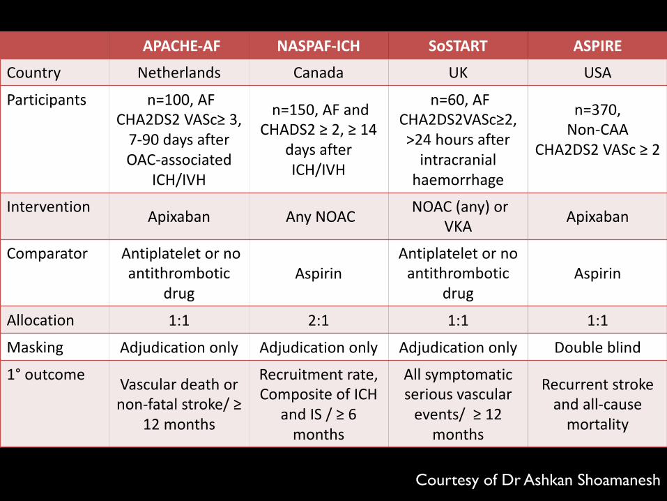
APACHE-AF NASPAF-ICH SoSTART ASPIRECountry Netherlands Canada UK USA
Participants n=100,AFCHA2DS2VASc≥3,7-90daysafterOAC-associated
ICH/IVH
n=150,AFandCHADS2≥2,≥14
daysafterICH/IVH
n=60,AFCHA2DS2VASc≥2,>24hoursafterintracranialhaemorrhage
n=370,Non-CAA
CHA2DS2VASc ≥2
Intervention Apixaban AnyNOAC NOAC(any)orVKA Apixaban
Comparator Antiplateletornoantithrombotic
drugAspirin
Antiplateletornoantithrombotic
drugAspirin
Allocation 1:1 2:1 1:1 1:1
Masking Adjudicationonly Adjudicationonly Adjudicationonly Doubleblind
1° outcome Vascular deathornon-fatalstroke/ ≥
12months
Recruitment rate,CompositeofICH
andIS/≥6months
Allsymptomaticseriousvascularevents/≥12
months
Recurrent strokeandall-causemortality
Courtesy of Dr Ashkan Shoamanesh
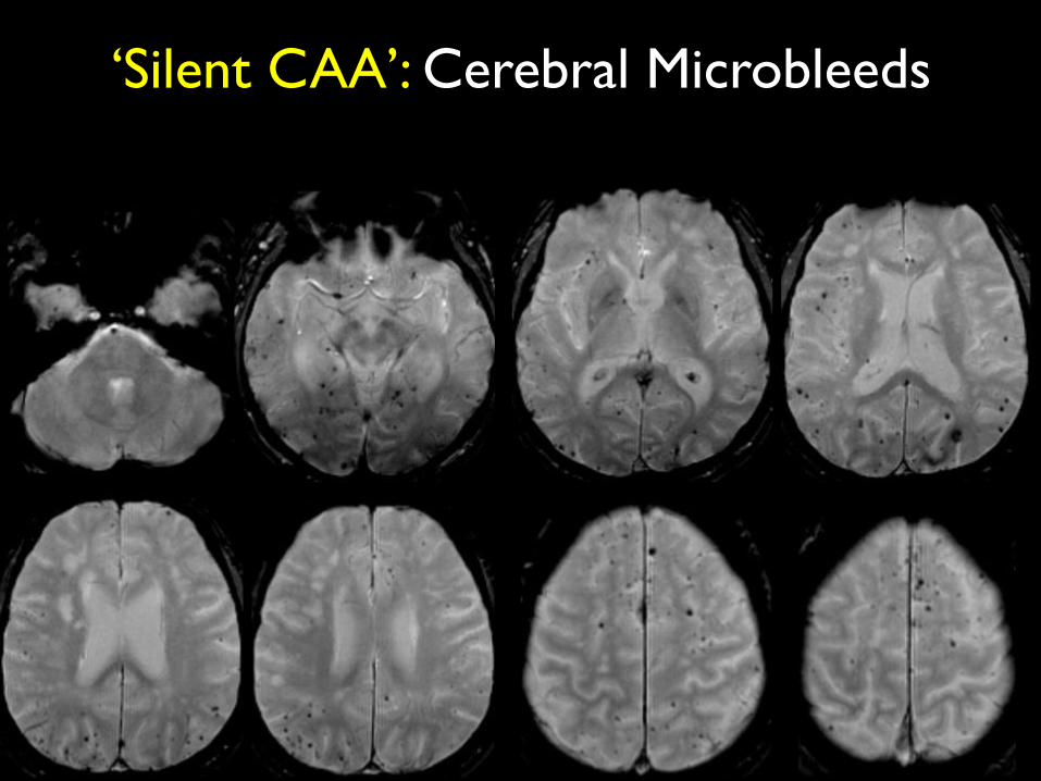
‘Silent CAA’: Cerebral Microbleeds
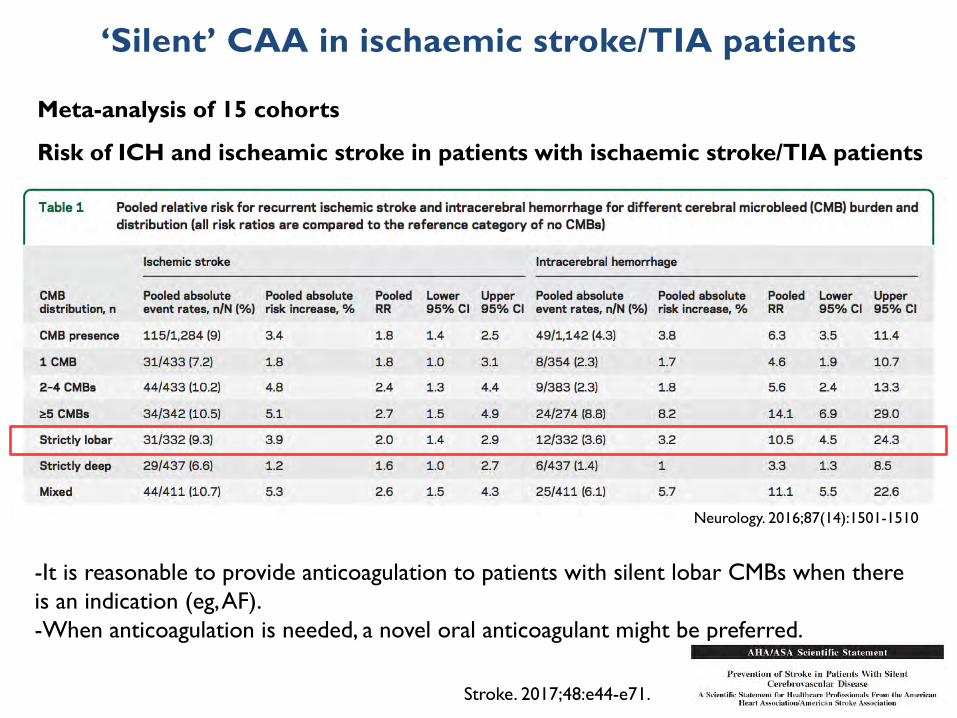
‘Silent’ CAA in ischaemic stroke/TIA patients
Meta-analysis of 15 cohorts
Risk of ICH and ischeamic stroke in patients with ischaemic stroke/TIA patients
Neurology. 2016;87(14):1501-1510
-It is reasonable to provide anticoagulation to patients with silent lobar CMBs when there is an indication (eg, AF).-When anticoagulation is needed, a novel oral anticoagulant might be preferred.
Stroke. 2017;48:e44-e71.
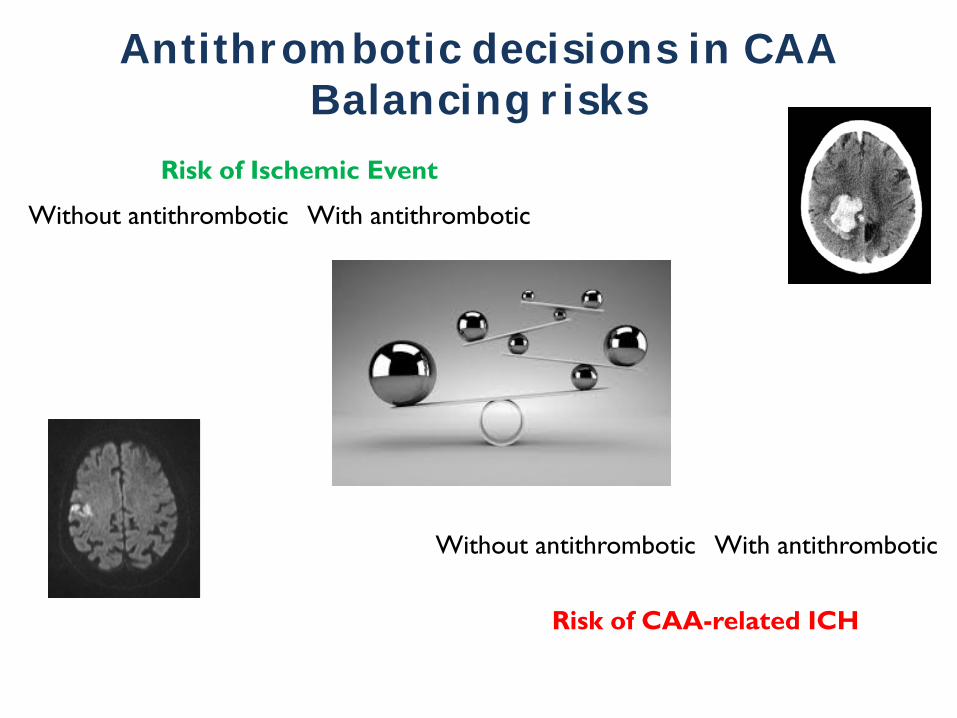
Risk of Ischemic Event
Without antithrombotic With antithrombotic
Risk of CAA-related ICH
Without antithrombotic With antithrombotic
Antithrombotic decisions in CAABalancing risks
§ CAA/CMBs literature over-emphasises ICH risk§ Data on CAA come from observational studies vs. RCTs for OAC§ Don’t mistake statistical certainty for size of effect
§ Absolute risks (%/year) for future ICH and ischaemic stroke instead§ Conflating overall risk (ischemic stroke vs. ICH) with risk reductions• Instead assess risk reduction for cardiac/cerebrovascular events
with antithrombotics vs. ICH risk increase
§ Future ICH risk is not uniform - Clinical context: • CAA-related syndrome, CAA-ICH vs. non-ICH CAA, silent CAA
§ Haemorrhagic CAA signatures: cSS
§ Consider the severity of the consequences• ICH has higher mortality than ischemic stroke
Antithrombotic decisions in CAASome basic principles – Avoid fuzzy logic
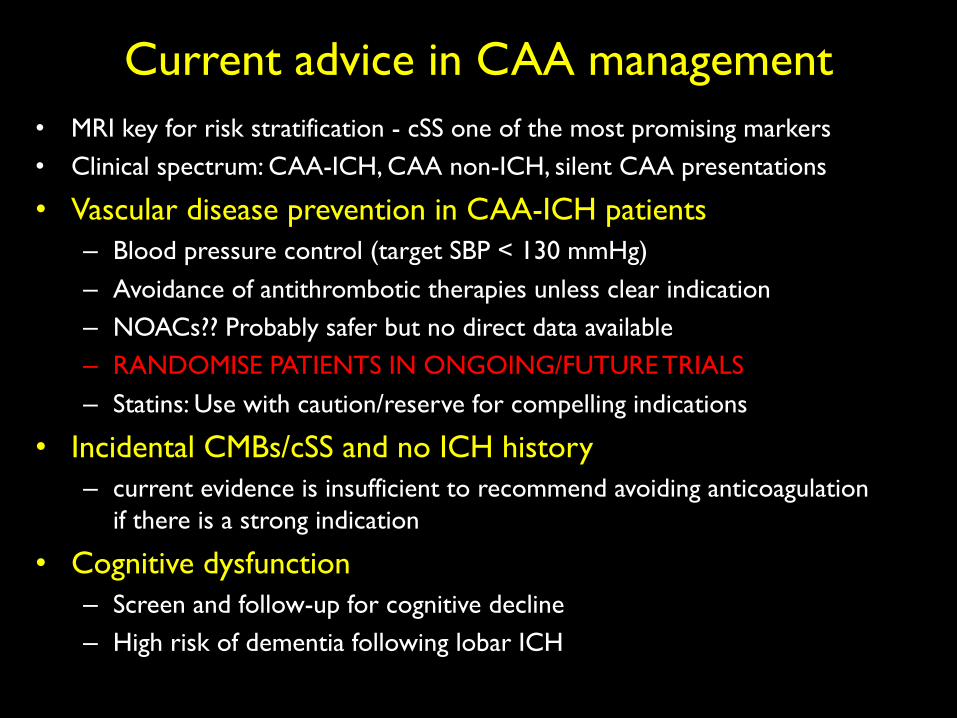
Current advice in CAA management• MRI key for risk stratification - cSS one of the most promising markers• Clinical spectrum: CAA-ICH, CAA non-ICH, silent CAA presentations
• Vascular disease prevention in CAA-ICH patients– Blood pressure control (target SBP < 130 mmHg)– Avoidance of antithrombotic therapies unless clear indication– NOACs?? Probably safer but no direct data available– RANDOMISE PATIENTS IN ONGOING/FUTURE TRIALS– Statins: Use with caution/reserve for compelling indications
• Incidental CMBs/cSS and no ICH history– current evidence is insufficient to recommend avoiding anticoagulation
if there is a strong indication
• Cognitive dysfunction– Screen and follow-up for cognitive decline– High risk of dementia following lobar ICH
https://caaforum.org/
Many thanks to…
@microbleeds
Collaborators
Ashkan ShoamaneshJean-Claude BaronKarim FaridEric SmithDavid J. WerringSalim Elyas
MGH Stroke Research Group
Steve GreenbergAnandViswanathan Jonathan RosandEdip GurolMatthew FroschGoldstein JoshuaGregoire BoulouisSergi Martinez-RamirezPanos FortiadisLi Xiong
Pasi MarcoSusanne van VeluwEllis van EttenAlison AyresKristin Schwab
Geneva Brain collectionDr Eniko KovariProf Costas BourasMiss Evgenia Daskalou


![Medicine > Neurology > Stroke Stroke - Initial assessment ... · Stroke caused by intracerebral haemorrhage is defined as [1][L2]: ... and emboli may be generated. • The most common](https://img.pdfslide.net/doc/110x75/5c8ae1ae09d3f2fa728b79a9/medicine-neurology-stroke-stroke-initial-assessment-stroke-caused.jpg)