Embed Size (px)
Citation preview
CEREBRAL PROTECTION INCEREBRAL PROTECTION IN NEUROSURGERY
PRESENTOR:PRESENTOR:DR SAURABH SHARMA
• The term ‘Neuroprotection’ signifies treatments used toprotect neural tissue from cellular events induced bydeprivation of oxygen or glucose or both to the brain.
• Treatment initiated before onset of ischemia, intended todif i i h i ll l d l bi l i lmodify intra-ischemic cellular and vascular biological
responses to deprivation of energy supply so as to increasetolerance of tissue to ischemia resulting in improvedtolerance of tissue to ischemia resulting in improvedoutcome.
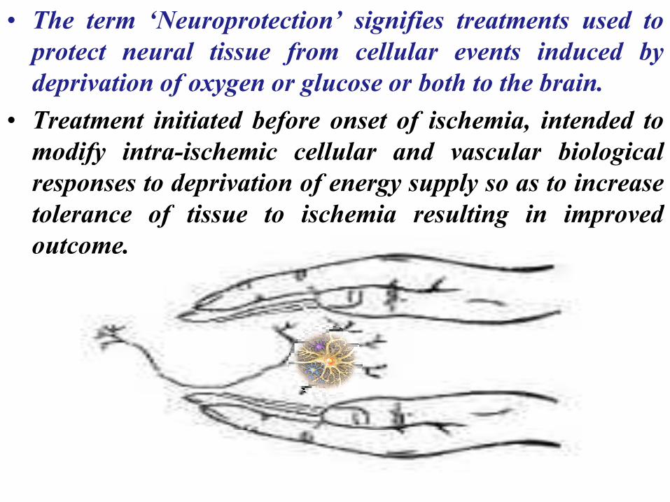
• Cerebral protection-physiological and pharmacological interventions that precede cerebral insult.
• Cerebral resuscitation-similar interventions after the insult has occurred and is a process of damage limitation.
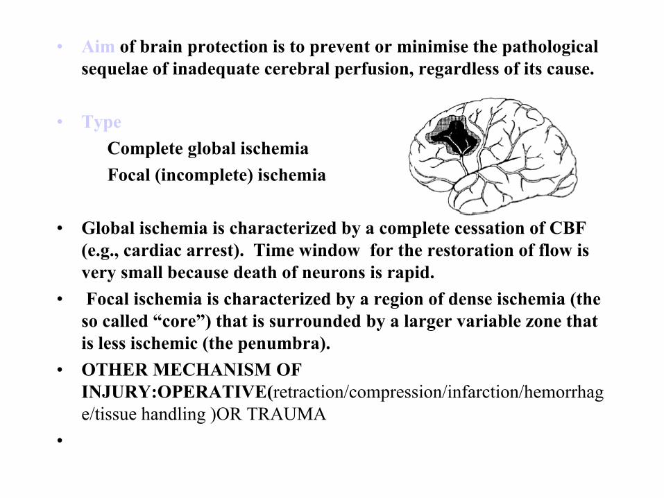
• Aim of brain protection is to prevent or minimise the pathological sequelae of inadequate cerebral perfusion regardless of its causesequelae of inadequate cerebral perfusion, regardless of its cause.
• TypeC l l b l i h iComplete global ischemiaFocal (incomplete) ischemia
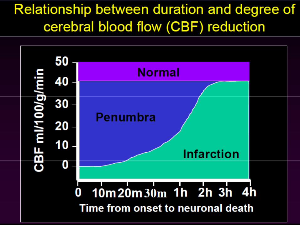
• Global ischemia is characterized by a complete cessation of CBF (e.g., cardiac arrest). Time window for the restoration of flow is very small because death of neurons is rapid.
• Focal ischemia is characterized by a region of dense ischemia (the so called “core”) that is surrounded by a larger variable zone that is less ischemic (the penumbra).
• OTHER MECHANISM OF INJURY:OPERATIVE(retraction/compression/infarction/hemorrhage/tissue handling )OR TRAUMA
•
Complete Global ischemiaischemia
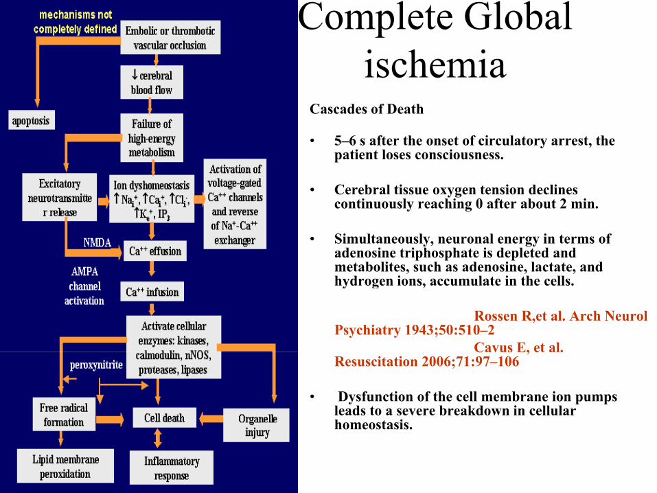
Cascades of Death
• 5 6 s after the onset of circulatory arrest the• 5–6 s after the onset of circulatory arrest, the patient loses consciousness.
• Cerebral tissue oxygen tension declines continuously reaching 0 after about 2 min.y g
• Simultaneously, neuronal energy in terms of adenosine triphosphate is depleted and metabolites, such as adenosine, lactate, and hydrogen ions accumulate in the cellshydrogen ions, accumulate in the cells.
Rossen R,et al. Arch NeurolPsychiatry 1943;50:510–2
Cavus E, et al.Cavus E, et al. Resuscitation 2006;71:97–106
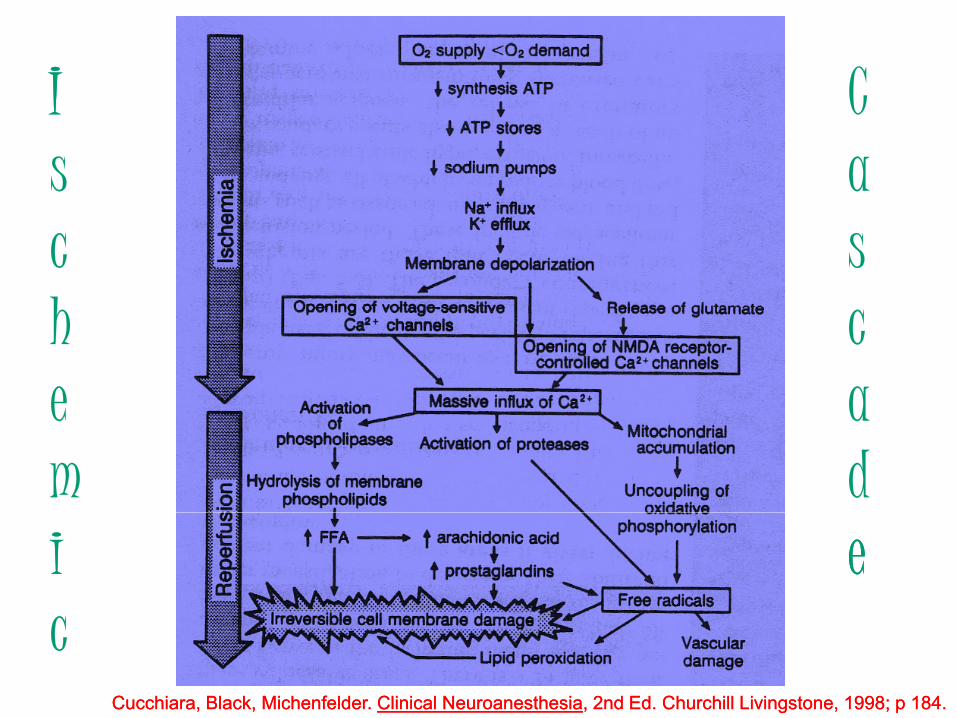
• Dysfunction of the cell membrane ion pumps leads to a severe breakdown in cellular homeostasishomeostasis.
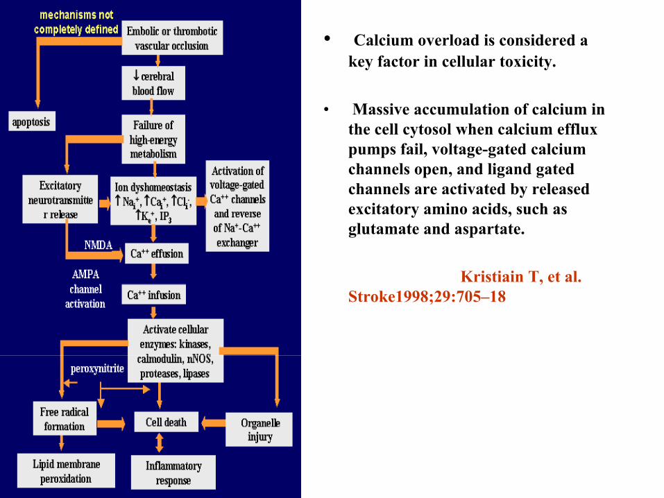
• Calcium overload is considered a key factor in cellular toxicity.
• Massive accumulation of calcium in the cell cytosol when calcium efflux pumps fail, voltage-gated calcium channels open, and ligand gated channels are activated by released excitatory amino acids, such asexcitatory amino acids, such as glutamate and aspartate.
Kristiain T, et al. ,Stroke1998;29:705–18
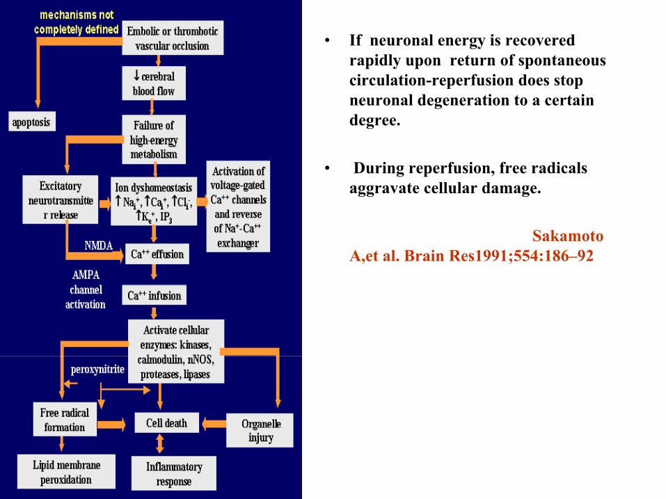
• If neuronal energy is recovered rapidly upon return of spontaneous circulation-reperfusion does stop neuronal degeneration to a certain degree.
• During reperfusion, free radicals aggravate cellular damage.
Sakamoto A,et al. Brain Res1991;554:186–92
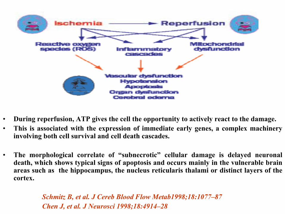
• During reperfusion, ATP gives the cell the opportunity to actively react to the damage.g p , g pp y y g• This is associated with the expression of immediate early genes, a complex machinery
involving both cell survival and cell death cascades.
Th h l i l l t f “ b ti ” ll l d i d l d l• The morphological correlate of “subnecrotic” cellular damage is delayed neuronaldeath, which shows typical signs of apoptosis and occurs mainly in the vulnerable brainareas such as the hippocampus, the nucleus reticularis thalami or distinct layers of thecortex.
Schmitz B, et al. J Cereb Blood Flow Metab1998;18:1077–87Chen J, et al. J Neurosci 1998;18:4914–28
II CIIs
Ca
ch
she
cae
mad
ii eec
CucchiaraCucchiara, Black, , Black, MichenfelderMichenfelder. . Clinical Clinical NeuroanesthesiaNeuroanesthesia, 2nd Ed. Churchill Livingstone, 1998; p 184., 2nd Ed. Churchill Livingstone, 1998; p 184.
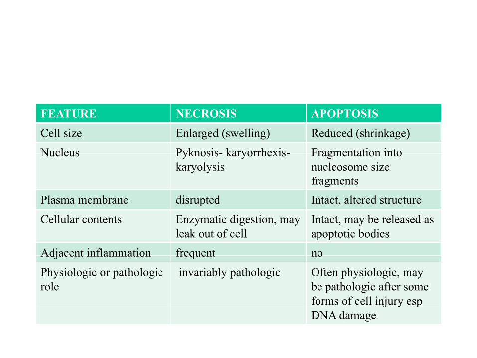
FEATURE NECROSIS APOPTOSISCell size Enlarged (swelling) Reduced (shrinkage)Nucleus Pyknosis- karyorrhexis- Fragmentation intoNucleus Pyknosis- karyorrhexis-
karyolysisFragmentation into nucleosome size fragments
Plasma membrane disrupted Intact, altered structurep ,Cellular contents Enzymatic digestion, may
leak out of cellIntact, may be released as apoptotic bodies
Adjacent inflammation frequent noAdjacent inflammation frequent noPhysiologic or pathologic role
invariably pathologic Often physiologic, may be pathologic after some forms of cell injury espj y pDNA damage
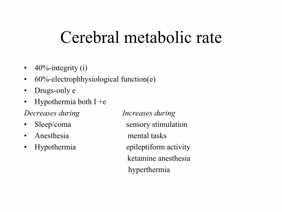
Cerebral metabolic rate• 40%-integrity (i)• 60%-electrophhysiological function(e)• Drugs only e• Drugs-only e• Hypothermia both I +eDecreases during Increases during• Sleep/coma sensory stimulation• Anesthesia mental tasks• Hypothermia epileptiform activity
ketamine anesthesiahyperthermia
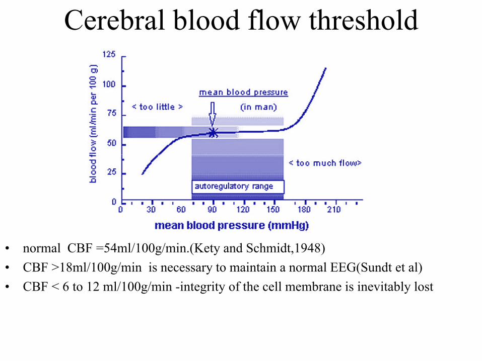
Cerebral blood flow threshold
• normal CBF =54ml/100g/min.(Kety and Schmidt,1948) • CBF >18ml/100g/min is necessary to maintain a normal EEG(Sundt et al)• CBF < 6 to 12 ml/100g/min -integrity of the cell membrane is inevitably lost
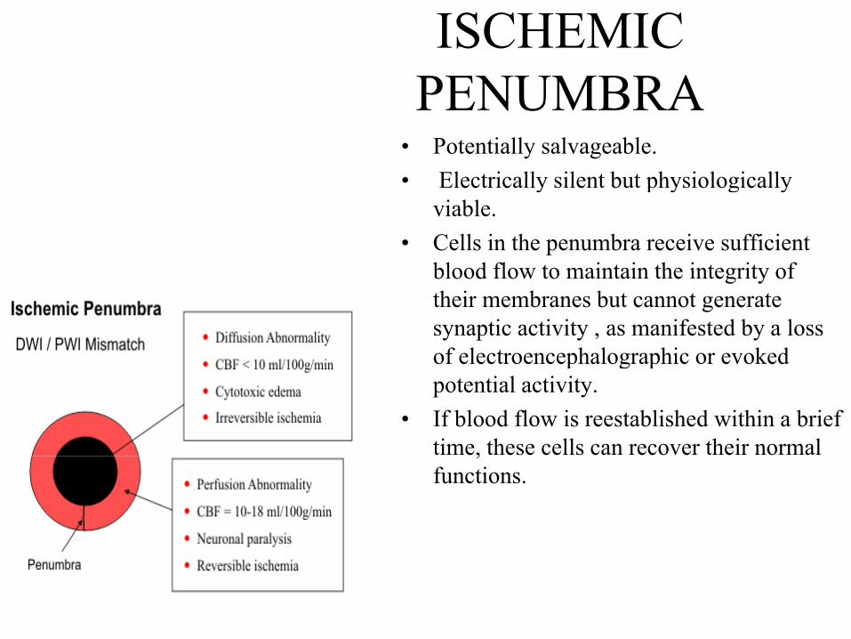
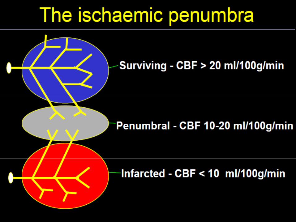
ISCHEMIC PENUMBRAPENUMBRA
• Potentially salvageable.• Electrically silent but physiologically• Electrically silent but physiologically
viable. • Cells in the penumbra receive sufficient
blood flow to maintain the integrity ofblood flow to maintain the integrity of their membranes but cannot generate synaptic activity , as manifested by a loss of electroencephalographic or evoked p g ppotential activity.
• If blood flow is reestablished within a brief time, these cells can recover their normal ,functions.
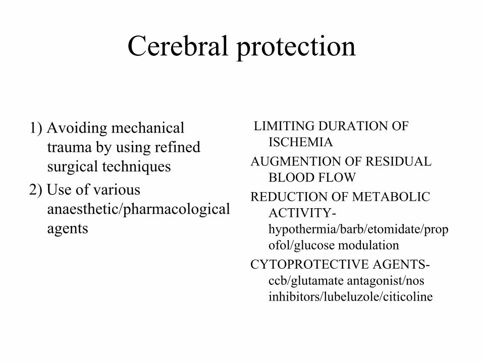
Cerebral protectionCerebral protection
1) Avoiding mechanical trauma by using refined
LIMITING DURATION OF ISCHEMIAy g
surgical techniques2) Use of various
anaesthetic/pharmacological
AUGMENTION OF RESIDUAL BLOOD FLOW
REDUCTION OF METABOLIC anaesthetic/pharmacological agents
ACTIVITY-hypothermia/barb/etomidate/propofol/glucose modulation
CYTOPROTECTIVE AGENTS-ccb/glutamate antagonist/nos inhibitors/lubeluzole/citicoline
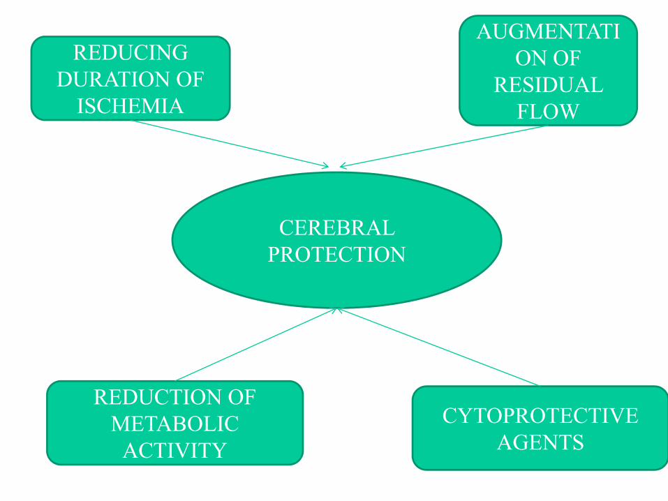
REDUCING A O O
AUGMENTATION OF
DURATION OF ISCHEMIA
RESIDUAL FLOW
CEREBRAL PROTECTION
REDUCTION OF METABOLIC CYTOPROTECTIVE METABOLIC
ACTIVITY AGENTS
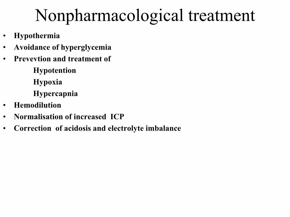
Nonpharmacological treatment• Hypothermia• Avoidance of hyperglycemia• Prevevtion and treatment of
HypotentionHypoxiaHypercapniaHypercapnia
• Hemodilution• Normalisation of increased ICP• Correction of acidosis and electrolyte imbalance
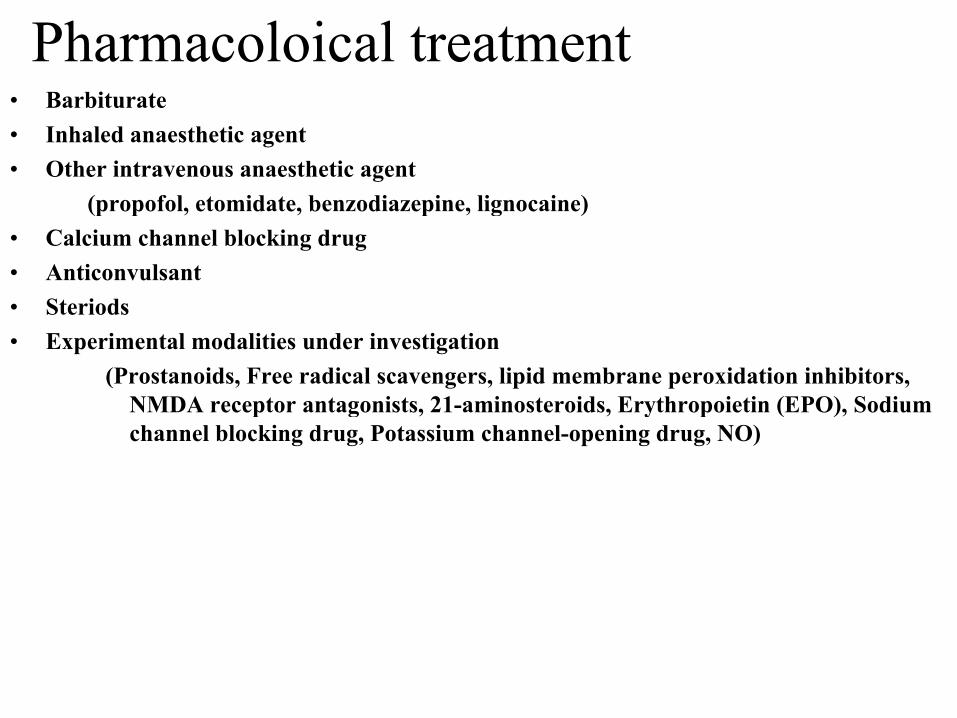
Pharmacoloical treatment• Barbiturate• Barbiturate • Inhaled anaesthetic agent• Other intravenous anaesthetic agent
(propofol etomidate benzodiazepine lignocaine)(propofol, etomidate, benzodiazepine, lignocaine)• Calcium channel blocking drug• Anticonvulsant• Steriods• Steriods• Experimental modalities under investigation
(Prostanoids, Free radical scavengers, lipid membrane peroxidation inhibitors, NMDA receptor antagonists, 21-aminosteroids, Erythropoietin (EPO), SodiumNMDA receptor antagonists, 21 aminosteroids, Erythropoietin (EPO), Sodium channel blocking drug, Potassium channel-opening drug, NO)
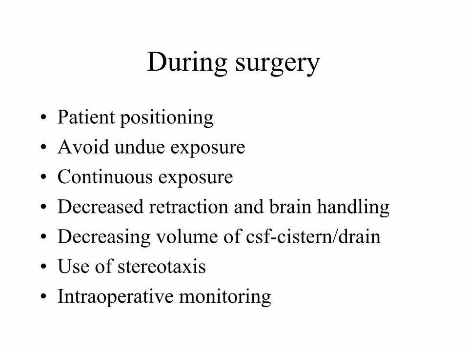
During surgery
• Patient positioning• Avoid undue exposureAvoid undue exposure• Continuous exposure
D d i d b i h dli• Decreased retraction and brain handling• Decreasing volume of csf-cistern/drain• Use of stereotaxis• Intraoperative monitoringIntraoperative monitoring
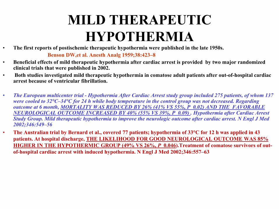
MILD THERAPEUTIC HYPOTHERMIA
• The first reports of postischemic therapeutic hypothermia were published in the late 1950s.Benson DW,et al. Anesth Analg 1959;38:423–8
• Beneficial effects of mild therapeutic hypothermia after cardiac arrest is provided by two major randomized clinical trials that were published in 2002.
• Both studies investigated mild therapeutic hypothermia in comatose adult patients after out-of-hospital cardiac arrest because of ventricular fibrillation.
• The European multicenter trial - Hypothermia After Cardiac Arrest study group included 275 patients, of whom 137 were cooled to 32°C–34°C for 24 h while body temperature in the control group was not decreased. Regarding outcome at 6 month, MORTALITY WAS REDUCED BY 26% (41% VS 55%, P 0.02) AND THE FAVORABLE NEUROLOGICAL OUTCOME INCREASED BY 40% (55% VS 39%, P 0.09) . Hypothermia after Cardiac Arrest Study Group Mild therapeutic hypothermia to improve the neurologic outcome after cardiac arrest N Engl J MedStudy Group. Mild therapeutic hypothermia to improve the neurologic outcome after cardiac arrest. N Engl J Med 2002;346:549–56
• The Australian trial by Bernard et al., covered 77 patients; hypothermia of 33°C for 12 h was applied in 43 patients. At hospital discharge, THE LIKELIHOOD FOR GOOD NEUROLOGICAL OUTCOME WAS 85% HIGHER IN THE HYPOTHERMIC GROUP (49% VS 26%, P 0.046).Treatment of comatose survivors of out-of-hospital cardiac arrest with induced hypothermia. N Engl J Med 2002;346:557–63
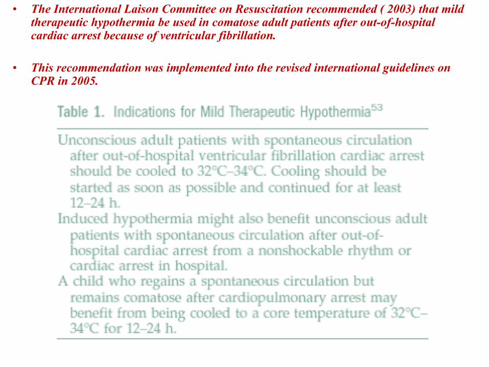
• The International Laison Committee on Resuscitation recommended ( 2003) that mild therapeutic hypothermia be used in comatose adult patients after out-of-hospital cardiac arrest because of ventricular fibrillation.
• This recommendation was implemented into the revised international guidelines on CPR in 2005.
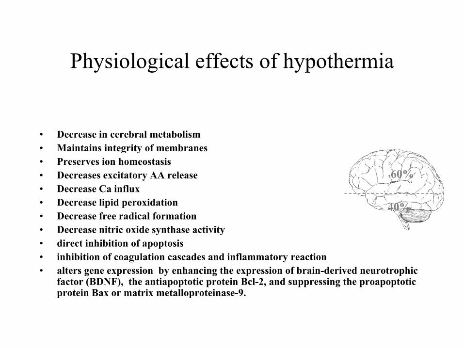
Physiological effects of hypothermia
• Decrease in cerebral metabolism• Maintains integrity of membranesMaintains integrity of membranes • Preserves ion homeostasis• Decreases excitatory AA release• Decrease Ca influx
i i i i
60%
• Decrease lipid peroxidation• Decrease free radical formation• Decrease nitric oxide synthase activity• direct inhibition of apoptosis
40%
p p• inhibition of coagulation cascades and inflammatory reaction• alters gene expression by enhancing the expression of brain-derived neurotrophic
factor (BDNF), the antiapoptotic protein Bcl-2, and suppressing the proapoptotic protein Bax or matrix metalloproteinase-9.p p
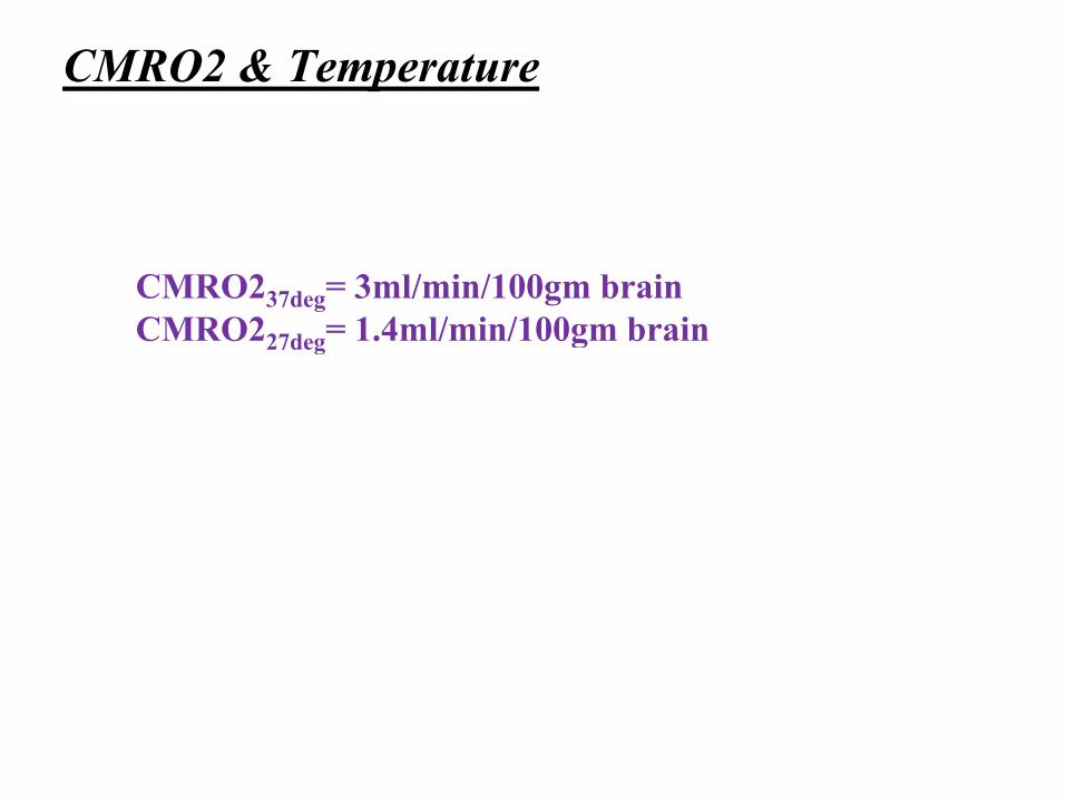
CMRO2 & Temperature
CMRO237deg= 3ml/min/100gm brainCMRO227deg= 1.4ml/min/100gm brain27deg g
• Hypothermia can be induced by different methods,surface cooling, ice-cold infusions or endovascular
cooling catheterscooling catheters.
• Recommendation is that hypothermia should be initiated with minimal delay after cardiac arrest.
Circulation 2005;112:IV1–IV211
S rface cooling or ice cold inf sions can be sed preclinicall• Surface cooling or ice-cold infusions can be used preclinically.
• Kim et al. conducted a randomized clinical trial in which patients were assigned to either receiving 4°C normal saline or not in the out-of hospital g g psetting,Survival rates was higher in patients who had received out-of-hospital cooling treatment.
Circulation 2007;115:3064–70
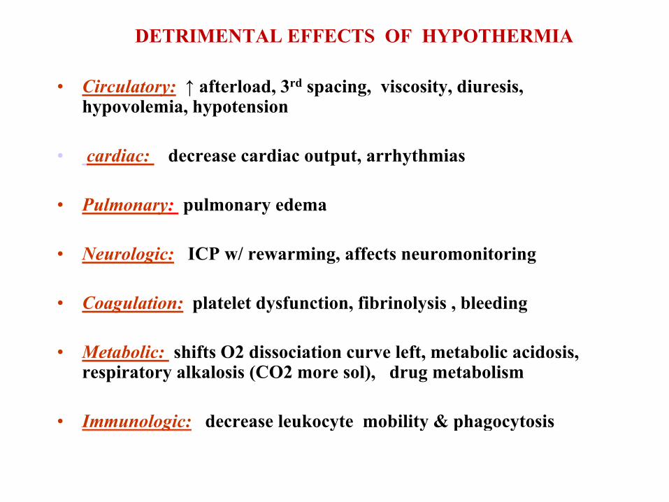
DETRIMENTAL EFFECTS OF HYPOTHERMIA
• Circulatory: ↑ afterload, 3rd spacing, viscosity, diuresis, hypovolemia, hypotension
• cardiac: decrease cardiac output, arrhythmias
• Pulmonary: pulmonary edema
• Neurologic: ICP w/ rewarming, affects neuromonitoring
• Coagulation: platelet dysfunction, fibrinolysis , bleeding
• Metabolic: shifts O2 dissociation curve left, metabolic acidosis, , ,respiratory alkalosis (CO2 more sol), drug metabolism
• Immunologic: decrease leukocyte mobility & phagocytosisg y y p g y
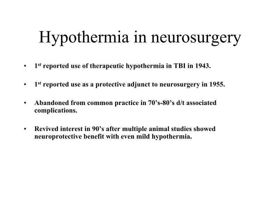
Hypothermia in neurosurgery • 1st reported use of therapeutic hypothermia in TBI in 1943.
• 1st reported use as a protective adjunct to neurosurgery in 1955.
• Abandoned from common practice in 70’s-80’s d/t associated complications.
• Revived interest in 90’s after multiple animal studies showed neuroprotective benefit with even mild hypothermia.
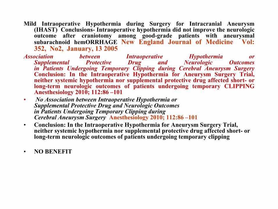
Mild Intraoperative Hypothermia during Surgery for Intracranial Aneurysm(IHAST) Conclusions Intraoperative hypothermia did not improve the neurologic(IHAST) Conclusions- Intraoperative hypothermia did not improve the neurologicoutcome after craniotomy among good-grade patients with aneurysmalsubarachnoid hemORRHAGE New England Journal of Medicine Vol:352, No2, January, 13 2005
Association between Intraoperative Hypothermia orp ypSupplemental Protective Drug and Neurologic Outcomesin Patients Undergoing Temporary Clipping during Cerebral Aneurysm SurgeryConclusion: In the Intraoperative Hypothermia for Aneurysm Surgery Trial,neither systemic hypothermia nor supplemental protective drug affected short- orlong-term neurologic outcomes of patients undergoing temporary CLIPPINGAnesthesiology 2010; 112:86 –101
• No Association between Intraoperative Hypothermia orSupplemental Protective Drug and Neurologic Outcomesin Patients Undergoing Temporary Clipping duringCerebral Aneurysm Surgery Anesthesiology 2010; 112:86 –101Ce eb al eu ys Su ge y es es o ogy 0 0; :86 0
• Conclusion: In the Intraoperative Hypothermia for Aneurysm Surgery Trial, neither systemic hypothermia nor supplemental protective drug affected short- or long-term neurologic outcomes of patients undergoing temporary clipping
• NO BENEFIT
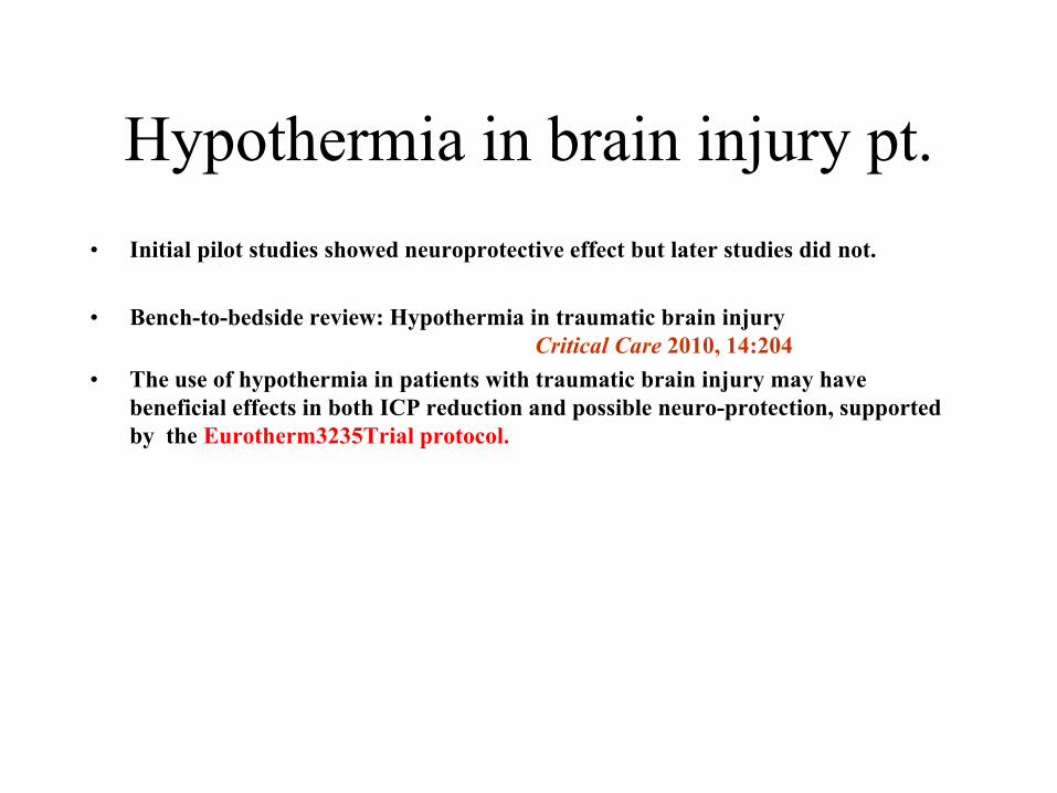
Hypothermia in brain injury pt.• Initial pilot studies showed neuroprotective effect but later studies did not.
• Bench-to-bedside review: Hypothermia in traumatic brain injuryC iti l C 2010 14 204Critical Care 2010, 14:204
• The use of hypothermia in patients with traumatic brain injury may have beneficial effects in both ICP reduction and possible neuro-protection, supported by the Eurotherm3235Trial protocol.
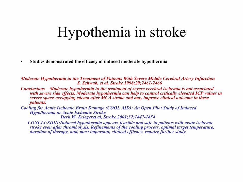
Hypothemia in stroke
• Studies demonstrated the efficacy of induced moderate hypothermia
Moderate Hypothermia in the Treatment of Patients With Severe Middle Cerebral Artery InfarctionModerate Hypothermia in the Treatment of Patients With Severe Middle Cerebral Artery InfarctionS. Schwab, et al. Stroke 1998;29;2461-2466
Conclusions—Moderate hypothermia in the treatment of severe cerebral ischemia is not associated with severe side effects. Moderate hypothermia can help to control critically elevated ICP values in severe space-occupying edema after MCA stroke and may improve clinical outcome in these patients.p
Cooling for Acute Ischemic Brain Damage (COOL AID): An Open Pilot Study of Induced Hypothermia in Acute Ischemic Stroke
Derk W. Kriegeret al, Stroke 2001;32;1847-1854CONCLUSION:Induced hypothermia appears feasible and safe in patients with acute ischemic stroke even after thrombolysis. Refinements of the cooling process, optimal target temperature, d i f h d i li i l ffi i f h dduration of therapy, and, most important, clinical efficacy, require further study.
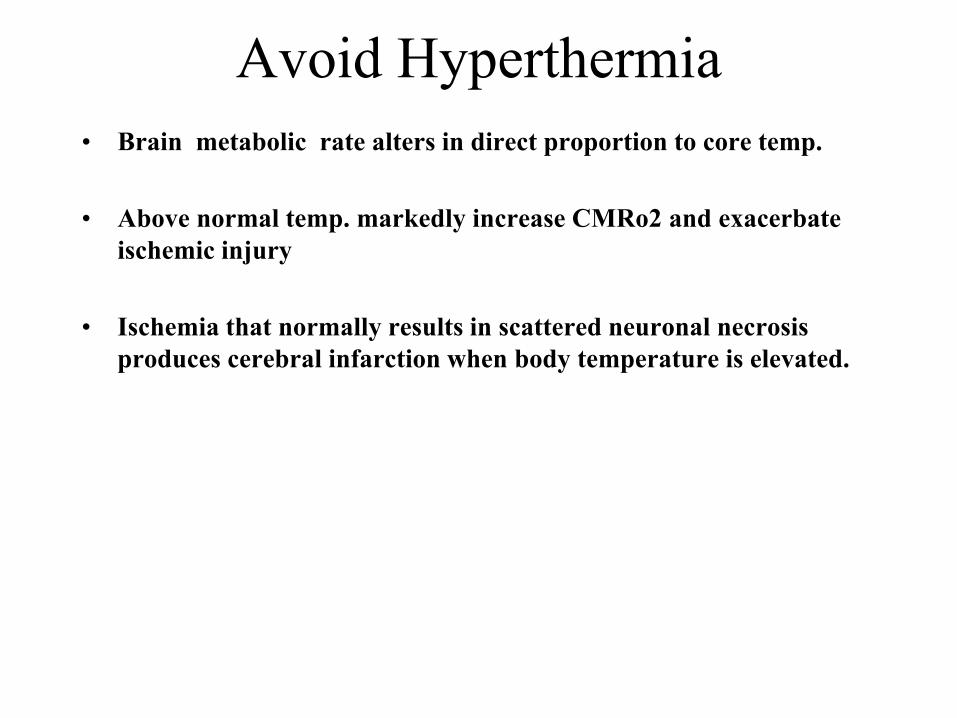
Avoid Hyperthermia• Brain metabolic rate alters in direct proportion to core temp.
• Above normal temp. markedly increase CMRo2 and exacerbate ischemic injury
• Ischemia that normally results in scattered neuronal necrosis produces cerebral infarction when body temperature is elevated.
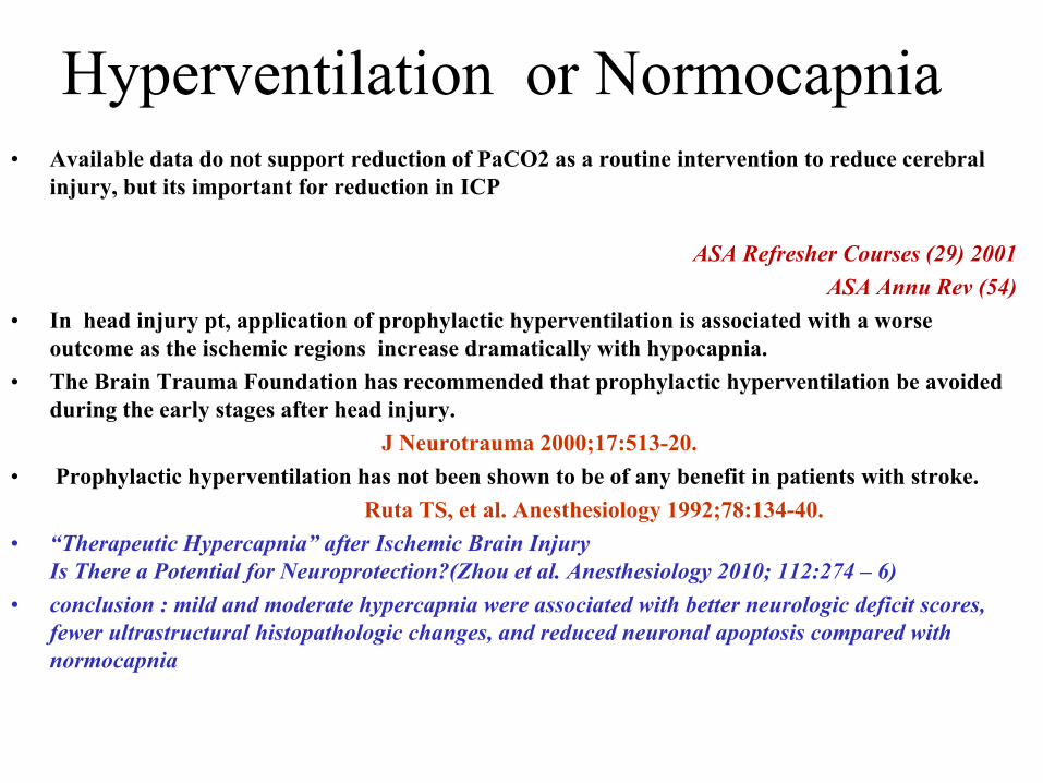
Hyperventilation or Normocapnia• Available data do not support reduction of PaCO2 as a routine intervention to reduce cerebral
injury, but its important for reduction in ICP
ASA Refresher Courses (29) 2001ASA Annu Rev (54)
• In head injury pt, application of prophylactic hyperventilation is associated with a worse outcome as the ischemic regions increase dramatically with hypocapnia.
• The Brain Trauma Foundation has recommended that prophylactic hyperventilation be avoided during the early stages after head injury.
J Neurotrauma 2000;17:513-20.J Neurotrauma 2000;17:513 20.• Prophylactic hyperventilation has not been shown to be of any benefit in patients with stroke.
Ruta TS, et al. Anesthesiology 1992;78:134-40.• “Therapeutic Hypercapnia” after Ischemic Brain Injury
Is There a Potential for Neuroprotection?(Zhou et al. Anesthesiology 2010; 112:274 – 6)• conclusion : mild and moderate hypercapnia were associated with better neurologic deficit scores,
fewer ultrastructural histopathologic changes, and reduced neuronal apoptosis compared with normocapniap
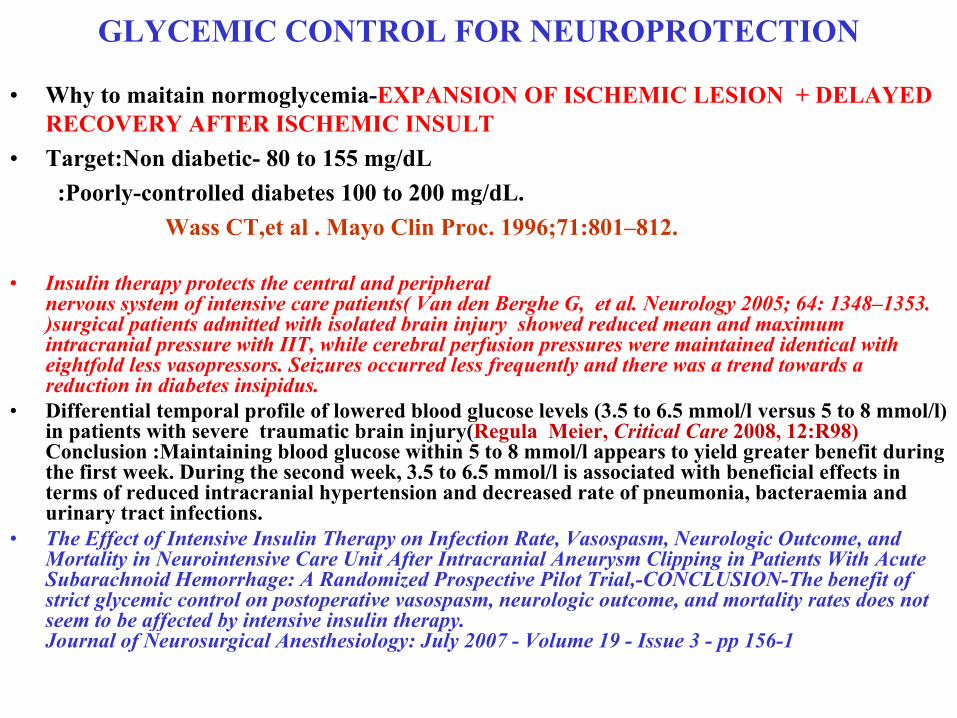
GLYCEMIC CONTROL FOR NEUROPROTECTION
• Why to maitain normoglycemia-EXPANSION OF ISCHEMIC LESION + DELAYEDWhy to maitain normoglycemia EXPANSION OF ISCHEMIC LESION DELAYED RECOVERY AFTER ISCHEMIC INSULT
• Target:Non diabetic- 80 to 155 mg/dL:Poorly-controlled diabetes 100 to 200 mg/dL.y g
Wass CT,et al . Mayo Clin Proc. 1996;71:801–812.
• Insulin therapy protects the central and peripheralnervous system of intensive care patients( Van den Berghe G, et al. Neurology 2005; 64: 1348–1353.nervous system of intensive care patients( Van den Berghe G, et al. Neurology 2005; 64: 1348 1353. )surgical patients admitted with isolated brain injury showed reduced mean and maximum intracranial pressure with IIT, while cerebral perfusion pressures were maintained identical with eightfold less vasopressors. Seizures occurred less frequently and there was a trend towards a reduction in diabetes insipidus.
• Differential temporal profile of lowered blood glucose levels (3 5 to 6 5 mmol/l versus 5 to 8 mmol/l)• Differential temporal profile of lowered blood glucose levels (3.5 to 6.5 mmol/l versus 5 to 8 mmol/l) in patients with severe traumatic brain injury(Regula Meier, Critical Care 2008, 12:R98) Conclusion :Maintaining blood glucose within 5 to 8 mmol/l appears to yield greater benefit during the first week. During the second week, 3.5 to 6.5 mmol/l is associated with beneficial effects in terms of reduced intracranial hypertension and decreased rate of pneumonia, bacteraemia and urinary tract infectionsurinary tract infections.
• The Effect of Intensive Insulin Therapy on Infection Rate, Vasospasm, Neurologic Outcome, and Mortality in Neurointensive Care Unit After Intracranial Aneurysm Clipping in Patients With Acute Subarachnoid Hemorrhage: A Randomized Prospective Pilot Trial,-CONCLUSION-The benefit of strict glycemic control on postoperative vasospasm, neurologic outcome, and mortality rates does not
t b ff t d b i t i i li thseem to be affected by intensive insulin therapy. Journal of Neurosurgical Anesthesiology: July 2007 - Volume 19 - Issue 3 - pp 156-1
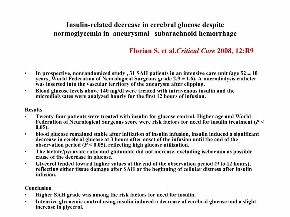
Insulin-related decrease in cerebral glucose despitel i i l b h id h hnormoglycemia in aneurysmal subarachnoid hemorrhage
Florian S, et al.Critical Care 2008, 12:R9
• In prospective, nonrandomized study , 31 SAH patients in an intensive care unit (age 52 ± 10 years, World Federation of Neurological Surgeons grade 2.9 ± 1.6). A microdialysis catheter was inserted into the vascular territory of the aneurysm after clipping.
• Blood glucose levels above 140 mg/dl were treated with intravenous insulin and the g gmicrodialysates were analyzed hourly for the first 12 hours of infusion.
Results • Twenty-four patients were treated with insulin for glucose control. Higher age and World
Federation of Neurological Surgeons score were risk factors for need for insulin treatment (P < g g (0.05).
• blood glucose remained stable after initiation of insulin infusion, insulin induced a significant decrease in cerebral glucose at 3 hours after onset of the infusion until the end of the observation period (P < 0.05), reflecting high glucose utilization.
• The lactate/pyruvate ratio and glutamate did not increase, excluding ischaemia as possible f icause of the decrease in glucose.
• Glycerol tended toward higher values at the end of the observation period (9 to 12 hours), reflecting either tissue damage after SAH or the beginning of cellular distress after insulin infusion.
Conclusion • Higher SAH grade was among the risk factors for need for insulin.• Intensive glycaemic control using insulin induced a decrease of cerebral glucose and a slight
increase in glycerol.
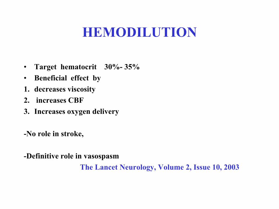
HEMODILUTIONHEMODILUTION
• Target hematocrit 30%- 35%• Beneficial effect by1 decreases viscosity1. decreases viscosity2. increases CBF3. Increases oxygen delivery
-No role in stroke,
-Definitive role in vasospasmThe Lancet Neurology, Volume 2, Issue 10, 2003
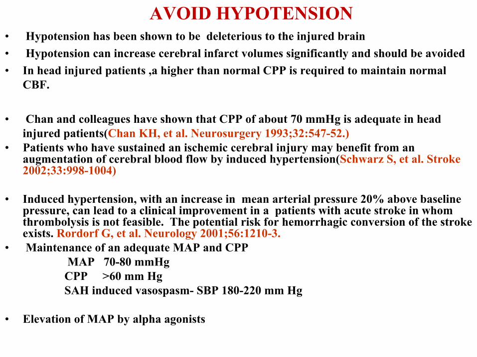
AVOID HYPOTENSION• Hypotension has been shown to be deleterious to the injured brain
H t i i b l i f t l i ifi tl d h ld b id d• Hypotension can increase cerebral infarct volumes significantly and should be avoided• In head injured patients ,a higher than normal CPP is required to maintain normal
CBF.
• Chan and colleagues have shown that CPP of about 70 mmHg is adequate in head injured patients(Chan KH, et al. Neurosurgery 1993;32:547-52.)
• Patients who have sustained an ischemic cerebral injury may benefit from an j y yaugmentation of cerebral blood flow by induced hypertension(Schwarz S, et al. Stroke 2002;33:998-1004)
• Induced hypertension, with an increase in mean arterial pressure 20% above baseline yp , ppressure, can lead to a clinical improvement in a patients with acute stroke in whom thrombolysis is not feasible. The potential risk for hemorrhagic conversion of the stroke exists. Rordorf G, et al. Neurology 2001;56:1210-3.
• Maintenance of an adequate MAP and CPPMAP 70-80 mmHg CPP >60 mm HgSAH induced vasospasm- SBP 180-220 mm Hg
• Elevation of MAP by alpha agonists
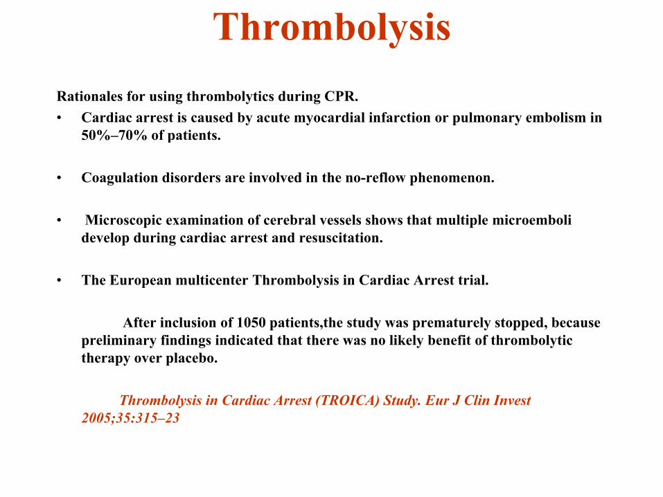
ThrombolysisRationales for using thrombolytics during CPR. • Cardiac arrest is caused by acute myocardial infarction or pulmonary embolism in
50%–70% of patients.p
• Coagulation disorders are involved in the no-reflow phenomenon.
• Microscopic examination of cerebral vessels shows that multiple microemboli develop during cardiac arrest and resuscitation.
• The European multicenter Thrombolysis in Cardiac Arrest trial.The European multicenter Thrombolysis in Cardiac Arrest trial.
After inclusion of 1050 patients,the study was prematurely stopped, because preliminary findings indicated that there was no likely benefit of thrombolytic th l btherapy over placebo.
Thrombolysis in Cardiac Arrest (TROICA) Study. Eur J Clin Invest 2005;35:315–23
HYPERTONIC, HYPERONCOTIC INFUSIONS TO PROMOTE MICROCIRCULATION
• Several animal studies have shown that hypertonic-hyperoncotic solutions given during CPR,or immediately after restoration of spontaneous circulation, decrease cerebral no-reflow. ,
Krep H, et al. Resuscitation 2004; 63:73–83Noppens RR,et al . Crit Care Med 2006;34:2194–200
• Besides having positive effects on cerebral microcirculation, hypertonic saline also seems to ameliorate cardiac function during and after CPR.
• Benderet al. randomized 66 patients who suffered out-of-hospital cardiac arrest into two groups. The patients received 2 mL / kg/ 10 min of either hypertonic saline with HES (7.2% NaCl with 6% HES 200,000/0.5) or HES alone during continuous CPR.
• Resuscitation success tended to be higher in patients receiving hypertonic saline with HES (66.7%vs 51.5%, P 0 21) d h it l d i i t l i d (57 6% 39 4% P 0 14)0.21) and hospital admission rates were also increased (57.6% vs 39.4%, P 0.14)
Resuscitation 2007;72:74–81
SENDAI COCKTAIL
• 20% MANNITOL+VITAMIN E+ DEXAMETHASONE
• Surgical treatment of AVMs occluding these feeders during removal--utilizing thethese feeders during removal utilizing the intraoperative balloon catheter and brain protective substances ("Sendaiprotective substances ( Sendai cocktail"]Takahashi A, Suzuki J, Sugawara T, Yoshimoto TSugawara T, Yoshimoto T

BARBITURATESBARBITURATES
• The Brain Resuscitation Clinical Trial failed to demonstrate any improved outcome due to thiopental therapy following cardiac arrest.
• Thiopentone, methohexital- Do not improve outcome in global or complete p , p g pischemia after cardiac arrest
• Pentobarbital(mechanism similar to thiopental(long acting ):Current clinical indication- BARBITURATE COMA
• Barbiturates have been found to be efficacious in the treatment of focal ischemia and can reduce the extent of cerebral injury.ischemia and can reduce the extent of cerebral injury.
Warner DS, et L. J Cereb Blood Flow Metab 1991;11:794-802.Drummond JC. Anesthesiology 1993;78:611-3.
BARBITURATES IN HEAD TRAUMA B bit t id fBARBITURATES IN HEAD TRAUMA: Barbiturates provide a means of reducing and maintaining ICP but not necessarily corresponds to improved outcome. (Ward JD, et al. J Neurosurg 1985; 62: 383-8)
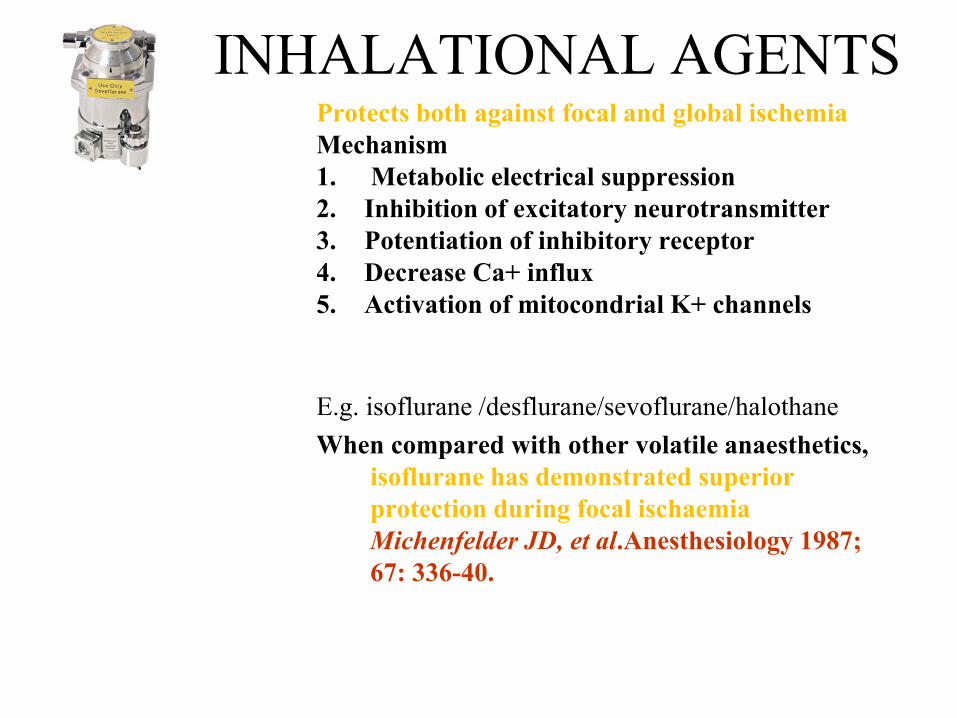
INHALATIONAL AGENTSProtects both against focal and global ischemiaMechanism1. Metabolic electrical suppression2 Inhibition of excitatory neurotransmitter2. Inhibition of excitatory neurotransmitter3. Potentiation of inhibitory receptor4. Decrease Ca+ influx5 Activation of mitocondrial K+ channels5. Activation of mitocondrial K+ channels
E g isoflurane /desflurane/sevoflurane/halothaneE.g. isoflurane /desflurane/sevoflurane/halothaneWhen compared with other volatile anaesthetics,
isoflurane has demonstrated superior protection during focal ischaemiaprotection during focal ischaemiaMichenfelder JD, et al.Anesthesiology 1987; 67: 336-40.
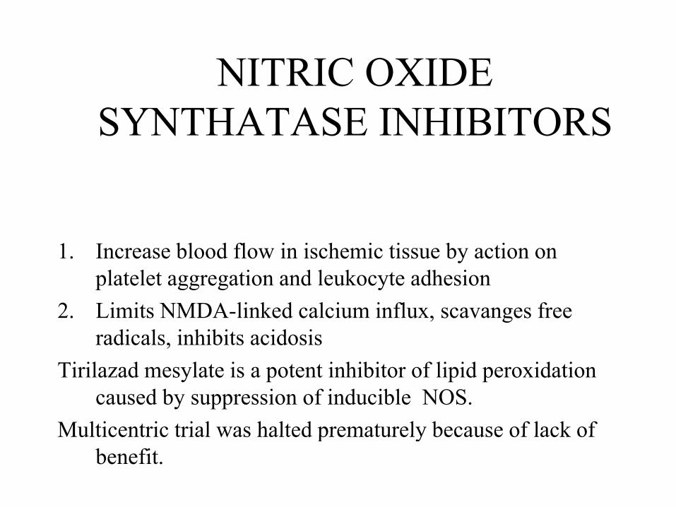
NITRIC OXIDENITRIC OXIDESYNTHATASE INHIBITORS
1. Increase blood flow in ischemic tissue by action on platelet aggregation and leukocyte adhesionp gg g y
2. Limits NMDA-linked calcium influx, scavanges free radicals, inhibits acidosis
i il d l i i hibi f li id id iTirilazad mesylate is a potent inhibitor of lipid peroxidation caused by suppression of inducible NOS.
Multicentric trial was halted prematurely because of lack ofMulticentric trial was halted prematurely because of lack of benefit.
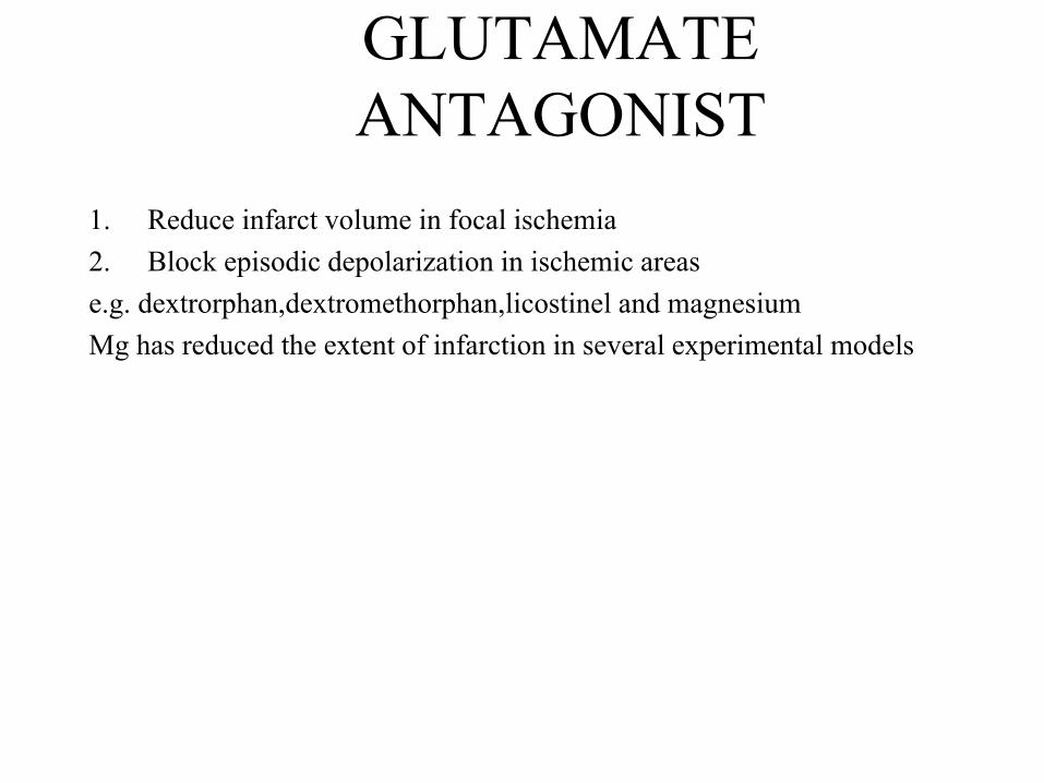
GLUTAMATE ANTAGONISTANTAGONIST
1 Reduce infarct volume in focal ischemia1. Reduce infarct volume in focal ischemia2. Block episodic depolarization in ischemic areas e.g. dextrorphan,dextromethorphan,licostinel and magnesiumM h d d h f i f i i l i l d lMg has reduced the extent of infarction in several experimental models
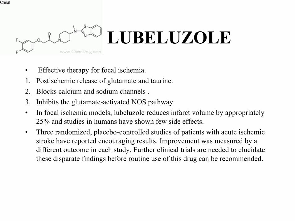
LUBELUZOLELUBELUZOLE• Effective therapy for focal ischemia.1. Postischemic release of glutamate and taurine. 2. Blocks calcium and sodium channels .3. Inhibits the glutamate-activated NOS pathway.• In focal ischemia models, lubeluzole reduces infarct volume by appropriately
25% and studies in humans have shown few side effects.• Three randomized, placebo-controlled studies of patients with acute ischemic
stroke have reported encouraging results. Improvement was measured by a different outcome in each study. Further clinical trials are needed to elucidate these disparate findings before routine use of this drug can be recommendedthese disparate findings before routine use of this drug can be recommended.
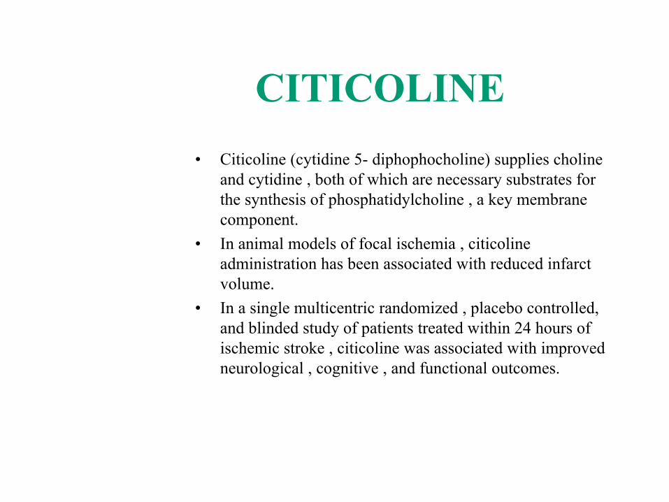
CITICOLINE• Citicoline (cytidine 5- diphophocholine) supplies choline
and cytidine , both of which are necessary substrates for the synthesis of phosphatidylcholine , a key membrane
tcomponent. • In animal models of focal ischemia , citicoline
administration has been associated with reduced infarct volumevolume.
• In a single multicentric randomized , placebo controlled, and blinded study of patients treated within 24 hours of ischemic stroke , citicoline was associated with improved neurological , cognitive , and functional outcomes.
Etomidate• Reduces CMRO2(50%)• Decreases CBF• Decreases ICP• But maintains cardiovascular stability and CPP• CO2 reactivity is preserved• Batjer’s group report using etomidate 1 mgkg-1 as a bolus followed by an infusion of
10 mgkg-1min-1 to maintain burst suppression during temporary arterial occlusion10 mgkg 1min 1 to maintain burst suppression during temporary arterial occlusion for complex intracranial aneurysms. This regimen was well tolerated. Batjer HH. Cerebral protective effects of etomidate: experimental and clinical aspects. Cerebrovascular and brain metabolism reviews 1993; 5: 17-32
• Models of focal ischemia revealed that etomidate actually increased the volume of• Models of focal ischemia revealed that etomidate actually increased the volume of brain infarction by reducing nitric oxide levels in ischemic brain tissue .
Drummond JC, et al. Anesth Analg 2005;100:841-6
AVAILABLE DATA DO NOT SUPPORT THE USE OF ETOMIDATE AS A NEUROPROTECTIVE AGENT.
Benzodiazepine• Stimulate inhibitory neurotransmitter GABA
• Decreases CMRO2,CBF while preserving CO2 reactivity, p g y
• Commonly used are diazepam, midazolam, lorazepam
• Diazepam improved the oxygen supply: demand ratio
• Reduces energy required for synaptic transmissiongy q y pMarana E,et al. Resuscitation1982 10: 89-100. Cotev S,et al.Anesthesiology 1975; 43:117-22
May be neuroprotectant in both global and focal ischaemia.
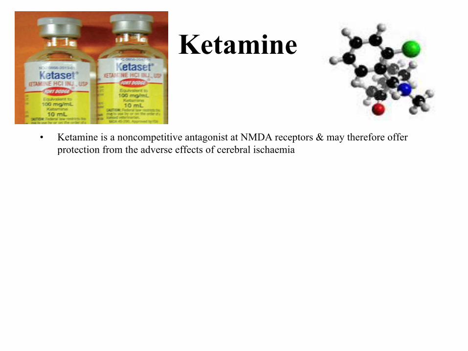
KetamineKetamine
• Ketamine is a noncompetitive antagonist at NMDA receptors & may therefore offer protection from the adverse effects of cerebral ischaemiaprotection from the adverse effects of cerebral ischaemia
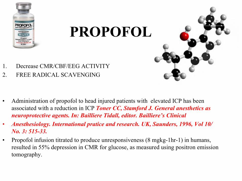
PROPOFOL
1. Decrease CMR/CBF/EEG ACTIVITY2. FREE RADICAL SCAVENGING
• Administration of propofol to head injured patients with elevated ICP has been p p j passociated with a reduction in ICP Toner CC, Stamford J. General anesthetics as neuroprotective agents. In: Bailliere Tidall, editor. Bailliere’s Clinical
• Anesthesiology. International pratice and research. UK, Saunders, 1996, Vol 10/ N 3 515 33No. 3: 515-33.
• Propofol infusion titrated to produce unresponsiveness (8 mgkg-1hr-1) in humans, resulted in 55% depression in CMR for glucose, as measured using positron emission tomographytomography.
• Antiinflammatory properties, alter the normal response to injury and may alter the neurological outcome following an ischaemic insult.
STERIODS• Insufficient evidence to define role of
glucocorticoids in focal ischemia(Cochrane Database Syst
Rev 2002)
• In double-blind study, administration of dexamethasone to acute stroke victims concluded that dexamethasone can be a useful adjunct to the treatment of the patient with a severe stroke and thepatient with a severe stroke and the beneficial effects of steroids are in part due to their ability to decrease brain oedema secondary to massive brain infarction.
Patten BM, et al. Neurology 22: 377-83.
Glucocorticoids exacerbate injury from global ischemia by increasing plasma
lglucose
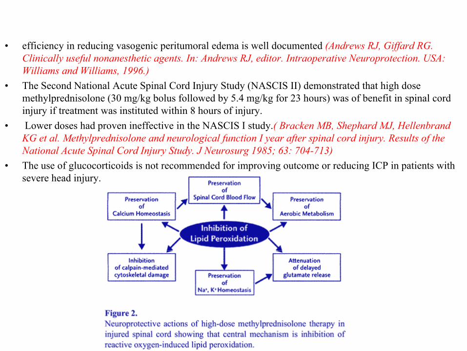
• efficiency in reducing vasogenic peritumoral edema is well documented (Andrews RJ, Giffard RG. y g g p ( , ffClinically useful nonanesthetic agents. In: Andrews RJ, editor. Intraoperative Neuroprotection. USA: Williams and Williams, 1996.)
• The Second National Acute Spinal Cord Injury Study (NASCIS II) demonstrated that high dose methylprednisolone (30 mg/kg bolus followed by 5 4 mg/kg for 23 hours) was of benefit in spinal cordmethylprednisolone (30 mg/kg bolus followed by 5.4 mg/kg for 23 hours) was of benefit in spinal cord injury if treatment was instituted within 8 hours of injury.
• Lower doses had proven ineffective in the NASCIS I study.( Bracken MB, Shephard MJ, Hellenbrand KG et al. Methylprednisolone and neurological function I year after spinal cord injury. Results of the National Acute Spinal Cord Injury Study J Neurosurg 1985; 63: 704 713)National Acute Spinal Cord Injury Study. J Neurosurg 1985; 63: 704-713)
• The use of glucocorticoids is not recommended for improving outcome or reducing ICP in patients with severe head injury.
Tirilazad mesylate-LAZAROIDSLAZAROIDS
• 21-aminosterid (lazaroid) that was developed specifically to maximize the inhibition of lipid21 aminosterid (lazaroid) that was developed specifically to maximize the inhibition of lipid peroxidation by glucocorticoids such as methylprednisolone, but eliminate the unwanted glucocorticoids effects. .( Andrews RJ, Giffard RG. Clinically useful nonanesthetic agents.)
• potent antioxidants, 100 times more potent than the corticosteroids• In animal experiments, TM has been of benefit in both focal and global ischaemia.• Double-blind, randomized, vehicle-controlled study of high-dose tirilazad mesylate in
women with aneurysmal subarachnoid hemorrhage. Part I. A cooperative study in Europe, A li N Z l d d S h Af i A ( ) / A h ( )L i /K l lAustralia, New Zealand, and South AfricaAuteur(s) / Author(s)Lasingo/Kassel et al Departments of Neurological Surgery and Virginia Neurological Institute, University of Virginia, Charlottesville, Virginia, ETATS-UNIS,Westmead Hospital and University of,Sydney, Sydney, AUSTRALIE,Ospedale Civile di Verona, Verona, ITALIE,University f, y y, y y, , p , , , yHospital, Lund, SUEDE,Klinikum Mannheim, University of Heidelberg, Heidelberg, ALLEMAGNEconclude that high-dose tirilazad mesylate is well tolerated in women with aneurysmal SAH. Although a significant reduction in the incidence of symptomatic
b d i th t t t THE PRIMARY END POINTvasospasm was observed in the treatment group, THE PRIMARY END POINT (MORTALITY RATE AT 3 MONTHS POST-SAH) WAS NOT AFFECTED BY THE STUDY DRUG
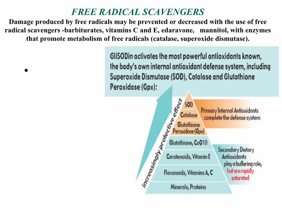
FREE RADICAL SCAVENGERSDamage produced by free radicals may be prevented or decreased with the use of free
radical scavengers barbiturates vitamins C and E edaravone mannitol with enzymesradical scavengers -barbiturates, vitamins C and E, edaravone, mannitol, with enzymes that promote metabolism of free radicals (catalase, superoxide dismutase).
•
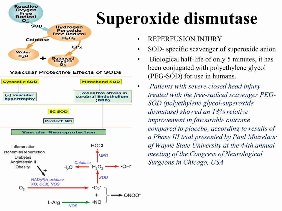
Superoxide dismutase• REPERFUSION INJURY• SOD- specific scavenger of superoxide anion • Biological half-life of only 5 minutes it hasBiological half life of only 5 minutes, it has
been conjugated with polyethylene glycol (PEG-SOD) for use in humans.
• Patients with severe closed head injury treated with the free-radical scavenger PEG-SOD (polyethylene glycol-superoxide dismutase) showed an 18% relative improvement in favourable outcomeimprovement in favourable outcome compared to placebo, according to results of a Phase III trial presented by Paul Muizelaar of Wayne State University at the 44th annual meeting of the Congress of Neurological Surgeons in Chicago, USA
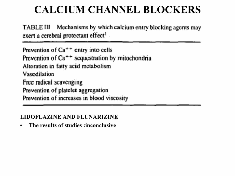
CALCIUM CHANNEL BLOCKERS
LIDOFLAZINE AND FLUNARIZINE• The results of studies :inconclusive• The results of studies :inconclusive
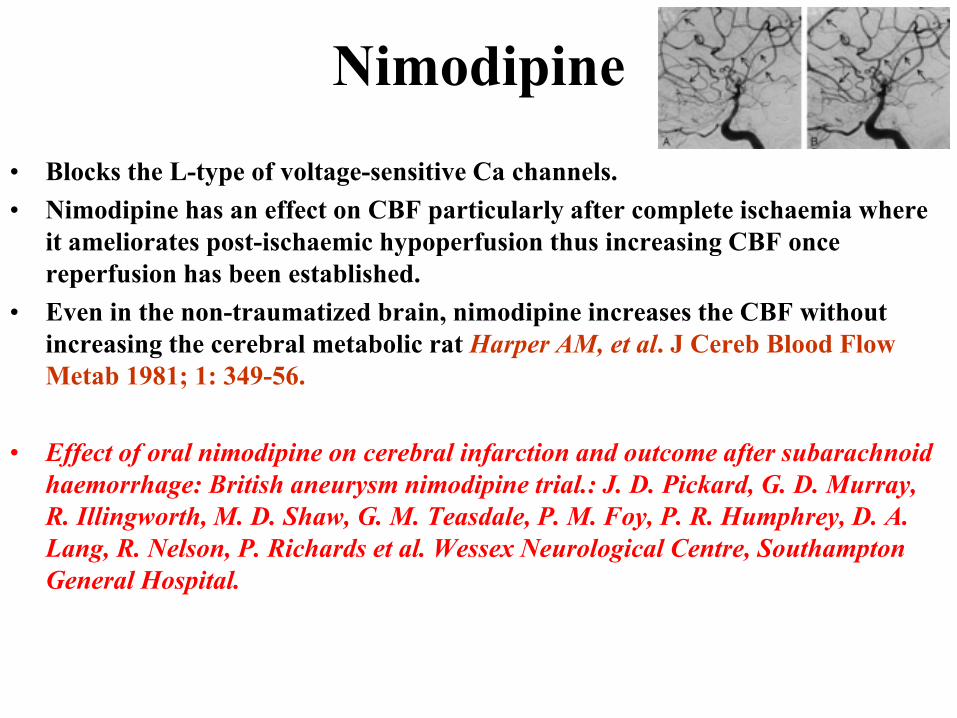
Nimodipine• Blocks the L-type of voltage-sensitive Ca channels.
Ni di i h ff t CBF ti l l ft l t i h i h• Nimodipine has an effect on CBF particularly after complete ischaemia where it ameliorates post-ischaemic hypoperfusion thus increasing CBF once reperfusion has been established. E i th t ti d b i i di i i th CBF ith t• Even in the non-traumatized brain, nimodipine increases the CBF without increasing the cerebral metabolic rat Harper AM, et al. J Cereb Blood Flow Metab 1981; 1: 349-56.
• Effect of oral nimodipine on cerebral infarction and outcome after subarachnoid haemorrhage: British aneurysm nimodipine trial.: J. D. Pickard, G. D. Murray, R Illi th M D Sh G M T d l P M F P R H h D AR. Illingworth, M. D. Shaw, G. M. Teasdale, P. M. Foy, P. R. Humphrey, D. A. Lang, R. Nelson, P. Richards et al. Wessex Neurological Centre, Southampton General Hospital.
Nimodipine in Sub-arachnoid haemorrhage
• The protective effect was attributed to the inhibition of cerebral arterial spasm by• The protective effect was attributed to the inhibition of cerebral arterial spasm by nimodipine.
• Allen et al. demonstrated a beneficial effect due to nimodipine as the occurrence of neurologic deficits and death were significantly reduced in treated patients.g g y p
Allen GS, et al. N Engl J Med ; 308: 619-24.
Nimodipine in head injuryp j y
• Nimodipine is efficacious in treating patients with severe head trauma but without producing adverse changes in ICP or systemic blood pressure.
Kostron H, et al. Neurol Res 1984; 6: 29-32.Nimodipine in stroke
• In a double-blind, placebo-controlled prospective study, nimodipine significantly d d t lit f ll d i t i h i t k ireduced mortality from all causes during acute ischaemic stroke in man.
• During a six-month follow-up, patients in the nimodipine group continued to show significant improvement when compared with the placebo group.
Gelmers H J, et al. N Engl J Med 1988; 318: 203-7.
LidocainePossible mechanisms for cerebral protection by lidocaine include1. deceleration of ischaemic transmembrane ion shifts2 reduction in CMR2. reduction in CMR3. modulation of leukocyte activity4. reduction of ischaemic excitotoxin release5. reduces intracranial hypertension
This effects of lidocaine (i.e., metabolic inhibition beyond that hi bl ith i l t i EEG l d d l i thachievable with an isoelectric EEG alone and a delay in the
ischaemic potassium efflux) resemble those of hypothermia.It was demonstrated by Astrup et al. in a canine global ischaemia
model that in the functionally arrested brain (i e an isoelectricmodel that in the functionally arrested brain (i.e., an isoelectricEEG induced by barbiturates), lidocaine can further reduce the metabolic rate by 15-20 per cent.
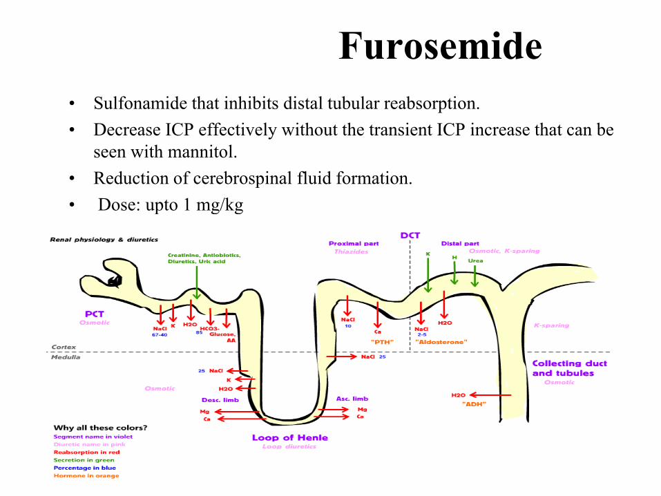
Furosemide• Sulfonamide that inhibits distal tubular reabsorption. • Decrease ICP effectively without the transient ICP increase that can be
seen with mannitol.• Reduction of cerebrospinal fluid formation.• Dose: upto 1 mg/kgp g g
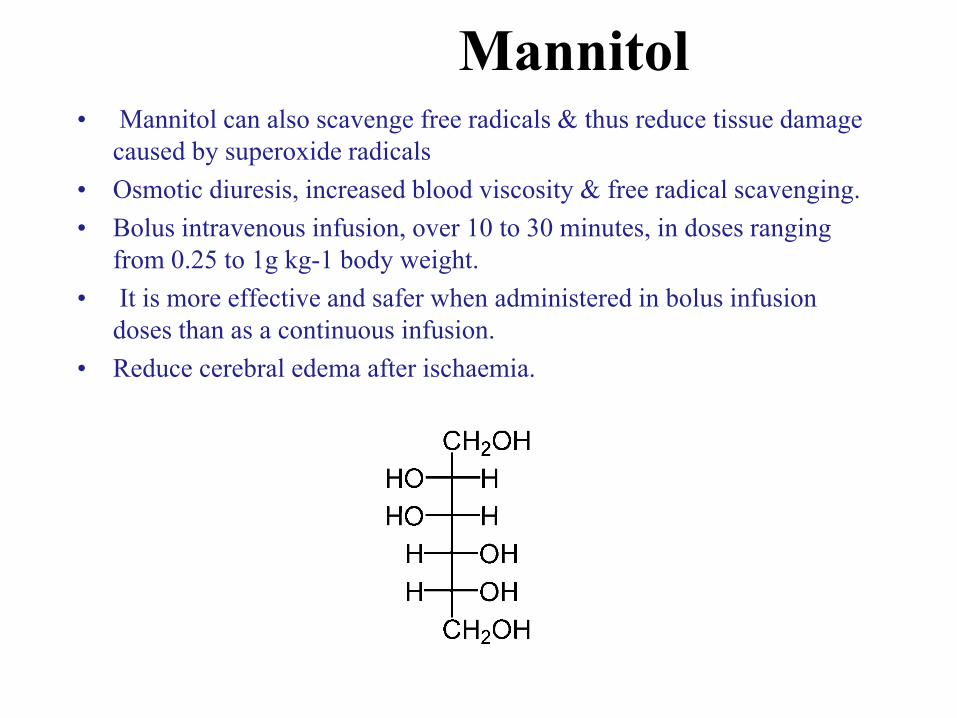
Mannitol• Mannitol can also scavenge free radicals & thus reduce tissue damage
caused by superoxide radicals• Osmotic diuresis, increased blood viscosity & free radical scavenging. • Bolus intravenous infusion, over 10 to 30 minutes, in doses ranging
from 0.25 to 1g kg-1 body weight.• It is more effective and safer when administered in bolus infusion
doses than as a continuous infusion.• Reduce cerebral edema after ischaemia.
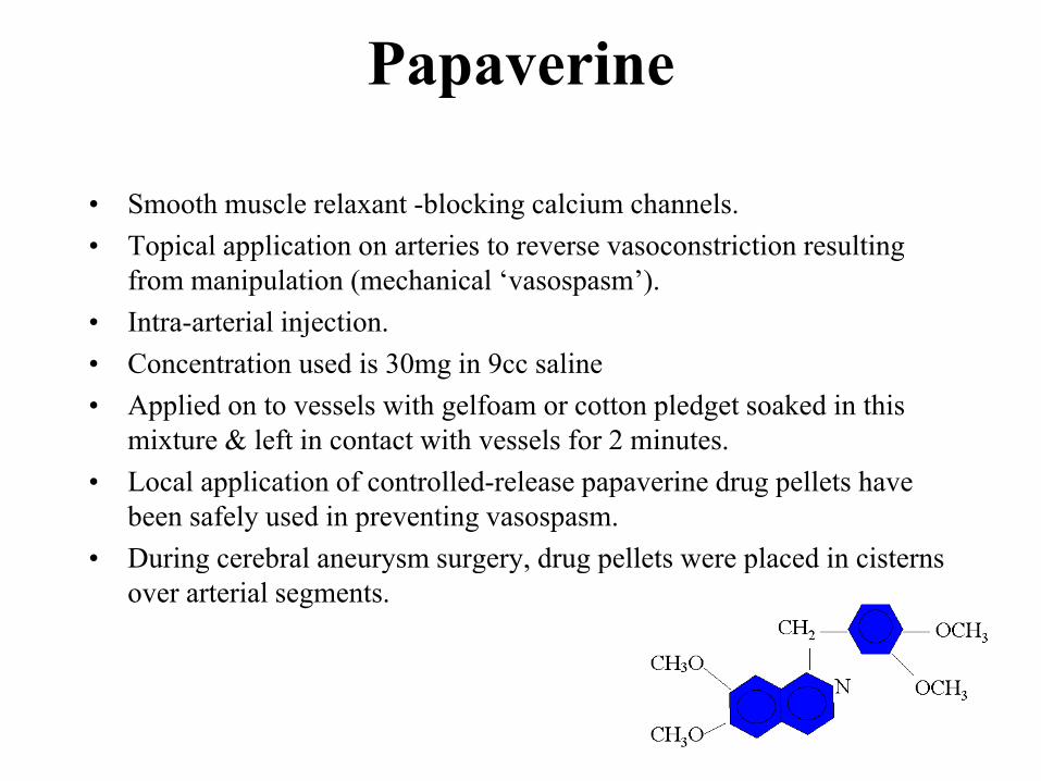
Papaverine
• Smooth muscle relaxant -blocking calcium channels.• Topical application on arteries to reverse vasoconstriction resulting
from manipulation (mechanical ‘vasospasm’).• Intra-arterial injection. j• Concentration used is 30mg in 9cc saline• Applied on to vessels with gelfoam or cotton pledget soaked in this
mixture & left in contact with vessels for 2 minutesmixture & left in contact with vessels for 2 minutes.• Local application of controlled-release papaverine drug pellets have
been safely used in preventing vasospasm.• During cerebral aneurysm surgery drug pellets were placed in cisterns• During cerebral aneurysm surgery, drug pellets were placed in cisterns
over arterial segments.
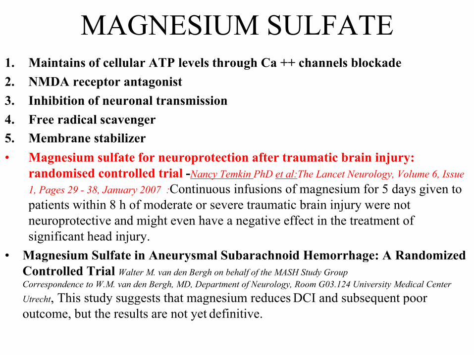
MAGNESIUM SULFATE1. Maintains of cellular ATP levels through Ca ++ channels blockade 2. NMDA receptor antagonist3 Inhibition of neuronal transmission3. Inhibition of neuronal transmission4. Free radical scavenger5. Membrane stabilizer• Magnesium sulfate for neuroprotection after traumatic brain injury:
randomised controlled trial -Nancy Temkin PhD et al:The Lancet Neurology, Volume 6, Issue 1, Pages 29 - 38, January 2007 :Continuous infusions of magnesium for 5 days given to
ti t ithi 8 h f d t t ti b i i j tpatients within 8 h of moderate or severe traumatic brain injury were not neuroprotective and might even have a negative effect in the treatment of significant head injury.
• Magnesium Sulfate in Aneurysmal Subarachnoid Hemorrhage: A Randomized• Magnesium Sulfate in Aneurysmal Subarachnoid Hemorrhage: A Randomized Controlled Trial Walter M. van den Bergh on behalf of the MASH Study GroupCorrespondence to W.M. van den Bergh, MD, Department of Neurology, Room G03.124 University Medical CenterUtrecht, This study suggests that magnesium reduces DCI and subsequent poor , y gg g q poutcome, but the results are not yet definitive.
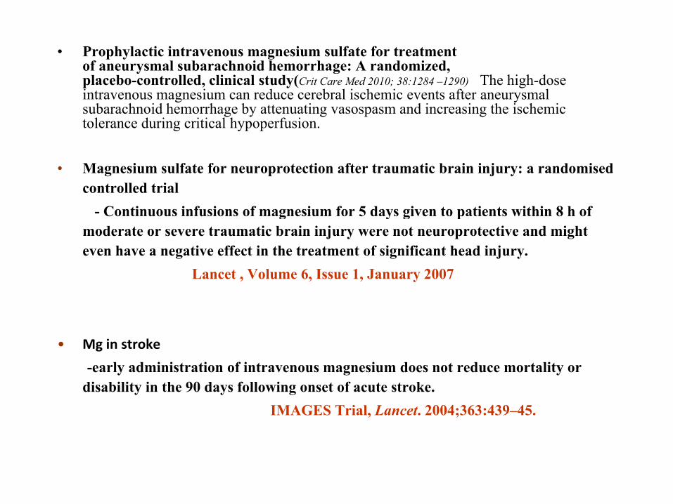
• Prophylactic intravenous magnesium sulfate for treatmentof aneurysmal subarachnoid hemorrhage: A randomized,y g ,placebo-controlled, clinical study(Crit Care Med 2010; 38:1284 –1290) The high-dose intravenous magnesium can reduce cerebral ischemic events after aneurysmal subarachnoid hemorrhage by attenuating vasospasm and increasing the ischemic tolerance during critical hypoperfusion.
• Magnesium sulfate for neuroprotection after traumatic brain injury: a randomised controlled trial
- Continuous infusions of magnesium for 5 days given to patients within 8 h ofContinuous infusions of magnesium for 5 days given to patients within 8 h of moderate or severe traumatic brain injury were not neuroprotective and might even have a negative effect in the treatment of significant head injury.
Lancet , Volume 6, Issue 1, January 2007
• Mg in stroke
-early administration of intravenous magnesium does not reduce mortality or disability in the 90 days following onset of acute stroke.
IMAGES Trial, Lancet. 2004;363:439–45.
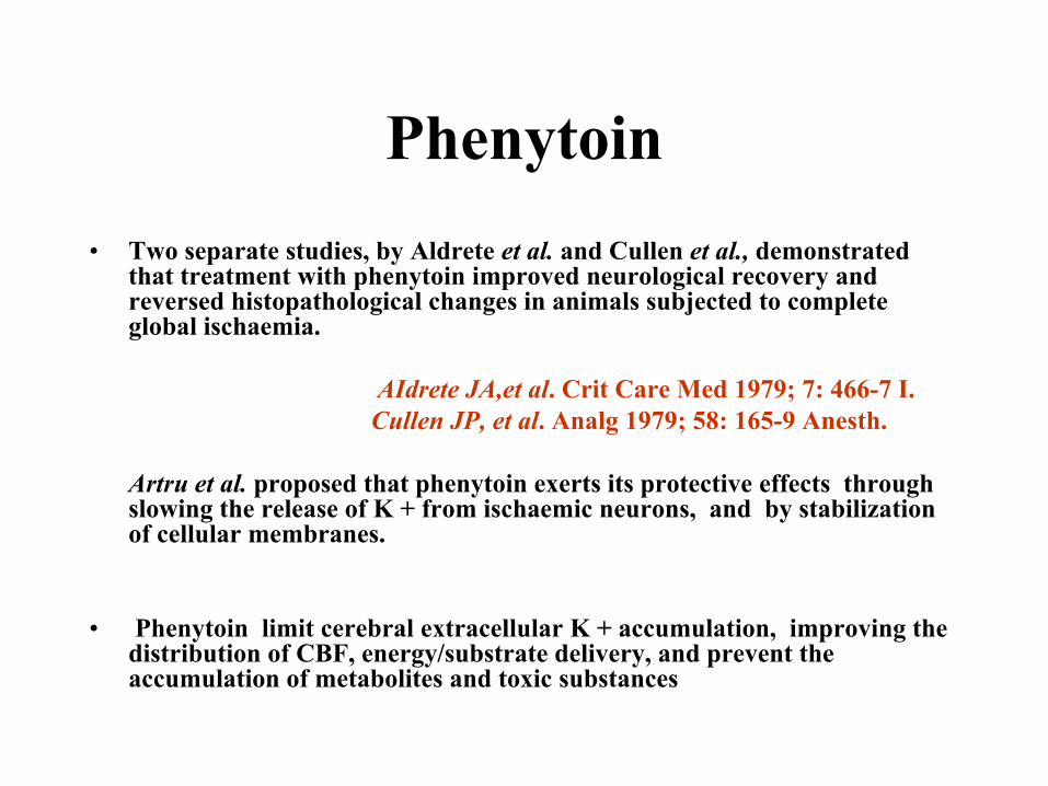
Phenytoin• Two separate studies, by Aldrete et al. and Cullen et al., demonstrated
that treatment with phenytoin improved neurological recovery and reversed histopathological changes in animals subjected to complete global ischaemia. g
AIdrete JA,et al. Crit Care Med 1979; 7: 466-7 I.Cullen JP, et al. Analg 1979; 58: 165-9 Anesth.
Artru et al. proposed that phenytoin exerts its protective effects through slowing the release of K + from ischaemic neurons, and by stabilization of cellular membranes.
• Phenytoin limit cerebral extracellular K + accumulation, improving the distribution of CBF, energy/substrate delivery, and prevent the
l ti f t b lit d t i b taccumulation of metabolites and toxic substances
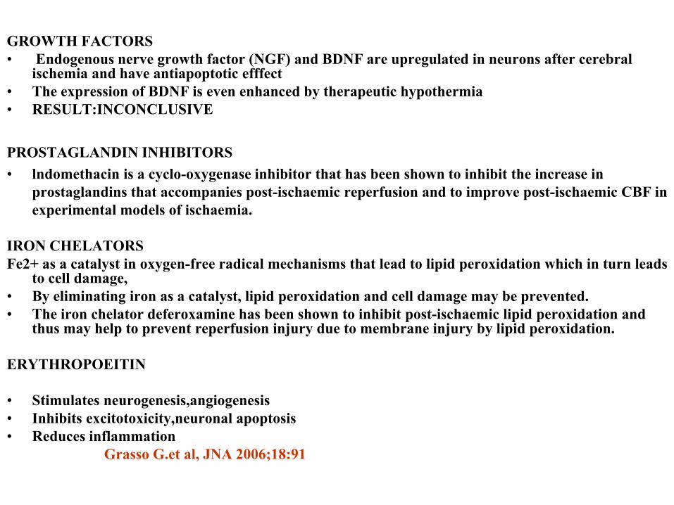
GROWTH FACTORS• Endogenous nerve growth factor (NGF) and BDNF are upregulated in neurons after cerebral
ischemia and have antiapoptotic efffectischemia and have antiapoptotic efffect• The expression of BDNF is even enhanced by therapeutic hypothermia• RESULT:INCONCLUSIVE
PROSTAGLANDIN INHIBITORS• lndomethacin is a cyclo-oxygenase inhibitor that has been shown to inhibit the increase in
prostaglandins that accompanies post-ischaemic reperfusion and to improve post-ischaemic CBF in experimental models of ischaemia.p
IRON CHELATORSFe2+ as a catalyst in oxygen-free radical mechanisms that lead to lipid peroxidation which in turn leads
to cell damage, • By eliminating iron as a catalyst, lipid peroxidation and cell damage may be prevented. • The iron chelator deferoxamine has been shown to inhibit post-ischaemic lipid peroxidation and
thus may help to prevent reperfusion injury due to membrane injury by lipid peroxidation.
ERYTHROPOEITINERYTHROPOEITIN
• Stimulates neurogenesis,angiogenesis• Inhibits excitotoxicity,neuronal apoptosis
R d i fl ti• Reduces inflammationGrasso G.et al, JNA 2006;18:91
xenon
• NMDA antagonist• Upregulation of genes and synthesis of the CREB-dependent survival proteins Bcl-p g g y p p
2 ,BDNF,Ras protein).D Ma et al, Journal of Cerebral Blood Flow & Metabolism (2006) 26, 199–208
Gene therapy for neuroprotection• Direct application of neurotropic factors (VEGF,GDGF,ILGF-1 )through
adenoviral construct(Ad-p65).
• ZFP transcription factor gene therapy to increase expression of the full complement of VEGF-A splice variants is a promising avenue for the treatment of nerve injury and neuro degeneration.
G Th (2009) 16 1292 1299Gene Therapy (2009) 16, 1292–1299
T i l li i f GDNF i l d d h i f i d• Topical application of GDNF protein greatly reduced the infarct size and brain edema at 24 hr of continuous MCAO in rats. GDNF protein showed a direct protective effect against ischemic brain damage, but not secondary by improving CBF.y y p g
Clinical Neurology.43;11;894-896(2003)

THANK YOU