Embed Size (px)
Citation preview
Cestodes
Tapeworms from man and animals
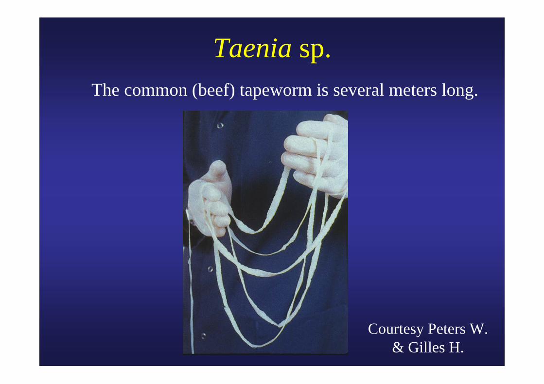
Taenia sp. The common (beef) tapeworm is several meters long.
Courtesy Peters W. & Gilles H.
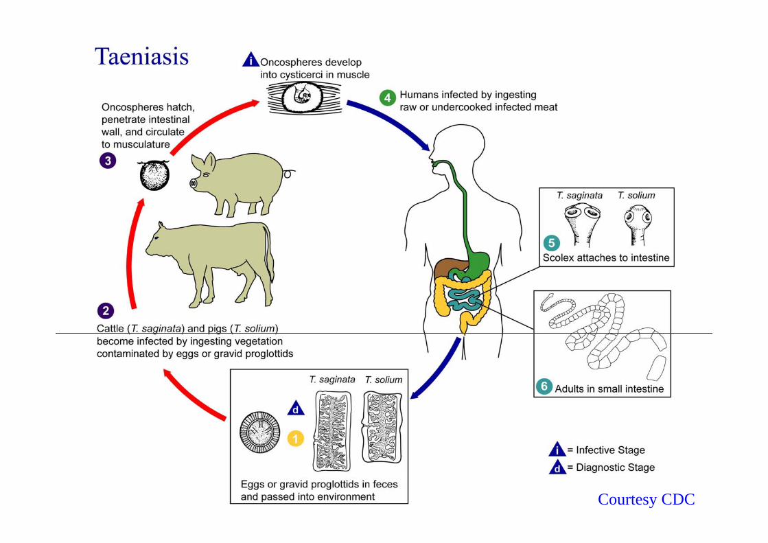
Courtesy CDC
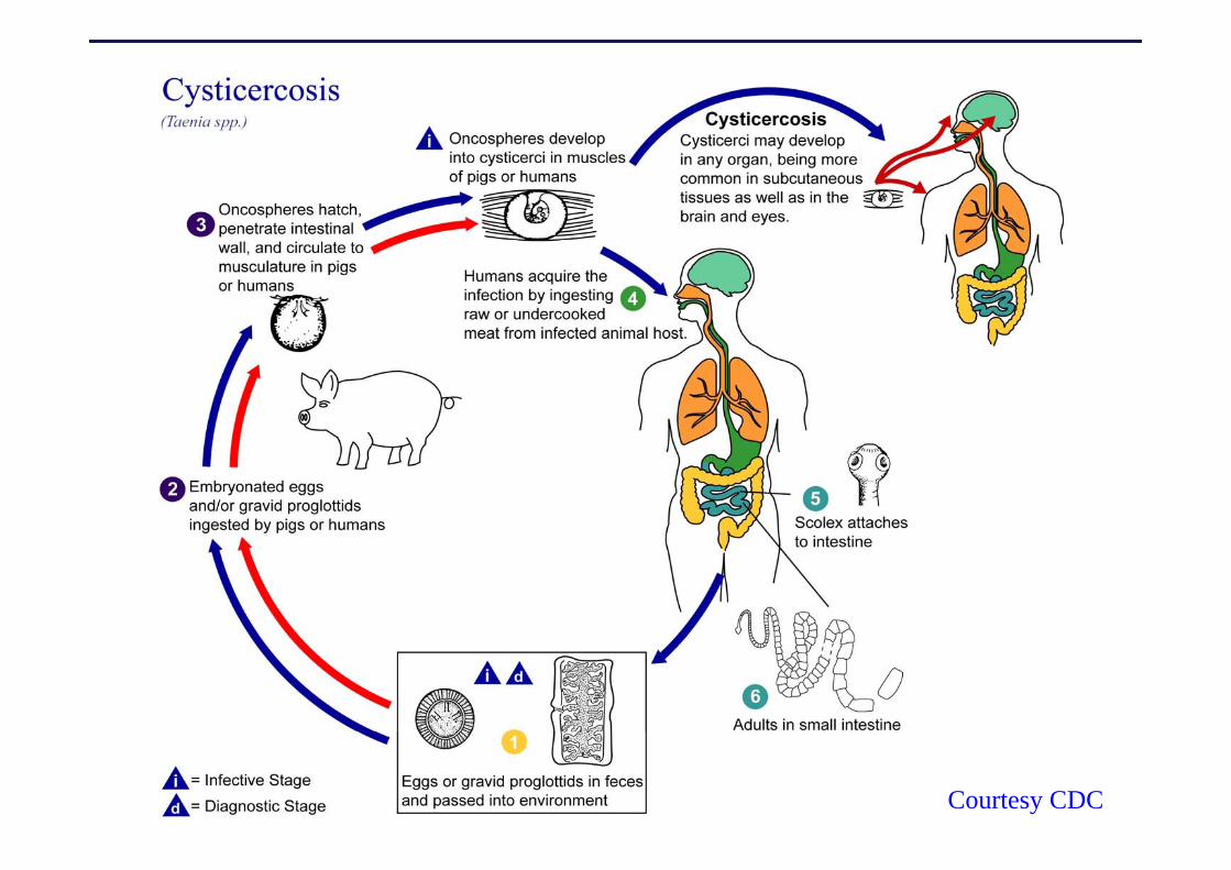
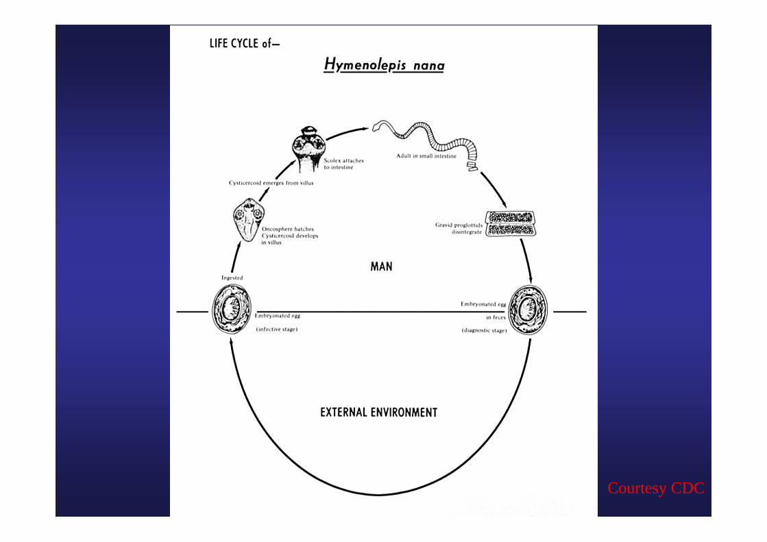
Courtesy CDC
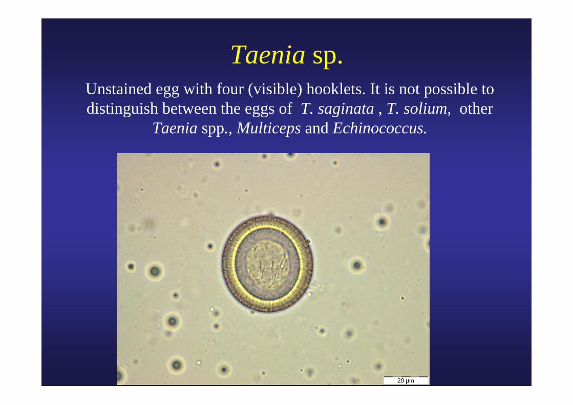
Taenia sp.Unstained egg with four (visible) hooklets. It is not possible to distinguish between the eggs of T. saginata , T. solium, other
Taenia spp., Multiceps and Echinococcus.
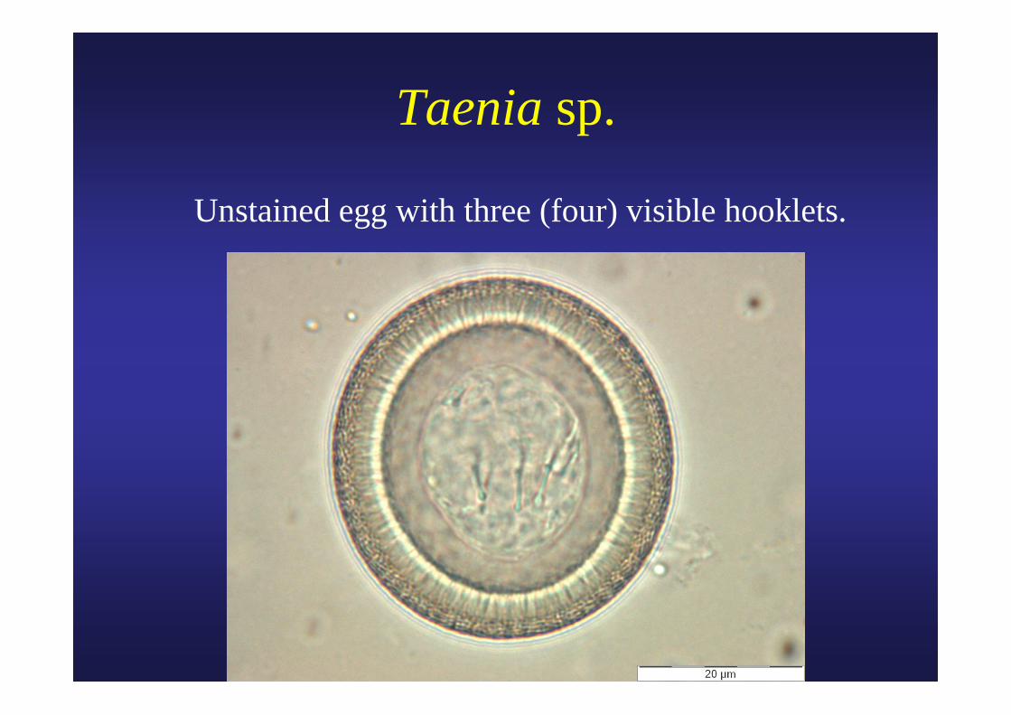
Taenia sp.
Unstained egg with three (four) visible hooklets.
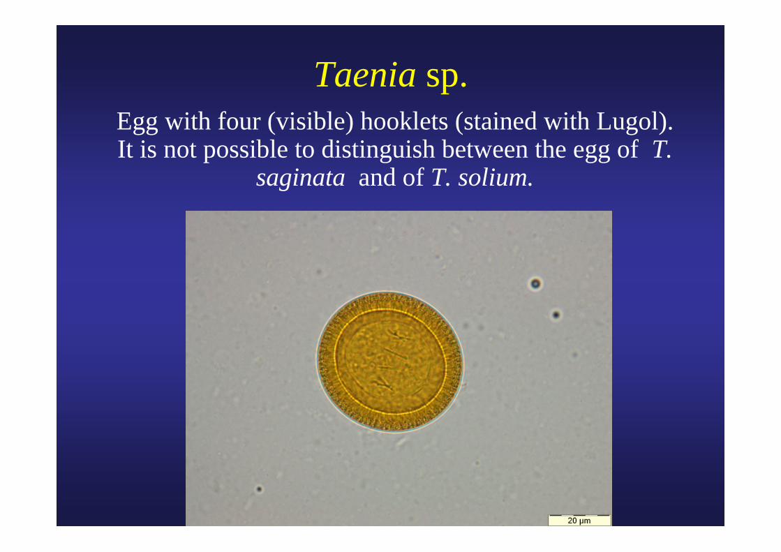
Taenia sp.Egg with four (visible) hooklets (stained with Lugol). It is not possible to distinguish between the egg of T.
saginata and of T. solium.
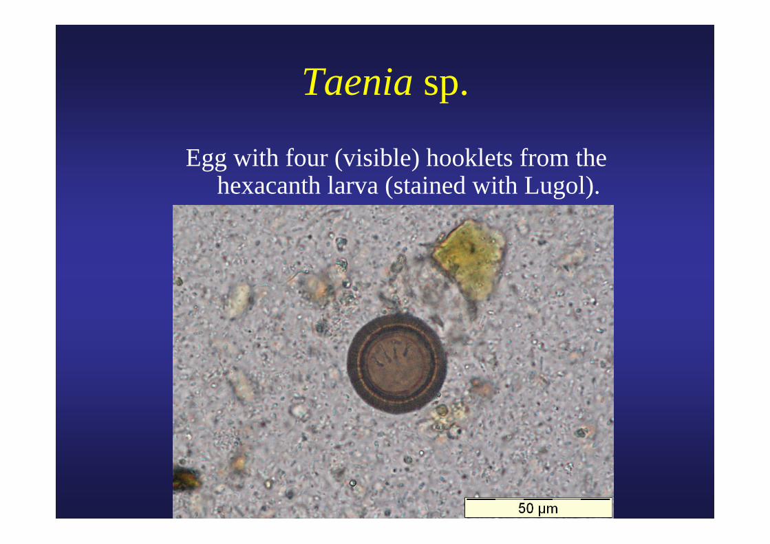
Taenia sp.
Egg with four (visible) hooklets from the hexacanth larva (stained with Lugol).
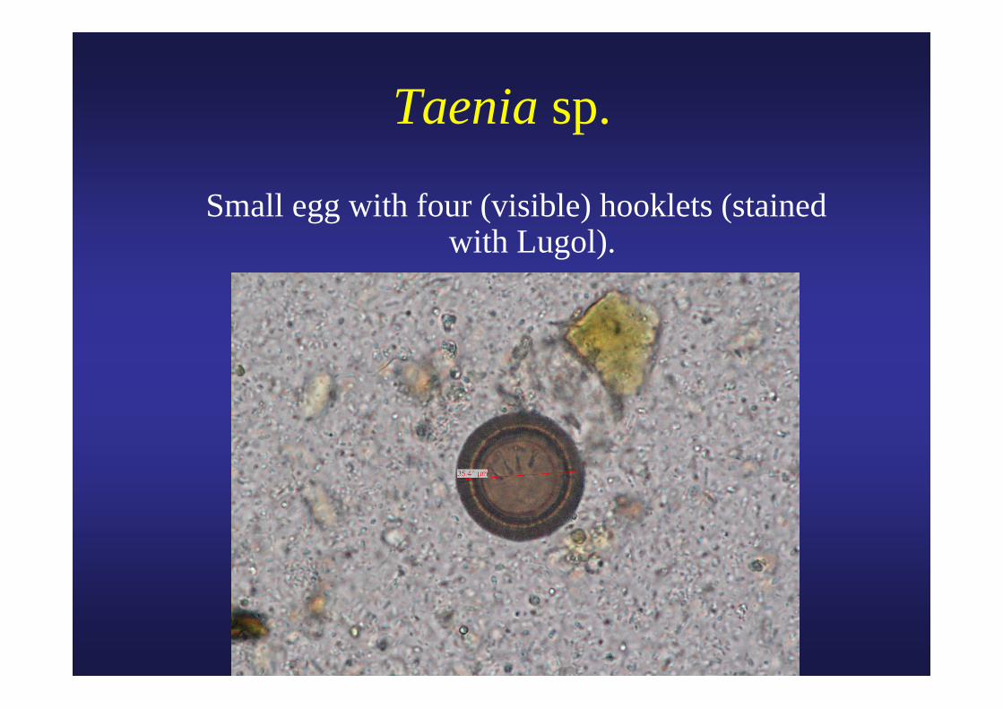
Taenia sp.
Small egg with four (visible) hooklets (stained with Lugol).
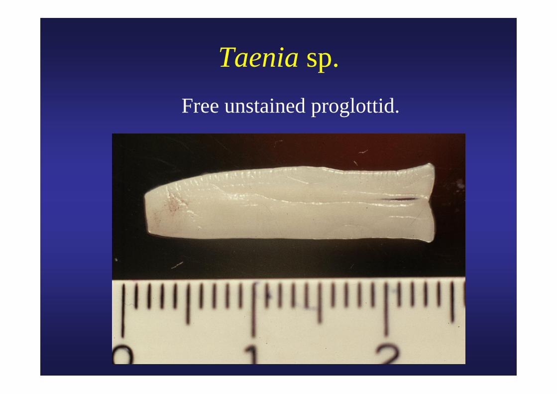
Taenia sp.Free unstained proglottid.
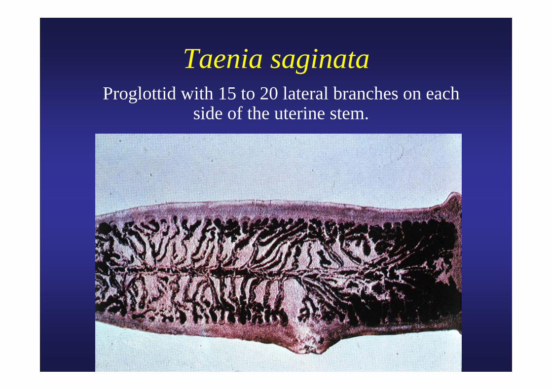
Taenia saginataProglottid with 15 to 20 lateral branches on each
side of the uterine stem.
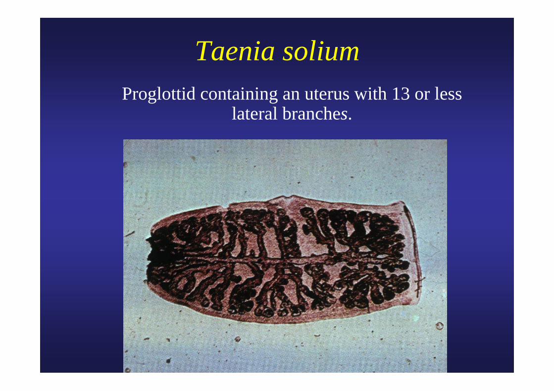
Taenia soliumProglottid containing an uterus with 13 or less
lateral branches.
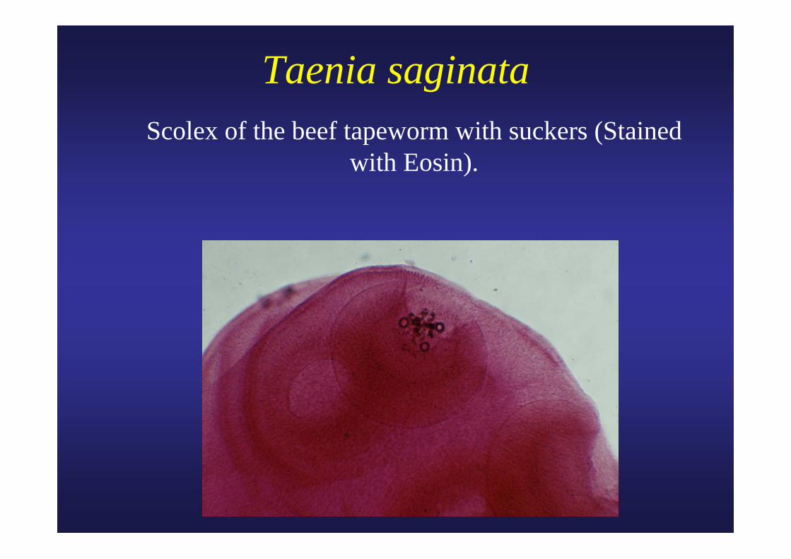
Taenia saginata Scolex of the beef tapeworm with suckers (Stained
with Eosin).
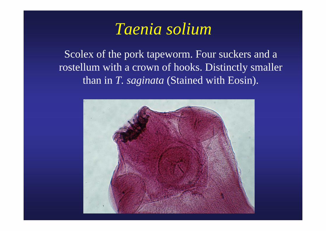
Taenia solium Scolex of the pork tapeworm. Four suckers and a rostellum with a crown of hooks. Distinctly smaller
than in T. saginata (Stained with Eosin).
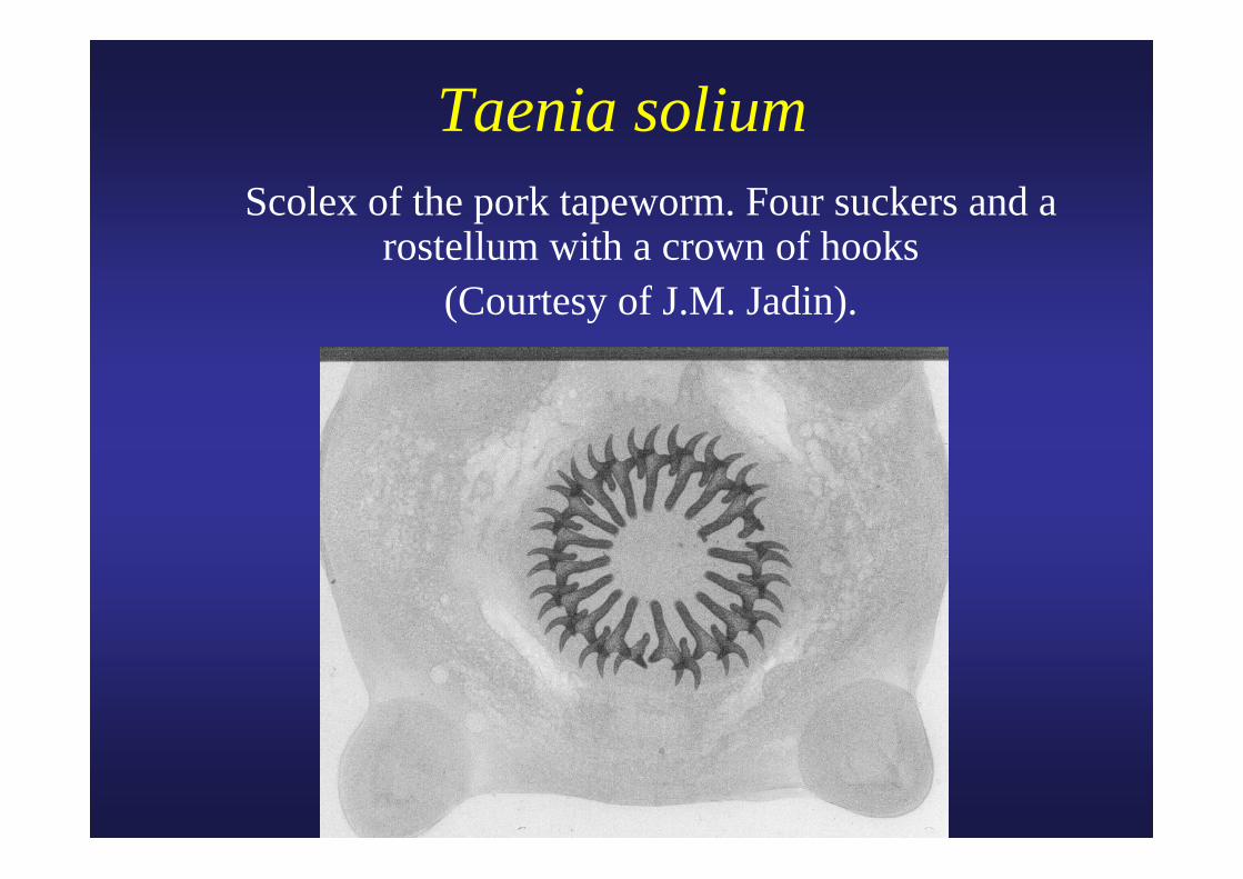
Taenia solium Scolex of the pork tapeworm. Four suckers and a
rostellum with a crown of hooks (Courtesy of J.M. Jadin).
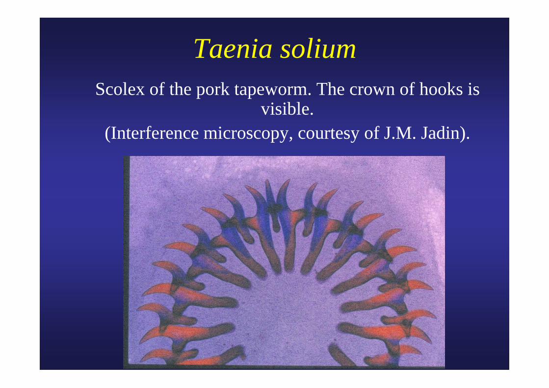
Taenia solium Scolex of the pork tapeworm. The crown of hooks is
visible. (Interference microscopy, courtesy of J.M. Jadin).
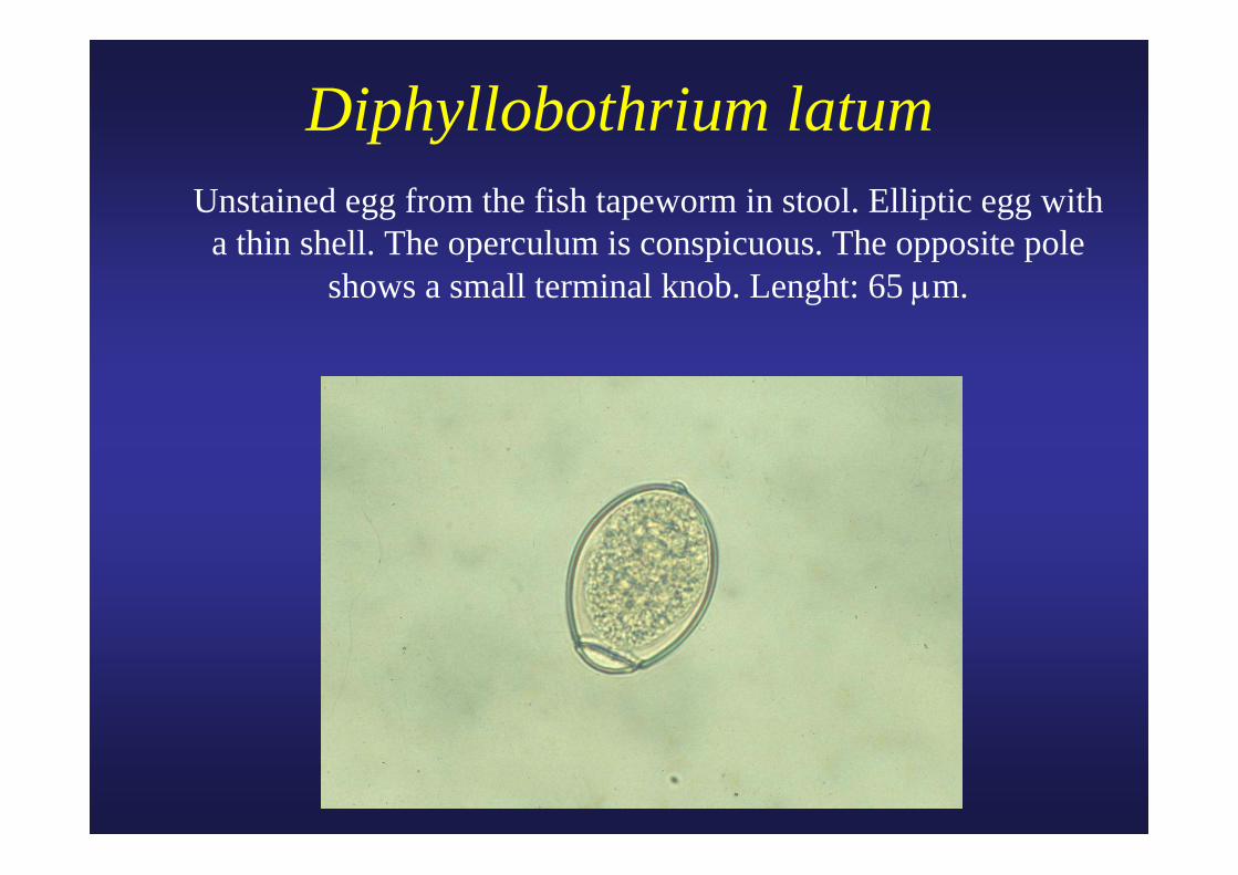
Diphyllobothrium latum Unstained egg from the fish tapeworm in stool. Elliptic egg with
a thin shell. The operculum is conspicuous. The opposite pole shows a small terminal knob. Lenght: 65 μm.
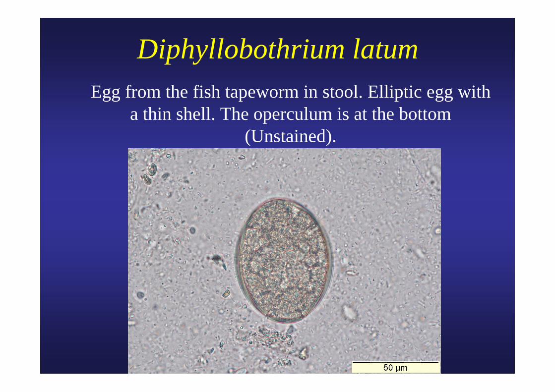
Diphyllobothrium latum Egg from the fish tapeworm in stool. Elliptic egg with
a thin shell. The operculum is at the bottom (Unstained).
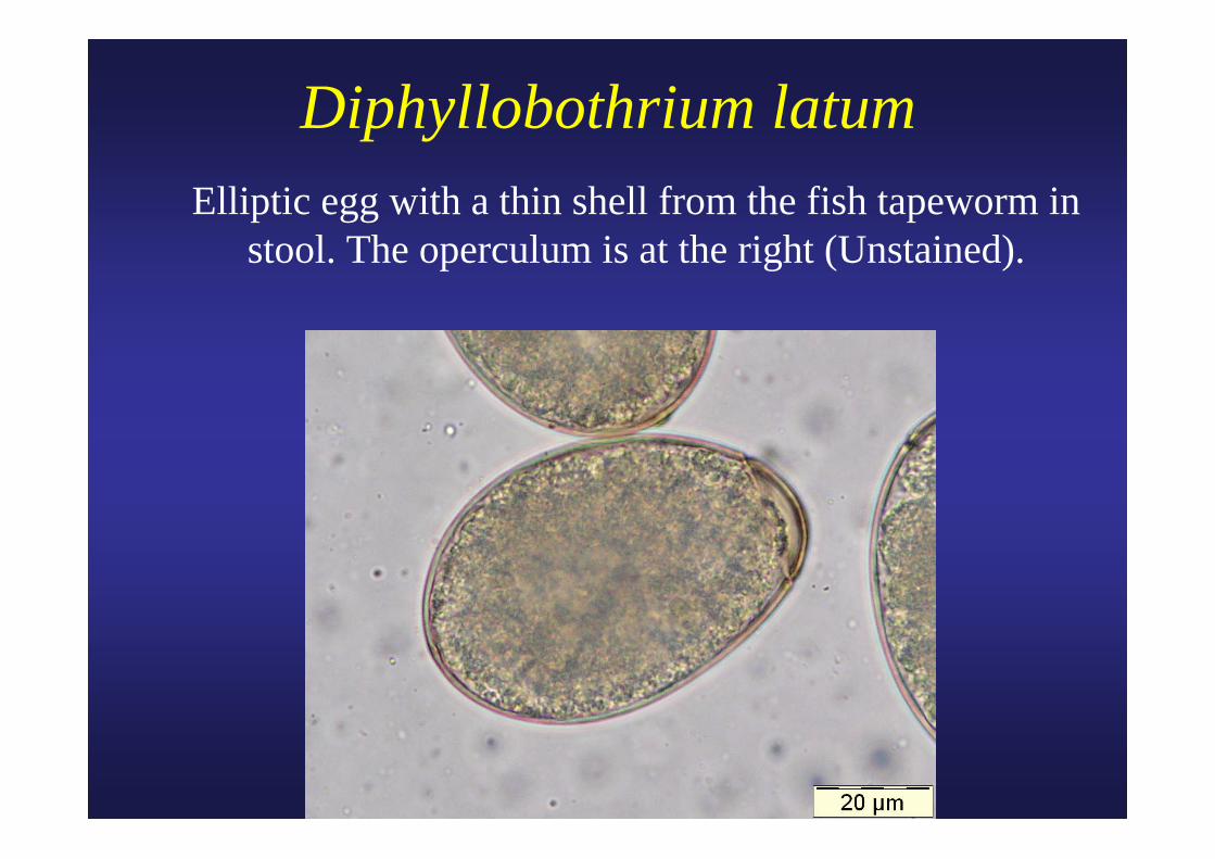
Diphyllobothrium latum Elliptic egg with a thin shell from the fish tapeworm in
stool. The operculum is at the right (Unstained).
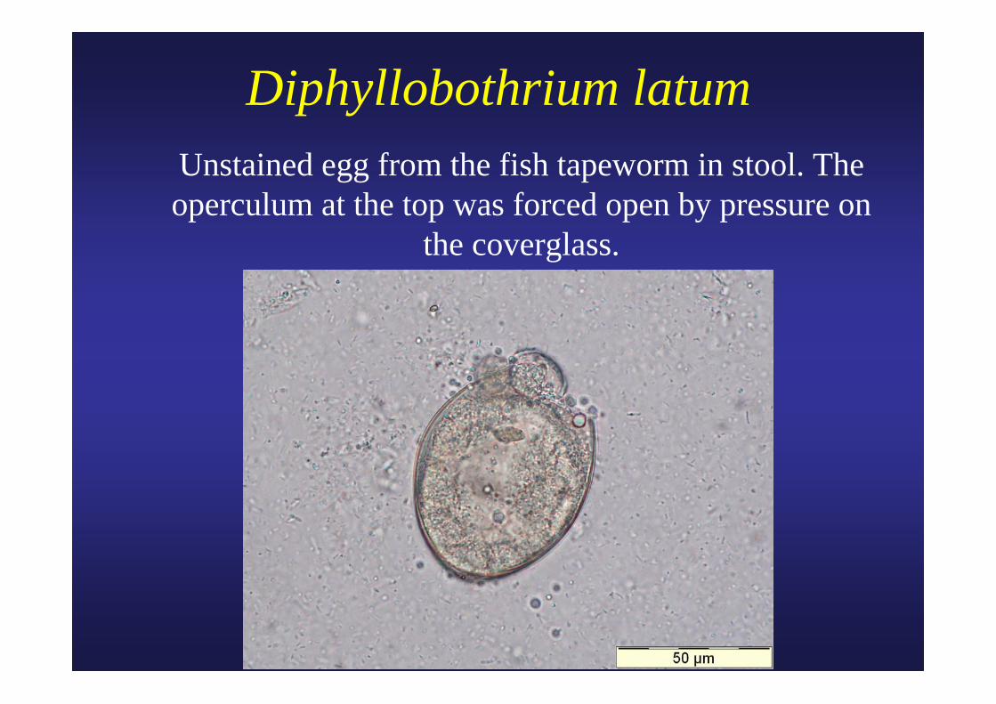
Diphyllobothrium latum Unstained egg from the fish tapeworm in stool. The
operculum at the top was forced open by pressure on the coverglass.
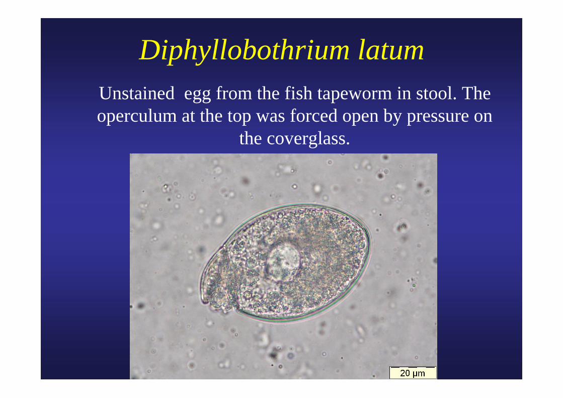
Diphyllobothrium latum Unstained egg from the fish tapeworm in stool. The
operculum at the top was forced open by pressure on the coverglass.
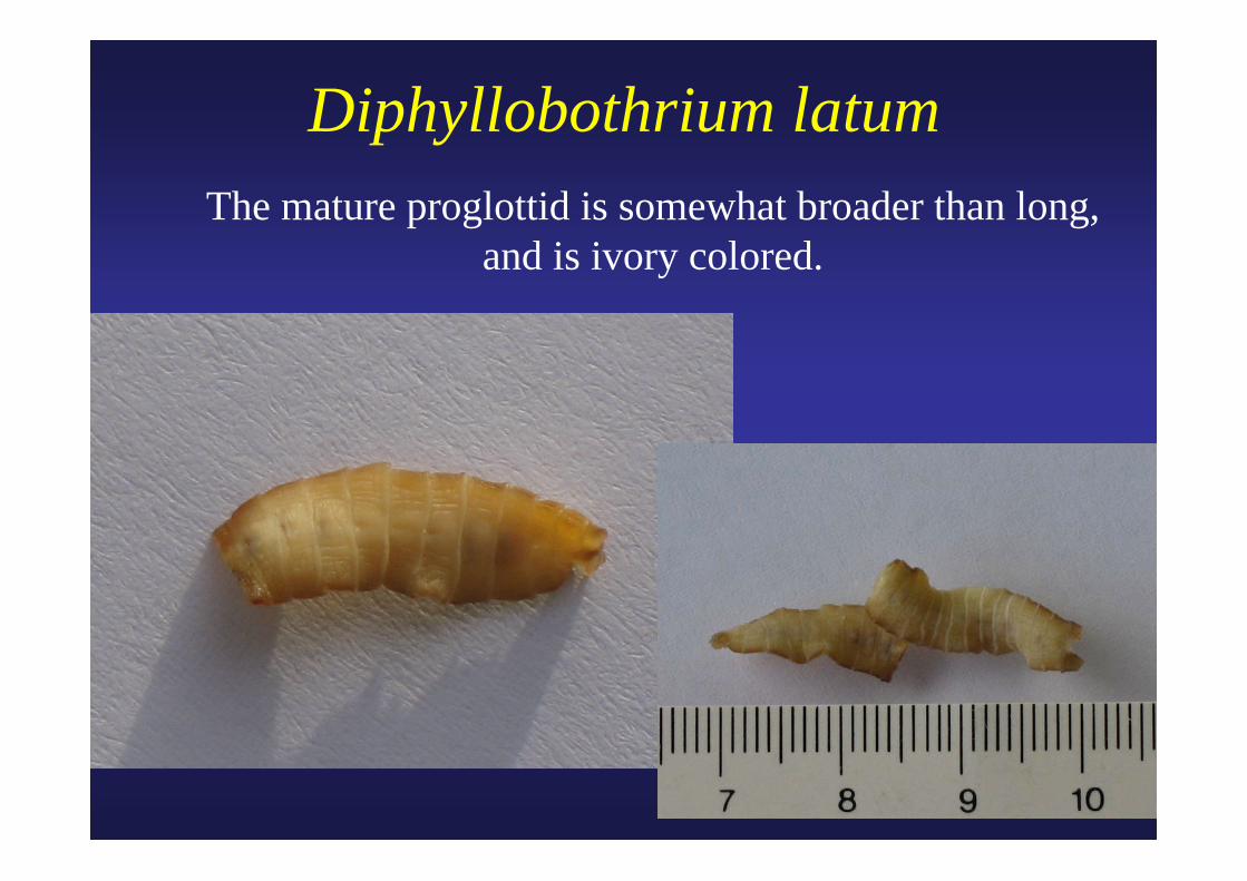
Diphyllobothrium latum The mature proglottid is somewhat broader than long,
and is ivory colored.
Courtesy CDC
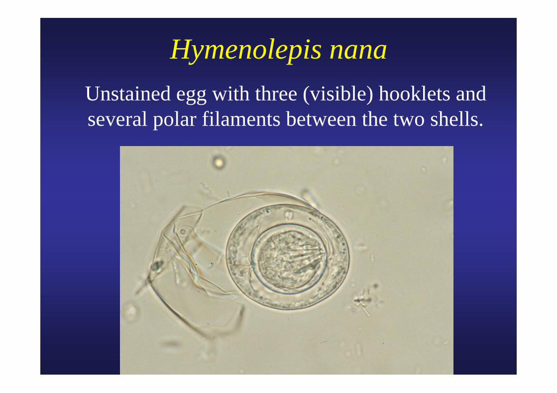
Hymenolepis nanaUnstained egg with three (visible) hooklets and several polar filaments between the two shells.
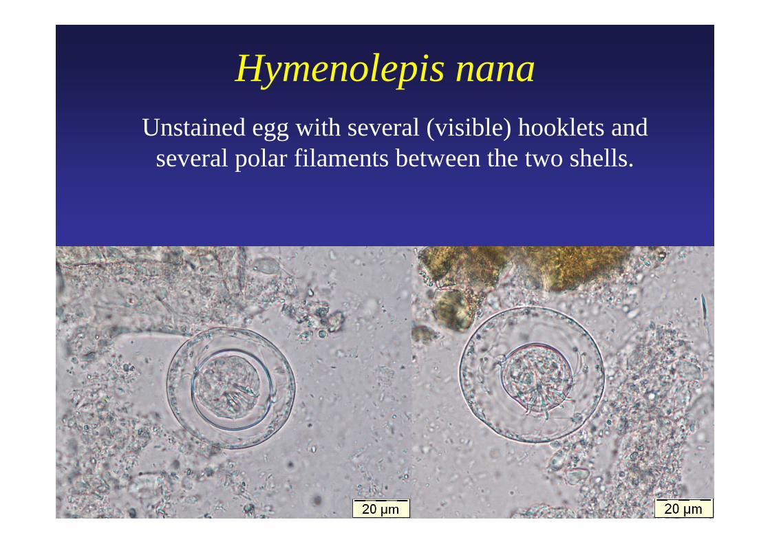
Hymenolepis nanaUnstained egg with several (visible) hooklets and
several polar filaments between the two shells.
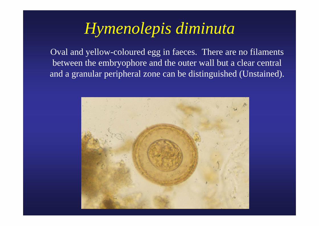
Hymenolepis diminuta Oval and yellow-coloured egg in faeces. There are no filaments
between the embryophore and the outer wall but a clear central and a granular peripheral zone can be distinguished (Unstained).
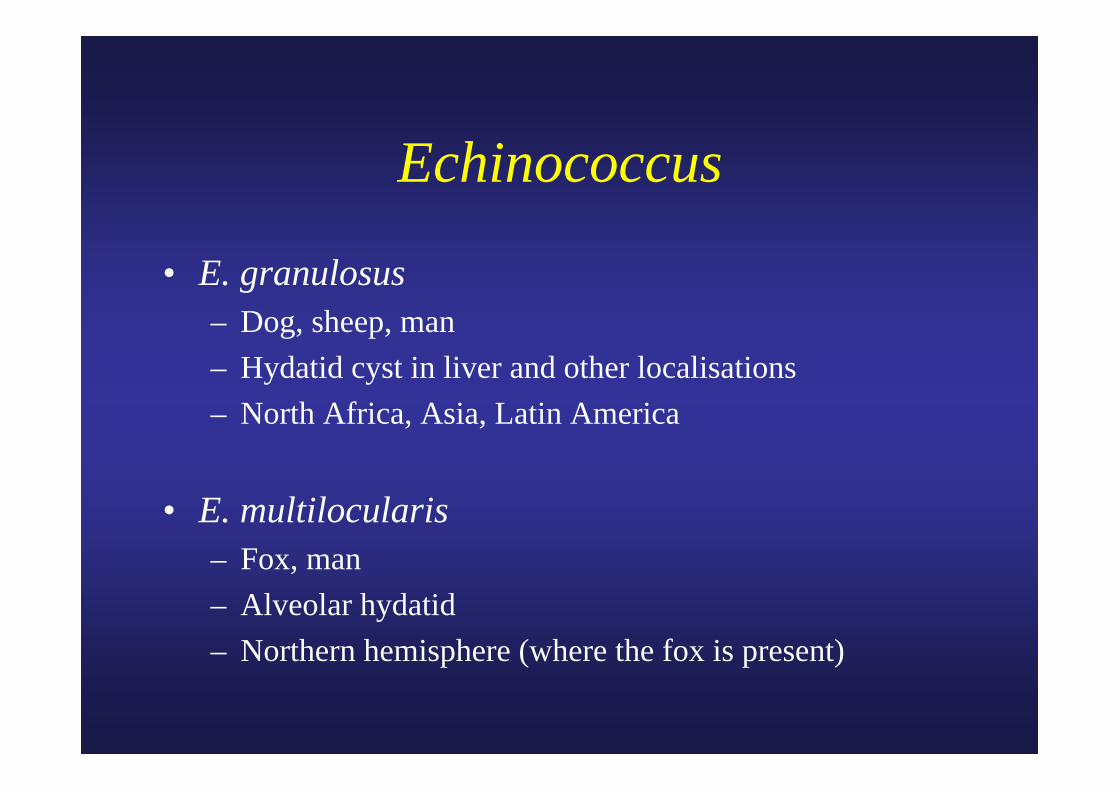
Echinococcus
• E. granulosus– Dog, sheep, man– Hydatid cyst in liver and other localisations– North Africa, Asia, Latin America
• E. multilocularis– Fox, man– Alveolar hydatid– Northern hemisphere (where the fox is present)
The free brood capsules and the free scolices are collectively referred to as “hydatid sand”.
However recurrence of the disease, due primarily to the development of secondary cysts from scolices spilled from the primary cyst at the time of operation, may be anticipated in 50% of the cases...
Beaver P.C. et al. 1984.
Echinococcus granulosus
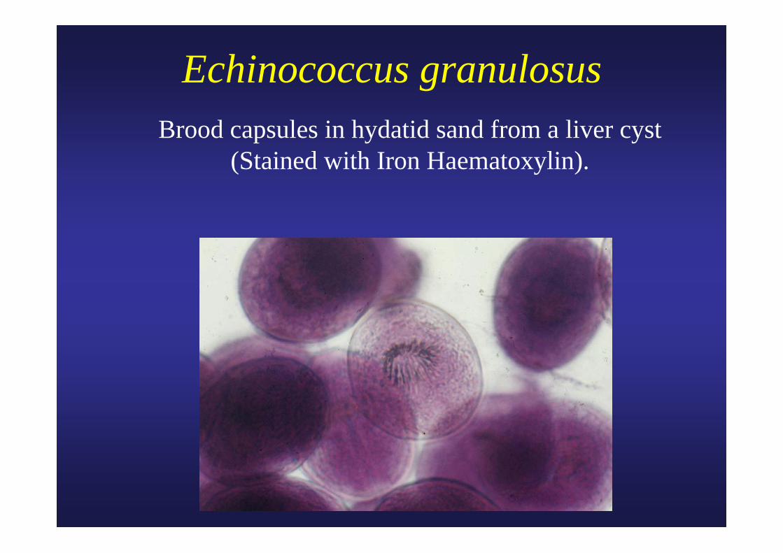
Echinococcus granulosus Brood capsules in hydatid sand from a liver cyst
(Stained with Iron Haematoxylin).
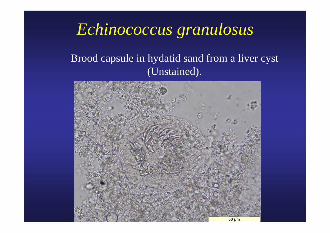
Echinococcus granulosus Brood capsule in hydatid sand from a liver cyst
(Unstained).
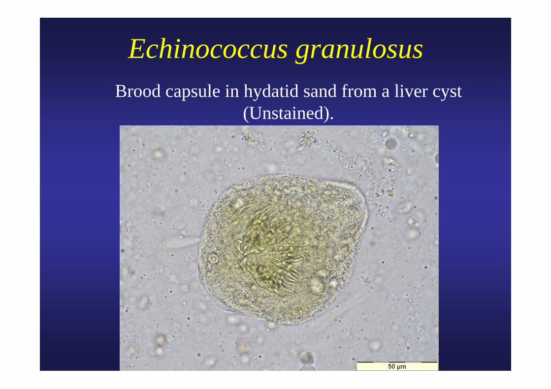
Echinococcus granulosus Brood capsule in hydatid sand from a liver cyst
(Unstained).
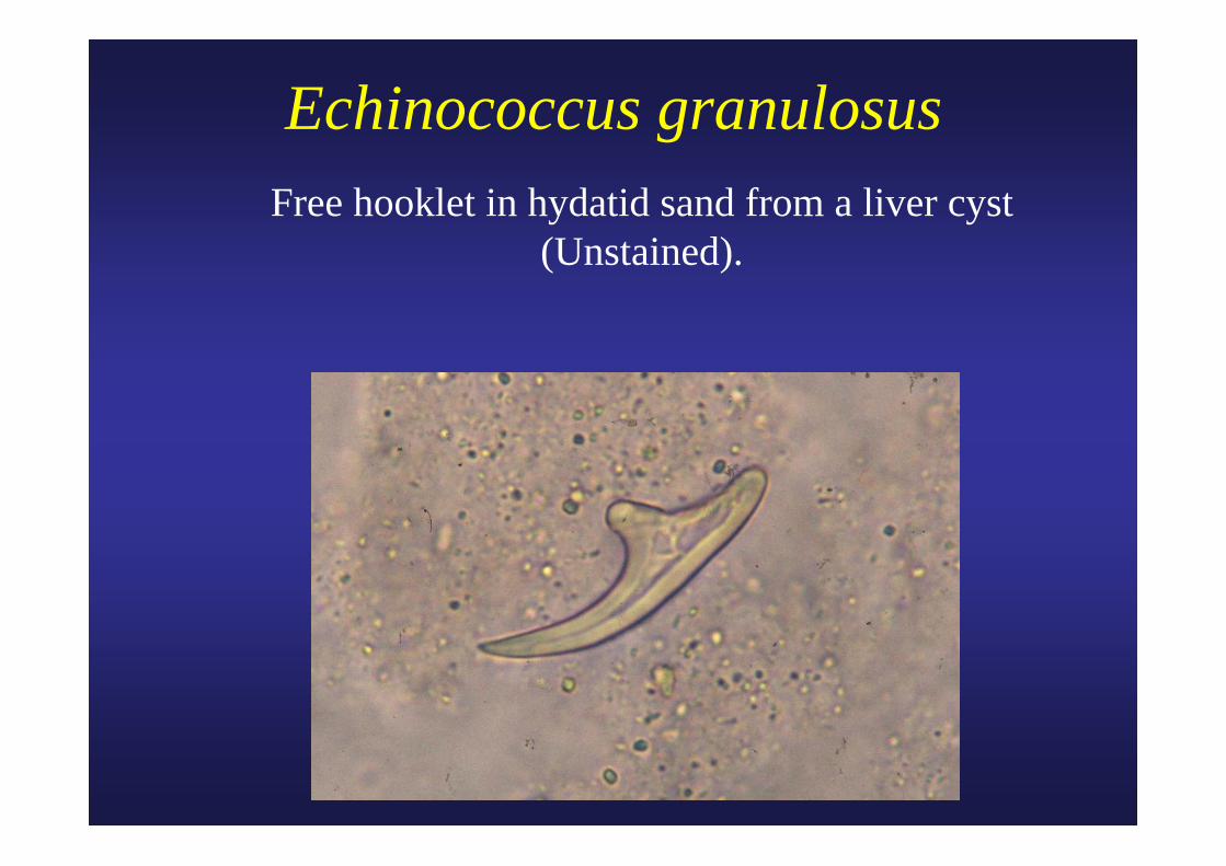
Echinococcus granulosus Free hooklet in hydatid sand from a liver cyst
(Unstained).
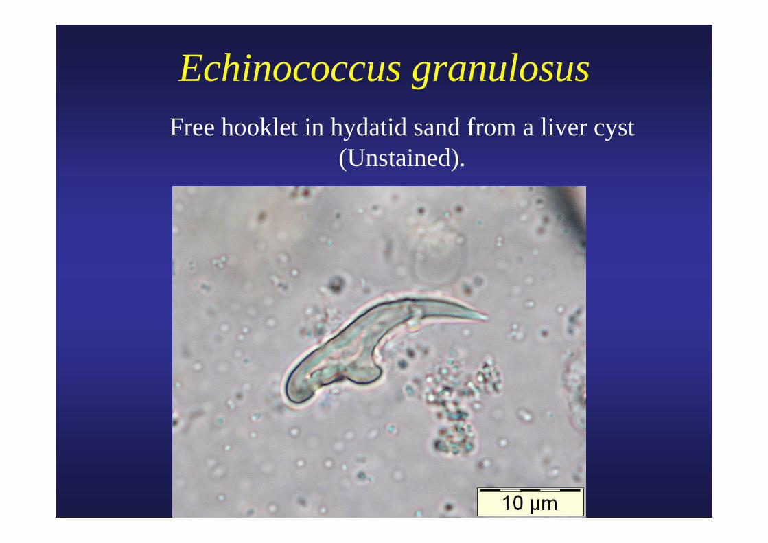
Echinococcus granulosus Free hooklet in hydatid sand from a liver cyst
(Unstained).
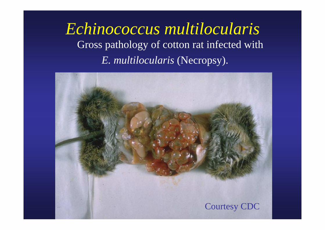
Echinococcus multilocularis Gross pathology of cotton rat infected with
E. multilocularis (Necropsy).
Courtesy CDC