Embed Size (px)
Citation preview
Ch. 15Special Senses: Vision
Slides mostly © Marieb & Hoehn 9th ed.Other slides by WCR
© 2013 Pearson Education, Inc.
Light And Optics: Wavelength And Color
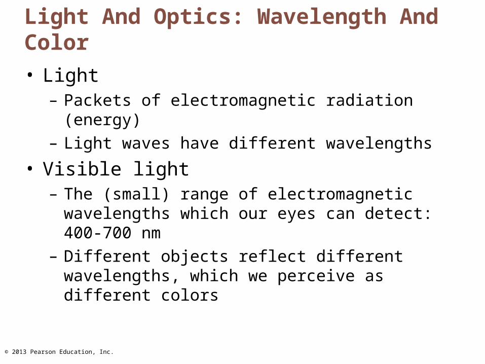
• Light– Packets of electromagnetic radiation (energy)– Light waves have different wavelengths
• Visible light– The (small) range of electromagnetic wavelengths
which our eyes can detect: 400-700 nm– Different objects reflect different wavelengths, which
we perceive as different colors
© 2013 Pearson Education, Inc.
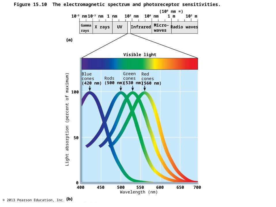
Figure 15.10 The electromagnetic spectrum and photoreceptor sensitivities.
10–5 nm
Gamma
raysX rays UV Infrared Micro-
wavesRadio waves
Visible light
Bluecones(420 nm)
Rods(500 nm)
Greencones(530 nm)
Redcones(560 nm)
Light
ab
sorp
tion
(perc
ent
of
maxim
um
)
100
50
0400
10–3 nm 1 nm 103 nm 106 nm 1 m 103 m(109 nm =)
Wavelength (nm)450 500 550 600 650 700
© 2013 Pearson Education, Inc.
Light And Optics: Refraction And Lenses
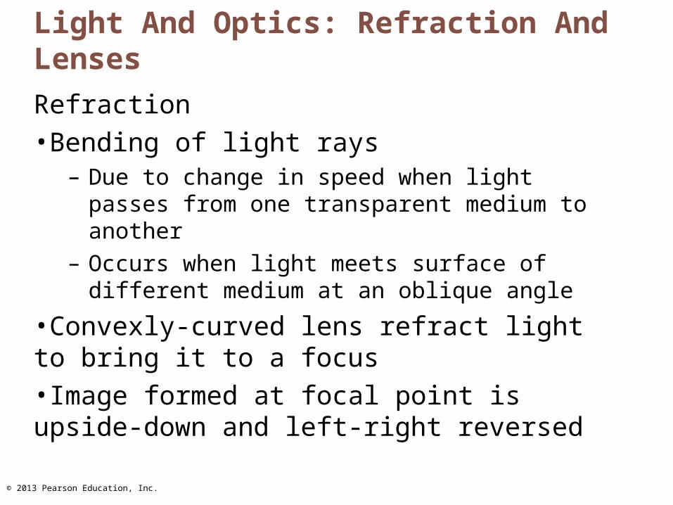
Refraction•Bending of light rays
– Due to change in speed when light passes from one transparent medium to another
– Occurs when light meets surface of different medium at an oblique angle
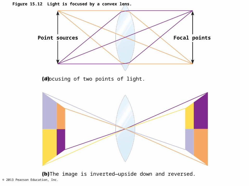
•Convexly-curved lens refract light to bring it to a focus•Image formed at focal point is upside-down and left-right reversed

© 2013 Pearson Education, Inc.
Figure 15.11 Refraction.
© 2013 Pearson Education, Inc.
Figure 15.12 Light is focused by a convex lens.
Point sources Focal points
Focusing of two points of light.
The image is inverted—upside down and reversed.
© 2013 Pearson Education, Inc.
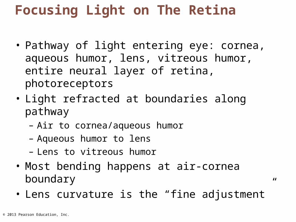
Focusing Light on The Retina
• Pathway of light entering eye: cornea, aqueous humor, lens, vitreous humor, entire neural layer of retina, photoreceptors
• Light refracted at boundaries along pathway– Air to cornea/aqueous humor– Aqueous humor to lens– Lens to vitreous humor
• Most bending happens at air-cornea boundary• Lens curvature is the “fine adjustment”

© 2013 Pearson Education, Inc.
Focusing For Distant Vision
• Eyes best adapted for distant vision• Far point of vision
– Distance beyond which no change in lens shape needed for focusing
• 20 feet for emmetropic (normal) eye• Cornea and lens focus light precisely on retina
• Ciliary muscles relaxed• Lens stretched flat by tension in ciliary zonule
© 2013 Pearson Education, Inc.
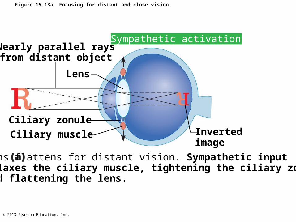
Figure 15.13a Focusing for distant and close vision.
Sympathetic activationNearly parallel raysfrom distant object
Lens
Ciliary zonule
Ciliary muscle Invertedimage
Lens flattens for distant vision. Sympathetic inputrelaxes the ciliary muscle, tightening the ciliary zonule,and flattening the lens.
© 2013 Pearson Education, Inc.
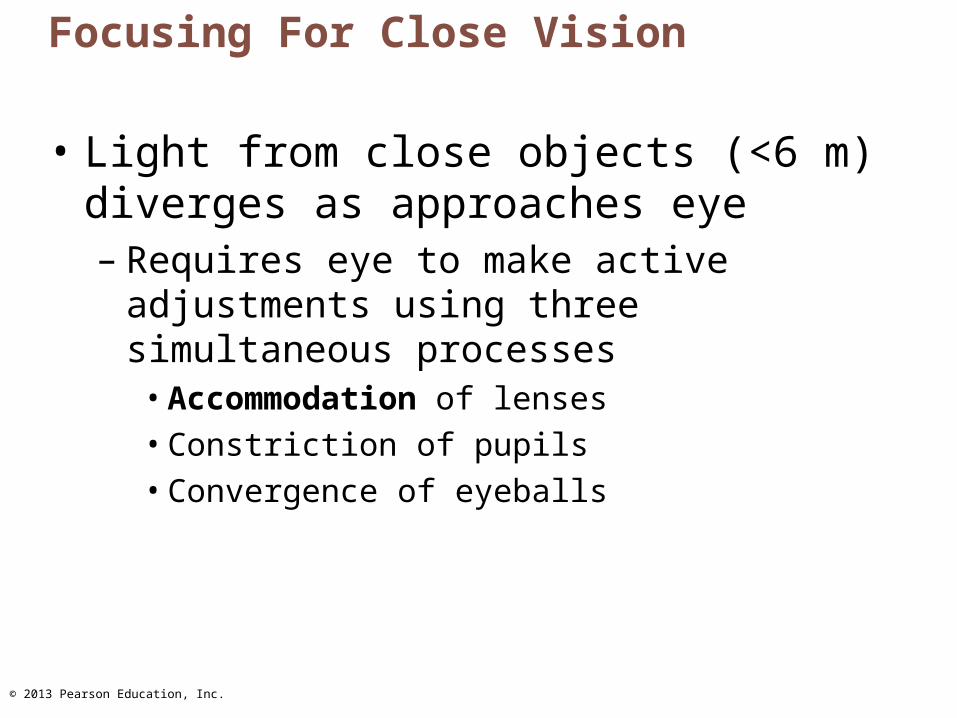
Focusing For Close Vision
• Light from close objects (<6 m) diverges as approaches eye– Requires eye to make active adjustments
using three simultaneous processes• Accommodation of lenses• Constriction of pupils• Convergence of eyeballs
© 2013 Pearson Education, Inc.
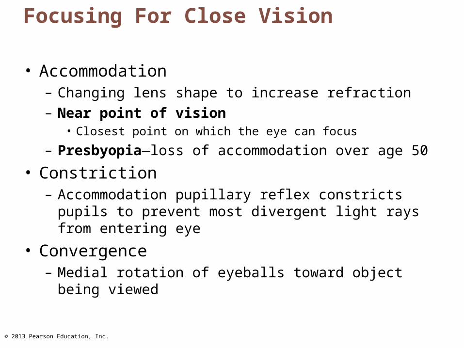
Focusing For Close Vision
• Accommodation– Changing lens shape to increase refraction– Near point of vision
• Closest point on which the eye can focus
– Presbyopia—loss of accommodation over age 50
• Constriction– Accommodation pupillary reflex constricts pupils to
prevent most divergent light rays from entering eye
• Convergence– Medial rotation of eyeballs toward object being viewed
© 2013 Pearson Education, Inc.
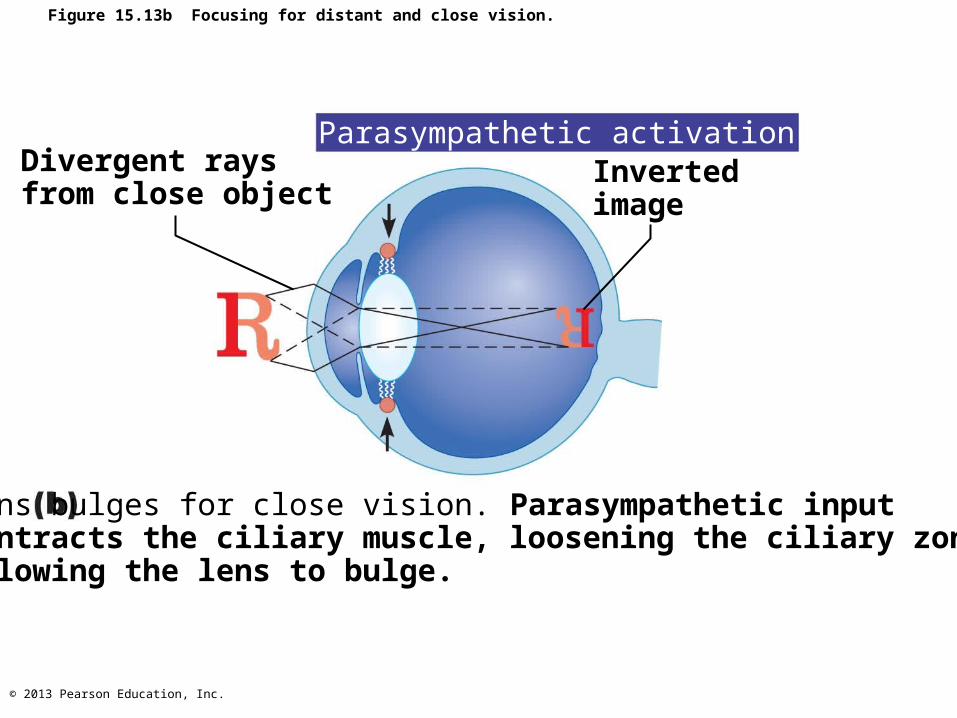
Figure 15.13b Focusing for distant and close vision.
Parasympathetic activationInvertedimage
Divergent raysfrom close object
Lens bulges for close vision. Parasympathetic inputcontracts the ciliary muscle, loosening the ciliary zonule,allowing the lens to bulge.
© 2013 Pearson Education, Inc.
Problems Of Refraction
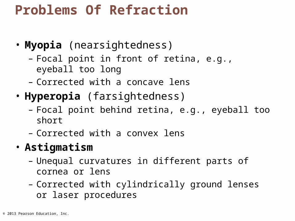
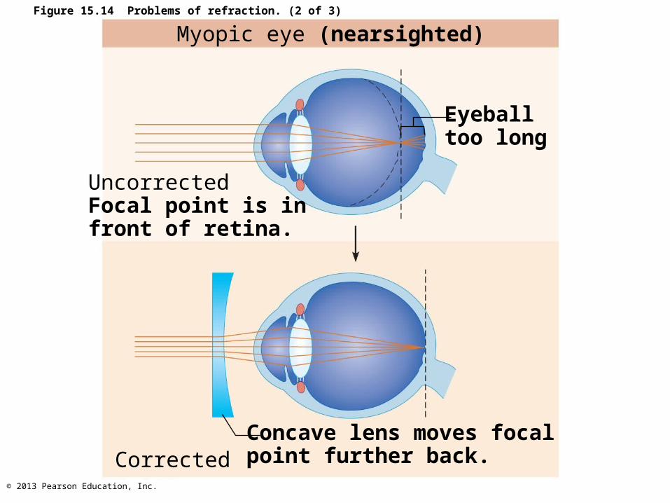
• Myopia (nearsightedness)– Focal point in front of retina, e.g., eyeball too long– Corrected with a concave lens
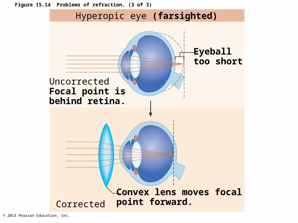
• Hyperopia (farsightedness)– Focal point behind retina, e.g., eyeball too short– Corrected with a convex lens
• Astigmatism– Unequal curvatures in different parts of cornea or lens– Corrected with cylindrically ground lenses or laser
procedures
© 2013 Pearson Education, Inc.
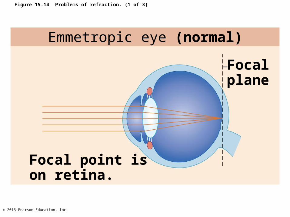
Figure 15.14 Problems of refraction. (1 of 3)
Emmetropic eye (normal)
Focalplane
Focal point ison retina.
© 2013 Pearson Education, Inc.
Myopic eye (nearsighted)
UncorrectedFocal point is infront of retina.
Concave lens moves focalpoint further back.
Eyeballtoo long
Corrected
Figure 15.14 Problems of refraction. (2 of 3)
© 2013 Pearson Education, Inc.
Hyperopic eye (farsighted)
Eyeballtoo short
UncorrectedFocal point isbehind retina.
CorrectedConvex lens moves focalpoint forward.
Figure 15.14 Problems of refraction. (3 of 3)
© 2013 Pearson Education, Inc.
Functional Anatomy Of Photoreceptors
• Rods and cones– Modified neurons– Receptive regions called outer segments
• Contain visual pigments (photopigments)– Molecules change shape as absorb light
– Inner segment of each joins cell body
© 2013 Pearson Education, Inc.
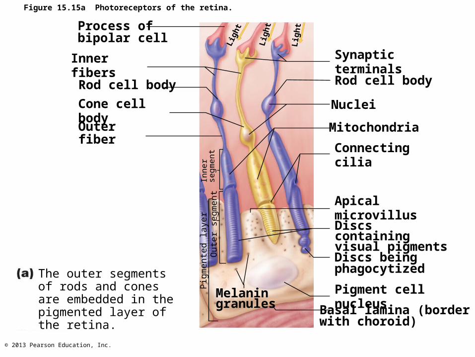
Figure 15.15a Photoreceptors of the retina.
Process of bipolar cell Li
ght
Synaptic terminals
Rod cell body
Inner fibers
NucleiCone cell bodyMitochondria
Connecting cilia
Outer fiber
Apical microvillus
Discs containingvisual pigmentsDiscs being phagocytized
Melanin granules
Pigment cell nucleusBasal lamina (border with choroid)
Inner
segm
ent
Pig
mente
d layer
Oute
r se
gm
ent
Lig
ht
Lig
ht
The outer segments of rods and cones are embedded in the pigmented layer of the retina.
Rod cell body
© 2013 Pearson Education, Inc.
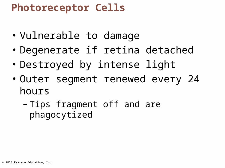
Photoreceptor Cells
• Vulnerable to damage
• Degenerate if retina detached
• Destroyed by intense light
• Outer segment renewed every 24 hours– Tips fragment off and are phagocytized
© 2013 Pearson Education, Inc.
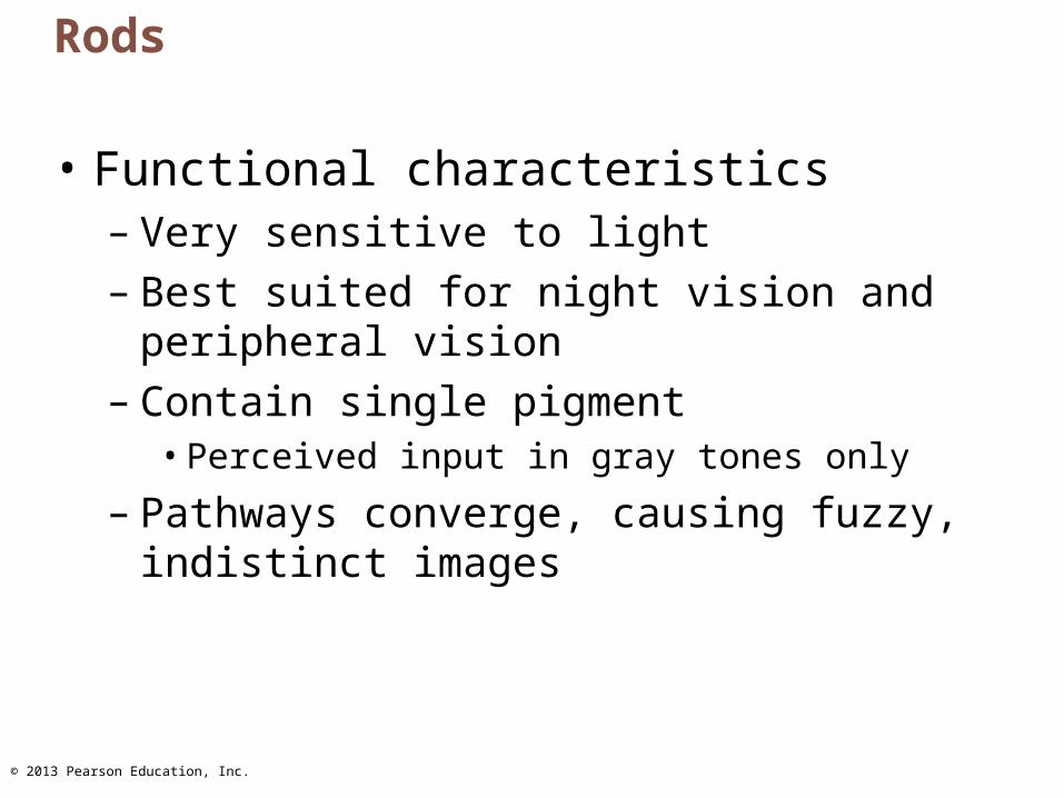
Rods
• Functional characteristics– Very sensitive to light– Best suited for night vision and peripheral
vision– Contain single pigment
• Perceived input in gray tones only
– Pathways converge, causing fuzzy, indistinct images
© 2013 Pearson Education, Inc.
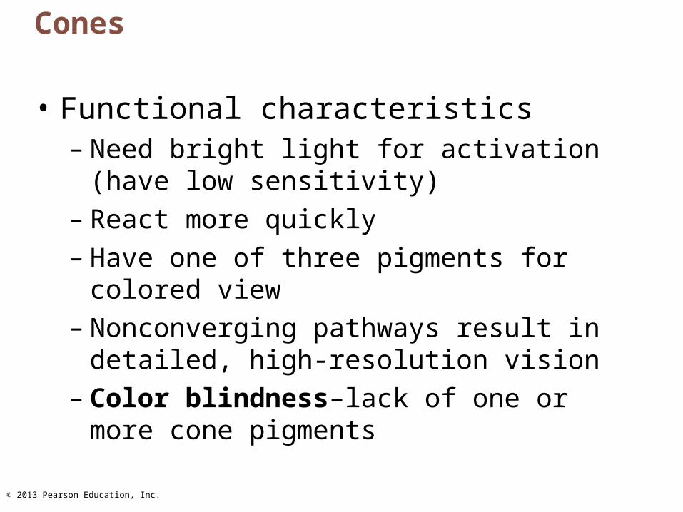
Cones
• Functional characteristics – Need bright light for activation (have low
sensitivity)– React more quickly– Have one of three pigments for colored view– Nonconverging pathways result in detailed,
high-resolution vision– Color blindness–lack of one or more cone
pigments
© 2013 Pearson Education, Inc.
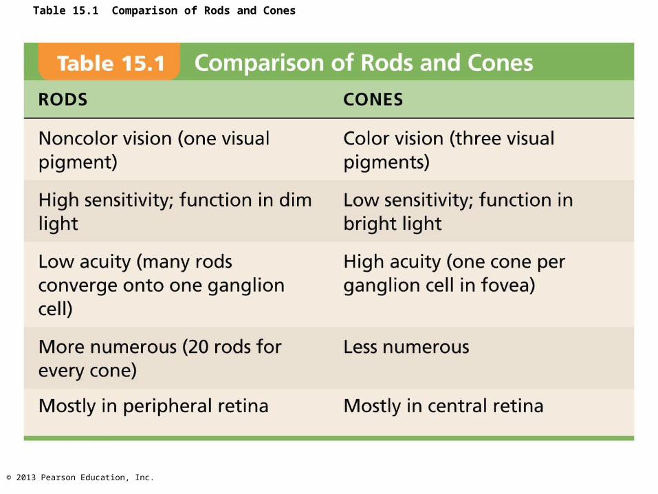
Table 15.1 Comparison of Rods and Cones
© 2013 Pearson Education, Inc.
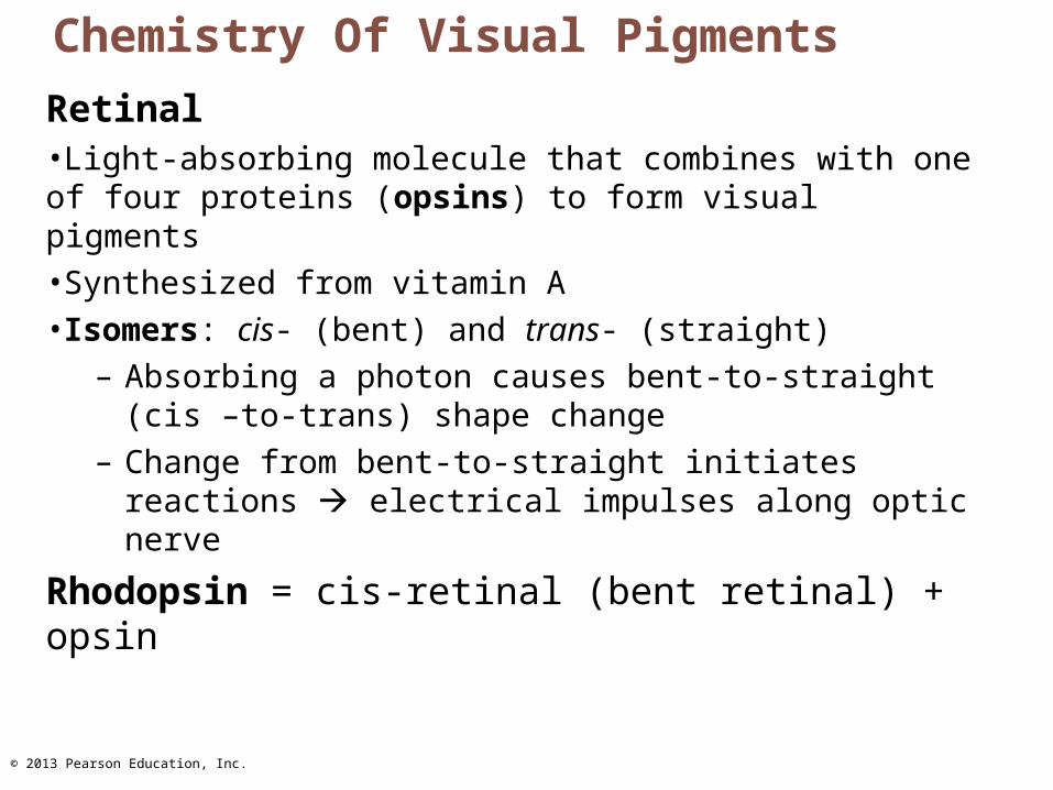
Chemistry Of Visual Pigments
Retinal•Light-absorbing molecule that combines with one of four proteins (opsins) to form visual pigments•Synthesized from vitamin A•Isomers: cis- (bent) and trans- (straight)
– Absorbing a photon causes bent-to-straight (cis –to-trans) shape change
– Change from bent-to-straight initiates reactions electrical impulses along optic nerve
Rhodopsin = cis-retinal (bent retinal) + opsin
© 2013 Pearson Education, Inc.
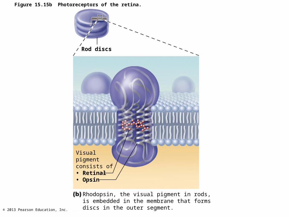
Figure 15.15b Photoreceptors of the retina.
Rod discs
Rhodopsin, the visual pigment in rods,is embedded in the membrane that formsdiscs in the outer segment.
Visualpigmentconsists of• Retinal• Opsin
© 2013 Pearson Education, Inc.
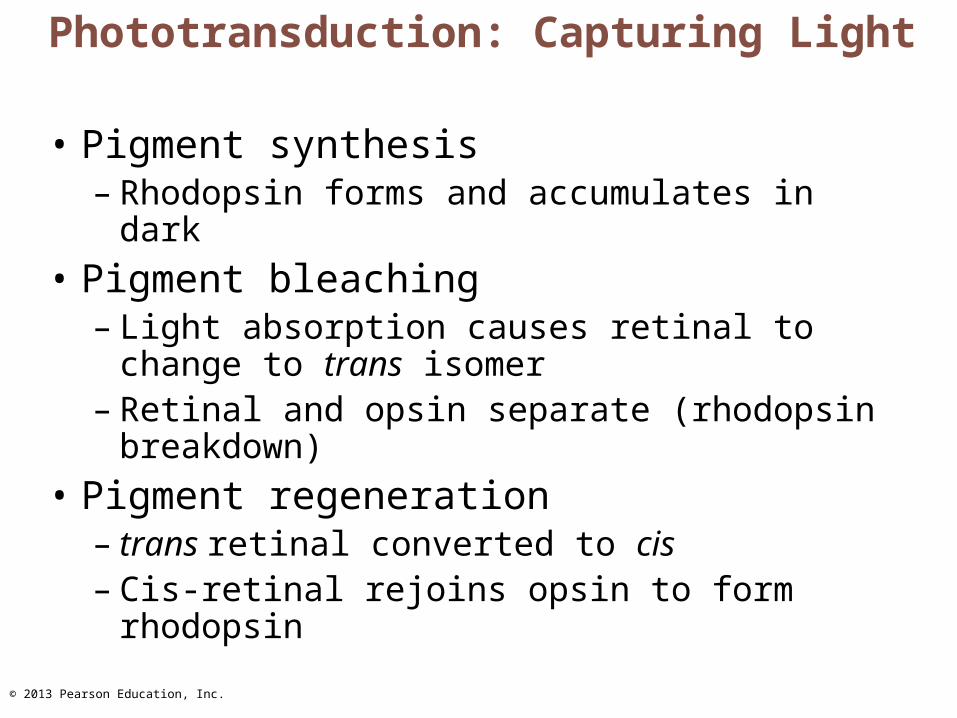
Phototransduction: Capturing Light
• Pigment synthesis– Rhodopsin forms and accumulates in dark
• Pigment bleaching– Light absorption causes retinal to change to
trans isomer– Retinal and opsin separate (rhodopsin
breakdown)
• Pigment regeneration– trans retinal converted to cis– Cis-retinal rejoins opsin to form rhodopsin
© 2013 Pearson Education, Inc.
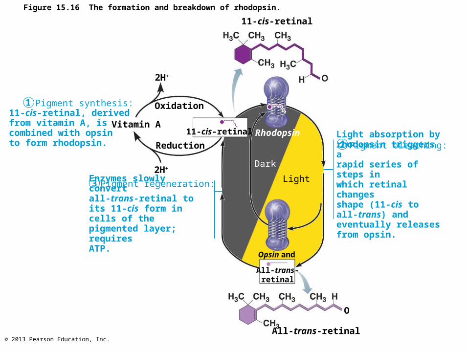
Figure 15.16 The formation and breakdown of rhodopsin.
Enzymes slowly convertall-trans-retinal to its 11-cis form in cells of thepigmented layer; requiresATP.
Pigment regeneration:
Light absorption byrhodopsin triggers arapid series of steps inwhich retinal changesshape (11-cis to all-trans) and eventually releases from opsin.
Pigment bleaching:
11-cis-retinal, derivedfrom vitamin A, iscombined with opsin to form rhodopsin.
Pigment synthesis:1
2H+
2H+
All-trans-retinal
All-trans-retinal
Rhodopsin
Dark
3
2
11-cis-retinalVitamin A
Oxidation
Reduction
Opsin and
Light
11-cis-retinal
O
© 2013 Pearson Education, Inc.
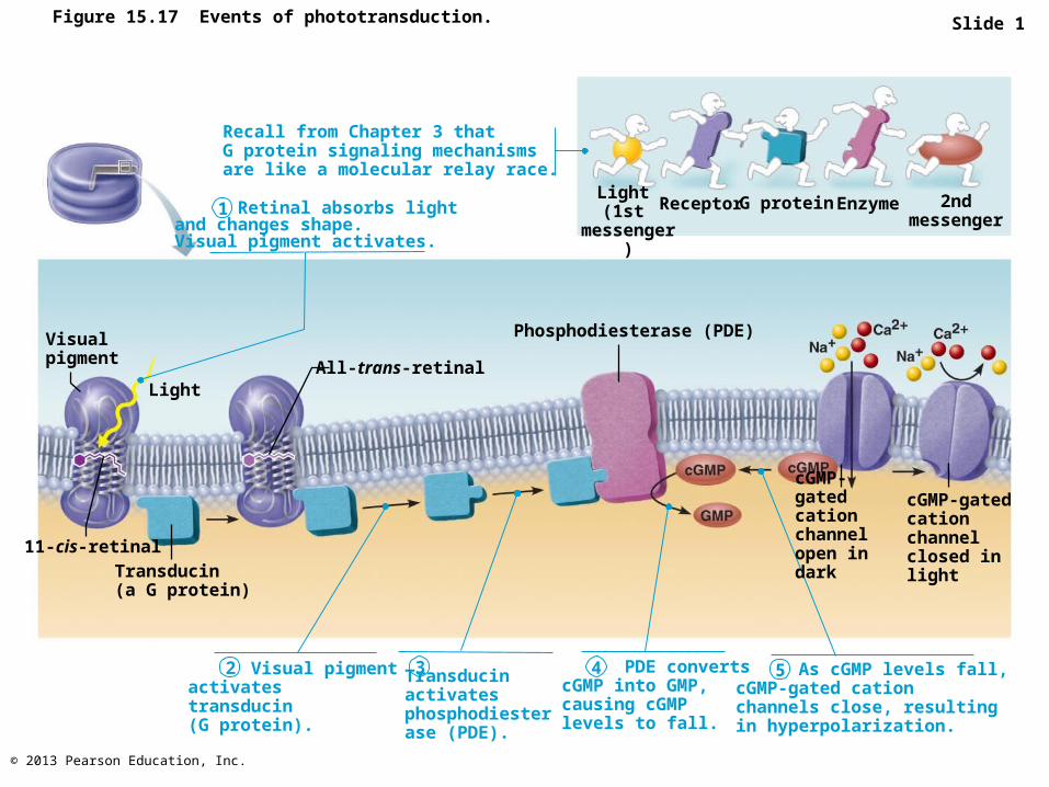
Figure 15.17 Events of phototransduction. Slide 1
Recall from Chapter 3 thatG protein signaling mechanismsare like a molecular relay race.
Retinal absorbs lightand changes shape.Visual pigment activates.
Light (1st
messenger)
Receptor G protein Enzyme 2ndmessenger
Visualpigment
1
Light
11-cis-retinal
Transducin(a G protein)
All-trans-retinal
2 3 Visual pigmentactivatestransducin(G protein).
Transducinactivatesphosphodiesterase (PDE).
4 5 PDE convertscGMP into GMP,causing cGMPlevels to fall.
As cGMP levels fall,cGMP-gated cationchannels close, resultingin hyperpolarization.
cGMP-gatedcation channelopen in dark
cGMP-gatedcation channelclosed in light
Phosphodiesterase (PDE)
© 2013 Pearson Education, Inc.
Phototransduction In Cones
• Similar as process in rods
• Cones far less sensitive to light– Takes higher-intensity light to activate cones
© 2013 Pearson Education, Inc.
Light Transduction Reactions
• Light-activated rhodopsin activates G protein transducin
• Transducin activates PDE, which breaks down cyclic GMP (cGMP)
• In dark, cGMP holds channels of outer segment open Na+ and Ca2+ depolarize cell
• In light cGMP breaks down, channels close, cell hyperpolarizes– Hyperpolarization is signal!
© 2013 Pearson Education, Inc.
Information Processing In The Retina
• Photoreceptors and bipolar cells only generate graded potentials (EPSPs and IPSPs)
• When light hyperpolarizes photoreceptor cells– Stop releasing inhibitory neurotransmitter
glutamate– Bipolar cells (no longer inhibited) depolarize,
release neurotransmitter onto ganglion cells– Ganglion cells generate APs transmitted in
optic nerve to brain
© 2013 Pearson Education, Inc.
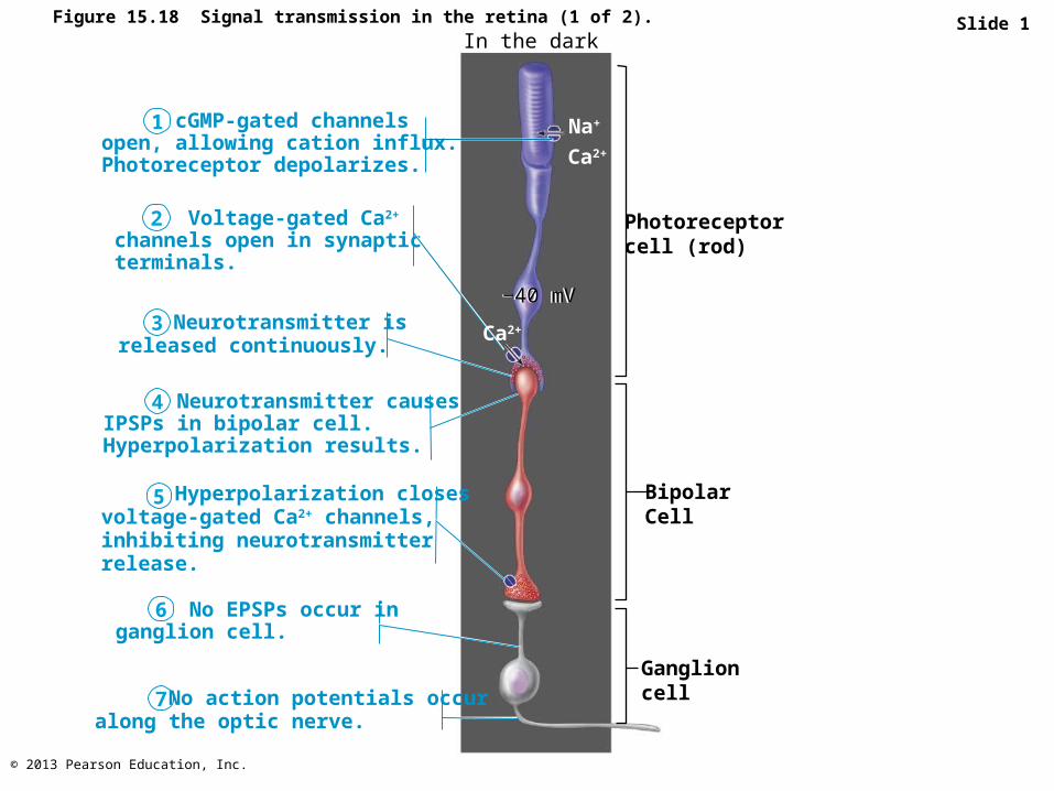
Figure 15.18 Signal transmission in the retina (1 of 2). Slide 1In the dark
cGMP-gated channelsopen, allowing cation influx.Photoreceptor depolarizes.
1
Voltage-gated Ca2+
channels open in synapticterminals.
Neurotransmitter isreleased continuously.
Neurotransmitter causesIPSPs in bipolar cell.Hyperpolarization results.
Hyperpolarization closesvoltage-gated Ca2+ channels,inhibiting neurotransmitterrelease.
No EPSPs occur inganglion cell.
No action potentials occuralong the optic nerve.
Photoreceptorcell (rod)
BipolarCell
Ganglioncell
Ca2+
−40 mV−40 mV
2
3
4
5
6
7
Ca2+
Na+
© 2013 Pearson Education, Inc.
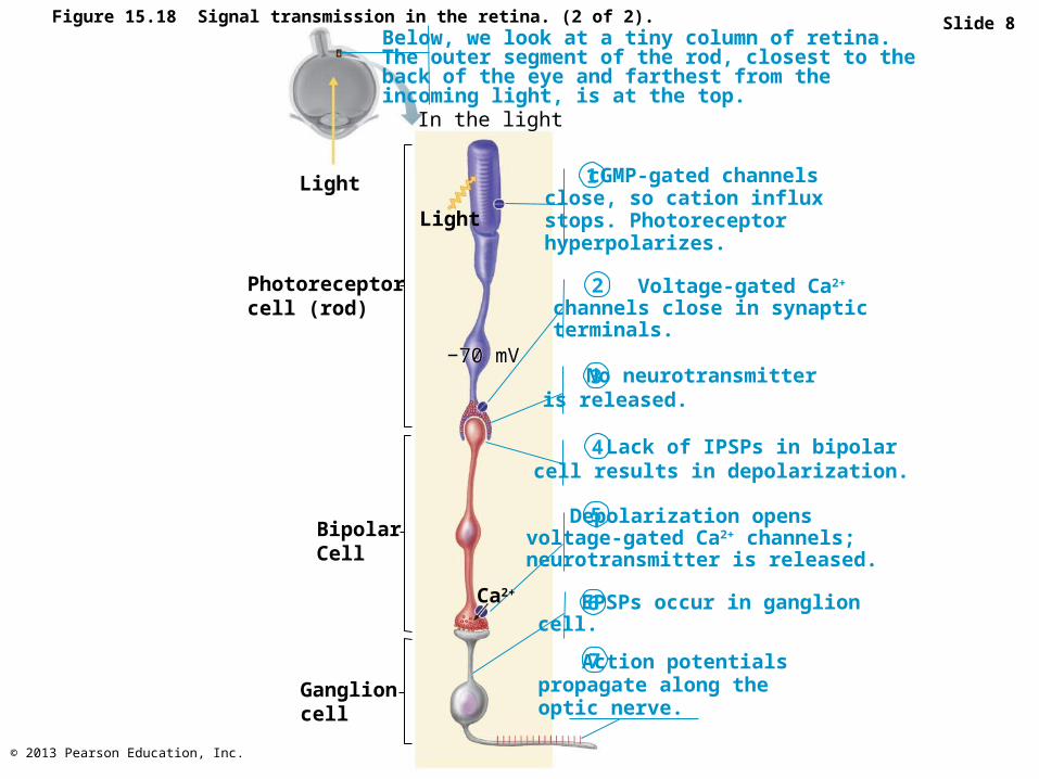
Figure 15.18 Signal transmission in the retina. (2 of 2). Slide 8
−70 mV No neurotransmitter
is released.
Depolarization opensvoltage-gated Ca2+ channels;neurotransmitter is released.
EPSPs occur in ganglioncell.
Action potentialspropagate along theoptic nerve.
cGMP-gated channelsclose, so cation influxstops. Photoreceptorhyperpolarizes.
Lack of IPSPs in bipolarcell results in depolarization.
Voltage-gated Ca2+
channels close in synapticterminals.
1
Photoreceptorcell (rod)
BipolarCell
Ganglioncell
In the light
Light
Ca2+
−70 mV
2
3
4
5
6
7
Below, we look at a tiny column of retina.The outer segment of the rod, closest to theback of the eye and farthest from theincoming light, is at the top.
Light
© 2013 Pearson Education, Inc.
Visual Pathway To The Brain
• Axons of retinal ganglion cells form optic nerve • Half of the fibers (medial half) of each optic nerve cross
over at optic chiasm; optic tracts exit• Most optic tract fibers go to lateral geniculate nucleus of
thalamus• Fibers from thalamic (LGN) neurons form optic radiation
and project to primary visual cortex in occipital lobes• Other optic tract fibers go to superior colliculi in midbrain
(initiating visual reflexes) • A few ganglion cells contain melanopsin and project to
other brain areas– Regulate pupil diameter, daily rhythms
© 2013 Pearson Education, Inc.
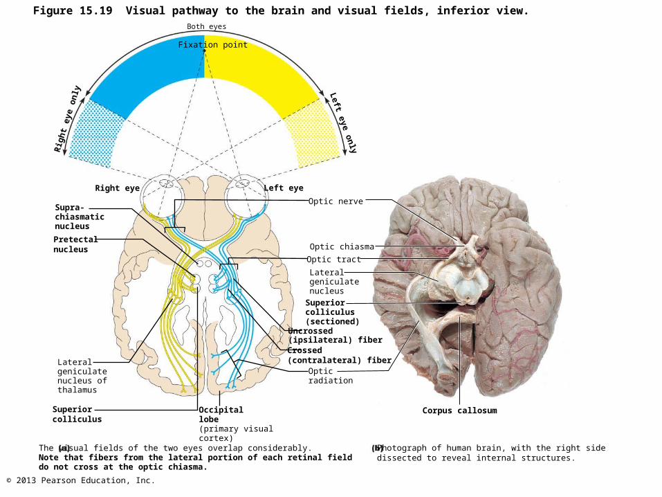
Figure 15.19 Visual pathway to the brain and visual fields, inferior view.
Rig
ht
eye
on
ly
Both eyes
Fixation point
Right eye
Supra-chiasmaticnucleus
Pretectalnucleus
Lateralgeniculatenucleus ofthalamus
Superiorcolliculus
The visual fields of the two eyes overlap considerably.Note that fibers from the lateral portion of each retinal fielddo not cross at the optic chiasma.
Occipitallobe(primary visualcortex)
Left eye
Left eye o
nly
Optic nerve
Optic chiasma
Optic tract
LateralgeniculatenucleusSuperiorcolliculus(sectioned)
Uncrossed(ipsilateral) fiberCrossed(contralateral) fiber
Opticradiation
Corpus callosum
Photograph of human brain, with the right sidedissected to reveal internal structures.
© 2013 Pearson Education, Inc.
Depth Perception
• Eyes see world from slightly different angles• Depth perception (three-dimensional vision)
results from detection of the small differences between R & L eye images
• Requires input from both eyes
© 2013 Pearson Education, Inc.
Visual Processing
• Retinal cells – Color, brightness, edge detection (by amacrine and
horizontal cells)
• Lateral geniculate nuclei of thalamus– Process for depth perception, cone input emphasized,
contrast sharpened
• Primary visual cortex (striate cortex)– Neurons detect edges, object orientation, movement– Provide form, color, motion inputs to visual
association areas (prestriate cortex)
© 2013 Pearson Education, Inc.
Cortical Processing
• Occipital lobe centers (prestriate cortex) continues processing form, color, movement
• Complex visual processing extends to other regions– "What" processing identifies objects in visual field– "Where" processing assesses spatial location of
objects– Output from both passes to frontal cortex